The suppressive role of miR-542-5p in NSCLC: The evidence from clinical data and in vivo validation using a chick chorioallantoic membrane model
Bạn đang xem bản rút gọn của tài liệu. Xem và tải ngay bản đầy đủ của tài liệu tại đây (8.57 MB, 15 trang )
He et al. BMC Cancer (2017) 17:655
DOI 10.1186/s12885-017-3646-1
RESEARCH ARTICLE
Open Access
The suppressive role of miR-542-5p in
NSCLC: the evidence from clinical data and
in vivo validation using a chick
chorioallantoic membrane model
Rong-quan He1†, Xiao-jiao Li2†, Lu Liang3, You Xie3, Dian-zhong Luo3, Jie Ma3, Zhi-gang Peng3,
Xiao-hua Hu1,3* and Gang Chen3*
Abstract
Background: Non-small cell lung cancer (NSCLC) has led to the highest cancer-related mortality for decades. To
enhance the efficiency of early diagnosis and therapy, more efforts are urgently needed to reveal the origins of
NSCLC. In this study, we explored the effect of miR-542-5p in NSCLC with clinical samples and in vivo models and
further explored the prospective function of miR-542-5p though bioinformatics methods.
Methods: A total of 125 NSCLC tissue samples were collected, and the expression of miR-542-5p was detected by
qRT-PCR. The relationship between miR-542-5p level and clinicopathological features was analyzed. The effect of
miR-542-5p on survival time was also explored with K-M survival curves and Cox’s regression. The effect of miR-5425p on the tumorigenesis of NSCLC was verified with a chick chorioallantoic membrane (CAM) model. The potential
target genes were predicted by bioinformatics tools, and relevant pathways were analyzed by GO and KEGG.
Several hub genes were validated by Proteinatlas.
Results: The expression of miR-542-5p was down-regulated in NSCLC tissues, and consistent results were also found in
the subgroups of adenocarcinoma and squamous cell carcinoma. Down-regulation of miR-542-5p was found
to be connected with advanced TNM stage, vascular invasion, lymphatic metastasis and EGFR. Survival analyses showed
that patients with lower miR-542-5p levels had markedly poorer prognosis. Both tumor growth and angiogenesis were
significantly suppressed by miR-542-5p mimic in the CAM model. The potential 457 target genes of miR-542-5p were
enriched in several key cancer-related pathways, such as morphine addiction and the cAMP signaling pathway
from KEGG. Interestingly, six genes (GABBR1, PDE4B, PDE4C, ADCY6, ADCY1 and GIPR) from the cAMP signaling pathway
were confirmed to be overexpressed in NSCLCs tissues.
Conclusions: This evidence suggests that miR-542-5p is a potential tumor-suppressed miRNA in NSCLC, which has the
potential to act as a diagnostic and therapeutic target of NSCLC.
Keywords: NSCLC, miR-542-5p, CAM, RT-qPCR, GO, KEGG
* Correspondence: ;
†
Equal contributors
1
Department of Medical Oncology, First Affiliated Hospital of Guangxi
Medical University, 6 Shuangyong Road, Nanning 530021, Guangxi Zhuang
Autonomous Region, People’s Republic of China
3
Department of Pathology, First Affiliated Hospital of Guangxi Medical
University, 6 Shuangyong Road, Nanning 530021, Guangxi Zhuang
Autonomous Region, People’s Republic of China
Full list of author information is available at the end of the article
© The Author(s). 2017 Open Access This article is distributed under the terms of the Creative Commons Attribution 4.0
International License ( which permits unrestricted use, distribution, and
reproduction in any medium, provided you give appropriate credit to the original author(s) and the source, provide a link to
the Creative Commons license, and indicate if changes were made. The Creative Commons Public Domain Dedication waiver
( applies to the data made available in this article, unless otherwise stated.
He et al. BMC Cancer (2017) 17:655
Background
Non-small cell lung cancer (NSCLC) is the most
frequent type of lung cancer with high mortality
worldwide [1]. Lung adenocarcinoma (LUAD) and
squamous cell carcinoma (LUSC) are the major subtypes of NSCLC, composing approximately 40% and
30% of NSCLC, respectively [2]. Like most malignancies, the patients with NSCLC who received diagnoses at an early stage achieved higher five-year
survival rates, compared to patients whose diagnoses
were made at an advanced stage [3]. However, only a
minority of NSCLC patients received early diagnosis
because of the lack of significant symptoms in early
stages [3]. Currently, therapeutic measures for advanced NSCLC patients are still limited. Although
the study of molecular targeted therapies is progressing, including EGFR and ALK-targeted therapies in
lung adenocarcinoma, which have had a successful
beginning, they are efficient in just 20% of patients
[4]. Given these results, high-performance biological
markers are critically needed to find and diagnose
NSCLC in early stages, to prevent NSCLC from advancing, and to help advanced patients achieve a
better prognosis.
MicroRNAs (miRNAs, miRs) are a type of non-coding
RNA with a short (less than 22 nucleotides), single
stranded nucleotide chain. MiRNAs can regulate the
generation of proteins by binding to the untranslated region of messenger RNAs, using complementary base
pairing. Through this mechanism, miRNAs can regulate
the differentiation, proliferation and apoptosis of cells
[5]. Many studies have found that the dysregulation of
miRNAs correlates with diseases, including lung cancer
[6, 7]. In NSCLC, hundreds of dysregulated miRNAs
have been detected from high-throughput experiments
[8, 9]. However, the clinicopathological significance and
related mechanisms of dysregulated miRNAs in NSCLC
remain largely unclear. In preliminary studies, we found
that miR-30a [10], miR-193a-3p and miR-133a-3p were
down-regulated in NSCLC tissues [11, 12], and all of
them have an effect on survival time of patients.
In the current study, we explored the expression of
miR-542-5p in NSCLC tissues, assessed the relationship
between miR-542-5p and clinicopathological parameters,
and verified the function of miR-542-5p on NSCLC in
vivo. Furthermore, the potential mechanism of miR-5425p action on NSCLC was predicted by bioinformatics
methods.
Methods
Tissue samples
The tissue samples fixed in this study were from 125
lung cancer patients who underwent surgeries at the
First Affiliated Hospital of Guangxi Medical University
Page 2 of 15
between January 2012 and February 2014. All tissues
were obtained before any cancer-related therapy was carried out. Adjacent noncancerous tissues were obtained
from at least two centimeters away from the edge of the
tumor node. All samples were prepared in the form of
formalin-fixed and paraffin-embedded (FFPE). The included lung cancer tissues were divided into 101 LUAD, 23
LUSC and 1 large cell lung cancer. Among 125 included
patients, 75 were males, and 50 were females. There were
57 younger (<60 years) patients and 68 older (>60 years)
patients. The subgroups were divided based on clinicopathological parameters, such as tumor size, smoking history,
and vascular invasion, which are displayed in Table 1.
Among 125 patients, 57 LUAD patients were followed-up
Table 1 Correlations between miR-542-5p expression and the
clinicopathological features of NSCLC
n
The expression of
miR-542-5p
(2-△Cq)
t
p
NSCLC
125
1.952 ± 1.507
−11.703
< 0.001*
Adjacent lung
125
4.568 ± 1.993
−0.052
0.958
0.279
0.783
0.183
0.504
−1.004
0.319
6.488
< 0.001*
3.905
< 0.001*
2.247
0.026*
7.753
< 0.001*
Parameters
Tissue
Age (years)
< 60
57
1.945 ± 1.382
≥ 60
68
1.959 ± 1.615
Male
75
1.983 ± 1.475
Female
50
1.906 ± 1.568
Gender
Tumor size (cm)
<3
60
1.979 ± 1.520
≥3
65
1.928 ± 1.506
Positive
30
2.622 ± 1.510
Negative
38
2.266 ± 1.407
I-II
54
2.822 ± 1.536
III-IV
71
1.292 ± 1.101
Smoking historya
TNM stage
Lymph node metastasis
Positive
69
1.504 ± 1.266
Negative
56
2.506 ± 1.604
Vascular invasion
Positive
35
1.475 ± 1.305
Negative
90
2.138 ± 1.545
low
17
0.739 ± 0.407
high
40
3.049 ± 1.194
EGFR proteinb
n The number of patients
*p < 0.05
a
The data were available from 68 patients
b
The data were available from 57 patients
He et al. BMC Cancer (2017) 17:655
until the manuscript deadline; 39 patients were alive, and
18 patients were dead. The study was permitted by the Ethical Committee of the First Affiliated Hospital of Guangxi
Medical University. Written informed agreements were obtained from the patients and clinicians for the samples
usage. Two pathological doctors reviewed all tissues
independently.
RNA extraction and expression of miR-542-5p in NSCLC
tissues
For FFPE tissues, five sections were acquired from each
tissue sample, at a thickness of 10 μm per section. Total
RNA was extracted by the RNeasy FFPE Kit (NO.
73504) as stated by the manufacturer’s instructions. The
concentration and purity of total RNA were confirmed
with a NanoDrop 2000 spectrophotometer (Wilmington,
DE, USA). Complimentary DNA was synthesized by
total RNA and the TaqMan MicroRNA Reverse Transcription Kit (NO. 4366596) as described previously [13].
Subsequently, an Applied Biosystems PCR7900 system
was used to carry out real-time quantitative reverse
Page 3 of 15
transcriptase-polymerase chain reactions (qRT-PCR) to
detect and analyze the expression of miR-542-5p. The
relative expression of miR-542-5p was calculated by 2△Cq
. RNA extraction and qRT-PCR were performed in
June of 2014.
Cell culture and expression of miR-542-5p in NSCLC cells
H460, H1299, PC9 and A549 cell lines were cultured in
this study, and all the cells were achieved from the Type
Culture Collection of the Chinese Academy of Sciences,
Shanghai, China. Cells were cultured in Dulbecco’s modified essential medium (DMEM, Invitrogen Corp., Grand
Island, NY, USA). Gentamicin, glutamine and fetal bovine
serum (10% heat-inactivated, Invitrogen Crop., Grand Island, NY, USA) were supplied in the culture medium. The
environment was set at 5% CO2, 37 °C and 100% humidity. The expression of miR-542-5p in cells was detected by
qRT-PCR. Total RNA was extracted with the TaKaRa
MiniBEST Universal RNA Extraction Kit (NO. 9767). The
Mir-X™ miRNA First-Strand Synthesis Kit was used to
synthesize cDNA: the total volume of the reaction system
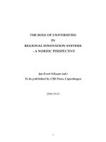
Fig. 1 The expression levels of miR-542-5p in NSCLC and adjacent lung tissues. a, b and c: Comparison of miR-542-5p levels in NSCLC (a), LUAD
(b), LUSC (c) and adjacent lung tissues; d, e and f: ROC curves of miR-542-5p to predict NSCLC (d), LUAD (e) and LUSC (f)
He et al. BMC Cancer (2017) 17:655
was 10 μl, and the parameters were set as follows: 37 °C
for 15 min, 85 °C for 5 min, and then 4 °C indefinitely.
The PCR reaction components included UItra SYBR Mixture (2X, 10 μl), forward primers (10 μm/μl, 1 μl), reverse
primers (10 μm/μl, 1 μl), ddH2O (7 μl) and cDNA (1 μl).
After 45 cycles (95 °C for 15 s, 60 °C for 16 s, 72 °C for
20 s), reactions were performed in 96-well plates using the
Roche LightCycle 480 Real-Time fluorescence quantitative
PCR system. The primers used in qRT-PCR were designed
with Prime 3.0 software and synthesized by Invitrogen
Company. The primer sequences were designed as follows: miR-542-5p (Forward: 5′-GCGGTCGGGGATCATCATGTC; Reverse: 5′-ATCCAGTGCAGGGTCCGAGG).
The relative expression of miR-542-5p was calculated
using 2-△Cq.
MiR-542-5p lentivirus construction
A lentivirus vector was constructed according to the
information of the miR-542-5p sequence and the Ubi-
Page 4 of 15
MCS-SV40-EGFP-IRES-puromycin (GV369) polyclone
site. The 293 T cell line and Lipofectamine 2000
(Invitrogen, USA) were used to package the lentivirus.
The construction and packaging of miR-542-5p lentivirus vector, as well as lentivirus infection, were performed by Shanghai Genechem Co. Ltd., and stored
at −80 °C.
Cell transfection
Among four types of NSCLC cells, the H460 cell line
showed the lowest level of miR-542-5p, so we chose
H460 to conduct the following experiments. Blank control, empty vector control (H460-Lv-vector) and experimental (H460-Lv-miR-542) groups were designed. H460
cells were seeded and infected with lentivirus in 6-well
plates at an MOI of 100 and selected by puromycin. The
green fluorescence expressed in cells served as a marker
to measure the infection efficiency.
Fig. 2 Relative expression of miR-542-5p in different clinicopathological groups of NSCLC patients. a, b, c and d: In NSCLC, different expression of miR-542-5p
in relative groups are divided by TNM, vascular invasion, lymphatic metastasis and EGFR status; e, f, g and h: In LUAD, different expression of miR-542-5p in
relative groups are divided by TNM, vascular invasion, lymphatic metastasis and EGFR status; i: In LUSC, different expression levels of miR-542-5p in relative
groups are divided by EGFR status
He et al. BMC Cancer (2017) 17:655
Chick chorioallantoic membrane model
The chick chorioallantoic membrane (CAM) model
was used to explore the effect of miR-542-5p on
tumor growth and angiogenesis of NSCLC cells. After
being obtained from local hatchery, fertilized eggs
were incubated in an incubator at 37 °C and 80% humidity and rotated every 5 h until day 7. On the 8th
day of fertilization, air space and embryo were
checked under the egg candler, and the head of the
embryo and large vessels in the CAM were marked
by pencil. To separate the chorioallantoic membrane
from the inner shell membrane, a tiny hole
(φ = 0.1 cm) in the middle of the air space was
made by a mini electric grinder, and an aurilave was
used to draw out the gas. A 1.5 cm2 window was
made in the shell where large vessels in the CAM
were marked. A silicone ring (φ = 0.5 cm,
height = 0.3 cm) was put at the crossing of large
vessels, and cells were then transferred into the silicone ring; the quantity of cells was 10 × 106 in each
egg. The eggs were randomly grouped into blank
control, empty vector control (H460-Lv-vector) and
experimental (H460-Lv-miR-542) groups. Pictures
were taken at 0, 24, 48, 72, 96 and 110 h. After every
time pictures were taken, the windows were resealed
by transparent tape. The silicone ring was removed
at 24 h, and transplant implanted tumors were harvested at 110 h. The vascular density was assessed by
Image Pro Plus. The harvested tumors were sliced
and examined by hematoxylin and eosin (HE) staining. Immunohistochemical (IHC) staining was used
to assess the expression of EGFR, VEGF and D2–40.
Specific pathogen free fertilized Guangxi Sanhuang
Page 5 of 15
chick eggs were purchased from the Experimental
Animal Center of Guangxi Medical University
(Guangxi, China) and all procedures involving animals and their care complied with the China National Institutes of Healthy Guidelines for the Care
and Use of Laboratory Animals. Ethical approval for
the study was granted by the Ethical Committee of
the First Affiliated Hospital of Guangxi Medical
University.
Biological informatics analysis of potential target genes
of miR-542-5p
The target genes of miR-542-5p were predicted by 13
platforms (miRWalk, Microt4, miRanda, mirbridge,
miRDB, miRMap, miRNAMap, Pictar2, PITA, RNA22,
RNAhybrid, Targetscan and mirTarbase). Genes copredicted on at least five platforms were collected. To
explore the potential function of miR-542-5p in
NSCLC, the target genes were analyzed by Gene
Ontology (GO) analysis, the Kyoto Encyclopedia of
Genes and Genomes (KEGG) pathways in Gorilla
( />and
DAVID
( String ( was used to analyze the connections among
genes and draw connected figures. The protein expression data from Proteinatlas ( was checked for the validation of several
potential target genes of miR-542-5p in NSCLCs.
Statistical analysis
The medium value of miR-542-5p was used to divide
the NSCLC patients into low expression and high
Fig. 3 Kaplan-Meier survival curves in 57 patients with lung adenocarcinoma with different miR-542-5p levels
He et al. BMC Cancer (2017) 17:655
Page 6 of 15
Fig. 4 The effect of miR-542-5p on tumor size and angiogenesis in the CAM model. a: Selected CAM pictures of a blank control and a miR-542-5p-transfected
group on the 0th, 3rd and 5th day of transfection; b: Different sizes of transfected tumors on blank control, negative control and miR-542-5p-transfected groups;
c: Different vascular density on blank control, negative control and miR-542-5p-transfected groups
expression groups. Student’s t test was selected to assess the different expression levels of miR-542-5p between NSCLC and adjacent lung tissues, trials and
control groups. Spearman method was used to assess
the relationship between miR-542-5p level and clinical
pathological parameters. Kaplan-Meier (KM) method,
log rank test as well as Cox’s regression were performed in the survival analyses. Adjusted hazard ratios (HRs) were calculated. p < 0.05 was considered
to be statistically significant. The data were calculated
by SPSS 22.0, and figures were constructed with
Graphpad Prism 5.0.
Fig. 5 Immunohistochemical staining showing the status of EGFR, VEGF and D2–40 in blank control and miR-542-5p-transfected CAM groups
He et al. BMC Cancer (2017) 17:655
Results
Page 7 of 15
The expression of miR-542-5p in NSCLC tissues
The relationship between miR-542-5p and clinicopathological features
MiR-542-5p showed evidently lower expression in
NSCLC when compared to adjacent normal lung tissues, and consistent results were also found in the
subgroups of LUAD and LUSC (Fig. 1a–c). The diagnostic value was assessed by ROC curves. The AUC
values were 0.859, 0.876 and 0.769 in NSCLC, LUAD
and LUSC, respectively (Fig. 1d–f ).
In 125 NSCLC patients, down-regulation of miR-5425p was correlated with advanced TNM stage, vascular
invasion and lymphatic metastasis (Fig. 2, Table 1). In
TNM I-II stages, the miR-542-5p level was
2.822 ± 1.536, prominently higher than that in III-IV
stages (1.292 ± 1.101, t = 6.488, p < 0.001). Of patients without lymph node metastasis, the miR-542-5p
Fig. 6 Connections between the predicted target genes of miR-542-5p
He et al. BMC Cancer (2017) 17:655
Page 8 of 15
Fig. 7 Gene ontology terms enriched by the potential target genes of miR-542-5p. (a: Cellular component terms of GO; b: Molecular function
terms of GO; c. Biological process terms of GO)
level was clearly overexpressed than those with lymph
node metastasis (2.506 ± 1.604 vs 1.504 ± 1.266,
t = 3.905, p < 0.001). Similarly, significantly higher
expression of miR-542-5p was also found in the samples without vascular invasion than that with invasion
(2.138 ± 1.545 vs 1.475 ± 1.305, t = 2.247, p < 0.001).
Spearman’s correlation analyses showed that the expression of miR-542-5p was negatively correlated with
TNM stage (r = −0.505, p < 0.001), lymph node metastasis (r = −0.332, p < 0.001), and vascular invasion
(r = −0.199, p = 0.026). In subgroups, downregulation of miR-542-5p was also found to be correlated with advanced TNM stage, vascular invasion,
lymphatic metastasis and EGFR expression in LUAD
patients (Fig. 2). In 101 LUAD cases, the expression
of miR-542-5p in stage III-IV patients was significantly lower than that of patients at stage I and II
(1.142 ± 0.935 vs 2.810 ± 1.516, t = 6.417, p < 0.001).
MiR-542-5p expression in patients with vascular invasion was significantly lower than that in patients
without vascular invasion (1.375 ± 1.30 vs
2.087 ± 1.497, t = 2.286, p = 0.024). And miR-542-5p
expression in patients with lymph node metastasis
was significantly lower than that in patients without
lymph node metastasis (1.333 ± 1.095 vs
2.085 ± 1.085, t = −2.477, p = 0.027). Additionally,
the expression of miR-542-5p was higher in patients
who smoke than that of patients without smoking
(3.065 ± 1.542 vs 2.535 ± 1.616, t = 4.265, p < 0.001).
Spearman’s correlation analyses showed that the expression of miR-542-5p was negatively correlated with
TNM stage (r = −0.564, p < 0.001), lymph node metastasis (r = −0.408, p < 0.001), and vascular invasion
(r = −0.224, p = 0.024). In 23 LUSC cases, there was
no significant association between the expression of
miR-542-5p and clinical parameters.
In tumor tissues of NSCLC patients, the relative
expression of miR-542-5p in patients with high
Table 2 The KEGG pathways enriched by the potential target
genes of miR-542-5p
Description
hsa05032
Morphine addiction
11
0.000111
hsa04024
cAMP signaling pathway
15
0.000587
hsa04261
Adrenergic signaling in
cardiomyocytes
12
0.001364
hsa04727
GABAergic synapse
9
0.001523
hsa05030
Cocaine addiction
7
0.001541
hsa04390
Hippo signaling pathway
12
0.001790
hsa04725
Cholinergic synapse
10
0.002243
hsa04724
Glutamatergic synapse
10
0.002693
hsa05200
Pathways in cancer
21
0.002768
hsa04923
Regulation of lipolysis in
adipocytes
7
0.003081
hsa05414
Dilated cardiomyopathy
8
0.005886
hsa04911
Insulin secretion
8
0.006278
hsa04921
Oxytocin signaling pathway
11
0.007747
hsa04976
Bile secretion
7
0.008632
hsa04520
Adherens junction
7
0.009886
Enriched by DAVID, p < 0.01
Count in gene set
p-value
ID
He et al. BMC Cancer (2017) 17:655
Page 9 of 15
Fig. 8 Connection among hub genes predicted on KEGG pathways. a. cAMP signaling pathway; b. Hippo signaling pathway; c. Pathways
in cancer
EGFR protein expression was significantly lower than
that of patients with low EGFR protein expression
(0.739 ± 0.407 vs 3.049 ± 1.194, t = 7.753,
p < 0.001). Spearman’s correlation analyses showed
that the expression of miR-542-5p was negatively
correlated with EGFR protein expression (r = −0.723,
p < 0.001). While in the groups of LUAD and LUSC,
the expression of miR-542-5p in patients with high
EGFR protein expression was significantly lower than
that of patients with low EGFR protein expression
(0.836 ± 0.373 vs 3.180 ± 0.952, t = 10.098,
p < 0.001, LUAD; 0.436 ± 0.345 vs 2.888 ± 1.448;
t = 6.543, p < 0.001, LUSC). Spearman’s correlation
analyses also showed that the expression of miR-542-
Fig. 9 Validation of the protein expression of GABBR1 in NSCLCs. GABBR1 protein was detected by the antibody of HPA050483. a, b: normal
lungs with pneumocytes being not detected. c: medium staining in LUAD. d: medium staining in LUSC. Immunohistochemistry, ×100
He et al. BMC Cancer (2017) 17:655
5p was negatively correlated with EGFR protein expression in LUAD (r = −0.818, p < 0.001) and LUSC
(r = −0.828, p < 0.001, Fig. 2).
Survival analysis
Based on the median expression level of miR-542-5p
in NSCLC patients (3.260 ± 2.197), we divided the
patients into two groups with high expression of
miR-542-5p and low expression of miR-542-5p
(4.568 ± 1.993 vs 1.953 ± 1.507). The results of KM
curve survival analyses showed that NSCLC patients
with lower miR-542-5p expression (n = 50,
11.274 ± 1.387 months) had a significantly poorer
prognosis than those patients with higher miR-5425p expression (n = 7, 35.714 ± 3.469 months)
(t = −6.219, p < 0.001, Fig. 3). The multivariate analysis showed that the HR of miR-542-5p was 0.948
(95% CI: 0.916–0.982, p = 0.003), which was adjusted by other clinical parameters as gender, age,
tumor size, clinical stage, and tumor grading.
Page 10 of 15
In vivo study using the CAM model
MiR-542-5p was also detected by RT-qPCR in NSCLC
cell lines H460, PC9, H1299 and A549. Among the four
different cell lines, the lowest level of miR-542-5p was
found in H460, and it was selected for transfection with
a miR-542-5p mimic to unveil the function of miR-5425p in NSCLC. The efficiency of lentiviral transfection
was higher than 90%. In CAM, the tumor size was
assessed after cells were transplanted and harvested on
day 5. Compared to the blank and negative control
groups, the trial group showed smaller tumor size,
and angiogenesis was suppressed (Fig. 4). The expression levels of EGFR, VEGF and D2–40 in the trial
group were weaker, when compared to the blank control group (Fig. 5).
Potential target genes of miR-542-5p and functional annotation analysis
A total of 457 target genes of miR-542-5p were predicted by the 12 platforms mentioned above. Close
Fig. 10 Validation of the protein expression of PDE4B in NSCLCs. PDE4B protein was detected by the antibody of HPA003005. a, b: pneumocytes
in normal lungs with low staining and macrophages with high expression. c: medium staining in LUAD. d: high staining in LUAD.
Immunohistochemistry, ×100
He et al. BMC Cancer (2017) 17:655
connections were found among the genes listed in
Fig. 6. These genes were analyzed by GO and KEGG
pathways. In the CC of GO analysis, the genes were significantly enriched in the plasma membrane and extrinsic components of the cytoplasmic side of the plasma
membrane (Fig. 7a). In MF, non-membrane spanning
protein tyrosine kinase activity, ATP binding and other
binding items were significantly enriched by genes (Fig.
7b). In BP, the adenylate cyclase-activating G-protein
coupled receptor signaling pathway was the most
significantly enriched pathway (Fig. 7c). The most significantly enriched KEGG pathway was morphine addiction; and cancer-related mechanisms, such as the
cAMP signaling pathway and the Hippo signaling pathway, were also significantly enriched in the 457 potential target genes of miR-542-5p (Table 2, Fig. 8). The
genes from cAMP signaling pathway were selected for
further protein expression validation in NSCLC tissues
by using data from Proteinatlas. Six genes showed particularly stronger expression pattern in lung cancer tissues, as compared to normal lungs (Figs. 9, 10, 11, 12,
13 and 14), which demonstrated that these six genes
Page 11 of 15
(GABBR1, PDE4B, PDE4C, ADCY6, ADCY1 and GIPR)
had more likelihood of being direct targets of miR-5425p in lung cancers. However, this hypothesis still needs
in vitro and in vivo verification.
Discussion
In this study, miR-542-5p was found to be predominantly down-regulated in NSCLC tissues, which had a
negative effect on the prognosis of NSCLC patients.
Exploration of the effect of miR-542-5p on NSCLC
with a CAM model confirmed the suppressive function of miR-542-5p on cell growth and angiogenesis
of NSCLC. Furthermore, the mechanism of miR-5425p predicted by bioinformatical approaches suggested
valuable pathways that might relate to tumorigenesis
and tumor development.
Many dysregulated miRNAs have been shown to relate to tumorigenesis or development in numerous
cancers. In published studies, miR-542-5p was described as a tumor-suppressed miRNA in endometrial
carcinosarcoma [14], neuroblastoma [15] and rectal
cancer [16]. However, in several cancers, miR-542-5p
Fig. 11 Validation of the protein expression of PDE4C in NSCLCs. PDE4C protein was detected by the antibody of HPA048975. a, b: pneumocytes
in normal lungs with no staining. c, d: high staining in LUAD. Immunohistochemistry, ×100
He et al. BMC Cancer (2017) 17:655
Page 12 of 15
Fig. 12 Validation of the protein expression of ADCY6 in NSCLCs. ADCY6 protein was detected by the antibody of CAB018365. a, b: pneumocytes
in normal lungs with no staining. c, d: medium staining in LUSC. Immunohistochemistry, ×100
was regarded as a cancer promotor. For example, in
osteosarcoma, miR-542-5p was overexpressed in cancer tissues and linked with poor prognosis [17]. In
lung cancer, the effect of miR-542-5p was reported in
only one publication [18]. In the study of Yamaguchi
et al. [18], miR-542-5p was found to be inversely
expressed with EGFR in lung cancer tissues when
tested by immunohistochemistry, which was consistent with our study. In addition, the expression of
VEGF and D2–40 in NSCLC tissues were also tested
in current study, and both of them were supported
the idea that miR-542-5p can suppress angiogenesis
in NSCLC. In vitro, miR-542-5p was reported to suppress the proliferation of the lung cancer cell line
A549, which was supported by our CAM assays. In
the study of Yamaguchi et al. [18], because of the lack
of normal lung tissues to compare with, the relative
level of miR-542-5p in lung cancer tissues was not
clear. To supplement this research, the differential expression of miR-542-5p between NSCLC and adjacent
normal lung tissues was tested in the current study,
and the results suggested that miR-542-5p was notably down-regulated in NSCLC tissues. It is also interesting to find that the lower miR-542-5p level in
NSCLC could predict the poorer prognosis with the
adjust HR being 0.948 (p = 0.003), which further confirms that miR-542-5p acts as a tumor-suppressive
miRNA in the pathogenesis and progression of
NSCLC, and higher level of miR-542-5p level could
act as a protective indicator of NSCLC. However, this
finding needs to be verified with larger sample size.
Although many miRNAs have been affirmed as tumor
suppressors or promoters in NSCLC, the functional
mechanism of miRNAs in NSCLC was still unclear. In
the current study, we explored the potential target genes
of miR-542-5p using 13 programs with different algorithms, then analyzed target genes by functional annotation. The enriched results of GO analysis and KEGG
pathways suggested that most potential target genes are
significantly related to message transfer. The trio of
enriched KEGG pathways, the cAMP signaling pathway,
the Hippo signaling pathway, and other cancer-related
He et al. BMC Cancer (2017) 17:655
Page 13 of 15
Fig. 13 Validation of the protein expression of ADCY1 in NSCLCs. ADCY1 protein was detected by the antibody of CAB018364. a, b: pneumocytes
in normal lungs with no staining, macrophages with medium staining. c: high staining in LUAD. d: high staining in LUSC.
Immunohistochemistry, ×100
pathways, hinted at the probable mechanism of miR542-5p in cancers. Cyclic adenosine monophosphate
(cAMP), as a second messenger, can regulate cellular responses by activated effectors [19]. The most famous effector of cAMP is the cAMP-dependent protein kinase
(PKA). Shaikh et al. [20] found that prevention of the activity of PKA could suppress the hypoxia-mediated
epithelial-mesenchymal transition (EMT), which is involved in invasion and migration in human lung cancer
cells. cAMP signaling can also down-regulate p300,
which is a transcriptional coactivator, through Epac and
p38 MARK [21]. The Hippo signaling pathway was another significant enriched KEGG pathway found in our
analysis. It has been reported to regulate the proliferation and apoptosis of cells, mediated by transcription
coactivators like yes-associated protein (YAP) [22]. Several studies of the Hippo signaling pathway in NSCLC
have been published [23–25]. You et al. [23] found that
Hippo/YAP signaling was inhibited after knockdown of
ERK1/2. In breast cancer cells, Zhang et al. [26] found
that the Hippo signaling pathway has an effect on EMT.
EMT is also a vital process in NSCLC, which is developed from normal lung epithelial cells [27]. Wnt signaling was reported to relate to Hippo signaling [28], and it
was notable that two members of the Wnt family
(Wnt10A and Wnt7B) were predicted as target genes of
miR-542-5p in the current study and were enriched in
the Hippo signaling pathway. In our functional annotation of genes, the cAMP and Hippo signaling pathways
were enriched in 15 and 12 genes, respectively. To test
the predicting power and validate the potential target
genes of miR-542-5p in NSCLC, the protein level of all
the genes involved in the cAMP pathway were checked
in Proteinatlas. Interestingly, six genes (GABBR1,
PDE4B, PDE4C, ADCY6, ADCY1 and GIPR) were confirmed to be overexpressed in NSCLCs tissues. These six
genes have greater possibility to be real target genes of
miR-542-5p in NSCLCs. This signaling pathway might
play an integral part in the potential mechanism of miR542-5p in NSCLC.
He et al. BMC Cancer (2017) 17:655
Page 14 of 15
Fig. 14 Validation of the protein expression of GIPR in NSCLCs. GIPR protein was detected by the antibody of CAB022710. a, b: pneumocytes in
normal lungs with no staining. c: medium staining in LUAD. d: low staining in LUSC. Immunohistochemistry, ×100
Conclusions
In conclusion, the current study suggests that miR-542-5p
acts as an anti-oncogene in NSCLC and that the cAMP
and Hippo signaling pathways may be the most likely
mechanisms regulated by miR-542-5p. However, because
of the limited amount of included tissues and lack of further investigation in vitro or of the mechanism of action
of miR-542-5p in NSCLC, more researches are needed to
clarify the effect of miR-542-5p on NSCLC in the future.
Funding
The study was supported by Funds of the National Natural Science
Foundation of China (NSFC81560469, NSFC81360327), the Natural Science
Foundation of Guangxi, China (2015GXNSFCA139009) and Guangxi Medical
University Training Program for Distinguished Young Scholars (2017). The
funding body had no role in the design of the study and collection, analysis,
and interpretation of data and in writing of this manuscript.
Abbreviations
CAM: Chick chorioallantoic membrane; cAMP: cyclic adenosine
monophosphate; EMT: Epithelial-mesenchymal transition; FFPE: Formalin-fixed
and paraffin-embedded; GO: Gene ontology; HE: Hematoxylin and eosin;
HRs: Hazard ratios; IHC: Immunohistochemical; KEGG: Kyoto encyclopedia of
genes and genomes; KM: Kaplan-Meier; LUAD: Lung adenocarcinoma;
LUSC: Lung squamous cell carcinoma; miRs: microRNAs; NSCLC: Non-small cell
lung cancer; PKA: Protein kinase; qRT-PCR: Quantitative reverse
transcriptase-polymerase chain reactions; YAP: Yes-associated protein
Authors’ contributions
This study was designed and organized by GC and XHH. Clinical samples
and data were collected by YX, RQH and JM. Clinical samples were reviewed
by DZL and GC. LL, RQH, XJL and YX were conductors of experiments and
data analysis. ZGP was the coordinating investigator of study and
contributed to design and interpretation of data. The manuscript was
written by LL, XJL and RQH. Manuscript was reviewed by DZL, ZGP, XHH
and GC. XJL and RQH performed most of the correction for revision. All
authors read and approved the final manuscript.
Acknowledgements
Sincerely thanks to the patients who were included in this study, the target
gene predicted tools (miRWalk, Microt4, miRanda, mirbridge, miRDB,
miRMap, miRNAMap, Pictar2, PITA, RNA22, RNAhybrid, Targetscan and
mirTarbase), the database of DAVID, Gorilla and String.
Ethics approval and consent to participate
The study was permitted by the Ethical Committee of the First Affiliated
Hospital of Guangxi Medical University. Written informed agreements were
obtained from the patients and clinicians for the samples usage.
Availability of data and materials
Presented within the manuscript.
He et al. BMC Cancer (2017) 17:655
Page 15 of 15
Consent for publication
Not applicable.
15.
Competing interests
The authors declare that they have no competing interests.
16.
Publisher’s Note
Springer Nature remains neutral with regard to jurisdictional claims in
published maps and institutional affiliations.
Author details
1
Department of Medical Oncology, First Affiliated Hospital of Guangxi
Medical University, 6 Shuangyong Road, Nanning 530021, Guangxi Zhuang
Autonomous Region, People’s Republic of China. 2Department of PET-CT,
First Affiliated Hospital of Guangxi Medical University, Nanning, Guangxi,
Zhuang Autonomous Region, People’s Republic of China. 3Department of
Pathology, First Affiliated Hospital of Guangxi Medical University, 6
Shuangyong Road, Nanning 530021, Guangxi Zhuang Autonomous Region,
People’s Republic of China.
17.
18.
19.
20.
Received: 13 February 2017 Accepted: 13 September 2017
21.
References
1. Martino EC, Misso G, Pastina P, Costantini S, Vanni F, Gandolfo C, Botta C,
Capone F, Lombardi A, Pirtoli L, et al. Immune-modulating effects of
bevacizumab in metastatic non-small-cell lung cancer patients. Cell Death
Discov. 2016;2:16025.
2. Lemjabbar-Alaoui H, Hassan OU, Yang YW, Buchanan P. Lung cancer: biology
and treatment options. Biochim Biophys Acta. 2015;1856(2):189–210.
3. Zaporozhchenko IA, Morozkin ES, Skvortsova TE, Ponomaryova AA, Rykova
EY, Cherdyntseva NV, Polovnikov ES, Pashkovskaya OA, Pokushalov EA,
Vlassov VV, et al. Plasma miR-19b and miR-183 as potential biomarkers of
lung cancer. PLoS One. 2016;11(10):e0165261.
4. Wang D, Narula N, Azzopardi S, Smith RS, Nasar A, Altorki NK, Mittal V, Somwar
R, Stiles BM, Du YN. Expression of the receptor for hyaluronic acid mediated
motility (RHAMM) is associated with poor prognosis and metastasis in nonsmall cell lung carcinoma. Oncotarget. 2016;7(26):39957–69.
5. Bartel DP. MicroRNAs: target recognition and regulatory functions. Cell.
2009;136(2):215–33.
6. Cristobal I, Madoz-Gurpide J, Rojo F, Garcia-Foncillas J. Potential therapeutic
value of miR-425-5p in metastatic colorectal cancer. J Cell Mol Med. 2016;
20(11):2213–4.
7. Lin CH, Tsai CH, Yeh CT, Liang JL, Hung WC, Lin FC, Chang WL, Li HY, Yao
YC, Hsu TI, et al. MiR-193a-5p/ERBB2 act as concurrent chemoradiation
therapy response indicator of esophageal squamous cell carcinoma.
Oncotarget. 2016;7(26):39680–93.
8. Leidinger P, Brefort T, Backes C, Krapp M, Galata V, Beier M, Kohlhaas J,
Huwer H, Meese E, Keller A. High-throughput qRT-PCR validation of blood
microRNAs in non-small cell lung cancer. Oncotarget. 2016;7(4):4611–23.
9. Molina-Pinelo S, Gutierrez G, Pastor MD, Hergueta M, Moreno-Bueno G,
Garcia-Carbonero R, Nogal A, Suarez R, Salinas A, Pozo-Rodriguez F, et al.
MicroRNA-dependent regulation of transcription in non-small cell lung
cancer. PLoS One. 2014;9(3):e90524.
10. Tang R, Liang L, Luo D, Feng Z, Huang Q, He R, Gan T, Yang L, Chen G.
Downregulation of MiR-30a is associated with poor prognosis in lung
cancer. Med Sci Monit. 2015;21:2514–20.
11. Ren F, Ding H, Huang S, Wang H, Wu M, Luo D, Dang Y, Yang L, Chen G.
Expression and clinicopathological significance of miR-193a-3p and its
potential target astrocyte elevated gene-1 in non-small lung cancer tissues.
Cancer Cell Int. 2015;15:80.
12. Lan D, Zhang X, He R, Tang R, Li P, He Q, Chen G. MiR-133a is
downregulated in non-small cell lung cancer: a study of clinical significance.
Eur J Med Res. 2015;20:50.
13. Chen G, Umelo IA, Lv S, Teugels E, Fostier K, Kronenberger P, Dewaele A,
Sadones J, Geers C, De Greve J. miR-146a inhibits cell growth, cell migration
and induces apoptosis in non-small cell lung cancer cells. PLoS One. 2013;
8(3):e60317.
14. Castilla MA, Moreno-Bueno G, Romero-Perez L, Van De Vijver K, Biscuola M,
Lopez-Garcia MA, Prat J, Matias-Guiu X, Cano A, Oliva E, et al. Micro-RNA
22.
23.
24.
25.
26.
27.
28.
signature of the epithelial-mesenchymal transition in endometrial
carcinosarcoma. J Pathol. 2011;223(1):72–80.
Bray I, Tivnan A, Bryan K, Foley NH, Watters KM, Tracey L, Davidoff AM,
Stallings RL. MicroRNA-542-5p as a novel tumor suppressor in
neuroblastoma. Cancer Lett. 2011;303(1):56–64.
Gaedcke J, Grade M, Camps J, Sokilde R, Kaczkowski B, Schetter AJ,
Difilippantonio MJ, Harris CC, Ghadimi BM, Moller S, et al. The rectal cancer
microRNAome–microRNA expression in rectal cancer and matched normal
mucosa. Clin Cancer Res. 2012;18(18):4919–30.
Cheng DD, Yu T, Hu T, Yao M, Fan CY, Yang QC. MiR-542-5p is a negative
prognostic factor and promotes osteosarcoma tumorigenesis by targeting
HUWE1. Oncotarget. 2015;6(40):42761–72.
Yamaguchi G, Takanashi M, Tanaka M, Fujita K, Ohira T, Kuroda M, Ikeda N.
Isolation of miRNAs that target EGFR mRNA in human lung cancer. Biochem
Biophys Res Commun. 2012;420(2):411–6.
Rahamim Ben-Navi L, Almog T, Yao Z, Seger R, Naor Z. A-Kinase anchoring
protein 4 (AKAP4) is an ERK1/2 substrate and a switch molecule between
cAMP/PKA and PKC/ERK1/2 in human spermatozoa. Sci Rep. 2016;6:37922.
Shaikh D, Zhou Q, Chen T, Ibe JC, Raj JU, Zhou G. cAMP-dependent protein
kinase is essential for hypoxia-mediated epithelial-mesenchymal transition,
migration, and invasion in lung cancer cells. Cell Signal. 2012;24(12):2396–406.
Jeong MJ, Kim EJ, Cho EA, Ye SK, Kang GH, Juhnn YS. cAMP signalling
decreases p300 protein levels by promoting its ubiquitin/proteasome
dependent degradation via Epac and p38 MAPK in lung cancer cells. FEBS
Lett. 2013;587(9):1373–8.
Yu FX, Zhao B, Guan KL. Hippo pathway in organ size control, tissue
homeostasis, and cancer. Cell. 2015;163(4):811–28.
You B, Yang YL, Xu Z, Dai Y, Liu S, Mao JH, Tetsu O, Li H, Jablons DM, You L.
Inhibition of ERK1/2 down-regulates the hippo/YAP signaling pathway in
human NSCLC cells. Oncotarget. 2015;6(6):4357–68.
Yuan Y, Zhong W, Ma G, Zhang B, Tian H. Yes-associated protein regulates
the growth of human non-small cell lung cancer in response to matrix
stiffness. Mol Med Rep. 2015;11(6):4267–72.
Zhao Z, Zheng N, Wang L, Hou Y, Zhou X, Wang Z. Rottlerin exhibits
antitumor activity via down-regulation of TAZ in non-small cell lung cancer.
Oncotarget. 2016; [Epub ahead of print]
Zhang X, Liu X, Luo J, Xiao W, Ye X, Chen M, Li Y, Zhang GJ. Notch3 inhibits
epithelial-mesenchymal transition by activating Kibra-mediated hippo/YAP
signaling in breast cancer epithelial cells. Oncogene. 2016;5(11):e269.
Denlinger CE, Ikonomidis JS, Reed CE, Spinale FG. Epithelial to mesenchymal
transition: the doorway to metastasis in human lung cancers. J Thorac
Cardiovasc Surg. 2010;140(3):505–13.
Lim SK, Lu SY, Kang SA, Tan HJ, Li Z, Adrian Wee ZN, Guan JS, Reddy
Chichili VP, Sivaraman J, Putti T, et al. Wnt Signaling promotes breast cancer
by blocking ITCH-mediated degradation of YAP/TAZ transcriptional
Coactivator WBP2. Cancer Res. 2016;76(21):6278–89.
Submit your next manuscript to BioMed Central
and we will help you at every step:
• We accept pre-submission inquiries
• Our selector tool helps you to find the most relevant journal
• We provide round the clock customer support
• Convenient online submission
• Thorough peer review
• Inclusion in PubMed and all major indexing services
• Maximum visibility for your research
Submit your manuscript at
www.biomedcentral.com/submit