Dilemmas in a pregnant woman with myelofibrosis secondary to signet ring adenocarcinoma: A case report
Bạn đang xem bản rút gọn của tài liệu. Xem và tải ngay bản đầy đủ của tài liệu tại đây (768.9 KB, 4 trang )
Guan et al. BMC Cancer (2017) 17:679
DOI 10.1186/s12885-017-3666-x
CASE REPORT
Open Access
Dilemmas in a pregnant woman with
myelofibrosis secondary to signet ring
adenocarcinoma: a case report
Pujun Guan1,2†, Zihang Chen1,3†, Li Zhang1* and Ling Pan1*
Abstract
Background: We describe the first reported case of myelofibrosis as an extremely rare complication of gastric
cancer during pregnancy; the clinical diagnosis and treatment of which is highly challenging due to nonspecific
symptoms coupled with the conflicting needs of immediate disease control and continuation of pregnancy.
Case presentation: We report a 36-year-old pregnant woman who presented with cytopenia, fatigue, vomiting,
and diarrhea for 20 days on the background of newly diagnosed myelofibrosis secondary to gastric signet ring
adenocarcinoma. She accepted palliative care and died several months after the delivery of a healthy newborn.
Conclusion: Signet ring gastric adenocarcinoma is an unusual cause of myelofibrosis during pregnancy. Treatment
remains a great challenge as clinicians have to consider the needs of immediate treatment against fetal well-being
while taking into account patient preference and fetus rights.
Keywords: Myelofibrosis, Pregnancy, Gastric cancer, Signet ring adenocarcinoma
Background
Myelofibrosis (MF) is a rare disease that can result from a
multitude of reactive and neoplastic disorders. Secondary
MF is commonly mistaken to be primary MF because the
severe hematopoietic features may mask symptoms
caused by the underlying primary disease(s) [1, 2]. The
diagnoses and treatment of secondary MF during
pregnancy are further complicated by a series of clinical
dilemmas. We describe a late pregnant woman with MF
secondary to metastatic bone marrow infiltration by signet
ring adenocarcinoma (SRC) of the stomach. She died
several months later while her newborn was safe and
healthy.
Case presentation
The patient was a 36-year-old G2P1 patient at 28 weeks’
gestation whose chief complaint was fatigue for more
than 20 days accompanied by vomiting, diarrhea, and
* Correspondence:
;
†
Equal contributors
1
Department of Hematology, West China Hospital, Sichuan University, No. 37
Guo-Xue Xiang, Chengdu, Sichuan 610041, China
Full list of author information is available at the end of the article
cough with sputum for 10 days and right limb weakness
for more than 3 days. She had a history of cesarean
section in 2004 and pelvic fracture in 2006. The physical
examination revealed significant ecchymosis in the right
inguinal region and mild weakness of the right extremities
(muscle strength grade 3/5) and normal muscle tone.
Right Babinski sign was positive. Routine blood tests
revealed thrombocytopenia (platelet count: 6 × 10^9/L;
reference range100–300 × 10^9/L), anemia (hemoglobin:
62 g/L; reference range115-150 g/L), and leukocytosis
(white blood cell count: 24.37 × 10^9/L; reference range
3.5–9.5 × 10^9/L) with 10% nucleus left shift. Fecal occult
blood test was positive. A peripheral blood smear revealed
increased red cell distribution width with basophilic
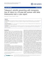
stippling. Bone marrow aspiration was not successful due
to dry tap while bone marrow biopsy showed grade 2 to 3
reticular fibrosis (Fig. 1). No JAK2 (Janus kinase 2)
mutations and cytogenetic abnormalities were detected.
Elevated levels of alkaline phosphatase (578 U/L) and
lactate dehydrogenase (507 U/L) were detected along with
a gradual one-month increase in tumor marker CA-125
(52.26 U/ml to 272.00 U/ml; reference range < 35 U/ml).
Thyroid function test and immunophenotyping revealed
slight hypothyroidism and suppression of cellular
© The Author(s). 2017 Open Access This article is distributed under the terms of the Creative Commons Attribution 4.0
International License ( which permits unrestricted use, distribution, and
reproduction in any medium, provided you give appropriate credit to the original author(s) and the source, provide a link to
the Creative Commons license, and indicate if changes were made. The Creative Commons Public Domain Dedication waiver
( applies to the data made available in this article, unless otherwise stated.
Guan et al. BMC Cancer (2017) 17:679
Page 2 of 4
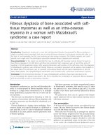
Fig. 1 Bone marrow biopsy before delivery. a HE (200×). b Fibrosis ++ ~ +++. Foot-Menard Stain (200×)
immunity respectively. Low-dose computed tomography
(CT) scan showed low density small areas in the left
insular lobe and besides left lateral ventricle angle, patchy
areas with uneven density in pelvis and spine, splenomegaly with some infarction and enlarged lymph
nodes around the stomach, and a little bit of the perioancreatic fat.
The patient was given supportive care to continue
pregnancy to 34 weeks. Three weeks later (the 31st week),
the patient complained of hematemesis accompanied by
unbearable abdominal pain and then a cesarean section
was operated. After that, positron emission tomographycomputed tomography (PET-CT) with 18F–FDG revealed
thickening of the gastric wall with accompanying increased uptake of glucose in the stomach and skeletal
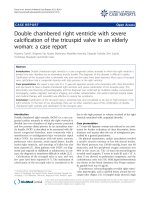
bones. Finally, a gastric biopsy was performed and the
patient was diagnosed with SRC (Fig. 2). A repeat bone
marrow biopsy revealed the presence of tumor metastasis
and confirmed the diagnosis of MF secondary to bone
marrow infiltration. The patient accepted palliative care
and died several months later.
Discussion
Pregnancy-associated cancer is a rare condition, with an
estimated incidence of 1:106 to 1:103 pregnancies, depending on the type of cancer [3]. Kaoru Sakamoto et al.
Fig. 2 a & b Bone marrow biopsy after delivery. a HE (200×). b The tumor cells are CK pan positive (brown region, CK pan: a broad spectrum
marker of epithelial cells; tumors which are originated from epithelium should be positive). (200×). c & d Gastric Mucosa Biopsy. c Signet ring cell
infiltrated into gastric mucosa. HE (40×). d Signet ring cell. HE (400×)
Guan et al. BMC Cancer (2017) 17:679
reported only 37 cases of pregnancy-associated gastric
cancer from 1988 to 2007 in Japan; with SRC accounting
for approximately 10% of cases [4]. We herein present
the first reported case of MF secondary to SRC during
pregnancy.
Primary MF is typically diagnosed in patients in their
fifth or sixth decades and shows a significant male predominance [5]. This 36-year old pregnant patient with
acute MF did not present with the cardinal signs of MF,
i.e., her spleen was not palpable (slight splenomegaly on
CT) and JAK2 mutational status was normal, thereby
ruling out primary MF. And it is noteworthy that nonspecific clinical features of gastric cancer can be masked
by pregnancy and easily ignored. Based on the patient’s
symptoms (vomiting and diarrhea) and an abnormal
stool guaiac test, we then screened for infectious and
metabolic causes to no avail but instead detected elevated levels of several tumor markers. The tumor
marker CA-125 was monitored weekly and found to be
increased from 52.26 U/ml to 272.00 U/ml within a
month. The most likely diagnosis was therefore MF
secondary to a gastrointestinal tumor.
Thus, the CT scan for brain, chest, abdomen and pelvis
was performed to screen for whether distant metastases
existed. However, pregnancy is a relative contraindication
to CT scans [6]. This case demonstrates the classic
maternal-fetal conflict. As physicians, we should balance
the interests of the mother and her fetus while determining the management strategy. A literature search reviewed
that the average uterine/fetal dose for chest CT
(0.17 mGy) and abdominal-pelvic CT (18-25 mGy) are
relatively low (less than 200-500 mGy) and are associated
with an acceptable risk of adverse radiobiological events,
although there is not a threshold dose for no injury [3]. A
lower-dose abdominal CT protocol was applied; the CT
dose index was 8.32 mGy (average of our center is
14 mGy) which might be lesser impact on the fetus wellbeing.
However, endoscopy was not performed accordingly
for this case during pregnancy. The previous study indicated that gastroscopy is innocuous and should be done
when clinically required during pregnancy, unless patients have obstetric complications such as placental
abruption, imminent delivery, ruptured membranes, or
eclampsia [3, 7]. This patient had thrombocytopenia
(platelet count persistently below 20 × 10^9/L), which is
a relative contraindication to gastroscopy. Additionally,
the patient refused to undergo gastroscopy without sedation because of pain intolerance. Painless gastroscopy
under sedation, on the other hand, would place endanger both the patient and her fetus due to maternal/fetal
hypoxia [8]. In order to protect the pregnant woman
and her fetus, endoscopy was delayed for 3 weeks after
parturition.
Page 3 of 4
Supportive therapy was finally applied, while abortion
was not practiced for this case. Although the decision to
continue on with the pregnancy was a tough one as the
patient’s condition was deteriorating, societal norms and
expectations shaped by cultural and religion play an
essential role in the decision-making process [9, 10].
Eventually, we managed the patient with supportive
therapies such as hemostasis, transfusion and nutritional
support up to the point of delivery and achieved an
acceptable outcome.
Conclusion
In general, we present a rare cause of MF secondary to
gastric SCR in a pregnant woman and provide both
clinical restrictions and ethical dilemmas surrounding
the management of this patient. The diagnosis may be
delayed as mild gastrointestinal symptoms are common
during pregnancy, while early detection of gastric cancer
is critical to ensure better outcomes.
Abbreviations
18
F–FDG: 2-Deoxy-2-fluoro-D-glucose; CT: Computed tomography;
JAK2: Janus kinase 2 gene; MF: Myelofibrosis; PET-CT: Positron emission
tomography–computed tomography; SRC: Signet ring adenocarcinoma
Acknowledgements
We appreciate Dr., Matthew Ho Zhi Guang, from School of Medicine,
University College Dublin, helped us modify the language of the paper.
Funding
This work was supported by the Research and Development Fund for
Hematopoietic Tumors, Chinese Anti-Cancer Association (312160342). The
funding body is not involved in the design of the study, collection and interpretation of data and writing the manuscript.
Availability of data and materials
All the data supporting the findings are presented within the manuscript.
Authors’ contributions
GPJ and CZH compiled all information relating to the patient and wrote the
manuscript. ZL and PL were involved in the treatment of the patient and
revised the manuscript. All authors read and approved the final manuscript.
Ethics approval and consent to participate
As it is a case report, ethics approval is not necessary after consulting the
Ethics Committee of West China Hospital.
Consent for publication
Patients’ written consent was obtained for publication of the case report.
Competing interests
The authors declare that they have no competing interests.
Publisher’s Note
Springer Nature remains neutral with regard to jurisdictional claims in
published maps and institutional affiliations.
Author details
1
Department of Hematology, West China Hospital, Sichuan University, No. 37
Guo-Xue Xiang, Chengdu, Sichuan 610041, China. 2Department of Radiology,
Huaxi Magnetic Resonance Research Centre (HMRRC), West China Hospital,
Sichuan University, No. 37 Guo-Xue Xiang, Chengdu, Sichuan 610041, China.
3
Department of Pathology, West China Hospital, Sichuan University, No. 37
Guo-Xue Xiang, Chengdu, Sichuan 610041, China.
Guan et al. BMC Cancer (2017) 17:679
Page 4 of 4
Received: 4 April 2017 Accepted: 28 September 2017
References
1. Thiele J, Kvasnicka HM. Myelofibrosis–what's in a name? Consensus on
definition and EUMNET grading. Pathobiology. 2007;74(2):89–96.
2. Devos T, Zachée P, Bron D, et al. Myelofibrosis patients in Belgium: disease
characteristics. Acta Clin Belg. 2015;70(2):105–11.
3. Pentheroudakis G, Pavlidis N. Cancer and pregnancy: poena magna, not
anymore. Eur J Cancer. 2006;42(2):126–40.
4. Sakamoto K, Kanda T, Ohashi M, et al. Management of patients with
pregnancy-associated gastric cancer in Japan: a mini-review. Int J Clin
Oncol. 2009;14(5):392–6.
5. Arber DA, Orazi A, Hasserjian R, et al. The 2016 revision to the World Health
Organization classification of myeloid neoplasms and acute leukemia. Blood.
2016;127(20):2391–405.
6. International Commission on Radiological Protection. Pregnancy and
medical radiation. Ann ICRP. 2000. 30(1): iii-viii, 1-43.
7. Shergill AK, Ben-Menachem T, Chandrasekhara V, et al. Guidelines for
endoscopy in pregnant and lactating women. Gastrointest Endosc.
2012;76(1):18–24.
8. Duncan PPW, Cohen M, et al. Foetal risk of anesthesia and surgery during
pregnancy. Anesthesiology. 1986;64(790):4.
9. Finnerty JJ, Pinkerton JV, Moreno J, Ferguson JE. Ethical theory and
principles: do they have any relevance to problems arising in everyday
practice. Am J Obstet Gynecol. 2000;183(2):301–6. discussion 306-8
10. Wallace R, Wiegand F, Warren C. Beneficence toward whom? Ethical
decision-making in a maternal-fetal conflict. AACN Clin Issues.
1997;8(4):586–94.
Submit your next manuscript to BioMed Central
and we will help you at every step:
• We accept pre-submission inquiries
• Our selector tool helps you to find the most relevant journal
• We provide round the clock customer support
• Convenient online submission
• Thorough peer review
• Inclusion in PubMed and all major indexing services
• Maximum visibility for your research
Submit your manuscript at
www.biomedcentral.com/submit