Evaluation of Slug expression is useful for predicting lymph node metastasis and survival in patients with gastric cancer
Bạn đang xem bản rút gọn của tài liệu. Xem và tải ngay bản đầy đủ của tài liệu tại đây (1.03 MB, 9 trang )
Lee et al. BMC Cancer (2017) 17:670
DOI 10.1186/s12885-017-3668-8
RESEARCH ARTICLE
Open Access
Evaluation of Slug expression is useful for
predicting lymph node metastasis and
survival in patients with gastric cancer
Han Hee Lee1, Sung Hak Lee2* , Kyo Young Song3*, Sae Jung Na4, Joo Hyun O5, Jae Myung Park1, Eun Sun Jung2,
Myung-Gyu Choi1 and Cho Hyun Park6
Abstract
Background: Slug is a transcription factor that activates the epithelial–mesenchymal transition (EMT) process in cancer
progression. The aim of our study was to evaluate the clinical significance of Slug expression in gastric cancer.
Methods: The expression of Slug in gastric cancer tissues of 456 patients who underwent gastrectomy was evaluated
by immunohistochemistry using tissue microarrays. Slug expression level was defined by the composite score
determined by multiplying the tumor staining scores for intensity and extent. The associations of Slug expression with
clinicopathological characteristics and overall and recurrence-free survival were analyzed.
Results: Patients were divided into three groups according to Slug composite score (≤4, 6, and 9). Low, mid, and high
expression of Slug was observed in 104 (22.7%), 130 (28.3%), and 225 (49.0%) of cases, respectively. Overall survival and
recurrence-free survival progressively increased from high to low Slug expression. In terms of lymph node metastasis, the
rate of positive lymph node metastasis was 38/104 (36.5%), 79/130 (60.8%), and 178/225 (79.1%) in low, mid, and high
Slug expression groups, respectively, displaying a tendency to increase with higher Slug expression. In a multivariate
analysis adjusting for patient age, tumor size, tumor depth, and histology, high Slug expression was associated with a
high rate of positive lymph node metastasis compared with low Slug expression (odds ratio 3.42; 95% confidence interval,
1.74–6.69). In a subgroup analysis of T1 cancer, patients with negative Slug expression (defined as <5% positive tumor
cells or no/weak staining) showed no lymph node metastasis (0/13), whereas those with positive Slug expression showed
15.9% (17/107) lymph node metastasis, with a negative predictive value of 100%.
Conclusions: High expression of Slug in gastric cancer tissue was associated with lymph node metastasis and poor survival.
Evaluation of Slug would be useful for discriminating patients at high risk of lymph node metastasis in early gastric cancer.
Keywords: Slug, Gastric cancer, Epithelial–mesenchymal transition, Tissue microarray, Prognosis
Background
Gastric cancer is the third leading cause of cancer death
worldwide, and almost 1 million new cases occur annually [1]. With the introduction of mass screening
methods such as endoscopy and upper gastrointestinal
series, the proportion of patients with early detection of
* Correspondence: ;
2
Department of Hospital Pathology, Seoul St. Mary’s Hospital, College of
Medicine, The Catholic University of Korea, 222, Banpo-daero, Seocho-gu,
Seoul 06591, Republic of Korea
3
Division of Gastrointestinal Surgery, Department of Surgery, Uijeongbu St.
Mary’s Hospital, College of Medicine, The Catholic University of Korea, Seoul,
Korea, 271, Cheonbo-ro Uijeongbu, Gyeonggi-do 480-717, Republic of Korea
Full list of author information is available at the end of the article
early gastric cancer (EGC) or precancerous adenoma has
been increasing [2, 3]. Endoscopic submucosal dissection
(ESD) has become the standard therapy for EGC
because it is minimally invasive and allows en bloc and
complete resection [4]. Recently, there has been an
attempt to expand the indications of ESD [5]. Along
with this, prediction of lymph node metastasis (LNM) in
EGC is becoming more important because LNM is one
of the most important factors for assessment of prognosis and decision of therapeutic modalities [6, 7]. Advanced gastric cancer (AGC) has a particularly poor
prognosis compared with EGC. AGC spreads locally by
breaking through the gastric wall into neighboring tissue
© The Author(s). 2017 Open Access This article is distributed under the terms of the Creative Commons Attribution 4.0
International License ( which permits unrestricted use, distribution, and
reproduction in any medium, provided you give appropriate credit to the original author(s) and the source, provide a link to
the Creative Commons license, and indicate if changes were made. The Creative Commons Public Domain Dedication waiver
( applies to the data made available in this article, unless otherwise stated.
Lee et al. BMC Cancer (2017) 17:670
and metastasizes to regional lymph nodes. The presence
of metastatic lymph nodes could be an outstanding
prognostic factor. Differences in the prognoses of
patients with negative lymph node metastasis versus
positive lymph node metastasis are especially robust in
surgically treated AGC [8–10].
Epithelial-mesenchymal transition (EMT) is a biologic
process by which epithelial cells lose their cell-cell junctions and apical-basal polarity and gain a highly motile
and invasive phenotype to become mesenchymal cells
[11]. EMT is integral to embryo formation and organ development [12] and has also been shown to occur during
wound healing and tissue fibrosis [13]. In cancer, EMT
contributes pathologically to cancer progression by enabling primary tumor cells to break through the basal
lamina and invade adjacent tissue, leading to tumor
metastasis [14].
Slug, also known as Snail2, is one of the key transcription factors that activate EMT process in cancer progression [15]. It contributes to repression of the epithelial
phonotype by binding to E-box DNA sequences in the
proximal promoter region of the E-cadherin gene [16–18].
This role as a strong E-cadherin repressor mediates loss of
tight junctions of epithelial cells and initiates EMT,
which facilitates cancer cell invasion and distant metastasis [18, 19]. Slug has been highly studied in various
cancers. In breast cancer patients, Slug is consistently
overexpressed in aggressive and basal-type breast tumors [20] and seems to be involved in breast tumorigenesis and metastasis through regulation of the EMT
[21]. It has also been demonstrated that Slug expression
is correlated with poor prognosis in pancreatic and
esophageal cancer patients [22, 23]. Recent studies have
revealed that Slug not only functions in cancer metastasis,
but also plays a role in cancer stemness [24, 25], implying
that Slug participates in early steps of cancer progression.
In gastric cancer, upregulation of Slug mRNA is associated with suppression of E-cadherin in intestinal and
diffuse type gastric carcinomas [26]. In a study focused
on protein expression, high Slug expression was correlated with advanced stages and worse clinical outcomes
[27]. However, there are only a few studies on the clinical significance of Slug in gastric cancer. In addition, the
significance of Slug expression in early gastric cancer has
not been proved.
Therefore, the purpose of our study was to evaluate
the clinical significance of Slug expression in gastric cancer using a tissue microarray method in a large series of
patients with resected gastric cancer.
Methods
Patients and clinical samples
A total of 459 patients (313 men and 146 women) were
randomly selected by random number generation from
Page 2 of 9
2495 consecutive patients with gastric cancer who had
undergone radical surgery at Seoul St. Mary’s Hospital,
The Catholic University of Korea, between 2000 and 2009.
Clinicopathological data were reviewed retrospectively
from the participants’ medical records and pathology reports at our institution. Variable factors including age,
gender, type of surgery, tumor size, location, pathologic
staging, histology, and lymphatic, venous, and perineural
invasion were analyzed. Tumor location was categorized
into upper, middle, and lower thirds of the stomach. The
gastric cancers were staged according to the pathological
tumor/node/metastasis (pTNM) classification (8th edition) of the Union for International Cancer Control [28].
The histological types of the gastric cancers were assessed
according to the 2010 World Health Organization classification [29]. Tumors were also classified into intestinal,
diffuse, and mixed types by Lauren classification [30].
Written informed consent was obtained from all patients.
Patient consent and specimen collection were conducted in accordance with protocols approved by the
Institutional Review Board of The Catholic University
of Korea (KC14SISI0158).
Tissue microarray construction and
immunohistochemistry
All gastric specimens were histologically reviewed, and tissue microarrays (TMAs) were constructed from each of
the formalin-fixed, paraffin-embedded (FFPE) tissue blocks
using a Manual Tissue Arrayer (Beecher Instruments, Sun
Prairie, WI, USA) with a 2.0-mm tip.
Immunohistochemical analysis was performed using
primary antibody against Slug (ab188875) (polyclonal;
1:150; Abcam, Cambridge, UK). We determined the optimal dilution of the Slug antibody using positive control
tissue such as normal gastric epithelial cells and placenta. Four-micrometer-thick tissue sections from the
TMA blocks were transferred to Probe On Plus slides
(Fisher Scientific, Pittsburgh, PA, USA) and baked for
2 h in a dry oven at 56 °C (Agilent Technologies, Santa
Clara, CA, USA). The FFPE sections were deparaffinized
in xylene three times and rehydrated through 100%, 90%,
80%, and 70% ethanol in Tris-buffered saline (pH 7.4).
Antigen retrieval was achieved by boiling in 10 mM sodium citrate buffer (pH 6.0) using a microwave oven for
20 min. After treatment with 3% H2O2 in phosphatebuffered saline, the tissues were incubated with primary
antibody at 4 °C overnight and then with diluted (1:100)
biotinylated anti-mouse antibody (Abnova, Walnut, CA,
USA) for 1 h at room temperature. The signal was amplified using diluted ExtrAvidin-peroxidase (1:50; SigmaAldrich, St. Louis, MO, USA) for 1 h at room temperature
and visualized using the liquid 3,3′-diaminobenzidine +
Substrate Chromogen system (Dako, Glostrup, Denmark).
Counterstaining was performed with hematoxylin.
Lee et al. BMC Cancer (2017) 17:670
Nonspecific staining was not observed in any negative
control sections.
Page 3 of 9
Table 1 Scoring methods of Slug expression
Measures
Number
Percent
0: negative (<5%)
15
3.3
1: sporadic (5–25%)
35
7.6
2: focal (25–50%)
141
30.7
3: diffuse (>50%)
268
58.4
0: no staining
2
0.4
1: weak staining
24
5.2
2: moderate staining
103
22.4
3: strong staining
330
71.9
Low (≤4)
104
22.7
Mid (6)
130
28.3
High (9)
225
49.0
Extent
Evaluation of immunohistochemical staining
Two pathologists (SH Lee and ES Jung) who were
blinded to the clinicopathological parameters independently reviewed the immunohistochemical staining for
the tissue sections. We used a semi-quantitative scoring
system based on the intensity and extent of stained cells
for each case. The staining intensity was graded from 0
to 3 (0 = no expression at all, 1 = weak, 2 = moderate,
3 = strong). The extent was graded from 0 to 3
(0 = <5%, 1 = 5–25%, 2 = 26–50%, 3 = >50%). The intensity scores and extent scores were multiplied to
obtain the composite score.
Intensity
Extent × Intensity
= Slug composite score (range 0–9)
Statistical analysis
Continuous data are presented as mean ± standard deviation, and categorical data are presented as quantity and
proportion. Pearson’s χ2 test for categorical variables and
Student’s t test for unpaired data for continuous variables were performed to compare clinicopathological
characteristics among the three Slug expression groups.
A P value <0.05 was considered significant. Survival
rates were calculated by the Kaplan–Meier method, with
the date of gastrectomy as the starting point. Patients
who were alive were censored at the time of the last
follow-up. Differences in survival were examined by the
log-rank test. Multivariable analysis was performed using
a Cox proportional hazards model with a backward
stepwise selection procedure. All analyses were performed by SAS for Windows software (version 8.02, SAS
Institute, Cary, NC, USA).
Results
Expression profile of Slug in gastric cancer
Table 1 shows overall immunohistochemical Slug expression in the gastric cancer tissue microarray. More
than half of the tissues showed diffuse Slug expression,
which corresponds to extent score 3, and 71.9% (330/
459) of tissues showed intensity score 3, indicating
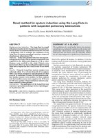
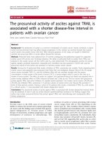
strong staining. Figure 1 shows representative images of
the range of Slug staining intensity. Multiplication of
these two variables yielded the Slug composite score,
which ranged from 0 to 9. Classification of the patients
according to Slug composite score yielded 104 (22.7%),
130 (28.3%), and 225 (49.0%) patients in the low, mid,
and high Slug groups, respectively.
Relationships between Slug expression and
clinicopathological parameters
Table 2 summarizes the clinicopathological characteristics of the 459 patients undergoing gastrectomy for gastric cancer. The mean age of the patients was 58.6 years
(range 23–86 years), and 68.2% (n = 313) were male.
Distal subtotal gastrectomy was the most commonly
performed surgery (63.2%). The high Slug group tended
to have large tumors and advanced tumor depth and
stages. They also had a high rate of positive perineural
invasion. Regarding histology, the proportion of poorly
differentiated adenocarcinoma tended to increase from
low to high Slug expression groups. However, the proportion of signet ring cell carcinoma was highest in the
low Slug group.
Slug expression and lymph node metastasis
The rate of positive lymph node metastasis was 36.5% in
the low group, 60.8% in the mid group, and 79.1% in the
high Slug expression group, thus displaying a tendency
to increase with increasing Slug expression (Table 3).
Positive lymph node ratio calculated by dividing number
of metastatic LNs by number of retrieved LNs was significantly higher in the high Slug group. The high Slug
group also showed a high proportion of positive lymphatic invasion.
In a multivariate logistic regression analysis for lymph
node metastasis, Slug composite score was identified as
an independent predictive factor for lymph node metastasis even after adjusting for age, tumor size, tumor
depth, and Lauren classification (Table 4). Compared
with patients with low Slug score, the adjusted odds ratio
in the high Slug group was 3.42 (95% confidence interval = 1.74–6.69). Tumor size and depth were also identified as predictive factors for lymph node metastasis.
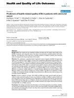
The recurrence rates of gastric cancer were compared
between the three Slug groups (Fig. 2). Patients with
high Slug score had the highest tumor recurrence rate.
Lee et al. BMC Cancer (2017) 17:670
Page 4 of 9
Fig. 1 Immunohistochemistry findings showing expression of Slug in gastric cancer tissue. a no staining. b weak staining. c moderate staining.
d strong staining
The rate of recurrence was significantly higher in the
high Slug group than in the low (P < 0.001) and mid
(P = 0.006) Slug groups. There was no statistically significant difference between the low and mid Slug groups
(P = 0.280).
The rate of lymph node metastasis in T1 tumor
was 14.2% (17/120). Patients with negative Slug expression showed no lymph node metastasis (0/13),
whereas those with positive Slug expression showed
15.9% (17/107) lymph node metastasis, with a negative predictive value of 100%.
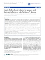
Slug expression and survival
Overall survival rates were determined with respect to
the Slug composite score using the log rank test (Fig. 3).
The 5-year overall survival rate was significantly worse
in the high Slug group compared with the mid (61.5%
versus 72.4%; P = 0.017) and low (61.5% versus 84.6%;
P < 0.001) Slug groups. The low Slug group had the best
5-year overall survival rate. In multivariable Cox regression analysis including age, gender, TNM stage, Lauren
classification, and Slug composite score, Slug score was
not significantly associated with overall survival, whereas
age and TNM stage remained independent prognostic
factors (Additional file 1: Table S1).
Subgroup analysis of T1 tumors
We conducted a subgroup analysis of T1 tumors
(Table 5). Negative Slug expression was defined as <5%
positive tumor cells or no/weak staining intensity.
Tumor depth and size were not significantly different
between negative and positive Slug expression. Approximately 60% of cases with negative Slug expression were
signet ring cell carcinoma.
Discussion
The present study aimed to determine the relationship
between Slug expression and prognosis in patients with
gastric cancer. High Slug expression according to our
composite score was observed in about 50% of gastric
cancer tissues. We demonstrated that the expression of
Slug is associated with tumor progression and poor
prognosis in gastric cancer. Especially, Slug expression
was highly correlated with various indicators reflecting
lymphatic progression such as lymph node metastasis,
lymphatic invasion, and positive lymph node ratio. As it
is reasonable to consider that advanced cancer has
greater migrating activity and invasiveness than EGC,
this finding supports the hypothesis that Slug, one of the
important EMT drivers, is involved in lymphatic metastasis of gastric cancer through the EMT process. In the
case of T1 tumor confirmed after surgical resection,
negative Slug expression might exclude lymph node metastasis of EGC.
To the best of our knowledge, there is only one previous study that investigated Slug protein expression in
Lee et al. BMC Cancer (2017) 17:670
Page 5 of 9
Table 2 Comparison of characteristics of the patients according to Slug composite score
Total patients (N = 459)
Low (n = 104)
Mid (n = 130)
High (n = 225)
P
Mean ± SD
58.6 ± 11.9
56.3 ± 12.3
58.9 ± 11.8
59.6 ± 11.6
0.064
Range
23–86
23–81
32–82
24–86
313 (68.2%)
65 (62.5%)
94 (72.3%)
154 (68.4%)
166 (36.2%)
30 (28.8%)
51 (39.2%)
85 (37.8%)
Measures
Age (years)
Male
0.276
Type of surgery
Total gastrectomy
Subtotal gastrectomy
290 (63.2%)
73 (70.2%)
78 (60.0%)
139 (61.8%)
Wedge resection
3 (0.7%)
1 (1.0%)
1 (0.8%)
1 (0.4%)
Mean ± SD
5.0 ± 2.9
4.1 ± 2.5
5.1 ± 3.3
5.4 ± 2.8
Range
0.2–19.0
0.4–12.5
0.5–19.0
0.2–15.5
Tumor size (cm)
0.001
Location
Upper third
80 (17.4%)
16 (15.4%)
22 (16.9%)
42 (18.7%)
Middle third
164 (35.7%)
43 (41.3%)
50 (38.5%)
71 (31.6%)
Lower third
206 (44.9%)
44 (42.3%)
55 (42.3%)
107 (47.6%)
Whole stomach
9 (2.0%)
1 (1.0%)
3 (2.3%)
5 (2.2%)
120 (26.1%)
56 (53.8%)
30 (23.1%)
34 (15.1%)
0.599
Tumor depth (pT)
T1
T2
62 (13.5%)
18 (17.3%)
26 (20.0%)
18 (8.0%)
T3
121 (26.4%)
14 (13.5%)
35 (26.9%)
72 (32.0%)
T4
156 (34.0%)
16 (15.4%)
39 (30.0%)
101 (44.9%)
<0.001
TNM Stage
I
131 (28.5%)
62 (59.6%)
37 (28.5%)
32 (14.2%)
II
122 (26.6%)
25 (24.0%)
45 (34.6%)
52 (23.1%)
III
206 (44.9%)
17 (16.3%)
48 (36.9%)
141 (62.7%)
<0.001
Venous invasiona
Negative
406 (88.5%)
97 (94.2%)
117 (90.0%)
192 (85.3%)
Positive
52 (11.3%)
6 (5.8%)
13 (10.0%)
33 (14.7%)
Negative
270 (58.8%)
81 (77.9%)
75 (57.7%)
114 (50.7%)
Positive
189 (41.2%)
23 (22.1%)
55 (42.3%)
111 (49.3%)
0.055
Perineural invasion
<0.001
Histology
0.005b
Adenocarcinoma
Well differentiated
38 (8.3%)
12 (11.5%)
5 (3.8%)
21 (9.3%)
Moderately differentiated
136 (29.6%)
23 (22.1%)
44 (33.8%)
69 (30.7%)
Poorly differentiated
189 (41.2%)
35 (33.7%)
53 (40.8%)
101 (44.9%)
Mucinous adenocarcinoma
19 (4.1%)
5 (4.8%)
7 (5.4%)
7 (3.1%)
Signet ring cell carcinoma
77 (16.8%)
29 (27.9%)
21 (16.2%)
27 (12.0%)
174 (37.9%)
40 (38.5%)
47 (36.2%)
87 (38.7%)
Lauren classification
Intestinal
Diffuse
177 (38.6%)
40 (38.5%)
58 (44.6%)
79 (35.1%)
Mixed
108 (23.5%)
24 (23.1%)
25 (19.2%)
59 (26.2%)
Where appropriate, data are shown as the mean ± SD
a
Lymphatic and venous invasion could not be evaluated in 2 and 1 cases, respectively
b
Linear-by-linear association
0.433
Lee et al. BMC Cancer (2017) 17:670
Page 6 of 9
Table 3 Association of lymphatic metastasis and Slug expression
Total patients (N = 459)
Low (n = 104)
Mid (n = 130)
High (n = 225)
P
164 (35.7%)
66 (63.5%)
51 (39.2%)
47 (20.9%)
<0.001
Positive
295 (64.3%)
38 (36.5%)
79 (60.8%)
178 (79.1%)
N1
98 (21.4%)
20 (19.2%)
30 (23.1%)
48 (21.3%)
N2
99 (21.6%)
15 (14.4%)
26 (20.0%)
58 (25.8%)
N3
98 (21.4%)
3 (2.9%)
23 (17.7%)
72 (32.0%)
Measures
Lymph node metastasis (pN)
Negative
N3a
94 (20.5%)
3 (2.9%)
20 (15.4%)
71 (31.6%)
N3b
4 (0.9%)
0 (0.0%)
3 (2.3%)
1 (0.4%)
Number of metastatic lymph nodes
3.7 ± 4.9 (0–42)
1.2 ± 2.3 (0–12)
3.6 ± 5.8 (0–42)
4.9 ± 4.7 (0–25)
Number of retrieved lymph nodes
42.4 ± 15.4 (6–106)
39.5 ± 13.3 (14–78)
44.9 ± 16.1* (8–97)
42.4 ± 15.8 (6–106)
<0.001
Positive lymph node ratio
0.09 ± 0.12
0.03 ± 0.06
0.08 ± 0.12
0.12 ± 0.13
<0.001
<0.001
0.028
Lymphatic invasiona
Negative
154 (33.6%)
59 (57.3%)
45 (34.9%)
50 (22.2%)
Positive
303 (66.0%)
44 (42.7%)
84 (65.1%)
175 (77.8%)
p < 0.05; when compared with “low Slug composite score group” using the ANOVA test with post-hoc Tukey-HSD test
Lymphatic invasion could not be evaluated in 2 cases
*
a
gastric cancer tissues by immunohistochemical staining
[27]. In that study, 30% of tissues showed positive Slug
expression, defined as detectable immunoreaction in the
perinuclear and other cytoplasmic regions of more than
10% of the cancer cells. This is in contrast to findings
from the current study showing that about 75% of gastric cancer patients had mid to high Slug expression. A
possible explanation for this finding is that many more
advanced cancers were included in our study compared
to the previous study; approximately 60% of patients in
the previous study were stage I, compared with only
about 30% in our study. In another previous study based
on mRNA expression of Slug by real-time quantitative
RT-PCR, 58% of gastric cancer patients showed Slug upregulation in the tumor, which is in close agreement
with our finding [26]. Moreover, the tendency for Slug
expression to be associated with advanced pTNM stages
was observed in both studies [26, 27]. The correlation of
Slug expression with increased tumor size and perineural
invasion was newly identified in the present study.
We focused on the association of lymphatic metastasis
and Slug expression because Slug can activate the EMT
Table 4 Multivariate analysis showing independence of the effect on lymph node metastasis
Odds ratio
95% CI
P
Age
1.01
0.99–1.04
0.277
Tumor size
1.12
1.00–1.25
0.049
Number of patients
Tumor depth (pT)
T1
120 (26.1%)
1 (ref)
T2
62 (13.5%)
17.14
7.70–38.17
<0.001
T3
121 (26.4%)
33.99
14.87–77.71
<0.001
T4
156 (34.0%)
13.35
6.22–28.64
<0.001
0.825
Lauren classification
Intestinal
174 (37.9%)
1 (ref)
Diffuse + Mixed
285 (62.1%)
1.07
0.61–1.88
104 (22.7%)
1 (ref)
1.09–1.76
Slug composite score
Low
Mid
130 (28.3%)
1.33
0.67–2.63
0.413
High
225 (49.0%)
3.42
1.74–6.69
<0.001
Lee et al. BMC Cancer (2017) 17:670
Fig. 2 Cumulative recurrence rates according to Slug expression
after gastrectomy
process. For this purpose, we used the Slug composite
score to produce a more continuous scale (low, mid, and
high Slug groups) instead of dichotomizing the patient
groups. As expected, higher Slug expression was associated with more prevalent lymph node metastasis and
lymphatic invasion. In addition, the positive lymph node
ratio gradually increased with increasing Slug score. This
ratio represents lymph node metastasis density [31]. Much
study has focused on this ratio because it has global
Fig. 3 Overall survival according to Slug expression after gastrectomy
Page 7 of 9
prognostic relevance in gastric cancer regardless of stage
in multivariable analysis and is more sophisticated than
conventional nodal metastasis in TNM staging for predicting prognosis [32]. In addition, we demonstrated that
Slug expression is an independent prognostic factor for
lymph node metastasis in gastric cancer patients even
after adjustment for well-known prognostic factors including tumor size and depth of tumor invasion.
The current study indicates that Slug expression correlates well with overall survival as well as tumor recurrence. The high Slug expression group had the worst
long-term survival rate and the highest tumor recurrence rate. These results correspond well with previous
studies, in which positive Slug expression was associated
with distant metastasis and poor postoperative 5-year
survival [26, 27]. To our knowledge, this is the first report of long-term survival and recurrence data according
to Slug expression and suggests that EMT signaling with
involvement of Slug could affect long-term prognosis
after gastrectomy of gastric cancer patients.
In a subgroup analysis of T1 tumors, we documented
that Slug expression is associated with unexpected lymph
node metastasis in EGC. EGC is defined as gastric cancer
that invades no more deeply than the submucosa,
irrespective of lymph node metastasis [33]. It has been reported that about 10–15% of patients with EGC have
lymph node metastasis [1, 34, 35]. Precise prediction of
lymph node metastasis status in EGC is a very important
issue because ESD has become increasingly popular as a
minimally invasive treatment for EGC [36]. We applied
strict criteria for negative Slug expression in order to increase the negative predictive value because false negative
results could be fatal when making the decision between
surgical resection and ESD. In our study, all patients with
T1 tumor and negative Slug expression showed no lymph
node metastasis even though some of them had submucosal tumor invasion (T1b) or undifferentiated (poorly
differentiated or signet ring cell) type histology. Tumor
depth beyond submucosa and histological differentiation
are well known independent risk factors for lymph node
metastasis of EGC [37, 38]. Interestingly, 8 of 13 Slug
negative T1 tumors were signet ring cell cancer. A previous study showed that signet ring foci of 8 patients with
hereditary diffuse gastric cancer had a low proliferative
index and there was no evidence for EMT [39]. This finding corresponds well with our result.
Our study has some strengths. First, a relatively large
number of patients were randomly selected from consecutive patients undergoing surgery for gastric cancer
for TMA and analyzed. Moreover, we present a novel
finding regarding greater than 5-year survival and tumor
recurrence according to Slug expression. In addition, this
is the first report to document the significance of Slug
expression in EGC.
Lee et al. BMC Cancer (2017) 17:670
Page 8 of 9
Table 5 Subgroup analysis of T1 tumor according to Slug expression
Total
Slug expression
P
Negative (n = 13)
Positive (n = 107)
Tumor depth
T1a
56
6 (46.2%)
50 (46.7%)
T1b
64
7 (53.8%)
57 (53.5%)
3.0 ± 1.9
2.7 ± 2.0
3.0 ± 1.9
0.670
Adenocarcinoma, WD
25
0 (0.0%)
25 (23.4%)
0.001a
Adenocarcinoma, MD
37
3 (23.1%)
34 (31.8%)
Tumor size
0.969
Histology
Adenocarcinoma, PD
31
2 (15.4%)
29 (27.1%)
Signet ring cell cancer
27
8 (61.5%)
19 (17.8%)
Negative
103
13 (100%)
90 (84.1%)
Positive
17
0 (0.0%)
17 (15.9%)
Lymph node metastasis
0.210b
WD well differentiated, MD moderately differentiated, PD poorly differentiated
a
Linear-by-linear association
b
Fisher’s exact test
Conclusions
Our data demonstrated that high expression of Slug in gastric cancer tissue was associated with higher tumor recurrence rate and poor long-term survival. In particular, in
cases with lymph node metastasis Slug expression was an
independent predictive factor regardless of tumor size or
depth of tumor invasion. Negative Slug expression showed
high negative predictive value for lymph node metastasis
in EGC, which could have potential for future use in discriminating patients with EGC at high risk of lymph node
metastasis.
Additional file
Additional file 1: Table S1. Multivariate analysis showing independence
of the effect on overall mortality. (DOCX 15 kb)
Abbreviations
AGC: Advanced gastric cancer; EGC: Early gastric cancer; EMT: Epithelial–
mesenchymal transition; ESD: Endoscopic submucosal dissection;
FFPE: Formalin-fixed, paraffin-embedded; LNM: Lymph node metastasis;
pTNM: Pathological tumor/node/metastasis; TMAs: Tissue microarrays
Acknowledgements
Not applicable
Funding
This study was supported by Research Fund of College of Medicine, The
Catholic University of Korea, by a program of Catholic Medical Center Research
Foundation made in 2014, and by the National Research Foundation of Korea
(NRF) grant funded by the Korea government (Ministry of Science, ICT & Future
Planning) (NRF-2016R1C1B2015185).
Availability of data and materials
Attributing to the privacy of patients, the patient information is publicly
inaccessible.
Authors’ contributions
HHL participated in the study design, performed the data collection and chart
review, and drafted the manuscript with help of other authors. SHL participated in
planning the study, performed histological examination of the samples, evaluated
IHC results and helped finalize the manuscript. KYS designed the treatment
protocol, performed the surgeries, and helped finalize the manuscript. SJN and
JHO participated in the study design. JMP participated in the study design and
helped finalize the manuscript. ESJ performed the pathological studies and made
pathology pictures. MGC participated in planning the study and helped to gather
clinical data. CHP participated in planning the study and performed the surgeries.
All authors have read and approved the final version of this manuscript.
Ethics approval and consent to participate
The study with human samples was approved by the Ethics Committee of
the Catholic University of Korea (KC14SISI0158). We clarify that all clinical
samples described here were gained from patients who had given written
informed consent.
Consent for publication
Not applicable
Competing interests
The authors declare that they have no competing interests.
Publisher’s Note
Springer Nature remains neutral with regard to jurisdictional claims in
published maps and institutional affiliations.
Author details
1
Department of Internal Medicine, The Catholic University of Korea, Seoul St.
Mary’s Hospital, Seoul, Korea. 2Department of Hospital Pathology, Seoul St.
Mary’s Hospital, College of Medicine, The Catholic University of Korea, 222,
Banpo-daero, Seocho-gu, Seoul 06591, Republic of Korea. 3Division of
Gastrointestinal Surgery, Department of Surgery, Uijeongbu St. Mary’s
Hospital, College of Medicine, The Catholic University of Korea, Seoul, Korea,
271, Cheonbo-ro Uijeongbu, Gyeonggi-do 480-717, Republic of Korea.
4
Department of Radiology, The Catholic University of Korea, Uijeongbu St.
Mary’s Hospital, Uijeongbu, Korea. 5Department of Radiology, The Catholic
University of Korea, Seoul St. Mary’s Hospital, Seoul, Korea. 6Department of
Surgery, The Catholic University of Korea, Seoul St. Mary’s Hospital, Seoul,
Korea.
Lee et al. BMC Cancer (2017) 17:670
Received: 31 May 2017 Accepted: 28 September 2017
References
1. Ferlay J, Soerjomataram I, Dikshit R, et al. Cancer incidence and mortality
worldwide: sources, methods and major patterns in GLOBOCAN 2012.
Int J Cancer. 2015;136(5):E359–86.
2. Mizoue T, Yoshimura T, Tokui N, et al. Prospective study of screening for
stomach cancer in Japan. Int J Cancer. 2003;106(1):103–7.
3. Suh M, Choi KS, Lee YY, et al. Cancer screening in Korea, 2012: results from
the Korean National Cancer Screening Survey. Asian Pac J Cancer Prev.
2013;14(11):6459–63.
4. Isomoto H, Shikuwa S, Yamaguchi N, et al. Endoscopic submucosal
dissection for early gastric cancer: a large-scale feasibility study. Gut. 2009;
58(3):331–6.
5. Gotoda T, Yanagisawa A, Sasako M, et al. Incidence of lymph node
metastasis from early gastric cancer: estimation with a large number of
cases at two large centers. Gastric Cancer. 2000;3(4):219–25.
6. Kunisaki C, Akiyama H, Nomura M, et al. Significance of long-term follow-up
of early gastric cancer. Ann Surg Oncol. 2006;13(3):363–9.
7. Ye BD, Kim SG, Lee JY, et al. Predictive factors for lymph node metastasis
and endoscopic treatment strategies for undifferentiated early gastric
cancer. J Gastroenterol Hepatol. 2008;23(1):46–50.
8. Yamashita K, Sakuramoto S, Kikuchi S, et al. Validation of staging systems for
gastric cancer. Gastric Cancer. 2008;11(2):111–8.
9. Kikuchi S, Futawatari N, Sakuramoto S, et al. Comparison of staging
between the old (6th edition) and new (7th edition) TNM classifications in
advanced gastric cancer. Anticancer Res. 2011;31(6):2361–5.
10. Nashimoto A, Akazawa K, Isobe Y, et al. Gastric cancer treated in 2002 in
Japan: 2009 annual report of the JGCA nationwide registry. Gastric Cancer.
2013;16(1):1–27.
11. Lamouille S, Xu J, Derynck R. Molecular mechanisms of epithelialmesenchymal transition. Nat Rev Mol Cell Biol. 2014;15(3):178–96.
12. Thiery JP, Acloque H, Huang RY, et al. Epithelial-mesenchymal transitions in
development and disease. Cell. 2009;139(5):871–90.
13. Kalluri R, Weinberg RA. The basics of epithelial-mesenchymal transition.
J Clin Invest. 2009;119(6):1420–8.
14. Tam WL, Weinberg RA. The epigenetics of epithelial-mesenchymal plasticity
in cancer. Nat Med. 2013;19(11):1438–49.
15. Barrallo-Gimeno A, Nieto MA. The Snail genes as inducers of cell movement
and survival: implications in development and cancer. Development. 2005;
132(14):3151–61.
16. Peinado H, Olmeda D, Cano A. Snail, Zeb and bHLH factors in tumour
progression: an alliance against the epithelial phenotype? Nat Rev Cancer.
2007;7(6):415–28.
17. Hajra KM, Chen DY, Fearon ER. The SLUG zinc-finger protein represses Ecadherin in breast cancer. Cancer Res. 2002;62(6):1613–8.
18. Bolos V, Peinado H, Perez-Moreno MA, et al. The transcription factor Slug
represses E-cadherin expression and induces epithelial to mesenchymal
transitions: a comparison with Snail and E47 repressors. J Cell Sci. 2003;
116(Pt 3):499–511.
19. Gupta PB, Kuperwasser C, Brunet JP, et al. The melanocyte differentiation
program predisposes to metastasis after neoplastic transformation. Nat
Genet. 2005;37(10):1047–54.
20. Phillips S, Kuperwasser CSLUG. Critical regulator of epithelial cell identity in
breast development and cancer. Cell Adhes Migr. 2014;8(6):578–87.
21. Liu T, Zhang X, Shang M, et al. Dysregulated expression of Slug, vimentin,
and E-cadherin correlates with poor clinical outcome in patients with basallike breast cancer. J Surg Oncol. 2013;107(2):188–94.
22. Hotz B, Arndt M, Dullat S, et al. Epithelial to mesenchymal transition:
expression of the regulators snail, slug, and twist in pancreatic cancer. Clin
Cancer Res. 2007;13(16):4769–76.
23. Uchikado Y, Natsugoe S, Okumura H, et al. Slug Expression in the E-cadherin
preserved tumors is related to prognosis in patients with esophageal
squamous cell carcinoma. Clin Cancer Res. 2005;11(3):1174–80.
24. Storci G, Sansone P, Trere D, et al. The basal-like breast carcinoma phenotype is
regulated by SLUG gene expression. J Pathol. 2008;214(1):25–37.
25. Yao C, Su L, Shan J, et al. IGF/STAT3/NANOG/Slug Signaling Axis
Simultaneously Controls Epithelial-Mesenchymal Transition and Stemness
Maintenance in Colorectal Cancer. Stem Cells. 2016;34(4):820–31.
Page 9 of 9
26. Castro Alves C, Rosivatz E, Schott C, et al. Slug is overexpressed in gastric
carcinomas and may act synergistically with SIP1 and Snail in the downregulation of E-cadherin. J Pathol. 2007;211(5):507–15.
27. Uchikado Y, Okumura H, Ishigami S, et al. Increased Slug and decreased Ecadherin expression is related to poor prognosis in patients with gastric
cancer. Gastric Cancer. 2011;14(1):41–9.
28. Washington K. 7th edition of the AJCC cancer staging manual: stomach.
Ann Surg Oncol. 2010;17(12):3077–9.
29. Hu B, El Hajj N, Sittler S, et al. Gastric cancer: Classification, histology and
application of molecular pathology. J Gastrointest Oncol. 2012;3(3):251–61.
30. Lauren P. The two histological main types of gastric carcinoma: diffuse and
so-called intestinal-type carcinoma. An attempt at a Histo-clinical
classification. Acta Pathol Microbiol Scand. 1965;64:31–49.
31. Yamashita K, Ooki A, Sakuramoto S, et al. Lymph node metastasis density
(ND)-factor association with malignant degree and ND40 as “non-curative
factor” in gastric cancer. Anticancer Res. 2008;28(1b):435–41.
32. Yamashita K, Hosoda K, Ema A, et al. Lymph node ratio as a novel and
simple prognostic factor in advanced gastric cancer. Eur J Surg Oncol. 2016;
42(9):1253–60.
33. Sano T, Aiko T. New Japanese classifications and treatment guidelines for
gastric cancer: revision concepts and major revised points. Gastric Cancer.
2011;14(2):97–100.
34. Roviello F, Rossi S, Marrelli D, et al. Number of lymph node metastases and
its prognostic significance in early gastric cancer: a multicenter Italian study.
J Surg Oncol. 2006;94(4):275–80. discussion 4
35. Pelz J, Merkel S, Horbach T, et al. Determination of nodal status and
treatment in early gastric cancer. Eur J Surg Oncol. 2004;30(9):935–41.
36. Soetikno R, Kaltenbach T, Yeh R, et al. Endoscopic mucosal resection for
early cancers of the upper gastrointestinal tract. J Clin Oncol. 2005;23(20):
4490–8.
37. Hyung WJ, Cheong JH, Kim J, et al. Application of minimally invasive treatment
for early gastric cancer. J Surg Oncol. 2004;85(4):181–5. discussion 6
38. Kunisaki C, Takahashi M, Nagahori Y, et al. Risk factors for lymph node
metastasis in histologically poorly differentiated type early gastric cancer.
Endoscopy. 2009;41(6):498–503.
39. Barber ME, Save V, Carneiro F, et al. Histopathological and molecular
analysis of gastrectomy specimens from hereditary diffuse gastric cancer
patients has implications for endoscopic surveillance of individuals at risk.
J Pathol. 2008;216(3):286–94.
Submit your next manuscript to BioMed Central
and we will help you at every step:
• We accept pre-submission inquiries
• Our selector tool helps you to find the most relevant journal
• We provide round the clock customer support
• Convenient online submission
• Thorough peer review
• Inclusion in PubMed and all major indexing services
• Maximum visibility for your research
Submit your manuscript at
www.biomedcentral.com/submit