Prospective study of 11C–methionine PET for distinguishing between recurrent brain metastases and radiation necrosis: Limitations of diagnostic accuracy and long-term results of salvage
Bạn đang xem bản rút gọn của tài liệu. Xem và tải ngay bản đầy đủ của tài liệu tại đây (742.66 KB, 9 trang )
Yomo and Oguchi BMC Cancer (2017) 17:713
DOI 10.1186/s12885-017-3702-x
RESEARCH ARTICLE
Open Access
Prospective study of 11C–methionine
PET for distinguishing between recurrent
brain metastases and radiation
necrosis: limitations of diagnostic accuracy
and long-term results of salvage treatment
Shoji Yomo1*
and Kazuhiro Oguchi2
Abstract
Background: On conventional diagnostic imaging, the features of radiation necrosis (RN) are similar to those of
local recurrence (LR) of brain metastases (BM). 11C–methionine positron emission tomography (MET-PET) is
reportedly useful for making a differential diagnosis between LR and RN. In this prospective study, we aimed to
investigate the diagnostic performance of MET-PET and the long-term results of subsequent patient management.
Methods: The eligible subjects had enlarging contrast-enhanced lesions (>1 cm) on MR imaging after any form of
radiotherapy for BM, suggesting LR or RN. However, it was difficult to differentiate LR from RN in these cases. From
August 2013 to February 2017, MET-PET was performed for 37 lesions in 32 eligible patients. Tracer accumulation in
the regions of interest was analysed as the standardised uptake value (SUV) and maximal lesion SUV/maximal
normal tissue SUV ratios (LNR) were calculated. The cut-off value for LNR was provisionally set at 1.40. Salvage
treatment strategies determined based on MET-PET diagnosis and treatment results were investigated. The
diagnostic accuracy of MET-PET was evaluated by receiver operating characteristic (ROC) curve analysis.
Results: The median interval from primary radiotherapy to MET-PET was 19 months and radiotherapy had been
performed twice or more for 13 lesions. The MET-PET diagnoses were LR in 19 and RN in 18 lesions. The mean values
and standard deviation of LNRs for each diagnostic category were 1.70 ± 0.30 and 1.09 ± 0.25, respectively. At the
median follow-up time of 18 months, final diagnoses were confirmed histologically for 17 lesions and clinically for 20
lesions. ROC curve analysis indicated the optimal LNR cut-off value to be 1.40 (area under the curve: 0.84), and the
sensitivity and specificity were 0.82 and 0.75, respectively. The median survival times of patient groups with LR and RN
based on MET-PET diagnosis were 14.8 months and 35.1 months, respectively (P = 0.035, log-rank test).
Conclusions: MET-PET showed apparently reliable diagnostic performance for distinguishing between LR and RN. The
provisional LNR cut-off value of 1.4 in our institution was found to be appropriate. Limitations of diagnostic accuracy
should be recognised in cases with LNR close to this cut-off value.
Keywords: 11C–methionine, Positron emission tomography, Brain metastases, Radiation necrosis, Local recurrence
* Correspondence:
1
Division of Radiation Oncology, Aizawa Comprehensive Cancer Center,
Aizawa Hospital, 2-5-1, Honjo, Matsumoto-city, Nagano-prefecturem, Japan
Full list of author information is available at the end of the article
© The Author(s). 2017 Open Access This article is distributed under the terms of the Creative Commons Attribution 4.0
International License ( which permits unrestricted use, distribution, and
reproduction in any medium, provided you give appropriate credit to the original author(s) and the source, provide a link to
the Creative Commons license, and indicate if changes were made. The Creative Commons Public Domain Dedication waiver
( applies to the data made available in this article, unless otherwise stated.
Yomo and Oguchi BMC Cancer (2017) 17:713
Background
The management of patients with brain metastases (BM)
has recently become more important because of the
increased incidence of these tumors and the prolonged
patient survival times that have accompanied improved
control of systemic cancers [1–3]. Gadolinium (Gd)-enhanced magnetic resonance (MR) imaging has become a
preferred imaging modality not only for early detection
of BM but also for evaluation of the efficacy of radiotherapy for BM. Local changes in the area of irradiation
application at follow-up, however, are not uncommonly seen on Gd-enhanced and T2-weighted MR
imaging [4, 5]. The interpretation of such changes is
often difficult and it may even be impossible to differentiate radiation-induced changes from local tumor
recurrence [6], which poses a critical dilemma in
decision-making for subsequent treatment.
Amino acid tracers such as 11C- methionine (MET)
are reportedly useful for positron emission tomography
(PET), particularly in the field of neuro-oncology, because of high amino acid uptake by tumor tissue with
low uptake by normal brain tissue, resulting in an enhanced tumor-to-background contrast [7, 8]. MET-PET
studies in primary brain tumors, especially gliomas, have
provided promising results, leading to an increase in
investigations in the twenty-first century [9, 10]. In contrast, there are few reported evaluations of MET-PET for
the imaging of BM [11–14]. Most previous studies investigated imaging changes within already treated BM by
focusing on assessment of the diagnostic accuracy of the
imaging modalities using receiver-operating characteristic (ROC) curve analysis [11, 12, 14].
The present study aimed to document our early experience with clinical use of MET-PET for distinguishing
radiation-induced changes from local tumor recurrence,
and to describe in detail the long-term clinical results of
modern salvage management based on MET-PET diagnosis. Thus, the diagnostic value and clinical utility of
MET-PET imaging for managing patients with BM were
critically appraised.
Methods
Patient eligibility
The present study was conducted in compliance with
the Declaration of Helsinki (sixth revision, 2008), and
fulfilled all of the requirements for patient anonymity.
The Aizawa Hospital Institutional Review Board (IRB)
approved this single center prospective clinical study in
July 2013 (No. 2013–049). Written permission was obtained prior to MET-PET from all patients and/or their
relatives, allowing the use of personal data for clinical
research. Patient records and information were anonymised and de-identified prior to analysis.
Page 2 of 9
The study candidates were limited to patients with
BM. Malignant gliomas were excluded from the present
study due to the possibility of there being a difference in
optimal cut-off values between BM and malignant gliomas [12, 15]. As the routine imaging protocol in our
institution, 3-dimensional volumetric gadoliniumenhanced T1-weighted MR images and T2-weighted MR
images were obtained for both radiotherapeutic intervention and follow-up imaging studies. In the course of
follow-up for BM treated with any type of radiotherapy,
including conventional fractionated radiotherapy, stereotactic radiosurgery and particle therapy, lesions with
continuous enlargement of Gd-enhanced areas documented on serial MR scans and suspected to be local recurrence (LR) or radiation necrosis (RN), which are
difficult to differentiate from each other, were studied
using MET-PET. The maximal diameter of a Gdenhanced area had to be at least 10 mm in order to exclude the possibility of false negative diagnostic errors
due to the relatively low spatial resolution of MET-PET.
The lesions in which neither LR nor RN could be definitively diagnosed because of insufficient follow-up data
were excluded from the present study.
MET-PET imaging
MET-PET was performed with a Discovery PET/CT 600
scanner (GE Healthcare, Milwaukee, USA) with a spatial
resolution of 5.1 mm full width at half maximum. After
intravenous injection of about 370 MBq of 11C–methionine, patients were placed in the scanner to assure that
slices parallel to the orbitomeatal line could be obtained.
After a transmission CT scan had been obtained, a 10min static emission scan was begun 20 min after the
injection. PET images were reconstructed by CT attenuation correction and a 3D ordered subset expected
maximisation algorithm (iteration 3, subset 16, field of
view 25.6 cm, matrix size of 128 × 128 and slice thickness 3.27 mm).
MET-PET interpretation
The region of interest (ROI) for lesions was manually located over the area corresponding to the Gd-enhanced
area on the MR images. As a normal control, a circular
ROI with a diameter of 10 mm was located within the
gray matter of the corresponding contralateral side. The
quantitative analysis was performed as follows. The maximum standardised uptake values (SUVmax) within the
suspected lesion and within the normal control were
measured. The lesion/normal ratios (LNR) were calculated by dividing the SUVmax of the lesion by the SUVmax of the normal control in order to give priority to
detection of a subtle LR mixed with RN. All scans were
assessed by an experienced, board-certified, nuclear
medicine physician (KO), not involved in any of the
Yomo and Oguchi BMC Cancer (2017) 17:713
Page 3 of 9
treatments for systemic cancer and BM. The cut-off
value of the LNR for diagnosis was provisionally set for
1.4, in accordance with previous studies [11, 12, 14]. A
LNR exceeding 1.4 was considered to represent LR, a
value below 1.4 to mean that the lesion was RN.
patients who had not visited our outpatient department
for more than three months. Inquiries about the latest
clinical and neuroimaging results and the date and mode
of death were made by directly corresponding with the
referring physicians and/or the families of deceased patients, with written permission obtained at the time of
undertaking MET-PET.
Subsequent management and follow-up
According to the MET-PET diagnosis, subsequent management was determined by a multidisciplinary team in
consideration of other clinical factors such as the patient’s age and performance status as well as the anatomical location of the lesion of interest (surgically
accessible or not). Details of subsequent management
and results were recorded (Fig. 1).
Final clinical diagnoses were determined from surgical
specimens, sequential neuroimaging changes and the
long-term clinical course secondary to salvage treatment.
Shrinkage of the lesion confirmed radiologically after
salvage radiotherapy was regarded as LR. A lesion that
either remained stable or showed spontaneous shrinkage
with no additional treatment on MR imaging follow-up
was assumed to be RN. A lesion in which the MET-PET
diagnosis could not be confirmed even after adequate
follow-up data had been obtained was regarded as a
diagnostic failure given the study aim of critical appraisal
of MET-PET.
Before closing the research database for analysis in
April 2017, the authors updated the follow-up data of
Statistical analysis
Patient characteristics were compared using Fisher’s exact
test for categorical variables and the Mann–Whitney U
test for quantitative variables. Receiver operating characteristic (ROC) curve analysis was performed to evaluate
the diagnostic capability of MET-PET for differentiating
between LR and RN and to determine the optimal cut-off
value in our institution, with the weights of false negative
and positive classifications being equivalent. The overall
survival rates were calculated by the Kaplan-Meier product limit method, based on the interval from the date of
MET-PET until the event date. The overall survival of
each patient group according to MET-PET diagnosis was
compared by log-rank test, wherein a patient with both
LR and RN was assigned to the LR group. Proportional
hazards regression analysis was not performed in the
present study because a too-small ratio of events per variable can lead to inaccurate regression estimates [16].
The statistical processing software package “R” version
3.0.1 (The R Foundation for Statistical Computing,
MET-PET
(n = 37)
LNR 1.4
(n = 19)
Observation
(n = 2)
Stabilized
(n = 1)
Progressed
(n = 1)
Salvage SRS
(n = 8)
Response +
(n = 5)
BV rescue
(n = 2)
Resection
(n = 1)
Stabilized
(n = 2)
(n = 8)
LR
(n = 14)
BV rescue
(n = 2)
(n = 3)
2nd MET-PET
LNR 1.4
(n = 1)
RN
(n = 2)
Resection
(n = 9)
LNR < 1.4
(n = 18)
Repeat BV
(n = 2)
Observation
(n = 1)
Observation
(n = 16)
Response+
(n = 2)
Stabilized
(n = 9)
Progressed
(n = 7)
Repeat BV
(n = 2)
Ommaya
(n = 2)
Stabilized
(n = 2)
Stabilized
(n = 2)
Stabilized
(n = 3)
Resection
(n = 5)
(n = 2)
RN
(n = 15)
(n = 3)
LR
(n = 3)
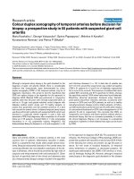
Fig. 1 Outcome tree diagram of 37 lesions for which MET-PET was performed to differentiate between LR and RN. Figures in parentheses indicate
number of lesions. Halftones indicate the lesions for which MET-PET diagnoses were incorrect or inconclusive
Yomo and Oguchi BMC Cancer (2017) 17:713
Page 4 of 9
Vienna, Austria) was used for all statistical analyses. A
P-value <0.05 was considered to indicate a statistically
significant difference.
Results
From August 2013 to February 2017, 33 patients with 38
BM were prospectively registered in the present study.
One patient who died of aspiration pneumonia soon
after MET-PET was excluded from the analysis. Patient
characteristics are presented in detail in Table 1. Of the
32 eligible patients, 19 were male and 13 were female.
The median age was 65 years (range: 14–87 years). The
primary cancers were of the lung in 22 patients, the
breast in 5, the digestive tract in 3, and one each had
ethmoid sinus carcinoma and soft tissue sarcoma. The
median Karnofsky performance status score at the time
of MET-PET was 90 (range: 50–100). All but one patient
had received a diagnosis of BM at one of the referring
regional hospitals. Twenty-seven patients (84%) had
undergone radiotherapeutic intervention using SRS at
our institution and the remaining five (16%) had been
treated at other institutions and referred to us for METPET diagnosis. Fifteen lesions in 13 patients (41%) had
received multiple radiotherapeutic interventions prior to
MET-PET. The median interval between primary radiotherapy and MET-PET was 18.8 months (range: 4–
120 months). Fifteen patients (47%) had been receiving
systemic chemotherapy at the time of MET-PET planning, but none had been administered bevacizumab
(BV), a monoclonal antibody against vascular endothelial
growth factor.
Table 1 Baseline demographic and clinical characteristics
MET-PET diagnosis and salvage management
Characteristic
Value
Sex (male/female)
19/13
Agea (years), median (range)
65 (14–87)
Nineteen tumors were diagnosed as LR and 18 as RN by
MET-PET. The mean value and standard deviation of
LNRs for each diagnostic category were 1.70 ± 0.30 and
1.09 ± 0.25, respectively. Comparison of baseline characteristics between the two groups revealed neurological
symptoms caused by the lesion of interest to be
significantly more frequent in the LR than in the RN
group (P = 0.011), while the time from primary radiotherapy to MET-PET was significantly longer in the RN
group (median: 24.9 months) than in the LR group
(median: 14.3 months) (P = 0.046) (Table 2).
Subsequent management based on MET-PET diagnosis is shown in Fig. 1. Of 19 LR, microsurgical resection
and SRS were performed in 9 and 8 lesions, respectively.
True tumor recurrence with various degrees of necrotic
tissue was histologically confirmed in 8 of 9 surgical
specimens. The rest of the lesion, previously irradiated
twice, was microscopically diagnosed as pure RN. After
salvage SRS, three of eight lesions showed no evident decrease in Gd-enhanced areas or perifocal oedema. Two
of these patients, one with HER-2 positive breast cancer
and other with EGFR wild-type lung adenocarcinoma,
experienced neurological worsening and needed salvage
therapy using repeat BV, resulting in immediate and durable symptomatic relief and radiological stabilisation for
more than 20 months (Fig. 2a). The other patients were
cautiously observed with temporary use of oral steroids,
showing Gd-enhanced areas and perifocal oedema which
remained stable for more than 3 years (Fig. 2b). We strategically chose an observation policy in two cases diagnosed as having LR on MET-PET. One eventually
showed disease progression and was confirmed to have a
true recurrence after surgical intervention, and the other
exhibited a self-limiting course after MET-PET, which
was consistent with RN. Of the 18 with RN, two patients
with moderate to severe neurological complications
Primary cancer
Non-small cell lung cancer
EGFR wild-type
14
EGFR mutant
5
Small cell lung cancer
3
Breast cancer
HER2-positive
4
HER2-negative
1
Gastrointestinal cancer
2
Oesophageal cancer
1
Sinonasal adenoid cystic carcinoma
1
Rhabdomyosarcoma
1
KPSa, median (range)
90 (50–100)
Neurological deficitsa
24 (75%)
a
RTOG-RPA Class (I/II/III)
8/18/6
Multiple BMa
16 (50%)
Prior radiotherapy (per lesion)
Proton therapy
1
SRS
21
WBRT + SRS
3
SRS × 2
9
SRS × 3
2
SRS × 4
1
Time from primary radiotherapy to MET-PET
(months), median (range)
18.8 (4–120)
EGFR epidermal growth factor receptor, HER human epidermal growth factor,
KPS Karnofsky performance status, RTOG radiation treatment oncology group,
RPA recursive partitioning analysis, BM brain metastases, aupdated status at
the time of MET-PET, SRS stereotactic radiosurgery, WBRT whole brain radiotherapy, MET-PET 11C–methionine positron emission tomography
Yomo and Oguchi BMC Cancer (2017) 17:713
Page 5 of 9
Table 2 Difference in clinical characteristics between MET-PET diagnosis groups
Characteristic
LNR ≥ 1.4 (n = 19)
LNR < 1.4 (n = 13)
Sex (male/female)
12/7
7/6
P value
0.72
Agea (years), median (range)
67 (14–87)
63 (49–79)
0.45
KPSa, median (range)
80 (50–100)
90 (60–100)
0.26
Neurological deficita
15 (79%)
9 (69%)
0.011
RTOG-RPA Classa (I/II/III)
4/11/4
4/7/2
0.79
Multiple BMa
11 (58%)
5 (38%)
0.47
Repeat prior radiotherapy
7 (37%)
6 (46%)
0.72
Time from primary radiotherapy to MET-PET (months), median (range)
14.3 (4–120)
24.9 (6–111)
0.046
11
MET-PET C–methionine positron emission tomography, LNR lesion/normal ratios, KPS Karnofsky performance status, RTOG radiation treatment oncology group,
RPA recursive partitioning analysis, BM brain metastases, aupdated status at the time of MET-PET
required repeat BV therapy, which produced rapid and
substantial symptom relief without relapse. Some of the
remaining 16 patients were initially observed or managed conservatively with low-dose oral steroids. Seven of
these cases, however, showed gradual progression and
ultimately needed salvage surgical treatment, and 3 of
these lesions were histologically confirmed to be LR.
ROC curve analysis
The ROC curve analysis for LNR is shown in Fig. 3. The
cut-off value for LNR of 1.40 provided the optimal sensitivity and specificity for differentiating LR from RN, 0.82 and
0.75, respectively. The highest area under the ROC curve
was 0.84 (95% CI: 0.71–0.97). Seven of the 8 lesions misdiagnosed on MET-PET had LNR values close to the
provisional cut-off point (within the range of 1.4 ± 0.2).
A second MET-PET was necessary in 4 patients because
of uncertainties in their clinical courses even after salvage
management based on the diagnosis made using the first
MET-PET. The LNR of the second scan (median: 3.55)
showed an obvious increase above that of the first
scan (median: 1.70) (P = 0.039, Paired t test) (Fig. 4)
and subsequent treatment confirmed true recurrence
in all 4 cases.
Fig. 2 Serial axial Gd-enhanced MR images and MET-PET images of two cases in which MET-PET diagnoses could not be confirmed even with
sufficient follow-up after salvage treatment. a: 60s–year-old-woman with multiple brain metastases from breast cancer. (a) Gd-enhanced MR image
obtained at the time of the initial SRS. (b) Gd-enhanced MR image obtained three months after SRS. (c) MET-PET image obtained 8 months after
initial SRS. The LNR was 1.67. (d) Gd-enhanced MR image obtained at the time of the second SRS. The yellow line represents the prescription
isodose volumes (12 Gy at 50%). (e) Gd-enhanced MR image obtained 1 month after the second SRS before BV rescue. Re-irradiation caused
neurological worsening and perifocal oedema. (f) Latest follow-up Gd-enhanced MR image obtained 20 months after MET-PET. Repeat BV therapy
resulted in symptomatic relief and radiological stabilisation. b: 70s–year-old-man with solitary cerebellar metastasis from gastric cancer. (a) Gdenhanced MR image obtained at the time of the initial SRS. (b) MET-PET image obtained 19 months after the initial SRS. The LNR was 1.50. (c)
Gd-enhanced MR image obtained at the time of the second SRS. The yellow line represents the prescription isodose volumes (22 Gy at 50%). (d)
Gd-enhanced MR image obtained 12 months after the second SRS. Re-irradiation caused cerebellar ataxia, requiring temporary conservative
treatment with oral steroids. (e) Gd-enhanced MR image obtained 26 months after the second SRS. (f) Latest follow-up Gd-enhanced MR image
obtained 36 months after MET-PET. Symptomatic relief and radiological stabilisation were maintained during long-term observation
Yomo and Oguchi BMC Cancer (2017) 17:713
Page 6 of 9
CI: 8.4 – NR) and 35.1 months (95% CI: 26.9 –NR), respectively (P = 0.035, log-rank test) (Fig. 5).
Fig. 3 ROC curve for LNR for MET-PET diagnosis. The LNR cut-off
value of 1.40 (dot) provided the best specificity and sensitivity for
differentiating between LR and LN, 0.75 and 0.82, respectively. The
highest area under the ROC curve was 0.84 (95% CI: 0.71–0.97)
Patient survival
The median follow-up interval of all patients after METPET was 17.5 months (range: 3–45). At the time of assessment, 20 patients (63%) were alive and 12 (37%) had
died. Nine of the 12 deceased patients had died of CNS
disease progression. The median survival time was
45.4 months (95% CI: 26.9 – NR (not reached)) (Fig. 5).
The median survival times in the LR and RN groups
based on MET-PET diagnosis were 14.8 months (95%
Fig. 4 Comparison of LNRs between the first and second MET-PET
in 4 patients. The LNR of the second scan (median: 3.55) showed a
marked increase as compared to that on the first scan (median: 1.70)
(P = 0.039, Paired t test)
Discussion
After any type of highly focused radiotherapy for BM,
one of the toxicities of greatest concern is delayed RN.
Because recent advancements in systemic therapy such
as targeted therapy have prolonged the survival of even
patients with BM [1–3], the importance of management
for delayed neurotoxicity and recurrence is anticipated
to further increase. In the follow-up of patients with BM
treated with radiotherapy, it is a matter of major importance to differentiate between delayed RN and LR either
clinically or with MR imaging [6]. The use of stereotactic
biopsy for histological assessment of post-radiotherapeutic
changes in MR imaging can be regarded as a viable diagnostic option [13], though it is somewhat invasive and not
always feasible. At present, many imaging methods are
practiced, including MR imaging (dynamic susceptibility
contrast perfusion [17], diffusion [18] and proton MR
spectroscopy [19]) and nuclear medicine techniques
(18F–fluorodeoxyglucose PET [20] and 201-thallium
single-photon emission computerised tomography [21]).
The active uptake of amino acids in viable tumor cells is
different from that in radiation-induced changes wherein
only passive diffusion across the damaged blood-brain
barrier occurs [7–10]. Thus, MET-PET is theoretically expected to reveal metabolic information in addition to morphological changes, resulting in high BM detection rates
and clear lesion delineation [22]. Our early experience
with MET-PET appeared to yield reliable and reproducible
diagnostic performance for differentiating between LR
and RN, when compared to areas under the ROC curves
of previous studies by other investigators, although there
were some differences in the calculation methods used between studies (Table 3). The provisional LNR cut-off value
of 1.4 in our institution was herein confirmed to be appropriate. Thus, we will not change our diagnostic criteria.
Several previous retrospective studies of MET-PET for
BM focused on its diagnostic accuracy and provided an
optimal cut-off value for diagnosis but, unfortunately,
did not provide subsequent management details. Such
specific details are often of critical importance to physicians caring for BM patients. The authors sought to
provide information useful for physicians on how to
manage such refractory situations, by investigating not
only the diagnostic accuracy of MET-PET but also the
long-term results of salvage management. We believe
this novel viewpoint to be the core value of the present
work.
This is the first report to demonstrate that MET-PET
can predict the patient’s survival as well as providing the
immediate diagnosis. This exploratory insight has, in our
opinion, clinical significance and can be regarded as
Yomo and Oguchi BMC Cancer (2017) 17:713
Page 7 of 9
Fig. 5 Kaplan –Meier curves showing the survival estimates for patients
with LR (dotted line) and RN (solid line) according to MET-PET diagnosis.
The median survival times in the LR and RN groups based on MET-PET
diagnosis were 14.8 months (95% CI: 8.4 – NR) and 35.1 months (95% CI:
26.9 –NR), respectively (P = 0.035, log-rank test)
relevant because RN follows a self-limited course in
most cases while, in contrast, LR can lead to neurological death. In fact, we observed that 9 patients diagnosed with LR on MET-PET ultimately succumbed to
central nervous system disease progression despite
multimodal treatments. The overall survival time after
MET-PET was also found to be prolonged in RN as
compared to LR cases. We speculate that this might, at
least in part, be attributable to the patients receiving
MET-PET having been self-selected to do well by virtue
of having had time to develop neuroimaging changes
and not dying of their systemic disease. This potential
patient population bias should be noted and caution
must be exercised in the generalisation of our present
findings.
There are limitations in the accuracy of imaging diagnosis. LNR values close to 1.4 should be regarded as a
“grey zone” with relatively low reliability and the possibility of the initial diagnosis ultimately being found to be
incorrect. As demonstrated herein, some of the lesions
diagnosed as RN by MET-PET were eventually confirmed to be LR, based on meticulous observation [23].
The second MET-PET was shown to be a meaningful
option for detecting longitudinal metabolic progression,
but given that the LNR values were significantly higher
on the second MET-PET, a lesion in such a case might
simply be regarded as LR. A delay in accurate diagnosis
can, in turn, lead to delayed initiation of salvage management. Given that the process of RN might, in part, represent a progressive destructive cascade, the timing of
MET-PET planning is of particular importance for effective salvage management aimed at preventing further
histologic injury. In addition to MET-PET imaging findings, we should also focus on patient’s progressively
worsening neurological symptoms and the shorter time
interval from radiotherapy to MET-PET as potentially
useful references for the diagnosis of LR, as shown in
Table 2.
Even with salvage management and long-term followup, the validity of MET-PET diagnosis could not be
determined in 3 patients (Fig. 2). We speculate that the
limitations of not only MET-PET but also other neuroimaging techniques for differentiation between LR and
RN might be attributable to there being three rather
than two possible diagnoses; LR, RN and pathology
combining these two. A series of microscopic analyses of
recurrent BM after SRS demonstrated that as many as
10 to 78% of lesions were diagnosed as showing mixed
pathology with various degrees of viable tumor and necrotic tissue [6, 24–27]. The management of both LR and
RN poses a significant therapeutic dilemma, if surgical
resection is not feasible, and effective therapies have yet
to be established. It is noteworthy that modern combined management using SRS followed by adjuvant BV
might be a viable and durable treatment option, even for
such complex conditions as in some of the patients
reported herein. Although evidence supporting the
remarkable effects of BV for refractory cerebral RN has
recently been accumulating [28–31], BV treatment is
not yet recognised as a standard of care for RN and is
not currently reimbursed by our public healthcare system, necessitating that the treatment indications be
strictly limited to cases having no alternative but to
undergo such an unproven treatment given its potential
Table 3 Comparison of qualitative tests of MET-PET for differentiation between LR and RN in BM
First author & year
No. of lesions
LNR cut-off value
Sensitivity (%)
Specificity (%)
AUC
Tsuyuguchi, 2005
21
1.42a
78
100
NR
Terakawa, 2008
51
1.41a
79
75
0.78
Minamimoto, 2015
42
1.30b
82
86
0.89
37
b
82
75
0.84
Present study, 2017
1.40
MET-PET 11C–methionine positron emission tomography, LR local recurrence, RN radiation necrosis, BM brain metastases, LNR lesion/normal ratios, AUC area
under the curve, NR not reported
a
SUVmean (lesion)/SUVmean (reference), bSUVmax (lesion)/SUVmax (reference)
Yomo and Oguchi BMC Cancer (2017) 17:713
toxicity. Optimisation of multimodal treatment using
antiangiogenic drugs merits further research.
The results of our present study must be interpreted
with caution. The subjects were a heterogeneous group.
The number of subjects was small and the lack of adequate statistical power may have resulted in the dataset
being underpowered to appropriately assess hypotheses
and potential prognostic factors. It should also be noted
that it remains difficult to make an accurate diagnosis in
some cases with the clinical and radiographic methods
employed herein. Thus, we plan to accumulate further
experience with this imaging modality, in hopes of establishing more efficient diagnostic and salvage treatment
regimens.
Conclusions
MET-PET appeared to have a reliable diagnostic capability for distinguishing between LR and RN after radiotherapy for BM. The provisional LNR cut-off value of
1.4 in our institution was found to be appropriate.
Diagnostic accuracy limitations should be recognised in
cases with LNR close to this cut-off value. An exploratory analysis raised the possibility of MET-PET diagnosis
predicting patient survival.
Abbreviations
AUC: Area under the curve; BM: Brain metastases; CI: Confidence interval;
EGFR: Epidermal growth factor receptor; Gd: Gadolinium; HER: Human
epidermal growth factor; KPS: Karnofsky performance status; LINAC: Linear
accelerator; LNR: Lesion/normal ratios; LR: Local recurrence; MET-PET: 11C–
methionine positron emission tomography; MR: Magnetic resonance;
RN: Radiation necrosis; ROC: Receiver operating characteristic; RPA: Recursive
partitioning analysis; RTOG: Radiation treatment oncology group;
SRS: Stereotactic radiosurgery; SUV: Standardised uptake value; WBRT: Whole
brain radiotherapy
Acknowledgements
We are grateful to Bierta Barfod, M.D., M.P.H. for her help with the language
editing of this manuscript.
Funding
No funding was obtained for this study.
Availability of data and materials
The dataset supporting the conclusions of this article is available from the
corresponding author upon appropriate request.
Authors’ contributions
SY and KO developed the study concept and protocols. SY: data collection,
statistical analysis, writing of manuscript. KO: assessment of MET-PET, oversight
of study. Both authors have read and approved the final manuscript.
Ethics approval and consent to participate
The Institutional Review Board of Aizawa Hospital granted ethics approval for
our present study in July 2013 (No. 2013–049). Written permission was
obtained prior to MET-PET from all patients and/or their relatives, allowing
the use of personal data for this clinical research when patient records and
information were anonymised and de-identified.
Consent for publication
Not applicable because this manuscript does not contain any data from
identifiable individuals.
Page 8 of 9
Competing interests
The authors declare that they have no competing interests.
Publisher’s Note
Springer Nature remains neutral with regard to jurisdictional claims in
published maps and institutional affiliations.
Author details
1
Division of Radiation Oncology, Aizawa Comprehensive Cancer Center,
Aizawa Hospital, 2-5-1, Honjo, Matsumoto-city, Nagano-prefecturem, Japan.
2
Positron Imaging Center, Aizawa Hospital, 2-5-1, Honjo, Matsumoto-city,
Nagano-prefecturem, Japan.
Received: 8 July 2017 Accepted: 22 October 2017
References
1. Sperduto PW, Yang TJ, Beal K, Pan H, Brown PD, Bangdiwala A, Shanley R,
Yeh N, Gaspar LE, Braunstein S, et al. Estimating survival in patients with
lung cancer and brain metastases: an update of the graded prognostic
assessment for lung cancer using molecular markers (lung-molGPA). JAMA
Oncol. 2016;
2. Cagney DN, Martin AM, Catalano PJ, Redig AJ, Lin NU, Lee EQ, Wen PY,
Dunn IF, Bi WL, Weiss SE, et al. Incidence and prognosis of patients with
brain metastases at diagnosis of systemic malignancy: a population-based
study. Neuro-Oncology. 2017;
3. Martin AM, Cagney DN, Catalano PJ, Warren LE, Bellon JR, Punglia RS, Claus
EB, Lee EQ, Wen PY, Haas-Kogan DA, et al. Brain metastases in newly
diagnosed breast cancer: a population-based study. JAMA Oncol. 2017;
4. Sneed PK, Mendez J, Vemer-van den Hoek JG, Seymour ZA, Ma L, Molinaro
AM, Fogh SE, Nakamura JL, McDermott MW. Adverse radiation effect after
stereotactic radiosurgery for brain metastases: incidence, time course, and
risk factors. J Neurosurg. 2015:1–14.
5. Kohutek ZA, Yamada Y, Chan TA, Brennan CW, Tabar V, Gutin PH, Yang TJ,
Rosenblum MK, Ballangrud A, Young RJ, et al. Long-term risk of
radionecrosis and imaging changes after stereotactic radiosurgery for brain
metastases. J Neuro-Oncol. 2015;125(1):149–56.
6. Stockham AL, Tievsky AL, Koyfman SA, Reddy CA, Suh JH, Vogelbaum MA,
Barnett GH, Chao ST. Conventional MRI does not reliably distinguish
radiation necrosis from tumor recurrence after stereotactic radiosurgery.
J Neuro-Oncol. 2012;109(1):149–58.
7. Jager PL, Vaalburg W, Pruim J, de Vries EG, Langen KJ, Piers DA.
Radiolabeled amino acids: basic aspects and clinical applications in
oncology. J Nucl Med. 2001;42(3):432–45.
8. Coope DJ, Cizek J, Eggers C, Vollmar S, Heiss WD, Herholz K. Evaluation of
primary brain tumors using 11C-methionine PET with reference to a normal
methionine uptake map. J Nucl Med. 2007;48(12):1971–80.
9. Ullrich RT, Kracht L, Brunn A, Herholz K, Frommolt P, Miletic H, Deckert M,
Heiss WD, Jacobs AH. Methyl-L-11C-methionine PET as a diagnostic marker
for malignant progression in patients with glioma. J Nucl Med. 2009;50(12):
1962–8.
10. Glaudemans AW, Enting RH, Heesters MA, Dierckx RA, van Rheenen RW,
Walenkamp AM, Slart RH. Value of 11C-methionine PET in imaging brain
tumours and metastases. Eur J Nucl Med Mol Imaging. 2013;40(4):615–35.
11. Tsuyuguchi N, Sunada I, Iwai Y, Yamanaka K, Tanaka K, Takami T, Otsuka Y,
Sakamoto S, Ohata K, Goto T, et al. Methionine positron emission
tomography of recurrent metastatic brain tumor and radiation necrosis after
stereotactic radiosurgery: is a differential diagnosis possible? J Neurosurg.
2003;98(5):1056–64.
12. Terakawa Y, Tsuyuguchi N, Iwai Y, Yamanaka K, Higashiyama S, Takami T,
Ohata K. Diagnostic accuracy of 11C-methionine PET for differentiation of
recurrent brain tumors from radiation necrosis after radiotherapy. J Nucl
Med. 2008;49(5):694–9.
13. Kickingereder P, Dorn F, Blau T, Schmidt M, Kocher M, Galldiks N, Ruge MI.
Differentiation of local tumor recurrence from radiation-induced changes
after stereotactic radiosurgery for treatment of brain metastasis: case report
and review of the literature. Radiat Oncol. 2013;8:52.
14. Minamimoto R, Saginoya T, Kondo C, Tomura N, Ito K, Matsuo Y, Matsunaga
S, Shuto T, Akabane A, Miyata Y, et al. Differentiation of brain tumor
recurrence from post-radiotherapy necrosis with 11C-methionine PET: visual
assessment versus quantitative assessment. PLoS One. 2015;10(7):e0132515.
Yomo and Oguchi BMC Cancer (2017) 17:713
15. Takenaka S, Asano Y, Shinoda J, Nomura Y, Yonezawa S, Miwa K, Yano H,
Iwama T. Comparison of (11)C-methionine, (11)C-choline, and (18)Ffluorodeoxyglucose-PET for distinguishing glioma recurrence from radiation
necrosis. Neurol Med Chir (Tokyo). 2014;54(4):280–9.
16. Peduzzi P, Concato J, Feinstein AR, Holford TR. Importance of events per
independent variable in proportional hazards regression analysis. II.
Accuracy and precision of regression estimates. J Clin Epidemiol. 1995;
48(12):1503–10.
17. Hoefnagels FW, Lagerwaard FJ, Sanchez E, Haasbeek CJ, Knol DL, Slotman
BJ, Vandertop WP. Radiological progression of cerebral metastases after
radiosurgery: assessment of perfusion MRI for differentiating between
necrosis and recurrence. J Neurol. 2009;256(6):878–87.
18. Jakubovic R, Zhou S, Heyn C, Soliman H, Zhang L, Aviv R, Sahgal A. The
predictive capacity of apparent diffusion coefficient (ADC) in response
assessment of brain metastases following radiation. Clin Exp Metastasis.
2016;33(3):277–84.
19. Chernov M, Hayashi M, Izawa M, Ochiai T, Usukura M, Abe K, Ono Y,
Muragaki Y, Kubo O, Hori T, et al. Differentiation of the radiation-induced
necrosis and tumor recurrence after gamma knife radiosurgery for brain
metastases: importance of multi-voxel proton MRS. Minimally invasive
neurosurgery : MIN. 2005;48(4):228–34.
20. Belohlavek O, Simonova G, Kantorova I, Novotny J Jr, Liscak R. Brain
metastases after stereotactic radiosurgery using the Leksell gamma knife:
can FDG PET help to differentiate radionecrosis from tumour progression?
Eur J Nucl Med Mol Imaging. 2003;30(1):96–100.
21. Serizawa T, Saeki N, Higuchi Y, Ono J, Matsuda S, Sato M, Yanagisawa M,
Iuchi T, Nagano O, Yamaura A. Diagnostic value of thallium-201 chloride
single-photon emission computerized tomography in differentiating tumor
recurrence from radiation injury after gamma knife surgery for metastatic
brain tumors. J Neurosurg. 2005;102(Suppl):266–71.
22. Matsuo M, Miwa K, Shinoda J, Kako N, Nishibori H, Sakurai K, Yano H, Iwama T,
Kanematsu M. Target definition by C11-methionine-PET for the radiotherapy of
brain metastases. Int J Radiat Oncol Biol Phys. 2009;74(3):714–22.
23. Yamane T, Sakamoto S, Senda M. Clinical impact of (11)C-methionine PET
on expected management of patients with brain neoplasm. Eur J Nucl Med
Mol Imaging. 2010;37(4):685–90.
24. Dequesada IM, Quisling RG, Yachnis A, Friedman WA. Can standard
magnetic resonance imaging reliably distinguish recurrent tumor from
radiation necrosis after radiosurgery for brain metastases? A radiographicpathological study. Neurosurgery. 2008;63(5):898–903. discussion 904
25. Kano H, Kondziolka D, Lobato-Polo J, Zorro O, Flickinger JC, Lunsford LD. T1/T2
matching to differentiate tumor growth from radiation effects after stereotactic
radiosurgery. Neurosurgery. 2010;66(3):486–91. discussion 491-482
26. Leeman JE, Clump DA, Flickinger JC, Mintz AH, Burton SA, Heron DE. Extent
of perilesional edema differentiates radionecrosis from tumor recurrence
following stereotactic radiosurgery for brain metastases. Neuro-Oncology.
2013;15(12):1732–8.
27. Nath SK, Sheridan AD, Rauch PJ, JB Y, Minja FJ, Vortmeyer AO, Chiang VL.
Significance of histology in determining management of lesions regrowing
after radiosurgery. J Neuro-Oncol. 2014;117(2):303–10.
28. Levin VA, Bidaut L, Hou P, Kumar AJ, Wefel JS, Bekele BN, Prabhu S, Loghin
M, Gilbert MR, Jackson EF. Randomized double-blind placebo-controlled trial
of bevacizumab therapy for radiation necrosis of the central nervous
system. Int J Radiat Oncol Biol Phys. 2011;79(5):1487–95.
29. Deibert CP, Ahluwalia MS, Sheehan JP, Link MJ, Hasegawa T, Yomo S, Feng
WH, Li P, Flickinger JC, Lunsford LD, et al. Bevacizumab for refractory
adverse radiation effects after stereotactic radiosurgery. J Neuro-Oncol.
2013;115(2):217–23.
30. Boothe D, Young R, Yamada Y, Prager A, Chan T, Beal K. Bevacizumab as a
treatment for radiation necrosis of brain metastases post stereotactic
radiosurgery. Neuro-Oncology. 2013;15(9):1257–63.
31. Yomo S, Hayashi M. Salvage stereotactic radiosurgery with adjuvant use of
bevacizumab for heavily treated recurrent brain metastases: a preliminary
report. J Neuro-Oncol. 2016;127(1):119–26.
Page 9 of 9
Submit your next manuscript to BioMed Central
and we will help you at every step:
• We accept pre-submission inquiries
• Our selector tool helps you to find the most relevant journal
• We provide round the clock customer support
• Convenient online submission
• Thorough peer review
• Inclusion in PubMed and all major indexing services
• Maximum visibility for your research
Submit your manuscript at
www.biomedcentral.com/submit