Optimal interval of endoscopic screening based on stage distributions of detected gastric cancers
Bạn đang xem bản rút gọn của tài liệu. Xem và tải ngay bản đầy đủ của tài liệu tại đây (762.62 KB, 8 trang )
Hamashima et al. BMC Cancer (2017) 17:740
DOI 10.1186/s12885-017-3710-x
RESEARCH ARTICLE
Open Access
Optimal interval of endoscopic screening
based on stage distributions of detected
gastric cancers
Chisato Hamashima1* , Rintaro Narisawa2, Kazuei Ogoshi3, Toshiyuki Kato4 and Kazutaka Fujita4
Abstract
Background: Although Korea and Japan have a national gastric cancer screening program, their screening intervals
are different. The optimal screening interval of endoscopic screening in Japan was investigated based on the stage
distributions of screen-detected gastric cancers.
Methods: Patients with gastric cancer detected by endoscopic and radiographic screenings were selected from the
Niigata City Medical Association database. The stage distributions of the detected gastric cancers were compared
among patients with different screening histories in both groups. Gastric cancer specific survival rates were
analyzed using the Kaplan-Meier method with the log-rank test.
Results: There were 1585 and 462 subjects in the endoscopic and radiographic screening groups, respectively. In
the endoscopic screening group, the stage IV proportion was lower in patients with screening history 1 and
2 years before diagnosis than in patients without screening history. Stage IV development was significantly
related to the absence of screening history (p < 0.001); however, there were no differences between patients
who had endoscopic screening history 2 and 3 years before diagnosis. The survival rates were not
significantly different between patients with endoscopic screening 1 and 2 years previously (p = 0.7763). The
survival rates were significantly higher in patients with endoscopic screening history 1 and 2 years before
diagnosis than in patients without screening history (p < 0.001), and in patients with endoscopic screaming
3 years before diagnosis (P < 0.0069).
Conclusion: The endoscopic screening interval for gastric cancer can be expanded to at least 2 years based
on the stage distributions of detected cancers and the patient survival rates.
Keywords: Gastric cancer screening, Upper gastrointestinal endoscopy, Screening interval, Stage distribution,
Survival rate
Background
Gastric cancer is the third leading cause of cancer death
all over the world. Despite the decrease in the incidence
of gastric cancer in recent years, it still remains a heavy
burden in eastern Asian and some European countries
[1]. Although national gastric cancer screening programs
have been effectively established in Asia, particularly in
Korea and Japan, upper gastrointestinal endoscopic
examination has already been performed as a standard
* Correspondence:
1
Division of Cancer Screening Assessment and Management, Center for
Public Health Science, National Cancer Center, 5-1-1 Tsukiji Chuo-ku, Tokyo
104-0045, Japan
Full list of author information is available at the end of the article
examination for stomach diseases, and this procedure is
also commonly used in the clinical setting worldwide
[2]. In Korea, endoscopic screening for gastric cancer
has been conducted since 1999 [3]. In Japan, endoscopic
screening for gastric cancer as a national program was
established only in 2016 based on the guidelines published by the National Cancer Center of Japan [4].
Before a new cancer screening technique is introduced in communities, the screening interval should
first be defined in consideration of the balance of
benefits and harms. Endoscopic screening is anticipated to have a high impact on mortality reduction of
gastric cancer; however, it can cause serious harms
© The Author(s). 2017 Open Access This article is distributed under the terms of the Creative Commons Attribution 4.0
International License ( which permits unrestricted use, distribution, and
reproduction in any medium, provided you give appropriate credit to the original author(s) and the source, provide a link to
the Creative Commons license, and indicate if changes were made. The Creative Commons Public Domain Dedication waiver
( applies to the data made available in this article, unless otherwise stated.
Hamashima et al. BMC Cancer (2017) 17:740
including complications, false-positive cases, and overdiagnosis [5]. Screening programs should maximize
mortality reduction in a defined screening interval for
the given resources [6]. The Korean guidelines have
defined the endoscopic screening interval as 2 years
based on the results of a case-control study [7].
However, the Japanese guidelines have not clearly defined the gastric cancer screening interval based on
conclusive evidence [8]. Moreover, as endoscopic
screening resources are limited, its rapid dissemination remains difficult [9]. Therefore, the efficient use
of resources should also be investigated to adequately
disseminate and provide equal access to endoscopic
screening for gastric cancer.
In Japan, Niigata City is considered to be the pioneer
city for endoscopic screening since it was implemented
there in 2003, much earlier than other municipalities
[10, 11]. The total number of participants has now
reached more than 45,000. Annual radiographic screening has also been provided in Niigata City.
In the present study, the optimal screening interval of
endoscopic screening for gastric cancer was investigated
according to the stage distributions of the screendetected gastric cancers and the survival rates of patients
with screen-detected gastric cancers using the Niigata
City Medical Association database.
Methods
Screening programs
Annual gastric cancer screening using the upper gastrointestinal series has been started and provided by the
local governments in Japan in accordance with the
Health Service Law for the Aged since 1983 [12]. Since
2003, endoscopic examination has been added to the
screening programs for gastric cancer in Niigata City
[10, 11]. Both photofluorography and regular radiographic screening using the upper gastrointestinal series
have been continued. Photofluorography has been performed as a mass screening program using mobile cars
mainly in the suburbs of Japan. On the other hand,
endoscopic and regular radiographic screenings have
been performed in clinical settings in the center of cities.
These screening programs have been basically performed
for asymptomatic individuals. Individuals who visited
regularly for disease treatment are often recommended
to undergo cancer screening by their own primary care
physicians. However, the underlying risk for the screened
individuals in terms of developing gastric cancer was not
clearly established. Individuals aged 40, 45, and 50 years
or over can undergo endoscopic and regular radiographic screenings. Individuals aged more than 40 years
can undergo photofluorography. Any screening method
can be selected based on the individual’s preference. For
all screening methods, there is no upper age limit and
Page 2 of 8
the screening interval is every year. Although the participation rate in gastric cancer screening has increased
since the introduction of endoscopic screening, the
screening rate has remained at approximately 25% [11].
The Niigata City Medical Association has provided
endoscopic screening and regular radiographic screening
to their member hospitals and clinics. Physicians who
perform endoscopic screening for gastric cancer in Niigata City have been approved by the local committee for
gastric cancer screening based on certain requirements
[11]. Although these endoscopic screenings have been
performed in clinical settings, the results have been
evaluated by the local committee which included experienced endoscopists on the basis of a monitor screen
review. In radiographic screening, a similar quality assurance system is used by the Niigata City Medical
Association.
Patients
The Niigata City Medical Association has developed a
database for detected gastric cancer according to the
screening method. The association has also systematically obtained detailed information related to gastric cancer from clinics and hospitals which have diagnosed and
provided treatment for gastric cancer in Niigata City.
The numbers of detected gastric cancer cases registered
from 2003 to 2012 were 2420 for endoscopic screening
and 572 for radiographic screening.
In the present study, the subjects were defined as individuals aged 40–79 years at the date of diagnosis of gastric cancer. All registered cases were ascertained by
linkage with the Niigata Prefectural Cancer Registry.
Even If there was no registration in the local cancer
registry, cases with pathological information on the database of the Niigata City Medical Association were included. Patients with other cancers such as malignant
lymphoma were excluded. The group was defined based
on the screening method at the year of diagnosis. In the
investigation of the screening interval for the same
method, cases which had different screening histories of
endoscopic and radiographic screenings were excluded.
Statistical analysis
The basic characteristics of the gastric cancers detected
by endoscopic and radiographic screenings were compared. Stage classification was based on the Japanese
Classification of Gastric Carcinoma [13]. Gastric cancers
were also classified histologically into intestinal and diffuse types according to Lauren’s criteria [14]. Differences
in the proportion of both screening groups were compared using the chi-square test and student t-test.
The patients with gastric cancer detected by endoscopic and radiographic screenings were divided into 4
categories: (1) patients without screening history; (2)
Hamashima et al. BMC Cancer (2017) 17:740
Page 3 of 8
patients who had screening 1 year before diagnosis; (3)
patients who had screening 2 years before diagnosis; (4)
patients who had screening 3 years before diagnosis.
Patients with no screening history were defined as those
who had no screening history within 3 years before their
diagnosis; however, patients who had screening history 4
or more years before their diagnosis were included. The
cancer stage distributions by different screening histories
in each screening group were compared using the chisquare test. Stage IV development was directly associated with gastric cancer death. The relationship between
screening history and stage IV development was evaluated by logistic regression analysis.
Gastric cancer specific survival analysis of both screening groups with different screening histories was
performed using the Kaplan-Meier method with the logrank test. The obtained curves show the proportion of
individuals alive over time from the time of screening.
All test statistics were two-tailed, and p-values <0.05
were considered to indicate a statistically significant difference. Analyses were performed using STATA 13.0
(STATA, College Station, TX, USA).
This study was approved by the Institutional Review
Board of the National Cancer Center of Japan.
Results
Subjects
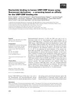
The selection procedure for the target population is
shown in Fig. 1. A total of 2922 subjects were selected
from the Niigata City Medical Association database, of
which 563 patients were not within the target age for
the analysis. Most of the subjects who were excluded
from the target group were more than 80 years old at
the date of diagnosis, which was not the actual target
age for cancer screening. On the basis of ascertainment
using the local cancer registry or pathology reports
which were collected from the hospitals and clinics,
2357 patients were verified as gastric cancer cases and
40 patients who had gastric cancer history before the
cancer registration were excluded. To investigate the optimal interval for each screening, individuals who had
different screening histories (mainly those who changed
from radiographic screening to endoscopic screening)
were excluded. Following the exclusion of patients with
different screening histories, patients with detected gastric cancer were finally divided into 2 groups: endoscopic
screening group (n = 1585) and radiographic screening
group (n = 462).
The characteristics of the patients with gastric cancers
detected by endoscopic and radiographic screenings
were compared (Table 1). The age group and female ratio were not significantly different between the 2 groups.
The distributions of screening histories for gastric cancer
patients were nearly equal between both groups
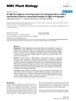
Fig. 1 Flowchart of the selection process for the target group. A total of
2922 subjects were selected from the Niigata City Medical Association
database, of which 563 patients were not within the target age for the
analysis. Based on the ascertainment by local cancer registry or pathology
reports which were collected from hospitals and clinics, 2357 patients were
verified to have gastric cancer and 40 patients who had a history of gastric
cancer before being registered as having cancer. To investigate the
optimal screening interval in each screening, individuals who had different
screening histories were excluded. Excluding those who had diffrent
screening histories, the patients in whom gastric cancer was detected by
gastric cancer screening were divided into 2 groups: endoscopic screening
group (n = 1585) and radiographic screening group (n = 462)
(p = 0.072). The cancer stage distributions were different
between both groups, although 26.9% of the endoscopic
screening-detected cancers lacked stage information.
Half of the gastric cancers detected by endoscopic
screening were treated by endoscopic submucosal
dissection.
Comparison of cancer stage distributions
The stage distributions of all detected gastric cancers between endoscopic and radiographic screenings were significantly different (p < 0.001). The stage distributions of
gastric cancers detected by endoscopic screening between patients aged 40–69 years and 70 years and older
were significantly different (p = 0.010). However, the
stage distributions of gastric cancers detected by radiographic screening between patients aged 40–69 years
and 70 years and older were not significantly different
(p = 0.545).
Hamashima et al. BMC Cancer (2017) 17:740
Page 4 of 8
Table 1 Characteristics of gastric cancer patients and their
gastric cancers detected by endoscopic and radiographic
screenings
Screening method
Endoscopic
screening
Total number
1585
Number
(%)
Radiographic
screening
Number
p-value
(%)
462
Sex
Men
1097
69.2
322
69.7
Women
488
30.8
140
30.3
40–49 years
3
0.2
1
0.2
50–59 years
69
4.4
20
4.3
60–69 years
614
38.7
164
35.5
70–79 years
899
56.7
277
60.0
0.842
Age group
0.647
Screening historya
No screening history
688
43.4
169
36.6
Screening 1 year before
715
45.1
231
50.0
Screening 2 years before
127
8.0
44
9.5
Screening 3 years before
55
3.5
18
3.9
0.072
Comparison of survival rates
Stage
Stage I
944
59.6
284
61.5
Stage II
66
4.2
45
9.7
Stage III
48
3.0
32
6.9
Stage IV
50
3.2
31
6.7
Unknown
427
26.9
70
15.2
1436
90.6
414
89.6
< 0.001
Pathology
Intestine
Diffuse
115
7.3
34
7.4
Others
13
0.8
6
1.3
Unknown
21
1.3
8
1.7
793
50.0
127
27.5
0.719
Treatment
Endoscopic submucosal
dissection
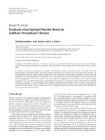
diagnosis and patients who had endoscopic screening
2 years before diagnosis. In the patients who had
endoscopic screening 3 years before diagnosis, the
proportion of stage IV was 1.8%.
In radiographic screening, the stage distributions of the
detected gastric cancers was not significantly different
among the various screening histories (Fig. 2b, I = 0.463),
and the proportion of stage I was 55.6% and that of stage
IV was 8.9% in the patients without screening history. The
proportion of stage IV was higher in patients who had
radiographic screening 2 years before diagnosis than in
patients without screening history.
In the patients with endoscopy-diagnosed cancer, although stage IV development was associated with no
screening history (odds ratio = 9.575, p < 0.001), there
were no differences between having screening history 2
and 3 years before diagnosis (Table 2). On the other
hand, in the patients with radiographic screeningdiagnosed cancer, any association with screening history
could not be found.
Surgery
703
44.4
293
63.4
Others
61
3.8
28
6.1
Unknown
28
1.8
14
3.0
< 0.001
a
Screening history was limited to the same method of which gastric cancer
was diagnosed
In endoscopic screening, the stage distribution of
the detected gastric cancers was significantly different among the different screening histories (Fig. 2a,
p < 0.001). More than 20% of the patients lacked
cancer stage information in all the groups. In the
first screening, the proportion of stage I was 61.0%
and that of stage IV was 6.3%. The proportion of
stage IV was not significantly different between patients who had endoscopic screening 1 year before
As information regarding the stage of the detected
gastric cancer was lacking, the survival rates among
different screening histories were compared. The mean
follow-up period was 67.6 ± 0.7 months (95% CI: 66.2–
69.1). The 5-year survival rate of the patients with gastric cancer detected by endoscopic screening at
95.1 ± 0.5% (95%CI: 93.9–96.0) was significantly different from the 5-year survival rate of patients with gastric
cancer detected by radiographic screening at 91.3 ± 0.1%
(95% CI: 88.1–93.6) (p = 0.005). In patients without
screening history, the survival rates were not significantly different between the endoscopic screening group
and the radiographic screening group (p = 0.1331).
In the endoscopic screening group, the survival rates
were significantly different among the 4 groups divided
by screening history (Fig. 3a, p < 0.001). The 5-year
survival rates among the 4 groups were follows: patients
without screening history = 92.1 ± 0.1% (95% CI: 89.7–
94.0); patients who had endoscopic screening 1 year
before diagnosis = 98.1 ± 0.6% (95% CI: 96.6–98.9);
patients who had endoscopic screening 2 years before
diagnosis = 97.2 ± 1.6% (95% CI: 91.4–99.1); patients who
had endoscopic screening 3 years before diagnosis = 92.6
± 0.4% (95% CI: 81.4–97.2). The survival rates were not
significantly different between patients with endoscopic
screening 1 and 2 years previously (p = 0.7763). The
survival rates were significantly higher in patients with
endoscopic screening history 1 and 2 years before diagnosis than in patients without screening history (p < 0.001),
and in patients with endoscopic screening 3 years before
diagnosis (P<0.0069).
Hamashima et al. BMC Cancer (2017) 17:740
Page 5 of 8
Fig. 2 Stage distributions of gastric cancer among patients with different screening histories. Patients with gastric cancer detected by
endoscopic and radiographic screenings were divided into 4 categories: (1) patients without screening history; (2) patients who had
screening 1 year before diagnosis; (3) patients who had screening 2 years before diagnosis; (4) patients who had screening 3 years
before diagnosis. Patients without screening history were included as well as individuals who had a screening history 4 or more years
before diagnosis. Screening history was limited to the same method of which gastric cancer was diagnosed. a Stage distributions of
gastric cancer detected by endoscopic screening. b Stage distributions of gastric cancer detected by radiographic screening
In the radiographic screening group, the survival rates
differed but not significantly among the 4 groups divided
by screening history (Fig. 3b, p = 0.2940). The 5-year
survival rates in the 4 groups were follows: patients
without screening history = 87.6 ± 2.7% (95% CI: 81.0–
92.0); patients who had radiographic screening 1 year
before diagnosis = 93.0 ± 1.7% (95% CI: 88.7–95.8);
patients who had radiographic screening 2 years before
diagnosis = 95.1 ± 3.4% (95% CI: 91.4–99.1); and
patients who had radiographic screening 3 years before
diagnosis = 92.9 ± 6.9% (95% CI: 59.1–99.0).
Discussion
The effectiveness of endoscopic screening for gastric
cancer has been increasingly reported in case-control
and cohort studies in Korea, China, and Japan [15–18].
However, before introducing endoscopic screening in
communities, the optimal screening interval must be defined to minimize harms and promote equal access to
endoscopic screening for gastric cancer. Radiographic
screening for gastric cancer has been performed since
1983 in Japan, and the screening interval has been defined annually [12]. In radiographic screening, the
Hamashima et al. BMC Cancer (2017) 17:740
Page 6 of 8
Table 2 Odds ratios for stage IV development by logistic regression analysis
Endoscopic screening
Odds ratio
Radiographic screening
95% CI
p-value
Odds ratio
95% CI
p-value
Sex
Men
1
Women
0.867
0.459–1.635
0.659
0.228
0.068–0.770
0.017
1.004
0.959–1.051
0.875
1.004
0.940–1.072
0.904
0.906
2.816
0.909–8.717
0.073
0.942–4.971
0.069
Age
1
Screening history
Screening 1 year before
1
Screening 2 years before
1.139
0.132–9.835
1
Screening 3 years before
2.613
0.300–22.773
0.385
–
No screening history
9.575
3.739–24.517
< 0.001
2.164
a
Screening history was limited to the same method of which gastric cancer was diagnosed
proportion of stage I was higher in patients who had
screening 2 years before diagnosis than in patients who
had no screening history. However, the survival rates of
patients were similar regardless of the screening interval
history among the patients whose gastric cancer was detected by radiographic screening. Based on the present
results, the interval of radiographic screening cannot be
easily expanded.
On the other hand, in endoscopic screening-detected
cancers, the proportion of stage IV was lower in patients
who had endoscopic screening 1 year or 2 years before
diagnosis than in patients who had no screening history.
The survival rates of patients who had endoscopic
screening were also higher, and the rates were nearly
equal between patients who had screening history 1 year
versus 2 years before diagnosis. Stage IV development
was strongly related to endoscopic screening history.
The difference in the survival rates between the
endoscopic and radiographic screening is caused by the
significant difference in the stage distribution of the detected gastric cancer and the different treatment
methods used by each screening methods. These results
suggest that the interval of endoscopic screening for gastric cancer can be expanded to at least 2 years.
Previous studies conducted in Korea have concluded
that the interval of endoscopic screening could be expanded to 2–3 years based on the stage distributions of
the detected cancers and the previous endoscopic examinations [19–21]. In a nested case-control study conducted under a Korean national program, reduction in
gastric cancer mortality could be confirmed even if the
patients were screened only once within 4 years or more
after the diagnosis of gastric cancer for case groups [16].
To define the screening interval, the natural history of
gastric cancer should also be considered. In Eastern
Asian countries, several studies and case reports have reported the natural history of gastric cancer based on a
long-term follow-up [22–26]. Tsukuma et al. described
the natural history of early-stage gastric cancer, and its
mean duration of progression was 44 months from the
early-stage to progress to the advanced stage [23]. The
survival rates of patients with interval cancer of annual
endoscopic screenings were nearly equal to the survival
rates of patients with cancer detected by annual endoscopic screening [27]. These results indicate that it may
be permissible to expand the endoscopic screening interval of gastric cancer from 1 year to 2 years or more.
The optimal interval for endoscopic screening of
gastric cancer should be clearly defined to avoid unnecessary harms and decrease total number of examinations [28]. The value of cancer screening is determined
by the difference of benefits and harms. As the intensity
of cancer screening increases, the benefits of cancer
screening rapidly increase. However, if the intensity of
cancer screening increases beyond an optimal level, the
harms and costs increase rapidly but not the benefits,
decreasing the value of care [29]. Endoscopic screening
also has harms such as false-negative results, falsepositive results, compliments, development of infection,
and overdiganosis [5]. As the upper age limit for endoscopic screening has not been defined in Japan, the target age should also be investigated to reduce harms and
obtain the maximum screening effectiveness.
The present study has several limitations. Firstly,
participation in radiographic screening has decreased
nationwide in Japan, and this trend has also been
observed in Niigata City. Therefore, patients with gastric
cancer as detected by radiographic screening were
limited in this study and the optimal screening interval
could not be specifically defined for radiographic screening. Secondly, as the number of patients who had cancer
screening 4 years or more before diagnosis was not sufficient, comparisons of the cancer stage distributions and
patient survival rates were not adequate. Thirdly, the
pathology of the detected cancers was mainly the intestinal type and their proportion was higher than the
Hamashima et al. BMC Cancer (2017) 17:740
Page 7 of 8
to be lacking from the Niigata City Medical Association
database. Fourthly, information regarding patients’
background and lifestyle was insufficient. Endoscopic
examination has been commonly performed regardless
of symptoms and has become commonly used as opportunistic screening. However, information as to whether
or not the patients participated in opportunistic screenings remains lacking. People who are visiting their family
physician have many opportunities to be screened. As
these people have diseases that have been treated, they
might constitute a high risk group for developing gastric
cancer. However, this assumption could not be verified
because of insufficient background information. Finally,
information regarding Helicobacter pylori infection in
the patients was unclear. Although the International
Agency for Research on Cancer recommended the establishment of H. pylori screening and eradication programs
in countries with a high incidence of gastric cancer, taking the local context into consideration is needed when
it is introduced [30]. Notably, progression to gastric
cancer differed according to the background of H. pylori
infection and atrophy [31]. When risk assessment is
combined with endoscopic screening, the screening
interval may be further expanded for the low-risk group
of gastric cancer. Further study is required on how to include risk assessment in gastric cancer screening.
Fig. 3 Survival rates of gastric cancer patients with different screening
histories. Patients with gastric cancer detected by endoscopic and
radiographic screenings were divided into 4 categories: (1) patients
without screening history; (2) patients who had screening 1 year before
diagnosis; (3) patients who had screening 2 years before diagnosis; (4)
patients who had screening 3 years before diagnosis. Patients without
screening history were included as well as individuals who had
screening history 4 or more years before diagnosis. Screening history
was limited to the same method of which gastric cancer was diagnosed.
a Survival rates of gastric cancer detected by endoscopic screening. The
5-year survival rates among the 4 groups were follows: patients without
screening history = 92.1 ± 0.1% (95% CI: 89.7–94.0); patients who had
endoscopic screening 1 year before diagnosis = 98.1 ± 0.6% (95% CI:
96.6–98.9); patients who had endoscopic screening 2 years before
diagnosis = 97.2 ± 1.6% (95% CI: 91.4–99.1); patients who had
endoscopic screening 3 years before diagnosis = 92.6 ± 0.4% (95% CI:
81.4–97.2). b Survival rates of gastric cancer detected by radiographic
screening. The 5-year survival rates in the 4 groups were follows: patients
without screening history = 87.6 ± 2.7% (95% CI: 81.0–92.0); patients who
had radiographic screening 1 year before diagnosis = 93.0 ± 1.7% (95% CI:
88.7–95.8); patients who had radiographic screening 2 years before
diagnosis = 95.1 ± 3.4% (95%CI: 91.4–99.1); and patients who had
radiographic screening 3 years before diagnosis = 92.9 ± 6.9%
(95% CI: 59.1–99.0)
proportion of other types of detected cancers in other
cities. Most of the patients in whom cancer was detected
by gastric cancer screening were aged 60 years and
above. In addition, data regarding a serious status appear
Conclusion
The interval of endoscopic screening for gastric cancer
can be expanded to at least 2 years based on the stage
distributions of the detected cancers and the patient survival rates. Further study regarding the optimal interval
of endoscopic screening and the target age group in
gastric cancer screening in Japan is warranted.
Abbreviations
CI: Confidence interval; H. pylori: Helicobacter pylori
Acknowledgements
We thank the Niigata Cancer Registry office, the local governments of
Niigata City, and the Niigata City Medical Association for their cooperation
with the study. We are grateful to Dr. Edward F. Barroga ( />0000-0002-8920-2607) for the editorial review of the manuscript. We
appreciate Ms. Etsuko Tanada, Ms. Kanoko Matsushima, Ms. Junko Asai, and
Ms. Ikuko Tominaga for research assistance.
Funding
This study was supported by the Japan Agency of Medical Research and
Development Tokyo, Japan (Grant number: 16,817,317). The funder had no
role in the conceptualization of the study design, data collection and
analysis, decision to publish, or preparation of the manuscript.
Availability of data and materials
We obtained data from the Niigata City Medical Association database. As
these data included personal information, the Niigata City government only
allowed their limited use specifically for evaluation studies on gastric cancer
screening. Dataset cannot also be made available in public. The data
analyzed in this study are kept by the Niigata City Medical Association (3–3-1
Shichikuyama Chuo-ku, Niigata 950–0914, Japan). Procedures for applying for
access to these data are available at the following web-site: Niigata City
Hamashima et al. BMC Cancer (2017) 17:740
Medical Association website: Please
contact Dr. Chisato Hamashima () for any specifics
regarding the data used for this study.
Authors’ contributions
CH designed the study. RN, KO, TK, and KF performed the follow-up survey
and collected the data. CH conducted the statistical analysis of the data. CH,
RN, KO, TK, and KF wrote, revised, and approved the manuscript for
submission. All authors read and approved the final manuscript.
Ethics approval and consent to participate
This study used the data from a local cancer registry and the results of
gastric cancer screening. This information was not included in the informed
consent for the collection of screening results and health data. Based on the
Japanese guidelines for epidemiological studies developed by the national
government, informed consent is not required for an observational study
using secondary data without human materials. In the present study,
secondary data from the local cancer registry and results of gastric cancer
screening were use, thus obtaining informed consent was waived. This study
was approved by the Institutional Review Board of the National Cancer
Center, Japan (Approval number: 2010–014).
Consent for publication
Not applicable
Competing interests
Chisato Hamashima, Rintaro Narisawa, Kazuei Ogoshi, Toshiyuki Kato, and
Kazutaka Fujita have no competing interests to disclose. Chisato Hamashma
is an Associate Editor of BMC Cancer.
Publisher’s Note
Springer Nature remains neutral with regard to jurisdictional claims in published
maps and institutional affiliations.
Author details
1
Division of Cancer Screening Assessment and Management, Center for
Public Health Science, National Cancer Center, 5-1-1 Tsukiji Chuo-ku, Tokyo
104-0045, Japan. 2Division of Gastroenterology, Niigata Cancer Center
Hospital, 2-15-3 Kawagishi-Cho Chuo-ku, Niigata 951-8566, Japan. 3Cancer
Registry Section, Niigata Cancer Center Hospital, 2-15-3 Kawagishi-Cho
Cyuo-ku, Niigata 951-8566, Japan. 4Committee of Gastrointestinal Cancer
Screening, Niigata City Medical Association, 3-3-1 Shichikuyama Chuo-ku,
Niigata 950-0914, Japan.
Received: 15 March 2017 Accepted: 30 October 2017
References
1. International Agency for Research on Cancer. GLOBOCAN 2012: Estimated
cancer incidence, mortality and prevalence worldwide in 2012. WHO. 2012.
Accessed 10 Jan 2017.
2. Leung WK, Wu MS, Kakugawa Y, et al. Screening for gastric cancer in Asia:
current evidence and practice. Lancet Oncol. 2008;9:279–87.
3. Kim Y, Jun JK, Choi KS, et al. Overview of the national cancer screening
programme and the cancer screening status in Korea. Asian Pac J Cancer
Prev. 2011;12:725–30.
4. Hamashima C, Fukao A. Quality assurance manual of endoscopic screening for
gastric cancer in Japanese communities. Jpn J Clin Oncol. 2016;46:1053–61.
5. Hamashima C. Benefits and harms of endoscopic screening for gastric
cancer. World J Gastroenterol. 2016;22:6385–92.
6. International Agency for Research on Cancer, WHO. IARC handbooks on
cancer prevention volume 10: cervical cancer screening. Lyon: IARC Press;
2005. p. 180–3.
7. Park HA, Nam SY, Lee SK, et al. The Korean guideline for gastric cancer
screening. J Korean Med Assoc. 2015;58:373–84.
8. Promotion of evidence based cancer screening. National Cancer Center, Japan.
The Japanese guidelines for gastric cancer screening 2015. Tokyo: National
Cancer Center; 2017. Accessed 15 Feb 2017
9. Hamashima C, Goto R. Potential capacity of endoscopic screening for
gastric cancer in Japan. Cancer Sci. 2017;108:101–7.
Page 8 of 8
10. Tashiro A, Sano M, Kinameri K, et al. Comparing mass screening techniques
for gastric cancer in Japan. World J Gastroenterol. 2006;12:4873–4.
11. Ogoshi K, Narisawa R, Kato T, et al. Endoscopic screening for gastric cancer
in Niigata city. Jpn J Endosc Forum Dig Dis. 2010;26:5–16.
12. Oshima A. A critical review of cancer screening programs in Japan. Int J
Technol Assess Health Care. 1994;10:346–58.
13. Japanese Gastric Cancer Association. Japanese classification of gastric
carcinoma. 3rd English edition. Gastric Cancer. 2011;14:101–12.
14. Lauren P. The two histological main types of gastric carcinoma: diffuse and
so-called intestinal-type carcinoma. An attempt at a histo-clinical
classification. Acta Pathol Microbiol Scand. 1965;64:31–49.
15. Hamashima C, Ogoshi K, Okamoto M, et al. A community-based, casecontrol study evaluating mortality reduction from gastric cancer by
endoscopic screening in Japan. PLoS One. 2013;8:e79088. doi: 10.1371/
journal.pone.0079088.
16. Jun JK, Choi KS, Lee HY, et al. Effectiveness of the Korean National Cancer
Screening Program in reducing gastric cancer mortality. Gastroenterol. 2017;
152(6):1319–28. [Epub ahead of print]
17. Chen Q, Yu L, Hao CQ, et al. Effectiveness of endoscopic gastric cancer
screening in a rural area of Linzhou, China: results from a case-control study.
Cancer Med. 2016;5:2615–22.
18. Hamashima C, Shabana M, Okada K, et al. Mortality reduction from gastric cancer
by endoscopic and radiographic screening. Cancer Sci. 2015;106:1744–9.
19. Nam JH, Choi IJ, Cho SJ, et al. Association of the interval between
endoscopies with gastric cancer stage at diagnosis in a region of high
prevalence. Cancer. 2012;118:4953–60.
20. Park CH, Kim EH, Chung H, et al. The optimal endoscopic screening interval
for detecting early gastric neoplasms. Gastrointest Endosc. 2014;80:253–9.
21. Kim J, Kim SM, Ha MH, et al. Does the interval of screening endoscopy
affect survival in gastric cancer patients? A cross-sectional study. Medicine
(Baltimore). 2016;95:e5490. doi: 10.1097/MD.0000000000005490.
22. Lee TY, Wang RC, Lee YC, et al. The Incidence of gastric adenocarcinoma
among patients with gastric intestinal metaplasia: A long-term cohort study.
J Clin Gastroenterol. 2016;50:532–7.
23. Tsukuma H, Oshima A, Narahara H, et al. Natural history of early gastric
cancer: a non-concurrent, long term, follow up study. Gut. 2000;47:618–21.
24. Yamada H, Ikegami M, Shimoda T, et al. Long-term follow-up study of
gastric adenoma/dysplasia. Endoscopy. 2004;36:390–6.
25. Fujisaki J, Nakajima T, Hirasawa T, et al. Natural history of gastric cancer―a
case followed up for eight years: early to advanced gastric cancer. Clin J
Gastroenterol. 2012;5:351–4.
26. Furukawa K, Yamada K, Konomi K, et al. Gastric carcinoma resected 95
months after being diagnosed: report of a case. Surg Today. 1994;24:756–8.
27. Hamashima C, Shabana M, Okamoto M, et al. Survival analysis of patients
with interval cancer undergoing gastric cancer screening by endoscopy.
PLoS One. 2015;10:e0126796. doi: 10.1371/journal.pone.0126796.
28. Wilt TJ, Harris RP, Qaseem A. Screening for cancer: advice for high-value care
from the American College of Physicians. Ann Intern Med. 2015;162:718–25.
29. Harris RP, Wilt TJ, Qaseem A. A value framework for cancer screening:
advice for high-value care from the American college of physicians. Ann
Intern Med. 2015;162:712–7.
30. IARC Working Group on the Evaluation of Carcinogenic Risks to Humans.
HELICOBACTER PYLORI. In: IARC working group reports volume 8.
Helicobacter pylori eradication as a strategy for preventing gastric cancer.
Lyon: IARC; 2014. p. 1–4.
31. Charvat H, Sasazuki S, Inoue M, et al. JPHC study group. Prediction of the
10-year probability of gastric cancer occurrence in the Japanese population:
the JPHC study cohort II. Int J Cancer. 2016;138:320–31.