Constitutional mutation in CDKN2A is associated with long term survivorship in multiple myeloma: A case report
Bạn đang xem bản rút gọn của tài liệu. Xem và tải ngay bản đầy đủ của tài liệu tại đây (838.52 KB, 6 trang )
Shah et al. BMC Cancer (2017) 17:718
DOI 10.1186/s12885-017-3715-5
CASE REPORT
Open Access
Constitutional mutation in CDKN2A is
associated with long term survivorship in
multiple myeloma: a case report
Vallari Shah1*, Kevin D. Boyd2, Richard S. Houlston1,3 and Martin F. Kaiser1
Abstract
Background: Multiple Myeloma is a cancer of plasma cells associated with significantly reduced survival. Long term
survivorship from myeloma is very rare and despite advances in its treatment the disease is generally considered
incurable. We report a patient diagnosed with myeloma carrying a germline mutation of a tumour suppressor gene
who has effectively been cured.
Case presentation: A 36-year-old woman was diagnosed with IgG lambda myeloma in 1985. She was treated with
melphalan chemotherapy followed by high-dose melphalan and autologous stem cell rescue and since remained
in complete remission despite not having received any additional therapy. After eliciting a prior history of multiple
primary melanomas and breast cancer, she was tested for and shown to be a carrier for a germline mutation in
CDKN2A.
Conclusions: This is the second case report of germline mutation of CDKN2A being associated with myeloma.
CDKN2A is a stabiliser of p53. Long term survivorship after high dose DNA damaging chemotherapy with melphalan
in this patient is compatible with an increased chemo-sensitivity due to impairment of the DNA repair pathway.
Keywords: Myeloma, Germline mutation, Survival, CDKN2A
Background
Multiple Myeloma (MM) is caused by the neoplastic
proliferation of somatically mutated plasma cells and is
associated with significant morbidity and mortality [1].
The use of alkylating agents such as melphalan to treat
MM four decades ago led to the first appreciable improvement in patient outcome with survival rates of between 24
and 48 months after diagnosis [2]. The subsequent introduction of immunomodulatory agents, proteasome inhibitors and high-dose autologous stem cell transplantation,
maintenance therapy, monoclonal antibodies and histone
deacetylase inhibitors more recently has led to further
improvements in patient outcome and median 5-year
survival rates are typically now around 50% [3]. Despite
these advances in treatment autologous stem cell transplantation has still been shown to be beneficial in extending survival [4].
* Correspondence:
1
Division of Molecular Pathology, The Institute of Cancer Research, London,
UK
Full list of author information is available at the end of the article
There is however significant variation in outcome
between patients with apparently same stage disease.
Staging systems such as the international staging system
(ISS) which uses serum albumin and β2-microglobulin
concentrations and the Revised-ISS (R-ISS) incorporating some adverse genetic markers and lactate dehydrogenase at diagnosis attempt to predict patients’ outcome.
These markers of adverse prognosis however cannot always accurately predict survival and there remain several
factors that are currently unknown with regards to
prognosis and response to treatment in myeloma. Of
considerable interest is understanding why a very small
number of patients have particularly long survivorship
for what is essentially an incurable disease.
It is increasingly being recognised that, as well as the
tumour profile, constitutional genotype also plays a role
in determining patient outcome [5]. Here we report on
an MM patient who has been in complete remission for
over 30 years after only receiving first-line standard of
care possibly being a consequence of also having hereditary Melanoma Syndrome.
© The Author(s). 2017 Open Access This article is distributed under the terms of the Creative Commons Attribution 4.0
International License ( which permits unrestricted use, distribution, and
reproduction in any medium, provided you give appropriate credit to the original author(s) and the source, provide a link to
the Creative Commons license, and indicate if changes were made. The Creative Commons Public Domain Dedication waiver
( applies to the data made available in this article, unless otherwise stated.
Shah et al. BMC Cancer (2017) 17:718
Case presentation
The patient, a 36-year-old woman, was diagnosed in 1985
with IgG lambda MM after presenting with tiredness and
recurrent infections. She was found to be anaemic with a
haemoglobin level of 73 g/l and thrombocytopenic with a
platelet count of 85 × 109/l. Further testing revealed a
markedly raised paraprotein of 62 g/l with positive urinary
Bence-Jones protein. There was evidence of immunosuppression with reduced levels of uninvolved IgA (0.1 g/l)
and IgM (0.2 g/l) immunoglobulins. Her renal function
was reduced as evidenced by a creatinine clearance of
57 ml/min. A skeletal survey revealed multiple lytic
lesions in both her humeri and femora. A bone marrow
biopsy confirmed a diagnosis of MM.
Since the patient met the established criteria for symptomatic MM [6] with end organ involvement as demonstrated by her anaemia, bony lytic lesions and
immunosuppression with recurrent infections, she was
commenced on standard chemotherapy advocated at the
time. This comprised three cycles of melphalan 10 mg
with prednisolone 60 mg for 4 days orally. After 1 cycle
of chemotherapy the patient’s paraprotein had fallen to
26 g/l. She subsequently received two further cycles of
melphalan and prednisolone which led to a further reduction in her paraprotein level to 7 g/l. This was
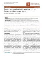
followed by a high-dose melphalan (140 mg/m2) and
autologous stem-cell transplant. Three months after her
bone marrow transplant the patient’s paraprotein was
undetectable and has never been detected again (Fig. 1).
The patient had been annually reviewed since diagnosis and has remained in complete remission 30 years
later with no further chemotherapy for her MM.
Fig. 1 Level of IgG lambda paraprotein (g/l) from diagnosis of myeloma
Page 2 of 6
Specifically, in April 2017, she continued to have a normal haematological profile with a haemoglobin of 135 g/
l, white cell count of 9.1 × 10⁹/l, and a platelet count of
191 × 109/l)). She is no longer immunosuppressed with
an IgA of 0.6 g/l and IgM of 0.5 g/l. She also has no
detectable paraprotein with a normal light chain ratio as
assessed by serum protein electrophoresis and serum
free light chain assay last assessed in April 2017. A repeat bone marrow biopsy 25 years ago showed that the
patient was in complete remission. Imaging by whole
body MRI in 2016 revealed no evidence of MM.
Five months prior to being diagnosed with MM she
had been diagnosed with a superficial spreading malignant melanoma on her right leg, which was successfully
treated by wide excision. She was subsequently diagnosed with three further primary melanomas at ages 53
(right buttock), 58 (right flank) and 62 (right forearm),
all also successfully treated by surgical excision. While
there was at that juncture no family history of melanomas or early onset pancreatic cancer, a diagnosis of
hereditary melanoma syndrome which can be caused by
germline mutations in the cyclin-dependent kinase Inhibitor 2A (CDKN2A) gene was considered in view of
the history of multiple melanomas. Genetic testing of
constitutional DNA extracted from EDTA venous blood
was performed by genomic DNA PCR amplification
using primers described previously of the 4 exons of
CDKN2A (exons 1α, 1β, 2 and 3) [7]. PCR fragments
were isolated by agarose gel electrophoresis and purified
prior to Sanger sequencing using QIAquick Gel Extraction Kit (Qiagen, Paisley, UK). This revealed the patient
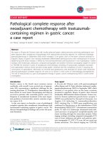
was a heterozygous carrier of the pathogenic c.213C > A
mutation in the CDKN2A gene. This mutation results in
a missense substitution of the amino acid asparagine to
lysine in the expressed INK4A protein at position
71(N71K) and a leucine to methionine substitution in
the expressed ARF protein (L86M) (Fig. 2). The patient’s
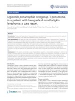
son has since been diagnosed with melanoma at the age
of 34 years but he has yet to be genetically tested (Fig. 3).
Otherwise the patient’s family history is unremarkable
and specifically there is no evidence for propensity to
pancreatic cancers in family members.
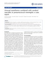
Aside from MM and melanoma the patient has also
been diagnosed with two other cancers. Firstly, in situ
breast cancer at the age of 50 incidentally discovered
during routine breast screening and which was treated
with a mastectomy. Secondly, stage T2bN1M0 adenocarcinoma of the lung at the age 66 which was diagnosed
following whole-body diffusion-weighted MRI investigation, performed as part of her MM follow-up investigation of hip pain. Her lung carcinoma has been treated by
lobectomy, adjuvant chemotherapy with carboplatin and
vinorelbine in addition to radiotherapy (Fig. 4). Mutation
testing of the patient’s lung cancer tissue by PCR
Shah et al. BMC Cancer (2017) 17:718
Page 3 of 6
Fig. 2 Chromatogram from Sanger sequencing showing pathogenic heterozygous c.213C > A mutation in CDKN2A of patient germline DNA,
the homozygous A allele at c.213 representing loss of heterozygosity in the patient’s lung cancer tissue compared to reference sequence with
diagrammatic representation of alternatively spliced products. The CDKN2A gene encodes both p14ARF (green exons) and p16INK4A (red exons),
generating two transcripts that are translated in alternative reading frames
amplification and Sanger sequencing as described above
revealed a loss of heterozygosity of the C.213C > A allele
compared to the patient’s germline DNA (Fig. 2). Paradoxically her MRI did not show any signs indicative of
active MM.
Discussion and conclusions
The pathogenic nature of the specific c.213C > A mutation in CDKN2A noted in this patient is suggested by the
fact that it has been described previously in several hereditary melanoma families [8–10] as well as a supraglottic
squamous cell carcinoma [11]. In silico predictions with
the algorithms used by Polyphen-2, SIFT and mutation
taster all indicate that this is a pathogenic mutation. Additionally, functional assays of the protein INK4A with this
mutation also suggest pathogenicity [12]. The loss of the
wild type allele in the patient’s lung cancer DNA as shown
in Fig. 2 also suggests that this is a pathogenic mutation
causing an increased susceptibility to tumours.
To our knowledge this is only the second case of a
germline mutation in CDKN2A being reported in association with MM. The previous report described a MM
patient who had a strong family history of melanoma
consistent with a diagnosis of hereditary Melanoma Syndrome caused by a pathogenic exon 1 CDKN2A mutation. Loss of the wild-type allele was detected in
malignant plasma cells consistent with CDKN2A acting
as a tumour suppressor in the context of MM in this
case report [13].
Typically, germline mutation of CDKN2A is associated
with a restricted spectrum of cancers; primarily melanoma and pancreatic carcinoma. However, an increased
risk of other cancers including childhood ones, lung,
oropharyngeal and breast have been reported albeit at
lower frequency [14]. Evidence for the association of the
CDKN2A gene and its association with myeloma susceptibility has been shown in genome wide association
studies which found a susceptibility locus for myeloma
Shah et al. BMC Cancer (2017) 17:718
Page 4 of 6
Fig. 3 Patient pedigree
at chromosome 9p21.3 variant rs2811710 of CDKN2A
[15]. A population based study in 1354 people with
multiple myeloma also suggests a link between multiple
myeloma, melanoma within first and second degree relatives [16]. This has been further confirmed in other
studies [17–19].
Such data implies a wider impact of CDKN2A in
tumour aetiology and although rare suggests the relationship with MM is not coincidental. It is perhaps
not surprising that CDKN2A impacts on the aetiology
of a wide range of tumour types. One of the gene
transcripts ARF functions as a stabiliser of p53
through interaction with E3 ubiquitin protein ligase
MDM2, thereby enhancing p53-dependent transactivation and apoptosis. Mutations in ARF result in
Fig. 4 Timeline of primary malignancies and therapy
destabilisation of p53. Abnormalities of p53 are
present in almost all cancers. This can be direct via
deletion/mutation or hypermethylation of the p53
promoter, altering its stabilisation through alterations/
deletions of ARF or overexpression of MDM2 [20, 21]
or via other mechanisms.
Alternate splice variant of CDKN2A, INK4A functions
is a member of the cyclin dependent kinase inhibitors. It
binds to CDK4 and CDK6 kinases and sequesters them
from their regulatory cyclin D subunits. As a result,
CDK4 cannot phosphorylate the retinoblastoma protein
(Rb) which is considered the gatekeeper of cell proliferation As a result of mutations in INK4A, there is resultant dysregulation of cell cycle control and tumour
proliferation [22].
Shah et al. BMC Cancer (2017) 17:718
Further evidence of the role of CDKN2A in tumour
development comes from mice lacking ARF and/or
INK4A which develop tumours early in life succumbing
to lymphomas and fibrosarcomas [23]. Additionally,
families with germline mutations of CDKN2A show increased rates of melanoma and pancreatic cancer but
also have increased rates of other malignancies such as
cancers of the breast, nervous system, GI tract, lymphoma and cervical cancers also suggesting that the
increased susceptibility to cancer is not restricted to
melanoma and pancreatic cancer alone [24, 25]. Furthermore, frequent somatic mutations and deletions of
CDKN2A have been noted in several cancers including
pancreatic adenocarcinoma, oesophageal and gastric carcinomas, leukemias and melanomas indicating its role in
cancer pathogenesis [26, 27]. Deletions as well as mutations of the Rb as well as TP53 are frequent in myeloma
indicating a critical role of these genes in its pathogenesis [28]. It is therefore feasible that genes altering the
function of these proteins will also increase the susceptibility to myeloma.
The case we report is striking in that after only melphalan therapy the patient has had a remission from
MM of over 30 years and in essence is cured. Although
speculative the observation is consistent with the
patient’s MM being especially sensitive to alkylating
chemotherapy. Melphalan causes DNA damage and subsequent cell death due to impairment in the DNA repair
pathway. Studies have shown that mutations in
CDKN2A increase sensitivity to chemotherapy [29, 30].
Moreover, MDM2 inhibitors increase sensitivity to
conventional chemotherapy in different cancers. MDM2
targets p53 protein for proteosomal degradation and its
function is inhibited by ARF. Mutations in ARF have
been shown to destabilise p53 through this mechanism and the increased susceptibility to chemotherapy
induced by MDM2 inhibitors may reflect why this
patient also had a high sensitivity to conventional
chemotherapy which causes DNA damage. Effectiveness of MDM2 inhibitors has also been demonstrated
in haematological malignancies such as MM, AML
and ALL. [31–33]. Of note in this regard is that AML
with the translocation of t(8;21) resulting in the
RUNX1-ETO fusion gene which directly inhibits ARF
is one of the few subtypes of AML which can be
cured by high dose chemotherapy alone [34].
In conclusion, outcome of high-dose chemotherapy in
the patient we report has resulted in cure suggesting that
such germline mutations may confer increased MM
sensitivity to chemotherapy. This finding raises the
possibility that long-term survivorship from MM in
other patients may be the consequence of carrier status
for tumour suppressor genes with biological relevance to
DNA damage.
Page 5 of 6
Abbreviations
ALL: acute lymphoblastic leukemia; AML: acute myeloid leukemia;
CDKN2A: cyclin-dependent kinase Inhibitor 2A; DNA: deoxyribonuelcic acid;
EDTA: Ethylanediaminetatraacetic acid; MDM2: mouse double minute 2
homolog/E3 ubiquitin-protein ligase MDM2; MM: Multiple Myeloma;
p53: tumour protein p53; RUNX1-ETO: Runt-related transcription factor 1Eight Twenty-One oncoprotein
Acknowledgements
We thank the patient for allowing us to write the case report. We also
acknowledge support from the National Institute of Health Biomedical
Research Centre at the Royal Marsden Hospital. We are grateful to Rebecca
Brown from the Leeds Genetics Laboratory for her advice and guidance with
regards to CDKN2A mutations. We would also like to thank Sidra Ellis, Fabio
Mirabella and Karen Menezes for their help and advice in the lab.
Funding
None.
Availability of data and materials
Data is included within the article.
Authors’ contributions
VS: design and draft of manuscript. KDB: design and draft of manuscript.
RSH: concept, design and draft of manuscript. MFK: concept, design and
draft of manuscript. All authors have read and approved the final version of
the manuscript.
Ethics approval and consent to participate
Not applicable.
Consent for publication
Written informed consent was obtained from the patient for publication of
the case report.
Competing interests
The authors declare that they have no competing interests.
Publisher’s Note
Springer Nature remains neutral with regard to jurisdictional claims in
published maps and institutional affiliations.
Author details
1
Division of Molecular Pathology, The Institute of Cancer Research, London,
UK. 2Department of Haemato-Oncology, Royal Marsden Hospital, London,
UK. 3Division of Genetics and Epidemiology, The Institute of Cancer Research,
London, UK.
Received: 27 June 2017 Accepted: 30 October 2017
References
1. Song X, Cong Z, Wilson K. Real-world treatment patterns, comorbidities, and
disease-related complications in patients with multiple myeloma in the
United States. Curr Med Res Opin. 2016;32(1):95–103.
2. Alexanian R, et al. Treatment for multiple myeloma. Combination
chemotherapy with different melphalan dose regimens. JAMA. 1969;
208(9):1680–5.
3. Rajkumar SV, Kumar S. Multiple myeloma: diagnosis and treatment. Mayo
Clin Proc. 2016;91(1):101–19.
4. Rosinol L, et al. Superiority of bortezomib, thalidomide, and dexamethasone
(VTD) as induction pretransplantation therapy in multiple myeloma: a
randomized phase 3 PETHEMA/GEM study. Blood. 2012;120(8):1589–96.
5. Johnson DC, et al. Genome-wide association study identifies variation at
6q25.1 associated with survival in multiple myeloma. Nat Commun.
2016;7:10290.
6. Rajkumar SV. Myeloma today: disease definitions and treatment advances.
Am J Hematol. 2016;91(1):90–100.
7. Harland M, et al. A comparison of CDKN2A mutation detection within
the melanoma genetics consortium (GenoMEL). Eur J Cancer.
2008;44(9):1269–74.
Shah et al. BMC Cancer (2017) 17:718
8.
9.
10.
11.
12.
13.
14.
15.
16.
17.
18.
19.
20.
21.
22.
23.
24.
25.
26.
27.
28.
29.
30.
31.
32.
33.
34.
Soufir N, et al. Prevalence of p16 and CDK4 germline mutations in 48
melanoma-prone families in France. The French familial melanoma study
group. Hum Mol Genet. 1998;7(2):209–16.
Bishop DT, et al. Geographical variation in the penetrance of CDKN2A
mutations for melanoma. J Natl Cancer Inst. 2002;94(12):894–903.
Goldstein AM, et al. Features associated with germline CDKN2A mutations: a
GenoMEL study of melanoma-prone families from three continents. J Med
Genet. 2007;44(2):99–106.
Fischer M. Analysis of exon 2 of MTS1 in HPV-positive and HPV-negative
tumors of the head and neck region. Eur Arch Otorhinolaryngol. 2007;
264(7):801–7.
McKenzie HA, et al. Predicting functional significance of cancer-associated
p16(INK4a) mutations in CDKN2A. Hum Mutat. 2010;31(6):692–701.
Dilworth D, et al. Germline CDKN2A mutation implicated in predisposition
to multiple myeloma. Blood. 2000;95(5):1869–71.
Mukherjee B, et al. Risk of non-melanoma cancers in first-degree relatives of
CDKN2A mutation carriers. J Natl Cancer Inst. 2012;104(12):953–6.
Mitchell JS, et al. Genome-wide association study identifies multiple
susceptibility loci for multiple myeloma. Nat Commun. 2016;7:12050.
Camp NJ, Werner TL, Cannon-Albright LA. Familial myeloma. N Engl J Med.
2008;359(16):1734–5. author reply 1735
Altieri A, et al. Familial risks and temporal incidence trends of multiple
myeloma. Eur J Cancer. 2006;42(11):1661–70.
Kristinsson SY, et al. Risk of solid tumors and myeloid hematological
malignancies among first-degree relatives of patients with monoclonal
gammopathy of undetermined significance. Haematologica.
2009;94(8):1179–81.
Lynch HT, et al. Familial multiple myeloma: a family study and review of the
literature. J Natl Cancer Inst. 2001;93(19):1479–83.
Zhang Y, Xiong Y. Mutations in human ARF exon 2 disrupt its nucleolar
localization and impair its ability to block nuclear export of MDM2 and p53.
Mol Cell. 1999;3(5):579–91.
Zhang Y, Xiong Y, Yarbrough WG. ARF promotes MDM2 degradation and
stabilizes p53: ARF-INK4a locus deletion impairs both the Rb and p53 tumor
suppression pathways. Cell. 1998;92(6):725–34.
Lin YC, et al. Human p16gamma, a novel transcriptional variant of
p16(INK4A), coexpresses with p16(INK4A) in cancer cells and inhibits cellcycle progression. Oncogene. 2007;26(49):7017–27.
Kamijo T, et al. Tumor suppression at the mouse INK4a locus mediated by
the alternative reading frame product p19ARF. Cell. 1997;91(5):649–59.
Borg A, et al. Novel germline p16 mutation in familial malignant melanoma
in southern Sweden. Cancer Res. 1996;56(11):2497–500.
Hewitt C, et al. Germline mutation of ARF in a melanoma kindred. Hum Mol
Genet. 2002;11(11):1273–9.
Caldas C, et al. Frequent somatic mutations and homozygous deletions of the
p16 (MTS1) gene in pancreatic adenocarcinoma. Nat Genet. 1994;8(1):27–32.
Liu Q, et al. MTS-1 (CDKN2) tumor suppressor gene deletions are a frequent
event in esophagus squamous cancer and pancreatic adenocarcinoma cell
lines. Oncogene. 1995;10(3):619–22.
Walker BA, et al. Mutational Spectrum, copy number changes, and outcome:
results of a sequencing study of patients with newly diagnosed myeloma. J
Clin Oncol. 2015;33(33):3911–20.
Simon M, et al. Role of p16 and p14ARF in radio- and chemosensitivity of
malignant gliomas. Oncol Rep. 2006;16(1):127–32.
Iwadate Y, et al. Alteration of CDKN2/p16 in human astrocytic tumors is
related with increased susceptibility to antimetabolite anticancer agents. Int
J Oncol. 2000;17(3):501–5.
Andreeff M, et al. Results of the phase I trial of RG7112, a small-molecule
MDM2 antagonist in leukemia. Clin Cancer Res. 2016;22(4):868–76.
Gu D, et al. Inhibition of the MDM2 E3 ligase induces apoptosis and
autophagy in wild-type and mutant p53 models of multiple myeloma, and
acts synergistically with ABT-737. PLoS One. 2014;9(9):e103015.
Reis B, et al. Acute myeloid leukemia patients' clinical response to
idasanutlin (RG7388) is associated with pre-treatment MDM2 protein
expression in leukemic blasts. Haematologica. 2016;101(5):e185–8.
Linggi B, et al. The t(8;21) fusion protein, AML1 ETO, specifically represses
the transcription of the p14(ARF) tumor suppressor in acute myeloid
leukemia. Nat Med. 2002;8(7):743–50.
Page 6 of 6
Submit your next manuscript to BioMed Central
and we will help you at every step:
• We accept pre-submission inquiries
• Our selector tool helps you to find the most relevant journal
• We provide round the clock customer support
• Convenient online submission
• Thorough peer review
• Inclusion in PubMed and all major indexing services
• Maximum visibility for your research
Submit your manuscript at
www.biomedcentral.com/submit