Carbon ion radiotherapy for 80 years or older patients with hepatocellular carcinoma
Bạn đang xem bản rút gọn của tài liệu. Xem và tải ngay bản đầy đủ của tài liệu tại đây (1.18 MB, 8 trang )
Shiba et al. BMC Cancer (2017) 17:721
DOI 10.1186/s12885-017-3724-4
RESEARCH ARTICLE
Open Access
Carbon ion radiotherapy for 80 years
or older patients with hepatocellular
carcinoma
Shintaro Shiba1, Takanori Abe1, Kei Shibuya1*, Hiroyuki Katoh2, Yoshinori Koyama3, Hirohumi Shimada2,
Satoru Kakizaki4, Ken Shirabe5, Hiroyuki Kuwano6, Tatsuya Ohno2 and Takashi Nakano1
Abstract
Background: To evaluate the safety and efficacy of carbon ion radiotherapy (C-ion RT) for 80 years or older patients
with hepatocellular carcinoma (HCC).
Methods: Eligibility criteria of this retrospective study were: 1) HCC confirmed by histology or typical hallmarks of HCC
by imaging techniques of four-phase multidetector-row computed tomography or dynamic contrast-enhanced magnetic
resonance imaging; 2) no intrahepatic metastasis or distant metastasis; 3) no findings suggesting direct infiltration of the
gastrointestinal tract; 4) performance status ≤2 by Eastern Cooperative Oncology Group classification; and 5) Child-Pugh
classification A or B. Patients received C-ion RT with 52.8 Gy (RBE) or 60.0 Gy (RBE) in four fractions for usual
cases and 60.0 Gy (RBE) in 12 fractions for close-to-gastrointestinal tract cases. Toxicities were classified using the National
Cancer Institute’s Common Terminology Criteria for Adverse Events (Version 4.0).
Results: Between March 2011 and November 2015, 31 patients were treated. The median follow-up period of all patients
was 23.2 months (range: 8.4–55.3 months). Median age at the time of registration of C-ion RT was 83 years
(range: 80–95 years). Child-Pugh grade A and B were 27 patients and 4 patients, respectively.
The 2-year estimated overall survival, local control, and progression-free survival rates were 82.3%, 89.2%, and
51.3%, respectively. No patients had Grade 2 or higher acute toxicities (within 3 months after C-ion RT). One patient
experienced progression in Child-Pugh classification from A to B within 3 months after C-ion RT. In late toxicities, Grade
3 encephalopathy was observed in 3 patients, and 2 improved with medication.
Conclusions: C-ion RT was effective with minimal toxicities for 80 years or older patients with hepatocellular carcinoma.
Trial registration: UMIN000020571: date of registration, 14 January 2016, retrospectively registered.
Keywords: Carbon-ion radiotherapy, Hepatocellular carcinoma, Elderly patients, Radiotherapy
Background
Hepatocellular carcinoma (HCC) is the sixth most common
cancer and the third major cause of cancer-related death
worldwide [1]. Most HCC patients have a background of
chronic liver disease resulting from alcohol abuse, infection
of hepatitis C virus or hepatitis B virus [2, 3]. According to
a 2005 report, the peak age range of HCC worldwide was
30 to 50 years [4]. On the other hand, in Japan, until 1990
the majority of HCC deaths were below the age of 69, but
* Correspondence:
1
Department of Radiation Oncology, Gunma University Graduate School of
Medicine, 3-39-22 Syowa-machi, Maebashi, Gunma 371-8511, Japan
Full list of author information is available at the end of the article
66% of patients with HCC were over 70 in 2006 [3]. In light
of the increased age of HCC patients, there is an urgent
need for less-invasive local treatments.
Surgical resection, local ablation therapies including
percutaneous radiofrequency ablation (RFA), and percutaneous ethanol infusion (PEI) are potentially curative
treatments [2, 5]. However, many patients are not amenable to surgery or local ablation therapy for medical or
anatomic reasons. Recently, proton therapy and stereotactic body radiotherapy (SBRT) with X-rays have been
applied for HCC as less-invasive procedures [6–9]. Also,
carbon ion radiotherapy (C-ion RT) has been used for
HCC because of its excellent dose localization property
© The Author(s). 2017 Open Access This article is distributed under the terms of the Creative Commons Attribution 4.0
International License ( which permits unrestricted use, distribution, and
reproduction in any medium, provided you give appropriate credit to the original author(s) and the source, provide a link to
the Creative Commons license, and indicate if changes were made. The Creative Commons Public Domain Dedication waiver
( applies to the data made available in this article, unless otherwise stated.
Shiba et al. BMC Cancer (2017) 17:721
and higher relative biological effectiveness based on the
characteristics of higher linear energy transfer beam
[10, 11]. Although the theoretical benefit of C-ion RT
would exist in elderly HCC patients with hepatic dysfunction. There was a lack of data on the clinical outcomes of
C-ion RT for elderly patients with HCC. In the current
study, we analyzed safety and efficacy of C-ion RT in elderly
HCC patients (80 years or older).
Methods
Patients
This retrospective analysis was performed using the
medical records of patients treated with C-ion RT for
eighty years or older patients with HCC at Gunma
University Heavy Ion Medical Center (GHMC) between
March 2011 and November 2015. All patients were treated
and monitored according to the protocol approved by the
Institutional Review Board. Eligibility criteria were: 1) HCC
confirmed by histology or typical hallmarks of HCC using
imaging techniques of four-phase multidetector-row
computed tomography (CT) or dynamic contrast-enhanced
magnetic resonance imaging (MRI) (hypervascular in arterial phase with washout in portal venous or delayed phases);
2) no intrahepatic metastasis or distant metastasis; 3) no
findings suggesting direct infiltration of the gastrointestinal
tract; 4) performance status (PS) ≤ 2 by Eastern Cooperative Oncology Group classification; and 5) Child-Pugh classification A or B. The disease stage according to the Union
for International Cancer Control (UICC) classification
(7th edition) [12] and The Barcelona Clinic Liver Cancer
(BCLC) classification [13] were determined by CT, MRI,
ultrasonography, and other variables. The model for endStage liver disease (MELD) score was calculated for evaluation of liver function in all patients [14]. HCC located
within 2 cm of the main portal vein was defined as a porta
hepatis group. In the current study, the uncontrolled tumors by transarterial chemoembolization (TACE) and/or
transarterial infusion (TAI) and/or RFA were included. The
treatment protocol for the current study was reviewed and
approved by Gunma university Institutional Review Board,
and all patients signed an informed consent form before
the initiation of therapy.
Carbon ion radiotherapy
Carbon ion beams were generated by synchrotron at
GHMC. Passive scattering technique was applied for
the treatment of HCC. Beam energies of 290 MeV/u,
380 MeV/u, and 400 MeV/u were employed. Beam energy
was chosen according to the depth of the tumor. At our
facility, XiO-N is used for treatment planning, which is
XiO (Elekta)-based software incorporating a dose engine
for ion beam radiotherapy (K2dose) [15–19] developed by
the National Institute of Radiological Sciences, Japan, with
interfaces from Mitsubishi Electric. Patients received C-
Page 2 of 8
ion RT once daily, 4 days per week (Tuesday to Friday).
Radiation dose calculated for the target volume and surrounding normal structures was expressed in Gy (RBE),
which was defined as the physical dose multiplied by the
relative biologic effectiveness (RBE) of carbon ions [20, 21].
Treatment planning and target delineation
Tailor-made fixation cushions and thermoplastic shells
were used for the immobilization of patients for acquiring
treatment planning CT images. After immobilization,
respiratory-gated CT and four-dimensional CT (4-D
CT) images were acquired. Images from the expiratory
phase were used for treatment planning. Contrast-enhanced
CT images were taken simultaneously and merged with
treatment planning CT to precisely delineate the gross
tumor volume (GTV). The clinical target volume (CTV)
margin, including microscopic disease progression, was
added to GTV, with an additional 5 mm in all directions.
The internal margin (IM) was added as the extent of
tumor motion shown in 4-D CT images. The planning
target volume (PTV) was defined as a summation of CTV,
IM, and setup margin. For daily patient position matching,
fiducial gold marker was inserted in the liver. In the cases
that lipiodol had been used in previous treatment, lipiodol
was used as a marker for position matching of C-ion RT.
Matching of the position of the fiducial marker was confirmed every day with two-directional X-ray images taken
immediately before treatment.
Dose prescription and fractionation
Prescribed doses were 52.8 Gy (RBE) or 60.0 Gy (RBE)
in four fractions for usual cases and 60.0 Gy (RBE) in
12 fractions for close-to-gastrointestinal-tract cases.
The planning aim was to cover PTV with at least 90%
of the prescribed dose. Dose constraints were: 1) D
1cm3 < 40 Gy (RBE) to the gastrointestinal tract; 2)
V20 < 35% to the liver [22, 23]. The dose to the portal vein
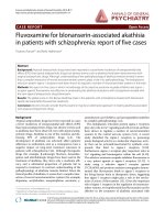
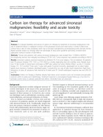
and bile duct was reduced as much as possible. Figure 1
shows a typical radiation field with dose distribution.
Evaluation during follow-up
After completion of C-ion RT, patients were followed up
one month after C-ion RT, and every 3 months thereafter. The examinations consisted of routine blood cell
counts, blood chemistry and abdominal diagnostic imaging such as four-phase multidetector-row CT, dynamic
contrast-enhanced MRI or contrast-enhanced ultrasonography. Acute and late toxicities were classified using
the National Cancer Institute’s Common Terminology
Criteria for Adverse Events, version 4.0 [24]. Acute toxicity was evaluated as the highest toxicity within 3 months
from the initiation of C-ion RT. Late toxicity was evaluated as the highest toxicity after 3 months from the initiation of C-ion RT. Local recurrence was defined as tumor
Shiba et al. BMC Cancer (2017) 17:721
Page 3 of 8
Fig. 1 An 80-year-old male with HCC treated with C-ion RT. a CT before treatment. b Dose distribution on axial CT images. The area within the
red outline is GTV and the area within the pink outline is CTV. c CT 3 months after treatment. d CT 18 months after treatment. There was a gastric
tube reconstruction of esophageal cancer after surgery to the right of the vertebrae
regrowth with enhancement of the contrast effect on CT
or MRI or ultrasonography in the irradiated field with or
without a continuous elevation of the tumor marker such
as alpha-fetoprotein (AFP), Lens culinaris agglutininreactive fraction of AFP, and protein induced by vitamin K
on blood test.
Statistical analysis
Survival was measured from the date of initiation of
treatment to the date of death or the most recent
follow-up. Progression-free survival (PFS) was measured
from the start of C-ion RT to the date of the first tumor
progression disease or death from any cause. Probabilities of overall survival (OS), local control (LC) and PFS
rates were calculated using the Kaplan-Meier method.
The log-rank test was used to compare between 2 survival curves for univariate analyses. The Cox proportional hazard regression analysis was used to determine
the implications of potential prognosticators. The statistical tests were two-sided, and a p < 0.05 was considered statistically significant. Factors with p < 0.1 in
univariate analyses were included in the multivariate
analyses. Variable risk was expressed as a hazard ratio
with a corresponding 95% confidence interval. Wilcoxon
signed ranks test was used for statistical analyses for
difference in Child-Pugh score between before and
3 months after C-ion RT. All statistical analyses were
performed using SPSS Statistics version 22 (SAS Institute,
Tokyo, Japan).
Results
Patient characteristics
A total of 31 patients were treated with C-ion RT, and
the patient characteristics are summarized in Table 1.
Median follow-up of all patients was 23.2 months (range:
8.4–55.3 months). Median age at the time of registration
for C-ion RT was 83 years (range: 80–95 years). Median
tumor size was 45 mm (range: 15–93 mm). Prior treatment for the target region of C-ion RT was TACE in 10
patients, RFA with TACE in 2 patients and TAI in 1 patient.
In patients with prior treatment, median duration between
prior treatment and C-ion RT was 3.1 months (range: 1.6–
13.2 months). There were no patients received a systemic
therapy before C-ion RT. Seventeen of 31 patients were
BCLC classification stage C. hree patients were due to
portal vein invasion, 11 were due to performance status
1–2 and 3 patients were due to portal vein invasion and
performance status 1–2. These patients were considered
as indications for local treatments. In contrast, they were
not to be indicated for systemic therapy and palliative
therapy [25, 26].
Three patients had 2 tumor lesions each, and they were
contained within one CTV. Dose fractionation schedule
was 52.8 Gy (RBE)/4 fractions in 15 patients, 60 Gy
(RBE)/4 fractions in 9 patients, and 60 Gy (RBE)/12 fractions in 7 patients. When there were calculated as the biologically equivalent dose using an α/β ratio of 10 (BED10),
C-ion RT of 52.8 Gy (RBE)/4 fractions, 60 Gy (RBE)/4
fractions and 60 Gy (RBE)/12 fractions were BED10 of
122.5 Gy, 150 Gy and 90 Gy. All patients completed C-ion
Shiba et al. BMC Cancer (2017) 17:721
Page 4 of 8
Table 1 Patient characteristics (n = 31)
Characteristics
No.
Gender
Male
22 (71%)
Female
9 (29%)
Performance status
0
17 (55%)
1
12 (39%)
2
2 (6%)
Child-Pugh classification
A
27 (87%)
B
4 (13%)
MELD score
6–7
17 (55%)
8–9
11 (35%)
10 ≤
3 (10%)
Viral marker
RT as scheduled. Median hospital stay from the start of Cion RT to discharge was 8 days (range: 5–23 days). Four
patients were treated as outpatients.
Overall survival and local control
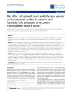
The OS, LC and PFS curves of all patients are shown in
Fig. 2. The 2-year estimated OS, LC and PFS rates were
82.3%, 89.2%, and 51.3%, respectively. At the time of
analysis, 5 patients had died of HCC, and 3 died from
intercurrent diseases (1 aspiration pneumonia, 1 pulmonary
embolism, and 1 bile duct cancer). Fourteen of 31 patients
were porta hepatis group. The 2-year OS and LC in porta
hepatis group were 50.9% and 82.5%.
In the univariate analyses, there were significant differences for overall survival in performance status, and ChildPugh classification (Table 2). In the multivariate analyses,
there was significant difference for overall survival in performance status (Table 3).
Toxicity
HBs-Ag (+), HCV-Ab (−)
2 (6%)
HBs-Ag (−), HCV-Ab (+)
18 (58%)
HBs-Ag (+), HCV-Ab (+)
1 (3%)
HBs-Ag (−), HCV-Ab (−)
10 (33%)
Co-morbidity
Diabetes mellitus
10 (33%)
Hypertension
16 (52%)
Cardiovascular disease
8 (26%)
Respiratory disease
5 (16%)
Chronic renal failure
2 (6%)
Brain disease
2 (6%)
Tumor size, mm, median [range]
45 [15–93]
Serum AFP level, ng/ml, median [range]
7.3 [1.3–48,058.3]
Serum AFP-L3, %, median [range]
3.1 [< 0.5–84.2]
Serum PIVKA-II level, mAU/ml, median [range]
85 [10–19,937]
Stage (UICC 7th edition)
I
24 (77%)
II
4 (14%)
III
3 (9%)
Stage (BCLC)
A
13 (42%)
B
1 (3%)
C
17 (55%)
Abbreviations: MELD = Model for end-stage liver disease, HBs-Ag = Hepatitis B
surface antigen, HCV-Ab = Hepatitis C virus antibody, AFP = Alpha-fetoprotein,
AFP-L3 = Lens culinaris agglutinin-reactive fraction of AFP, PIVKA-II = Protein
induced by vitamin K, AU = Arbitrary unit, UICC = Union for International Cancer
Control classification, BCLC = Barcelona Clinic Liver Classification
All of the observed acute and late toxicities are listed in
Table 4. No patients had Grade 2 or higher acute toxicities. One patient experienced progression in Child-Pugh
classification from A to B within 3 months. There was a
no significant difference in Child-Pugh score between
before and 3 months after C-ion RT (p = 0.803). As for
late toxicities, Grade 3 encephalopathy was observed in
3 patients. One of these patients had chronic renal failure
before C-ion RT. In this case, with the extensive progression of HCC and hepatic failure, encephalopathy did not
improve. The other two Grade 3 encephalopathy cases improved to baseline before C-ion RT but both patients then
developed intrahepatic metastasis. No patients had Grade
2 or higher other late toxicities such as dermatitis, pneumonitis, ascites, and rib fracture.
In porta hepatis group, no patients had Grade 2 or
higher acute toxicities. One patient experienced increasing 2 points of Child-Pugh score in acute and late phase.
Discussion
With the increasing elderly population of HCC patients
in Japan [3], a less-invasive and highly curable local
treatment strategy has to be explored.
HCC has a number of local treatment options. Surgical
resection is a well-established treatment, although its application has to be carefully selected in elderly patients.
A meta-analysis by Huang et al. presented clinical outcomes of hepatectomy for HCC in 67 elderly patients (≥
70 years old) and 268 control patients (< 70 years old)
[27]. In their report, the 3-year OS and disease-free survival between the elderly and control groups were 55%
and 40% (p = 0.017), and 58% and 41% (p = 0.157), respectively. The 2-year OS and disease-free survival rates,
according to the Kaplan-Meier method, between elderly
Shiba et al. BMC Cancer (2017) 17:721
Page 5 of 8
Fig. 2 Overall survival, local control, and disease-free survival curves. Overall survival (red line), local control (green line), and disease-free survival
(blue line) are shown for all patients treated with C-ion RT
and control groups were approximately 65% and 50%,
and approximately 60% and 52%, respectively. On the
other hand, 9.0% of patients developed postoperative
complications such as upper gastrointestinal hemorrhage
and liver failure in the elderly group. Nozawa et al., in a
report of surgical resection for HCC patients, divided
patients into super-elderly (≥ 80 years old, n = 20), elderly
(70–80 years old, n = 172) and younger (< 70 years old,
n = 239) groups [28]. The 5-year OS in the super-elderly,
elderly and younger groups were 67%, 60% and 65%,
Table 2 Local control and overall survival were analyzed by
clinical characteristics (univariate analyses)
Factor
No.
2y–LC
p-value
0.544
2y–OS
p-value
85.0%
0.034
Child-Pugh classification
A
27
85.9%
B
4
100%
37.5%
Performance status
0
17
90.9%
1, 2
14
82.5%
0.589
91.7%
0.015
64.6%
MELD score
6, 7
17
93.3%
8≤
14
80.8%
0.705
81.8%
0.707
75.5%
Stage (UICC 7th edition)
I
24
83.1%
II, III
7
100%
A, B
14
87.5%
C
17
87.1%
0.866
81.3%
0.762
71.4%
Table 3 Overall survival was analyzed by clinical characteristics
(multivariate analyses)
Stage (BCLC)
0.498
100%
respectively. The 3-year tumor-free survival rates in the
super-elderly, elderly and younger groups were 34%,
41% and 46%, respectively. The 2-year OS and 2-year
tumor-free survival rates in the super-elderly group by
Kaplan-Meier curves were approximately 100% and
10%, respectively. In these 2 studies, there was likely to
exist a selection bias of the patients in the elderly groups,
because inclusion criteria for resection was not fully described and because the elderly group generally presented
favorable results compared with the control group. Nozawa
et al. also reported a median postoperative hospital stay of
11 days in the super-elderly group [28]. Regarding complications, 30% of the super-elderly patients developed delirium ascribed to their long-term hospitalization, although
psychiatric support and/or premedication were provided
for the patients [28]. In addition, 10% of the patients developed cardiovascular disease and 5% of the patients developed abdominal infection and bile leakage. In the current
study, the 2-year estimated OS and PFS were comparable
with the result of surgery, although medically inoperable
cases were included in our population. The median hospital
stays with C-ion RT was 8 days, and 4 patients were treated
safely as outpatients. No patients developed delirium or
other severe complications, probably due to their shortterm treatment period.
0.097
Factor
A vs B
14
90.0%
45 mm ≤
17
86.2%
0.387
100%
p-value
Child-Pugh classification
67.2%
Tumor size
< 45 mm
Hazard ratio (95% confidence interval)
0.540
62.2%
Abbreviations: 2y–LC = 2-year local control rate, 2y–OS = 2-year overall survival
rate, MELD = Model for end-stage liver disease, UICC = Union for International
Cancer Control classification, BCLC = Barcelona Clinic Liver Classification
4.937 (0.818–29.796)
0.082
6.148 (1.189–31.807)
0.030
1.130 (0.277–4.605
0.864
Performance status
0 vs 1 and 2
Stage (BCLC)
A, B vs C
Shiba et al. BMC Cancer (2017) 17:721
Page 6 of 8
Table 4 Acute and late toxicities by CTCAE, version 4.0 (n = 31)
Acute toxicities
Organs involved
G0
G1
G2
G3
G4
Dermatitis
2
29
0
0
0
Pneumonitis
23
8
0
0
0
Encephalopathy
30
1
0
0
0
Ascites
31
0
0
0
0
G0
G1
G2
G3
G4
Late toxicities
Organs involved
Dermatitis
9
22
0
0
0
Pneumonitis
17
14
0
0
0
Encephalopathy
28
0
0
3
0
Ascites
26
5
0
0
0
Rib bone fracture
31
0
0
0
0
Change of Child-Pugh score after start of C-ion RT
≤0
+1
+2
+3
Acute phase
26
4
1
0
Late phase
29
5
1
0
Abbreviations: CTCAE = Common Terminology Criteria for Adverse Events, Cion RT = Carbon ion radiotherapy
RFA and PEI are performed to treat unresectable small
HCC. Tiong et al. reported a systemic review and metaanalysis of elderly patients with small HCC, 20–30 mm,
treated with RFA and PEI [29]. The 3-year disease-free
survival rates were 37–43% and 17–21%, respectively.
Nishikawa et al. reported clinical outcomes of RFA for
elderly (≥ 75 years) and control (< 75 years) patients with
HCC of 20–30 mm [30]. They reported that the 3-year OS
and recurrence-free survival rates between elderly and control groups were 64% and 84% (p = 0.001), and 21% and
40% (p = 0.001), respectively. The Kaplan-Meier method
showed that the 2-year OS and recurrence-free survival
rates between the elderly and control groups were approximately 75% and 90%, and approximately 35% and 55%,
respectively. On the other hand, there was no significant
difference in major adverse events related to RFA between
the two groups (p = 0.670). In the current study, clinical
outcomes were similar to those of HCC treated with
RFA, despite the inclusion of larger tumors (median
size, 45 mm). The indication for RFA is generally unresectable tumor of 30 mm or smaller, and is limited by
anatomical situation. In contrast, C-ion RT can be applied
for tumors larger than 30 mm or those anatomically untreatable with RFA [10].
Hata et al. reported proton therapy for 21 elderly patients
(≥ 80 years old) with HCC [31]. Their 3-year OS and 3-year
local progression-free rates were 62% and 100%, respectively, and no patient developed Grade 3 or higher toxicity
except for thrombocytopenia in 2 patients. Their median
fraction number was 22 (range: 10–35 fractions). In the
current study, the number of fractions was 4 or 12,
which was generally less than that of proton therapy.
Together with the safety of C-ion RT, in the present
study, C-ion RT also seems to be beneficial for elderly
patients in terms of avoiding long-term hospitalization
that can cause cognitive impairment.
There has been no analysis that focused on the outcome
of elderly patients with SBRT with X-rays. Andolino et al.
reported SBRT for HCC patients with a median age of
59 years and median tumor size of 31 mm [32]. Their 2year OS and 2-year LC were 67% and 90%, respectively.
There were no Grade 3 or higher acute non-hematologic
toxicities. However, 20% of the patients experienced progression in Child-Pugh class within 3 months of treatment,
7 of 36 patients with Child-Pugh class A progressing to
class B and 5 of 24 patients with class B progressing to class
C. On the other hand, only one patient experienced progression in Child-Pugh classification from A to B within
3 months in our study. Abe et al. previously reported the
results of dosimetric comparison between SBRT with Xrays and C-ion RT for HCC [22]. In their study, a low dose
volume such as 5 Gy (RBE) to 20 Gy (RBE) for normal liver
tissue was significantly lower with C-ion RT than SBRT.
Therefore, compared with SBRT with X-rays, C-ion RT
may have an advantage of conserving liver function.
Imada et al. reported comparison of efficacy and toxicity
of C-ion RT for HCC located in the porta hepatis. They
defined HCC located within 2 cm of the main portal vein
as a porta hepatis group. They reported that the 3-year
OS and LC in 18 patients were 44.4% and 87.8%. Acute
adverse events of Grade 3 or higher were developed in 9
cases. As to Child-Pugh score in late phase, cases with
changes in score increasing at least 2 points was five. In
the current study, 14 of 31 patients has HCC located
within 2 cm of the main portal vein as a porta hepatis
group. The 2-year OS and LC in porta hepatis group were
50.9% and 82.5%. No patients had Grade 2 or higher acute
toxicities. One patient experienced increasing 2 points of
Child-Pugh score in acute and late phase. In the current
study, clinical outcomes were comparable to results of
Imada et al. reported, although the current study analyzed
only elderly patients. Thus, this results suggested C-ion
RT for elderly patients with HCC located in porta hepatis
was effective and safe treatment.
There were some limitations to our study. First, this
study was a single institutional retrospective analysis with a
small number of patients and short follow-up duration.
However, the follow-up period of this study was considered
to be sufficient to confirm the safety because long-term
radiation-related adverse events are uncommon except for
radiation-induced malignancies. Second, the safety of C-ion
RT for elderly HCC patients with Child-Pugh class B
remained unclear due to small number of patients. Further
investigation is necessary to confirm the safety of C-ion RT
Shiba et al. BMC Cancer (2017) 17:721
for elderly HCC patients with Child-Pugh class B. Third,
clinical outcomes were not directly compared with other
treatment modalities including surgical resection and local
ablation therapies. However, the current study included not
amenable to surgery or local ablation therapies cases due to
the anatomical and medical reasons including poor PS and
co-morbidity. This patient selection would not have contributed to the favorable results of this study.
Conclusions
C-ion RT for patients 80 years or older with HCC was
effective with minimal toxicities. This result suggested
that C-ion RT may become an alternative treatment option for elderly HCC patients for whom surgery or local
ablation therapies are not a viable choice. Further accumulation of clinical data with larger cohorts is warranted.
Abbreviations
4-D CT: four-dimensional computed tomography; AFP: alpha-fetoprotein;
AFP-L3: Lens culinaris agglutinin-reactive fraction of alpha-fetoprotein;
AU: arbitary unit; BCLC: Barcelona Clinic Liver classification; C-ion RT: carbon
ion radiotherapy; CT: computed tomography; CTCAE: Common Terminology
Criteria for Adverse Events; CTV: clinical target volume; GHMC: Gunma
University Heavy Ion Medical Center; GTV: gross tumor volume; HBs-Ag: hepatitis B
surface antigen; HCC: hepatocellular carcinoma; HCV-Ab: hepatitis C virus
antibody; IM: internal margin; LC: local control; MELD: Model for end-Stage liver
disease; MRI: magnetic resonance imaging; OS: overall survival; PEI: percutaneous
ethanol infusion; PFS: progression-free survival; PIVKA-II: protein induced by
vitamin K; PS: performance status; PTV: planning target volume; RBE: relative
biologic effectiveness; RFA: percutaneous radiofrequency ablation;
SBRT: stereotactic body radiotherapy; TACE: transarterial chemoembolization;
TAI: transarterial infusion; UICC: Union for International Cancer Control classification
Acknowledgements
We wish to thank all patients who were involved in this study and our
colleagues at Department of Radiation Oncology, Gunma University
Graduate School of Medicine.
Funding
This work was supported by the Grants-in-Aid from the Ministry of Education,
Culture, Sports, Science, and Technology of Japan for programs for Leading
Graduate Schools, Cultivating Global Leaders in Heavy Ion Therapeutics and
Engineering. The funding body has no role in the design of the study, collection,
analysis, and interpretation of data nor in writing the manuscript.
Availability of data and materials
The datasets generated and/or analysed during the current study are not
publicly available because it contains personal information but are available
from the corresponding author on reasonable request.
Authors’ contributions
SS, TA, KShib and TO made substantial contributions to the conception and
design of the study. KShib, HKa, YK and TO treated and followed up the
patients. SS, TA, KShib, HKa, YK, and TO collected the data. SS TA, KShib and
TO drafted the manuscript and performed the statistical analysis. TO, HS, SK,
KShir, HKu and TN have been involved in revising it critically for important
intellectual content. SS, TA, KShib and TO participated in acquisition and
interpretation of data. All authors read and approved the final manuscript.
Ethics approval and consent to participate
The treatment protocol for the current study was reviewed and approved by
Gunma University Institutional Review Board, and all patients signed an
informed consent form before the initiation of therapy.
Consent for publication
Not applicable.
Page 7 of 8
Competing interests
The authors declare that they have no competing interests.
Publisher’s Note
Springer Nature remains neutral with regard to jurisdictional claims in
published maps and institutional affiliations.
Author details
1
Department of Radiation Oncology, Gunma University Graduate School of
Medicine, 3-39-22 Syowa-machi, Maebashi, Gunma 371-8511, Japan. 2Gunma
University Heavy Ion Medical Center, Maebashi, Gunma, Japan. 3Department
of Diagnostic Radiology, Shibukawa Medical Center, Shibukawa, Gunma,
Japan. 4Department of Medicine and Molecular Science, Gunma University
Graduate School of Medicine, Maebashi, Gunma, Japan. 5Department of
Hepato-Biliary and Pancreatic surgery, Gunma University Graduate School of
Medicine, Maebashi, Gunma, Japan. 6Department of Surgical Science, Gunma
University Graduate School of Medicine, Maebashi, Gunma, Japan.
Received: 28 October 2016 Accepted: 30 October 2017
References
1. Ferlay J, Shin HR, Bray F, Forman D, Mathers C, Parkin DM. Estimates of
worldwide burden of cancer in 2008: GLOBOCAN 2008. Int J Cancer.
2010;127(12):2893–917.
2. Forner A, Llovet JM, Bruix J. Hepatocellular carcinoma. Lancet (London,
England). 2012;379(9822):1245–55.
3. Umemura T, Ichijo T, Yoshizawa K, Tanaka E, Kiyosawa K. Epidemiology of
hepatocellular carcinoma in Japan. J Gastroenterol. 2009;44(Suppl 19):102–7.
4. Kumar V, Abbas Abdul K, Fausto NR. Cotran pathological basis of disease
7th edition. In: Saunders. 2005;
5. Cho YK, Kim JK, Kim MY, Rhim H, Han JK. Systematic review of randomized
trials for hepatocellular carcinoma treated with percutaneous ablation
therapies. Hepatology. 2009;49(2):453–9.
6. Bujold A, Massey CA, Kim JJ, Brierley J, Cho C, Wong RK, Dinniwell RE,
Kassam Z, Ringash J, Cummings B, et al. Sequential phase I and II trials of
stereotactic body radiotherapy for locally advanced hepatocellular
carcinoma. J Clin Oncol. 2013;31(13):1631–9.
7. Hata M, Tokuuye K, Sugahara S, Fukumitsu N, Hashimoto T, Ohnishi K,
Nemoto K, Ohara K, Matsuzaki Y, Akine Y. Proton beam therapy for
hepatocellular carcinoma with limited treatment options. Cancer.
2006;107(3):591–8.
8. Kimura T, Aikata H, Takahashi S, Takahashi I, Nishibuchi I, Doi Y, Kenjo M,
Murakami Y, Honda Y, Kakizawa H, et al. Stereotactic body radiotherapy for
patients with small hepatocellular carcinoma ineligible for resection or
ablation therapies. Hepatol Res. 2015;45(4):378–86.
9. Komatsu S, Fukumoto T, Demizu Y, Miyawaki D, Terashima K, Sasaki R, Hori
Y, Hishikawa Y, Ku Y, Murakami M. Clinical results and risk factors of proton
and carbon ion therapy for hepatocellular carcinoma. Cancer. 2011;117(21):
4890–904.
10. Imada H, Kato H, Yasuda S, Yamada S, Yanagi T, Kishimoto R, Kandatsu S,
Mizoe JE, Kamada T, Yokosuka O, et al. Comparison of efficacy and toxicity
of short-course carbon ion radiotherapy for hepatocellular carcinoma
depending on their proximity to the porta hepatis. Radiother Oncol.
2010;96(2):231–5.
11. Kamada T, Tsujii H, Blakely EA, Debus J, De Neve W, Durante M, Jakel O,
Mayer R, Orecchia R, Potter R, et al. Carbon ion radiotherapy in Japan:
an assessment of 20 years of clinical experience. The Lancet Oncology.
2015;16(2):e93–e100.
12. Sobin LH, Gospodarowicz MK, Wittekind C. TNM classification of malignant
tumours 7th ed: Wiley- Blackwell; 2011.
13. Llovet JM, Bru C, Bruix J. Prognosis of hepatocellular carcinoma: the BCLC
staging classification. Semin Liver Dis. 1999;19(3):329–38.
14. Singal AK, Kamath PS. Model for end-stage liver disease. J clin exp
hepatology. 2013;3(1):50–60.
15. Kanematsu N. Dose calculation algorithm of fast fine-heterogeneity correction
for heavy charged particle radiotherapy. Phys Med. 2011;27(2):97–102.
16. Kanematsu N, Akagi T, Takatani Y, Yonai S, Sakamoto H, Yamashita H.
Extended collimator model for pencil-beam dose calculation in proton
radiotherapy. Phys Med Biol. 2006;51(19):4807–17.
Shiba et al. BMC Cancer (2017) 17:721
Page 8 of 8
17. Kanematsu N, Endo M, Futami Y, Kanai T, Asakura H, Oka H, Yusa K. Treatment
planning for the layer-stacking irradiation system for three-dimensional
conformal heavy-ion radiotherapy. Med Phys. 2002;29(12):2823–9.
18. Kanematsu N, Torikoshi M, Mizota M, Kanai T. Secondary range shifting
with range compensator for reduction of beam data library in heavy-ion
radiotherapy. Med Phys. 2007;34(6):1907–10.
19. Kanematsu N, Yonai S, Ishizaki A. The grid-dose-spreading algorithm for
dose distribution calculation in heavy charged particle radiotherapy.
Med Phys. 2008;35(2):602–7.
20. Inaniwa T, Kanematsu N, Matsufuji N, Kanai T, Shirai T, Noda K, Tsuji H,
Kamada T, Tsujii H. Reformulation of a clinical-dose system for carbon-ion
radiotherapy treatment planning at the National Institute of Radiological
Sciences, Japan. Phys Med Biol. 2015;60(8):3271–86.
21. Kanai T, Endo M, Minohara S, Miyahara N, Koyama-ito H, Tomura H,
Matsufuji N, Futami Y, Fukumura A, Hiraoka T, et al. Biophysical
characteristics of HIMAC clinical irradiation system for heavy-ion radiation
therapy. Int J Radiat Oncol Biol Phys. 1999;44(1):201–10.
22. Abe T, Saitoh J, Kobayashi D, Shibuya K, Koyama Y, Shimada H, Shirai K,
Ohno T, Nakano T. Dosimetric comparison of carbon ion radiotherapy and
stereotactic body radiotherapy with photon beams for the treatment of
hepatocellular carcinoma. Radiat Oncol. 2015;10:187.
23. Abe T, Shibuya K, Koyama Y, Okamoto M, Kiyohara H, Katoh H, Shimada H,
Kuwano H, Ohno T, Nakano T. Initial results of Hypofractionated carbon ion
radiotherapy for Cholangiocarcinoma. Anticancer Res. 2016;36(6):2955–60.
24. Health UDo, Services H: National cancer institute: common terminology criteria
for adverse events (CTCAE), version 4.0. US Department of Health and Human
Services National Institutes of health National Cancer Institute 2009.
25. Cabibbo G, Maida M, Genco C, Parisi P, Peralta M, Antonucci M, Brancatelli G,
Camma C, Craxi A, Di Marco V. Natural history of untreatable hepatocellular
carcinoma: a retrospective cohort study. World J Hepatol. 2012;4(9):256–61.
26. Cheng AL, Kang YK, Chen Z, Tsao CJ, Qin S, Kim JS, Luo R, Feng J, Ye S,
Yang TS, et al. Efficacy and safety of sorafenib in patients in the Asia-Pacific
region with advanced hepatocellular carcinoma: a phase III randomised,
double-blind, placebo-controlled trial. Lancet Oncol. 2009;10(1):25–34.
27. Huang J, Li BK, Chen GH, Li JQ, Zhang YQ, Li GH, Yuan YF. Long-term
outcomes and prognostic factors of elderly patients with hepatocellular
carcinoma undergoing hepatectomy. J Gastrointest Surg. 2009;13(9):1627–35.
28. Nozawa A, Kubo S, Takemura S, Sakata C, Urata Y, Nishioka T, Kinoshita M,
Hamano G, Uenishi T, Suehiro S. Hepatic resection for hepatocellular
carcinoma in super-elderly patients aged 80 years and older in the first
decade of the 21st century. Surg Today. 2015;45(7):851–7.
29. Tiong L, Maddern GJ. Systematic review and meta-analysis of survival and
disease recurrence after radiofrequency ablation for hepatocellular
carcinoma. Br J Surg. 2011;98(9):1210–24.
30. Nishikawa H, Osaki Y, Iguchi E, Takeda H, Ohara Y, Sakamoto A, Hatamaru K,
Henmi S, Saito S, Nasu A, et al. Percutaneous radiofrequency ablation for
hepatocellular carcinoma: clinical outcome and safety in elderly
patients. J Gastrointestin Liver Dis. 2012;21(4):397–405.
31. Hata M, Tokuuye K, Sugahara S, Tohno E, Nakayama H, Fukumitsu N,
Mizumoto M, Abei M, Shoda J, Minami M, et al. Proton beam therapy for
aged patients with hepatocellular carcinoma. Int J Radiat Oncol Biol Phys.
2007;69(3):805–12.
32. Andolino DL, Johnson CS, Maluccio M, Kwo P, Tector AJ, Zook J, Johnstone
PA, Cardenes HR. Stereotactic body radiotherapy for primary hepatocellular
carcinoma. Int J Radiat Oncol Biol Phys. 2011;81(4):e447–53.
Submit your next manuscript to BioMed Central
and we will help you at every step:
• We accept pre-submission inquiries
• Our selector tool helps you to find the most relevant journal
• We provide round the clock customer support
• Convenient online submission
• Thorough peer review
• Inclusion in PubMed and all major indexing services
• Maximum visibility for your research
Submit your manuscript at
www.biomedcentral.com/submit