Detection of circulating miRNAs: Comparative analysis of extracellular vesicle-incorporated miRNAs and cell-free miRNAs in whole plasma of prostate cancer patients
Bạn đang xem bản rút gọn của tài liệu. Xem và tải ngay bản đầy đủ của tài liệu tại đây (1.32 MB, 13 trang )
Endzeliņš et al. BMC Cancer (2017) 17:730
DOI 10.1186/s12885-017-3737-z
RESEARCH ARTICLE
Open Access
Detection of circulating miRNAs:
comparative analysis of extracellular
vesicle-incorporated miRNAs and cell-free
miRNAs in whole plasma of prostate cancer
patients
Edgars Endzeliņš1†, Andreas Berger1†, Vita Melne1,2, Cristina Bajo-Santos1, Kristīne Soboļevska1, Artūrs Ābols1,
Marta Rodriguez3, Daiga Šantare4, Anastasija Rudņickiha1, Vilnis Lietuvietis1,2, Alicia Llorente3 and Aija Linē1*
Abstract
Background: Circulating cell-free miRNAs have emerged as promising minimally-invasive biomarkers for early
detection, prognosis and monitoring of cancer. They can exist in the bloodstream incorporated into extracellular
vesicles (EVs) and ribonucleoprotein complexes. However, it is still debated if EVs contain biologically meaningful
amounts of miRNAs and may provide a better source of miRNA biomarkers than whole plasma. The aim of this
study was to systematically compare the diagnostic potential of prostate cancer-associated miRNAs in whole
plasma and in plasma EVs.
Methods: RNA was isolated from whole plasma and plasma EV samples from a well characterised cohort of 50
patient with prostate cancer (PC) and 22 patients with benign prostatic hyperplasia (BPH). Nine miRNAs known to
have a diagnostic potential for PC in cell-free blood were quantified by RT-qPCR and the relative quantities were
compared between patients with PC and BPH and between PC patients with Gleason score ≥ 8 and ≤6.
Results: Only a small fraction of the total cell-free miRNA was recovered from the plasma EVs, however the EVincorporated and whole plasma cell-free miRNA profiles were clearly different. Four of the miRNAs analysed showed a
diagnostic potential in our patient cohort. MiR-375 could differentiate between PC and BPH patients when analysed in
the whole plasma, while miR-200c-3p and miR-21-5p performed better when analysed in plasma EVs. EV-incorporated
but not whole plasma Let-7a-5p level could distinguish PC patients with Gleason score ≥ 8 vs ≤6.
Conclusions: This study demonstrates that for some miRNA biomarkers EVs provide a more consistent source of RNA
than whole plasma, while other miRNAs show better diagnostic performance when tested in the whole plasma.
Keywords: Prostate cancer, Cell-free miRNAs, Extracellular vesicles, Exosomes, Microvesicles, Biomarkers, Liquid biopsy
* Correspondence:
†
Equal contributors
1
Latvian Biomedical Research and Study Centre, Ratsupites Str 1, k-1, Riga
LV-1067, Latvia
Full list of author information is available at the end of the article
© The Author(s). 2017 Open Access This article is distributed under the terms of the Creative Commons Attribution 4.0
International License ( which permits unrestricted use, distribution, and
reproduction in any medium, provided you give appropriate credit to the original author(s) and the source, provide a link to
the Creative Commons license, and indicate if changes were made. The Creative Commons Public Domain Dedication waiver
( applies to the data made available in this article, unless otherwise stated.
Endzeliņš et al. BMC Cancer (2017) 17:730
Background
Circulating cell-free micro-RNAs (miRNAs) have emerged
as promising biomarkers for the development of bloodbased assays for early detection, prognosis and monitoring
of cancer. In 2008, Mitchell et al. demonstrated for the first
time that miRNAs are released from prostate cancer (PC)
cells into the bloodstream, where they exist in a remarkably
stable form [1]. miRNAs were shown to remain stable after
incubation of plasma or serum at room temperature for up
to 24 h and to resist RNase A digestion, HCl and NaOH
treatment or multiple freeze-thaw cycles [1, 2]. Subsequently, the levels of circulating miRNAs have been studied
in patients with various cancers, including PC, resulting in
the discovery of individual miRNAs or miRNA signatures
with diagnostic and/or prognostic value [3].
PC is the most frequently diagnosed cancer in males in
Europe and the United States [4, 5]. Currently, the serum
PSA test is the most commonly used tool for organised
screening programs, opportunistic screening and monitoring of PC [6]. However, PSA is not cancer specific and the
high false-positive rate and low specificity leads to large
numbers of unnecessary prostate biopsies and emotional
morbidity [7]. Furthermore, PC is characterised by a highly
heterogeneous course - one part of the patients develops a
high-grade disease with extracapsular spread and distant
metastases requiring aggressive treatment, while others
have a relatively indolent, slowly progressing disease that
could have been managed by active surveillance [8]. The
current standard of care analyses, however, do not predict
whether a histologically proven tumour will give rise to a
clinically significant disease, leading to overtreatment of
indolent PC. Hence, the greatest unmet clinical needs in
the management of PC are sensitive and reliable noninvasive tools for differentiating between PC and benign
prostatic diseases, and between potentially fast progressing
PC requiring aggressive treatment and a relatively indolent
disease that can be managed by active surveillance.
More than 20 studies investigating levels of cell-free
miRNAs in plasma or serum of PC patients have been
published up to date [9, 10]. The majority of these studies
were focused on the identification of circulating miRNAs
that differentiate between patients with PC and benign
prostatic hyperplasia (BPH) or healthy controls. Some of
these studies have shown remarkably high diagnostic
value. For example, Chen et al. identified a 5 miRNA panel
that could differentiate PC from BPH with an AUC of
0.924 and PC from healthy controls with an AUC of 0.860
[11]. Some other studies have reported cell-free miRNAs
that differentiate between localised and metastatic castration resistant prostate cancer (mCRPC) or between lowgrade and high-grade PC. For example, Mihelich et al.
developed a “miRScore” that based on the serum levels of
14 miRNAs could predict absence of high-grade PC
among men with PC and BPH with a negative predictive
Page 2 of 13
value of 0.939 [12]. However, relatively few miRNA
biomarkers have been validated by several independent
studies, while many other miRNAs either have been
reported in a single study or show conflicting results [3,
10]. Therefore, the analysis of cell-free miRNAs is regarded
as a poorly reproducible technique [3, 13, 14].
Cell-free miRNAs circulating in the bloodstream have
been found to be enclosed into extracellular vesicles (EVs)
[15, 16], or to exist in a vesicle-free form associated with
high-density lipoproteins [17], Ago2 protein [18, 19] or
other RNA binding proteins [20]. The majority of the
studies has used whole plasma or serum as a source of
cell-free miRNAs. However, it has recently been hypothesised that cancer-derived EVs may be enriched with
miRNA signatures reminiscent of their cell of origin, contain rare yet highly specific RNA biomarkers and protect
their RNA cargo from degradation in the bloodstream and,
therefore, the analysis of EV-enclosed miRNAs may be superior to whole plasma/serum analysis [10, 21, 22]. Nevertheless, to the best of our knowledge, a direct comparison
of miRNA detection assays in whole plasma and plasma
EVs has not been reported so far.
In this study, we evaluated the performance of 9 miRNA
biomarkers previously reported to have a diagnostic or
prognostic significance in PC by quantifying them in the
whole plasma and plasma EVs in a cohort of 50 PC and
22 BPH patients.
Methods
Study population and sample collection
Patients with PC and BPH were recruited between
September 2011 and December 2013 at Riga East
University Hospital and subsequently were followed up
until December 2016. The diagnosis was established
using standard of care diagnostic examinations and
Gleason score was determined according to standard
histopathological criteria by an experienced pathologist.
Pre-treatment blood samples were collected into EDTAcoated tubes and processed at room temperature within
2 h of blood draw. Plasma samples were centrifuged twice
for 10 min at 2000 g, aliquoted and stored at −80 °C until
analysis. The samples were deposited into the Latvian
Genome Database. Biobanking procedures were approved
by the Committee of Medical Ethics of Latvia and the use
of clinical samples for the research was approved by the
Committee of Biomedical Ethics of Riga East University
Hospital. The blood samples were collected after the
patients’ informed written consent was obtained.
The following groups of patients were selected from
the Database: PC with Gleason score ≥ 8 (Gleason high,
n = 24), PC with Gleason score ≤6 (Gleason low, n = 26)
and BPH (absence of PC confirmed by histological
examination of ultrasound-guided needle biopsies and
no change in the diagnosis within the follow-up period,
Endzeliņš et al. BMC Cancer (2017) 17:730
Page 3 of 13
n = 22). Clinical data of the study population are
provided in Table 1. In addition, plasma samples from 5
PC patients and 5 healthy controls were used for the
quality control of EV isolation.
5 min. Then the samples were negatively stained with
1% uranyl formate (w/v) for 1 min, dried and examined
using JEM-1230 transmission electron microscope
(JEOL, USA).
Isolation of extracellular vesicles
Nanoparticle tracking analysis
EVs were isolated from 400 μl of plasma using size
exclusion chromatography (SEC). SEC columns were prepared by filling TELOS SPE columns (Kinesis, USA) with
10 ml (bed volume) of CL6B sepharose (GE Healthcare,
USA). Plasma samples were loaded on the columns and
gravity-eluted with PBS. The eluate was collected in 12
sequential 0.5 ml fractions. Each fraction was measured by
Zetasizer Nano ZS (Malvern, UK) and fractions containing particles larger than 30 nm were combined and concentrated to 100 μl using Amicon Ultra 3 kDa centrifugal
filters (Merck, Millipore, Germany).
Size distribution profile and concentration of EVs was determined using NanoSight NS500 instrument (Malvern,
UK). EV samples were diluted 1000–25,000 fold in PBS to
achieve particle concentration in range from 1×10⁸ to
1×10⁹ particles/ml. For each sample, five 30 s videos were
recorded with the following settings: 25C, 0.944–0.948 cP,
1259 slider shutter, 366 slider gain, and 11 camera level.
The data analysis was performed with NanoSight NTA
Software v3.1 Build 3.1.54 in the auto mode.
Transmission electron microscopy
Ten μl of EV suspension in PBS were applied to 300mesh carbon coated copper EM grid and incubated for
Western blot
EVs and PC-3 cells (used as a positive control) were
lysed in RIPA buffer (150 ml NaCl, 1% Triton X-100,
0.5% Na deoxycholate, 0.1% SDS, 50 ml Tris) and the
protein concentration was assessed using Pierce™ BCA
Table 1 Clinical characteristics of the study population
Characteristics
Prostate cancer, n = 50
Benign prostatic hyperplasia, n = 22
Age (years)
Mean ± SD
66 ± 7
61 ± 8
Median (range)
65 (54–85)
60 (44–75)
Missing
1
4
Serum PSA (ng/ml)
0–4.0
3 (6%)
9 (41%)
4.1–20.0
31 (62%)
13 (59%)
> 20.0
15 (30%)
0 (0%)
Missing
1 (2%)
0 (0%)
4–6
26 (52%)
–
8–9
24 (48%)
–
M0
39 (78%)
–
M1
3 (6%)
–
Missing
8 (16%)
–
G1
0 (0%)
–
G2
11 (22%)
–
G3
12 (24%)
–
Missing
27 (54%)
–
40 (80%)
11 (50%)
Gleason score
Metastasis status
Cancer grade
Prostatitis
–
+
8 (16%)
11 (50%)
Missing
2 (4%)
0 (0%)
Endzeliņš et al. BMC Cancer (2017) 17:730
Protein Assay Kit (Thermo Fisher Scientific, USA) following manufacturer’s instructions. Thirty micrograms
of EV and cell proteins were mixed with Laemmli buffer
under reduction conditions, denatured for 5 min at 100 °C
and loaded on 10% SDS-PAGE gel. Proteins were electroblotted to nitrocellulose membranes and the membranes
were blocked with 10% (w/v) fat-free milk and then incubated with the following primary antibodies: anti-TSG101
(Abcam, # ab125011), Calnexin (Abcam, # ab22595), CD9
(Santa Cruz Biotechnology, # sc-13118) and β-actin
(Abcam, # ab8224) in 1:1000 dilution. The blots were
washed and incubated with horseradish peroxidaseconjugated goat anti-rabbit IgG F(ab’)2-HRP (1:2000)
(Santa Cruz, #sc-3837) or chicken anti-mouse IgG-HRP
(1:2000) (Santa Cruz, #sc-2962) secondary antibodies, respectively. Protein expression was visualized using
Western Blotting Detection Reagent kit (GE HealthCare
Lifesciences, Germany).
Page 4 of 13
PCR primer sets and ExiLENT SYBR Green master mix
(Exiqon) according to the manufacturer’s protocol on ViiA
7 Real-Time PCR system (Thermo Fisher Scientific).
Statistical analysis
Ct values were averaged between duplicates and normalized
against UniSp6 spike-ins by subtracting them from average
spike-in Ct values in the same samples, resulting in log2
relative quantities (log2 RQ’s). The statistical analyses were
performed with GraphPadPrism 5 (GraphPad, USA). A
non-parametric Mann-Whitney U test was used to compare the RQ values of each miRNA between the groups of
samples. Multiple testing correction was done by false
discovery rate (FDR) estimation and adjusted (adj.) P-value
of ≤0.05 was considered to be significant. To assess the
diagnostic potential, the area under the ROC curve (AUC)
was calculated for each miRNA.
Results
Enzymatic treatment
Selection of miRNA biomarkers
Prior to RNA extraction, EVs samples were treated with
1 mg/ml proteinase K (Thermo Fisher Scientific, USA) for
30 min at 37 °C. Proteinase K was inactivated by incubating the samples for 10 min at 65 °C. Then the samples
were treated with 10 ng/μl RNase A (Thermo Fisher
Scientific, USA) for 15 min at 37 °C.
Nine miRNAs, whose levels in plasma or serum have been
reported to have a diagnostic or prognostic significance in
PC in at least two independent studies, were selected for
this study. Studies showing their relevance for the diagnosis
or prognosis of PC are summarised in Table 2. MiR-21-5p,
miR-200c-3p, miR-210-3p and miR-375 have been shown
to be increased in the blood of PC patients as compared to
BPH or healthy controls consistently by two or more studies, while miR-30c-5p and miR-223-3p were found to be
consistently decreased in the blood of PC patients. Inconsistent findings have been reported for Let-7a-5p, miR-1413p and miR-106a-5p.
RNA extraction
RNA was extracted from EV and whole plasma samples
using miRNeasy Micro Kit (Qiagen, USA) according to
the manufacturer’s instructions with slight modifications
of the protocol. Briefly, 5 volumes of QIAzol Lysis
Reagent were added to each sample. Subsequently, samples were spiked with 1 μl of UniSp6 (Exiqon, Denmark),
which was used as a normaliser in downstream analysis.
After adding 1 volume of chloroform samples were centrifuged for 15 min at 12000 g at 4 °C and the aqueous
phase was transferred to a new tube. Then, 1.5 volumes
of 100% ethanol were added to each sample and the
mixture was loaded onto a MinElute spin column.
Columns were centrifuged at 1000 g for 30 s at room
temperature in each round until entire sample was
loaded. RNA was eluted in 15 μl of RNase-free water
using low-bind tubes. The quantity and quality of RNA
was assessed using Agilent 2100 Bioanalyzer and RNA
6000 Pico Kit (Agilent technologies, # 5067–1513).
RT-qPCR analysis
One third of each RNA sample isolated from EVs and
whole plasma was reverse-transcribed using miRCURY
LNA Universal cDNA Synthesis kit II (Exiqon) according to the manufacturer’s protocol. cDNA reaction mixtures were diluted 1:40 and 4 μl were used for qPCR
reactions. qPCR was carried out using microRNA LNA
Yield and purity of EVs
In order to compare the levels of the selected miRNAs in
plasma EVs and whole plasma, each plasma sample was
divided into two 400 μl aliquots – one was used for the
isolation of EV-incorporated RNA, while another was
used directly for the isolation of cell-free RNA from whole
plasma according to the workflow shown in Fig. 1a.
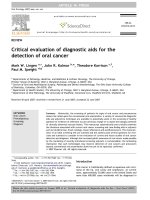
To assess the yield and purity of EVs, EV samples from
5 PC patients and 5 healthy controls (not included in
the miRNA analysis) were characterised by transmission
electron microscopy (TEM), nanoparticle tracking
analysis (NTA) and Western blot analysis. TEM images
revealed that the majority of particles were ranging in
size from 25 to 60 nm that corresponds to the size of
exosomes (Fig. 1b). However, as it has been shown that
SEC-based EV isolation methods do not result in
lipoprotein-free EV preparations [23], it cannot be
excluded that a fraction of the particles are lipoproteins.
NTA showed that the concentrations of EVs range from
3.14×10¹⁰ to 1.27×10¹² particles per ml of plasma
(Fig. 1c). The EV count was slightly increased in plasma
Endzeliņš et al. BMC Cancer (2017) 17:730
Page 5 of 13
Table 2 Circulating cell-free miRNA biomarkers for prostate cancer
miRNA
Let-7a5p
Expression in PC tissues
Level in blood
Direction
Ref. Sample
type
Patient groups and sample size Direction
Normalisation
Ref.
Down in PC vs adj.
Normal tissues
[45] Serum
PC (n = 75), BPH (n = 27)
Down in PC
RNA input and
miR-16, miR-425
[52]
Down in PC vs BPH
[44] Serum
High grade PC (n = 50),
low grade PC (n = 50),
BPH (n = 50)
Down in high grade PC
vs low grade PC, BPH
RNA input and
spike-ins
[12]
Disseminated PC (n = 20),
BPH (n = 13)
Up in disseminated PC
Spike-in and
miR-320a
[37]
Serum
miR-215p
Up in PC vs adj. Normal
(n = 10)
[55] Plasma
mCRPC (n = 25, pooled),
LPC (n = 25, pooled)
Up in mCRPC
miR-30e
[40]
Similar in PC and adj.
Normal tissues (n = 36)
[56] Serum
ADPC (n = 20), HRPC
(n = 10), LPC (n = 20),
BPH (n = 6)
Up in HRPC vs ADPC, LPC
U6 snRNA
[42]
Up in PC vs
normal tissues
[57] Plasma
PC (n = 51),
HC (n = 20)
Up in PC
RNU1A snRNA
[43]
miR-30c- Up in PC vs adj. Normal
5p
epithelium (n = 37)
[58] Serum
High grade PC (n = 50),
low grade PC (n = 50),
BPH (n = 50)
Down in high grade PC
vs low grade, BPH
RNA input and
spike-ins
[12]
Up in PC vs
normal tissues
[57] Plasma
PC (n = 80), BPH (n = 44),
HC (n = 54)
Down in PC vs BPH, HC
U6 snRNA
[11]
Serum
PC (n = 36), HC (n = 12)
Down in PC
RNA input
[51]
[57] Serum
High grade PC (n = 50),
low grade PC (n = 50),
BPH (n = 50)
Down in high grade PC
RNA input and
spike-ins
[12]
miR106a-5p
Up in PC vs
normal tissues
Serum
PC (n = 36), HC (n = 12)
Up in PC
RNA input
[51]
[53] Serum
High grade PC (n = 50),
low grade PC (n = 50),
BPH (n = 50)
Detectable in <50%
of patients
RNA input and
spike-ins
[12]
Up in PC vs BPH
[52] Serum
PC (n = 75), BPH (n = 27)
Up in PC
RNA input and
miR-16, miR-425
[52]
Up in BCR after RP
vs. no BCR after RP
[59] Plasma
mCRPC (n = 25, pooled),
LPC (n = 25, pooled)
Up in mCRPC
miR-30e
[40]
mCRPC (n = 26), low-risk LPC
(n = 28)
Up in mCRCP
U6 snRNA
[53]
PC (n = 78), HC (n = 28)
Up in PC
Spike-ins
[38]
Serum
EVs
mPC (n = 47), non-recurrent
PC (n = 72)
Up in mPC
Serum
71 PC: N1 (n = 48), N0 (n = 23), Up in N1 PC vs N0 PC;
GS ≥8 (n = 29), GS = 7 (n = 42) Up in GS ≥ 8 vs GS = 7
Spike-ins
[54]
Plasma
mPC (n = 25), LPC (n = 26)
Up in mPC vs LPC; Similar
in PC and HC
RNU1A snRNA
[43]
Serum
mPC (n = 25), HC (n = 25)
Up in mPC
Spike-ins
[60]
Serum
PC (n = 54), non-malignant
(n = 79)
Up in higher GS; Similar in
PC and non-malignant
RNU1–4 and
SNORD43
[61]
mCRPC (n = 25, pooled),
LPC (n = 25, pooled)
Up in mCRPC
miR-30e
[40]
mCRPC (n = 25), HC (n = 25)
Up in mCRCP
Spike-ins
[41]
PC (n = 31), BPH (n = 13)
Up in PC
Spike-in and miR320a
[37]
Up in mCRCP
Spike-ins
[41]
Down in high grade PC
vs low grade, BPH
RNA input and
spike-ins
[12]
miR-141- Up in mPC, PC vs
3p
normal tissues
Serum
Up in PC (n = 36)
vs normal tissue
(n = 36)
miR200c-3p
Up in PC vs
normal tissue
[54] Plasma
EVs
[62] Plasma
Serum
miR-210- Up in PC vs BPH
3p
[44] Serum
miR-223- Up in PC vs adj.
3p
Normal tissues (n = 10)
[63] Serum
Serum
mCRPC (n = 21), HC (n = 20)
Endzeliņš et al. BMC Cancer (2017) 17:730
Page 6 of 13
Table 2 Circulating cell-free miRNA biomarkers for prostate cancer (Continued)
miRNA
Expression in PC tissues
Direction
Level in blood
Ref. Sample
type
Patient groups and sample size Direction
Normalisation
Ref.
[51]
High grade PC (n = 50),
low grade PC (n = 50),
BPH (n = 50)
miR-375
Up in PC vs
normal tissues
[57] Serum
PC (n = 36), HC (n = 12)
Down in PC
RNA input
Up in mPC, PC
vs normal tissues
[53] Plasma
EVs
CRPC (n = 100)
High miRNA level
associated with poor OS
RNA input and miR- [39]
30a-5p, miR-30e-5p
Serum
PC (n = 31), BPH (n = 13)
Up in PC
Spike-in and miR320a
[37]
[54] Plasma
mCRPC (n = 25, pooled),
LPC (n = 25, pooled)
Up in mCRPC
miR-30e
[40]
Serum
mCRPC (n = 26), low-risk
LPC (n = 28)
Up in mCRCP
U6 snRNA
[53]
Serum
EVs
mPC after RP (n = 47),
non-recurrent PC after
RP (n = 72)
Up in mPC
Spike-ins
[38]
Serum
71 PC: N1 (n = 48), N0
(n = 23), GS ≥8 (n = 29),
GS = 7 (n = 42)
Up in N1 PC vs N0 PC;
Spike-ins
similar in GS ≥ 8 and GS = 7
[54]
Up in PC (n = 36) vs
normal tissue (n = 36)
ADPC androgen-dependent prostate cancer, BCR biochemical recurrence, BPH benign prostatic hyperplasia, CRPC castration resistant prostate cancer, EVs extracellular vesicles, HC healthy control, HRPC hormone-refractory prostate cancer, LPC localized prostate cancer, mCRPC metastatic castration resistant prostate cancer,
mPC metastatic prostate cancer, PC prostate cancer, RP radical prostatectomy
from PC patients as compared to the healthy controls
(mean count in PC 7.08×10¹¹ vs 4.15×10¹¹ in healthy
controls), although the difference didn’t reach statistical
significance in our sample set. The size distribution
analysis showed that the diameter for the majority of
particles was in the range from 50 to 150 nm with a
minor fraction reaching ~230 nm (Fig. 1d), which is
somewhat inconsistent with the TEM results. This
discrepancy likely has arisen due to the difference in the
minimum detectable EV size between both techniques
[24] and /or shrinking of EVs during fixation for TEM
[25]. Western blot analysis showed that the EVs were
positive for typical EV markers TSG101 and CD9, and
negative for the endoplasmic reticulum protein Calnexin
(Fig. 1e). Taken together, these results show that the EV
isolation method used in this study results in a relatively
high yield of exosome-enriched EV preparations without
detectable contamination of intracellular components.
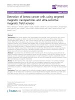
treatment with proteinase K prior to RNase A resulted
in the reduction of RQs by 50.4 to 69.3%. This suggests
that the proteinase K treatment is required for efficient
removal of extra-vesicular RNA. Therefore, in order to
study the intraluminal miRNAs, all EV preparations
were treated with proteinase K and RNase A prior to the
RNA extraction. RNA was extracted from EVs and
whole plasma using miRNeasy Micro kit, which is
designed for isolation of total RNA from small amounts
of sample. Typical RNA profiles obtained by Bioanalyzer
from whole plasma and EVs are shown in Fig. 2b. The
profiles show the presence of small RNA peaks of 25 to
200 nt both in whole plasma and EVs, while 18S and
28S rRNA peaks are present in whole plasma and EVs
without the enzymatic treatment (not shown) but not in
the treated EVs, thus suggesting that the majority of
rRNA is bound to the surface of EVs.
Relative abundance of EV-incorporated miRNAs
RNA profiles in EVs and whole plasma
As it has been suggested that EVs may associate with
lipoproteins or protein complexes that carry cell-free
miRNAs and protect them from degradation [18, 26], we
first tested the effect of proteinase K and RNase A treatment on the miRNA levels in plasma EVs from three
healthy individuals (Fig. 2a). Treatment of EVs with
RNase A alone reduced the relative quantity (RQ) values
by 15.5 to 43.6% for different miRNAs, while the
An equal proportion (one third) from the total RNA
amount obtained from the EV and whole plasma
samples of PC and BPH patients was used for the RTqPCR analysis of the 9 selected miRNA biomarkers.
Spike-ins were used to control for a variation in RNA
extraction, cDNA synthesis and PCR efficiency and they
typically varied less than by 1 Ct. In order to assess the
relative abundance of EV-enclosed miRNAs, a ratio
between EV-enclosed and total cell-free miRNAs in
Endzeliņš et al. BMC Cancer (2017) 17:730
Page 7 of 13
Fig. 1 Workflow of the study and characterisation of plasma EVs. a Workflow of the study. b Representative transmission electron microscopy
image of plasma EVs. c Quantification of EVs isolated from plasma of PC patients and healthy controls (HC) by nanoparticle tracking analysis. d
Average size distribution of EVs isolated from plasma of PC patients and healthy controls. e Western blot analysis of EV markers (TSG101, CD9),
endoplasmic reticulum protein Calnexin and β-actin in plasma EVs isolated from two healthy individuals and PC-3 cells (as a positive control)
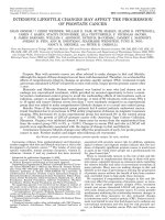
whole plasma was calculated (Fig. 3a). The results
showed that only a small fraction of the cell-free miRNA
was retrieved from the EVs. However, the EV-enclosed
fraction was not uniformly low – it varied from 6.36%
for Let-7a-5p to 0.65% for miR-210-3p. Spearman
correlation analysis revealed only weak to moderate
correlation between EV-enclosed and whole plasma cellfree miRNAs (Table 3). As an example, a paired dot plot
in Fig. 3b shows the discordance in the Let-7a-5p levels
in EVs and whole plasma from the same patients. These
data support the idea that EV-enclosed miRNA profile differs from cell-free miRNA profile in the whole plasma.
Clearly, the size of the EV-enclosed miRNA fraction
depends on the efficacy of the EV isolation method and
the obtained ratios are not expected to represent the
EV-enclosed: EV-free miRNA ratio. However, the NTA
Fig. 2 Effects of proteinase K and RNase A treatment on the relative quantity of EV-incorporated miRNAs and RNA profiles in whole plasma and
EVs. a RT-qPCR analysis of miRNA levels in EVs treated with RNase A alone or with a combination of proteinase K and RNase A relatively to untreated EVs.
Bars show the mean percentage in EVs from 3 healthy individuals. b A representative RNA profile from whole plasma and EVs treated with proteinase K
and RNase A obtained by Bioanlyzer RNA 6000 Pico chip
Endzeliņš et al. BMC Cancer (2017) 17:730
Page 8 of 13
Fig. 3 Relative abundance of EV-incorporated miRNAs. a Ratio between EV-incorporated and total cell-free miRNAs in whole plasma. Bars represent
the mean ratios in groups of patients with PC and BPH. b A paired dot plot shows the ranking of PC patients according to Let-7a-5p levels in EVs and
whole plasma; lines connect the samples from the same individual
data showed that the EV count recovered in this study
was similar or even higher than that reported by other
studies [27–30], therefore we assume that the EV yield
in our study is representative of that obtained by the
current standard EV isolation techniques.
These results show that although only a small fraction of
the total cell-free miRNA present in plasma was recovered
from EVs, the EV-incorporated miRNA profile is clearly different from that in the whole plasma.
Diagnostic potential of EV-enclosed and total cell-free
miRNAs
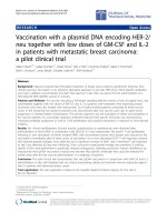
To assess the diagnostic potential of the selected miRNAs,
their relative quantity in EVs and whole plasma was compared between patients with PC (n = 50) and BPH
(n = 22). Three of the 9 miRNAs tested showed a diagnostic value in our sample set (Fig. 4). MiR-375 was significantly increased in PC patients as compared to BPH (FDR
adj. p = 0.03) and had an AUC of 0.68 (95% CI: 0.54–0.83,
p = 0.01), when tested in the whole plasma. The same tendency was observed for EV-enclosed miR-375, however it
didn’t reach statistical significance. On the contrary, miRTable 3 Spearman correlation coefficients of EV-enclosed and
whole plasma miRNAs
miRNA
Spearman r
95% confidence interval
p value
miR-375
0.37
0.15–0.56
0.0013
miR-141-3p
0.36
0.13–0.55
0.0018
miR-200c-30
0.37
0.13–0.56
0.0023
miR-21-5p
0.50
0.28–0.66
<0.0001
miR-30c-5p
0.42
0.19–0.60
0.0005
miR-106a-5p
0.37
0.13–0.57
0.0021
miR-223-3p
0.57
0.37–0.72
<0.0001
Let-7a-5p
0.27
0.02–0.48
0.03
miR-210-3p
0.28
0.05–0.049
0.01
200c-3p and miR-21-5p could differentiate between PC
and BPH better when tested in EVs than in the whole
plasma (AUC of 0.68, p = 0.01 and 0.67, p = 0.02, respectively, when tested in EVs and AUC of 0.62, p = 0.12 and
AUC of 0.61, p = 0.16, respectively, when tested in whole
plasma). The levels of the other miRNAs were not significantly different in PC samples compared to BPH neither
in EVs nor in whole plasma.
Next, we investigated the association of EV-enclosed
and whole plasma miRNA levels with PC aggressiveness.
We found that the level of Let-7a-5p was significantly
decreased in EVs from PC patients with high Gleason
score (≥8) compared to low Gleason score (≤6) and it
could differentiate between these groups with AUC of
0.68 (95% CI: 0.52–0.84, p = 0.03) (Fig. 5). Although the
same tendency was observed in whole plasma, the standard deviation was larger and statistical significance was
not reached. No other miRNA could differentiate
between PC patients with high and low Gleason scores.
Finally, none of the miRNAs was associated with the
presence of histologically confirmed prostatitis in PC
and BPH patients, thus showing that the alterations in
the miRNA levels are not due to prostatic inflammation.
Discussion
Cells can release miRNAs to the extracellular space
either incorporated into EVs [31, 32] or in a vesicle-free
form bound to various protein and lipoprotein complexes [17–20]. Quantification of these miRNAs in blood
from cancer patients may offer new opportunities for
diagnosis, prognosis, monitoring of treatment response
and early detection of recurrence in a minimally invasive
way. However, human blood contains a complex mixture
of miRNAs derived from various cell types and, therefore, robust quantification of cancer-derived cell-free
miRNAs has turned out to be a challenging task [14].
Currently, it is still debated if the EV-based miRNA
Endzeliņš et al. BMC Cancer (2017) 17:730
Page 9 of 13
Fig. 4 Circulating miRNA levels in patients with BPH and PC. Scatter plots show the log2RQ values of each miRNA tested in EVs and in whole
plasma. FDR-adjusted p values are show at the top of each graph. Area under the ROC curve (AUC), 95% confidence interval and p value for differentiating
between PC and BPH is shown below each graph
detection assays are superior to the whole plasma-based
assays. miRNA profiles in cancer-derived EVs have been
found to be reminiscent of their cell-of-origin [31, 33],
though due to selective RNA sorting mechanisms they
may be enriched or depleted of some specific miRNAs
[34]. The EV membrane protects the RNA cargo from
degradation in the bloodstream and the intraluminal
RNA content is thought to be relatively stable, therefore
EVs may provide a more consistent source of miRNA
biomarkers than whole plasma [15, 30]. On the other
hand, it has been calculated that there is far less than
one molecule of a given miRNA per EV [35], which
raises the question of whether all EVs contain miRNAs
and if the amounts are biologically meaningful.
Moreover, it can be argued that the EV isolation step
may introduce a higher variation and result in a low
RNA yield that in turn would lead to lower sensitivity,
higher standard deviations and poor reproducibility of the
EV-based miRNA assays as compared to whole plasma
assays.
Here, we have performed a systematic comparison of
miRNA levels in whole plasma and EVs isolated from the
same plasma samples in a well-characterised cohort of PC
and BPH patients. Our results show that EV-incorporated
miRNA constitutes only a minor fraction of whole plasma
miRNA. This is in line with a study by Chevillet et al.
showing that exosome fractions contained a small minority of the miRNA content of plasma [35]. Nevertheless,
Endzeliņš et al. BMC Cancer (2017) 17:730
Page 10 of 13
Fig. 5 Circulating Let-7a-5p levels in PC patients with low and high Gleason score. Scatter plots show the log2RQ values of Let-7a-5p tested in EVs
and in whole plasma of patients with Gleason score ≥ 8 (PC GH) and Gleason score ≤6 (PC GL). The mean log2RQ values and standard deviation is
shown above each scatter plot. Area under the ROC curve (AUC), 95% confidence interval and p value for differentiating between PC patients with
high and low Gleason score is shown below each graph
the miRNA levels in EVs and whole plasma were poorly
correlated, and the EV-incorporated and whole plasma
miRNA profile was clearly different. This finding is consistent with a NGS-based study by Cheng et al. that compared small RNA profiles in EVs, plasma and serum of 3
healthy individuals and showed that the miRNA levels differ remarkably between plasma and serum EVs and
between EVs and cell-free plasma and serum [30].
Three out of 9 miRNAs analysed could differentiate between PC and BPH patients in our cohort. MiR-375
showed a better diagnostic performance when tested in
whole plasma as compared to EVs. MiR-375 is an oncogenic miRNA that is overexpressed in tumours with high
Gleason score and more advanced pathological stage [36].
Increased plasma or serum levels of miR-375 in patients
with PC vs BPH or metastatic CRPC vs localised PC have
been reported before in several studies (Table 2), and the
AUC obtained in our study was similar to that reported before [37]. MiR-375 had one of the lowest EV to whole
plasma ratios among the miRNAs analysed in this study
and it was undetectable in a significant portion of EV samples. It still may have diagnostic properties in cases where it
is detectable, though proving its diagnostic value would require a larger cohort of samples. Two studies have reported
the presence of miR-375 in blood EVs from PC patients.
Bryant et al. showed that its level is increased in serum EVs
from patients with metastatic PC as compared to nonrecurring PC [38], and Huang et al. reported that high EVmiR-375 level is associated with a poor prognosis in CRPC
[39]. Hence, increased levels of EV-incorporated miR-375
appear to be associated with metastatic disease. As only 3
of the patients in our cohort had a metastatic disease at the
time of the blood draw, we reasonably detected it only in a
minority of PC patients in our cohort. Moreover, as these
studies did not describe treatment of EVs with proteinase
K, it is possible that the EV preparations also contained
protein-bound miRNAs co-isolated with EVs.
On the contrary, EV-incorporated miR-200c-3p and miR21-5p showed better diagnostic performance than in whole
plasma. Increased plasma or serum levels of miR-200c-3p
have been found before in patients with metastatic CRPC
as compared to localised PC or healthy controls [40, 41].
Similarly, miR-21-5p has been reported to be increased in
plasma or serum of patients with PC as compared to
healthy controls and patients with CRPC as compared to
localised PC [40, 42, 43]. However, to the best of our knowledge, an association of EV-incorporated miR-200c-3p and
miR-21-5p with PC has not been reported before. Hence,
our study shows for the first time that EVs provide a better
source for testing these miRNAs as PC biomarkers than
whole plasma.
The only miRNA biomarker that could differentiate
between PC patients with high vs low Gleason score was
EV-incorporated Let-7a-5p, whose level was decreased
in patients with Gleason score ≥ 8. This is in line with a
study by Mihelich et al. showing that serum levels of
Let-7a were decreased in PC patients with Gleason 4 + 5
grade tumours as compared with Gleason grade 3 [12].
Our study, though, shows that the whole plasma and EV
levels of Let-7a-5p are poorly correlated and that EVincorporated Let7a-5p level is more informative than
Let7a-5p in whole plasma.
The cellular origin of circulating miRNAs is unclear.
Although it seems likely that oncogenic miRNAs such as
miR-375, miR-200c-3p and miR-21-5p that are overexpressed in PC tissues are released in the bloodstream
from the tumour tissues, direct evidence for this is still
lacking. On the contrary, Let-7a-5p is a tumour suppressive miRNA that is downregulated in PC tissues as compared to normal or BPH tissues [44, 45]. Hence, the
decrease in Let-7a-5p plasma level in patients with
aggressive PC is unlikely to be due to the release from
cancer tissue. More plausibly, lower expression level or
reduced release of Let-7a-5p is genetically associated
Endzeliņš et al. BMC Cancer (2017) 17:730
with PC. Alternatively, it could be possible that signalling molecules produced by cancer cells actively downregulate the expression or release of this miRNA from
normal tissues. In fact, a recent study by Chen et al. has
demonstrated that breast cancer cells can downregulate
the expression and release of miR-486 from cardiac and
skeletal muscle in a TNFα-dependent manner [46], thus
providing evidence that miRNAs released from nontumour cells can have a diagnostic significance.
We did not observe diagnostic properties for circulating
miR-30c-5p, miR-106a-5p, miR-141-3p, miR-223-3p and
miR-210-3p in our patient cohort. The main reasons for
this could be a relatively low sample size, the usage of different RNA isolation methods and different sample storage and processing conditions that may affect miRNA
abundance and stability, and different normalisation
methods for RT-qPCR results. In most of these studies,
the results were normalised to the RNA input. Here, we
normalised the RT-qPCR data against plasma volume and
spike-ins that allow controlling for experimental variation.
We reasoned that the quantity of EV-RNA in our samples
is by far too small to be reliably measured by the currently
available RNA quantification methods (e.g. Nanodrop,
Qubit or Agilent Bioanalyzer), therefore the normalisation
against RNA input may lead to biased results. Moreover,
as EV levels have been found to be increased in cancer
patients as compared to healthy controls [47], it seems
likely that the levels of EV-enclosed RNA may also be increased, hence normalisation against the RNA input may
result in the loss of diagnostically relevant information.
Alternative approaches for RT-qPCR data normalisation
include normalisation to an individual endogenous reference gene or the geometric mean of a set of normalisers.
While the selection of housekeeping genes is relatively
straight-forward for miRNA expression analysis in cells or
tissues, the most commonly used internal control genes
have turned out to be highly variable in biofluids [48–50],
therefore, there is currently no consensus on appropriate
normalisers in biofluids. Possibly, the most reliable
normalisation strategy is a global geometric averaging of
multiple genes, however this is applicable only when large
panels of miRNAs are analysed. It should also be considered that EV-miRNA and cell-free miRNAs may require
different normalization genes.
Furthermore, miR-141-3p and miR-106a-5p had discordant results across publications. Increased miR-106a
serum levels were shown to correlate with increased
CAPRA scores in one study [51], while another study
showed that it is decreased in sera from PC patients with
Gleason grades 4 + 5 as compared to grade 3 and BPH
[12]. Increased serum or plasma levels of miR-141 have
been found in PC patients as compared to healthy controls or BPH [1, 52], and in patients with metastatic
CRPC as compared to localised PC [40, 53, 54]. At the
Page 11 of 13
same time, other studies reported that miR-141 was
detectable in less than 50% of patients or had similar
levels in PC patients and healthy controls [43, 54].
Conclusions
To the best of our knowledge, this is the first study providing a head-to-head comparison of diagnostically relevant
miRNA detection assays in whole plasma and plasma EVs
from cancer patients. We show that only a minor fraction
of the total cell-free miRNA could be recovered from the
plasma EVs, however the EV-incorporated and whole
plasma cell-free miRNA profiles were clearly different.
Whole plasma MiR-375 could differentiate between PC
and BPH, while miR-200c-3p and miR-21-5p performed
better when analysed in EVs. EV-incorporated but not
whole plasma Let-7a-5p level could distinguish patients
with aggressive and indolent PC. This shows that EVs provide a more consistent source of RNA than whole plasma
for the analysis of some miRNA biomarkers, while, possibly
due to specific sorting mechanisms, the abundance of other
miRNAs in EVs is very low and they show better diagnostic
performance in whole plasma.
Abbreviations
AUC: Area under the curve; BPH: Benign prostatic hyperplasia;
CRPC: Castration resistant prostate cancer; EV: Extracellular vesicle;
miRNA: MicroRNA; PSA: Prostate specific antigen; ROC: Receiver operator
curve
Acknowledgements
We thank all the patients who participated in this study and the staff of the
Latvian Genome Database for providing the samples and clinical data.
Funding
This study was supported by the Norwegian Financial Mechanism 2009–2014
under Project Contract No NFI/R/2014/045. The funding body had no role in
the design of the study and collection, analysis, and interpretation of data
and in writing the manuscript.
Availability of data and materials
The datasets analysed during the current study are available from the
corresponding author on reasonable request.
Authors’ contributions
AL, ALl and VL designed research, EE, AB, VM, CBS, KS, AĀ, MR and AR
performed research and participated in analysis and interpretation of the
results, VM, DŠ and VL contributed to the enrolment of patients, collection
and processing of clinical samples, and collection and analysis of clinical
data, AL wrote the manuscript, ALl revised the manuscript. All authors have
read and approved the manuscript.
Ethics approval and consent to participate
Biobanking procedures were approved by the Committee of Medical Ethics
of Latvia (decision No.5, 16.09.2010) and the use of clinical samples for
research was approved by the Committee of Biomedical Ethics of Riga East
University Hospital (decision No. 7-A/15, 04.06.2015). The blood samples were
collected after the patients’ informed written consent was obtained.
Consent for publication
Not applicable.
Competing interests
The authors declare that they have no competing interests.
Endzeliņš et al. BMC Cancer (2017) 17:730
Publisher’s Note
Springer Nature remains neutral with regard to jurisdictional claims in
published maps and institutional affiliations.
Author details
1
Latvian Biomedical Research and Study Centre, Ratsupites Str 1, k-1, Riga
LV-1067, Latvia. 2Riga Stradiņš University, Dzirciema Str 16, Riga LV-1007,
Latvia. 3Department of Molecular Cell Biology, Institute for Cancer Research,
Oslo University Hospital-The Norwegian Radium Hospital, 0379 Oslo, Norway.
4
Institute of Clinical and Preventive Medicine, Faculty of Medicine, University
of Latvia, Raina blvd. 19, Riga LV – 1586, Latvia.
Received: 24 June 2017 Accepted: 30 October 2017
References
1. Mitchell PS, Parkin RK, Kroh EM, Fritz BR, Wyman SK, Pogosova-Agadjanyan
EL, et al. Circulating microRNAs as stable blood-based markers for cancer
detection. Proc Natl Acad Sci USA. 2008;105:10513–8.
2. Chen X, Ba Y, Ma L, Cai X, Yin Y, Wang K, et al. Characterization of
microRNAs in serum: a novel class of biomarkers for diagnosis of cancer
and other diseases. Cell Res. 2008;18:997–1006.
3. Armand-Labit V, Pradines A. Circulating cell-free microRNAs as clinical
cancer biomarkers. Biomol Concepts. 2017;8:61–81.
4. Siegel RL, Miller KD, Jemal A. Cancer statistics, 2017. CA Cancer J Clin.
2017;67:7–30.
5. Ferlay J, Soerjomataram I, Dikshit R, Eser S, Mathers C, Rebelo M, et al.
Cancer incidence and mortality worldwide: sources, methods and major
patterns in GLOBOCAN 2012. Int J Cancer. 2015;136:E359–86.
6. Prensner JR, Rubin MA, Wei JT, Chinnaiyan AM. Beyond PSA: the next
generation of prostate cancer biomarkers. Sci Transl Med. 2012;4:127rv3.
7. Salman JW, Schoots IG, Carlsson SV, Jenster G, Roobol MJ. Prostate specific
antigen as a tumor marker in prostate cancer: biochemical and clinical
aspects. Adv Exp Med Biol. 2015;867:93–114.
8. Fitzpatrick JM, Bellmunt J, Fizazi K, Heidenreich A, Sternberg CN, Tombal B,
et al. Optimal management of metastatic castration-resistant prostate
cancer: highlights from a European expert consensus panel. Eur J Cancer.
2014;50:1617–27.
9. Bertoli G, Cava C, Castiglioni I. MicroRNAs as biomarkers for diagnosis,
prognosis and Theranostics in prostate cancer. Int J Mol Sci. 2016;17:421.
10. Endzelins E, Melne V, Kalnina Z, Lietuvietis V, Riekstina U, Llorente A, et al.
Diagnostic, prognostic and predictive value of cell-free miRNAs in prostate
cancer: a systematic review. Mol Cancer. 2016;15:41.
11. Chen ZH, Zhang GL, Li HR, Luo JD, Li ZX, Chen GM, et al. A panel of five
circulating microRNAs as potential biomarkers for prostate cancer. Prostate.
2012;72:1443–52.
12. Mihelich BL, Maranville JC, Nolley R, Peehl DM, Nonn L. Elevated serum
microRNA levels associate with absence of high-grade prostate cancer in a
retrospective cohort. PLoS One. 2015;10:e0124245.
13. Schwarzenbach H, Hoon DS, Pantel K. Cell-free nucleic acids as biomarkers
in cancer patients. Nat Rev Cancer. 2011;11:426–37.
14. Witwer KW. Circulating microRNA biomarker studies: pitfalls and potential
solutions. Clin Chem. 2015;61:56–63.
15. Huang X, Yuan T, Tschannen M, Sun Z, Jacob H, Du M, et al.
Characterization of human plasma-derived exosomal RNAs by deep
sequencing. BMC Genomics. 2013;14:319.
16. Valadi H, Ekstrom K, Bossios A, Sjostrand M, Lee JJ, Lotvall JO. Exosomemediated transfer of mRNAs and microRNAs is a novel mechanism of
genetic exchange between cells. Nat Cell Biol. 2007;9:654–9.
17. Vickers KC, Palmisano BT, Shoucri BM, Shamburek RD, Remaley AT.
MicroRNAs are transported in plasma and delivered to recipient cells by
high-density lipoproteins. Nat Cell Biol. 2011;13:423–33.
18. Arroyo JD, Chevillet JR, Kroh EM, Ruf IK, Pritchard CC, Gibson DF, et al.
Argonaute2 complexes carry a population of circulating microRNAs
independent of vesicles in human plasma. Proc Natl Acad Sci USA.
2011;108:5003–8.
19. Turchinovich A, Weiz L, Langheinz A, Burwinkel B. Characterization of
extracellular circulating microRNA. Nucleic Acids Res. 2011;39:7223–33.
20. Wang K, Zhang S, Weber J, Baxter D, Galas DJ. Export of microRNAs and
microRNA-protective protein by mammalian cells. Nucleic Acids Res.
2010;38:7248–59.
Page 12 of 13
21. Fabris L, Ceder Y, Chinnaiyan AM, Jenster GW, Sorensen KD, Tomlins S, et al.
The potential of MicroRNAs as prostate cancer biomarkers. Eur Urol.
2016;70:312–22.
22. Valentino A, Reclusa P, Sirera R, Giallombardo M, Camps C, Pauwels P, et al.
Exosomal microRNAs in liquid biopsies: future biomarkers for prostate
cancer. Clin Transl Oncol. 2017;19:651–7.
23. Sodar BW, Kittel A, Paloczi K, Vukman KV, Osteikoetxea X, Szabo-Taylor K,
et al. Low-density lipoprotein mimics blood plasma-derived exosomes and
microvesicles during isolation and detection. Sci Rep. 2016;6:24316.
24. van der Pol E, Coumans FA, Grootemaat AE, Gardiner C, Sargent IL, Harrison
P, et al. Particle size distribution of exosomes and microvesicles determined
by transmission electron microscopy, flow cytometry, nanoparticle tracking
analysis, and resistive pulse sensing. J Thromb Haemost. 2014;12:1182–92.
25. Théry C, Amigorena S, Raposo G, Clayton A. 2006. Isolation and Characterization
of Exosomes from Cell Culture Supernatants and Biological Fluids. Current
Protocols in Cell Biology. 30:3.22:3.22.1–3.22.29.
26. Li L, Zhu D, Huang L, Zhang J, Bian Z, Chen X, et al. Argonaute 2 complexes
selectively protect the circulating microRNAs in cell-secreted microvesicles.
PLoS One. 2012;7:e46957.
27. Mustapic M, Eitan E, Werner JK Jr, Berkowitz ST, Lazaropoulos MP, Tran J,
et al. Plasma extracellular vesicles enriched for neuronal origin: a potential
window into brain pathologic processes. Front Neurosci. 2017;11:278.
28. Hong CS, Muller L, Whiteside TL, Boyiadzis M. Plasma exosomes as markers
of therapeutic response in patients with acute myeloid leukemia. Front
Immunol. 2014;5:160.
29. Enderle D, Spiel A, Coticchia CM, Berghoff E, Mueller R, Schlumpberger M, et al.
Characterization of RNA from Exosomes and other extracellular vesicles
isolated by a novel spin column-based method. PLoS One. 2015;10:e0136133.
30. Cheng L, Sharples RA, Scicluna BJ, Hill AF. Exosomes provide a protective
and enriched source of miRNA for biomarker profiling compared to
intracellular and cell-free blood. J Extracell Vesicles. 2014;3:23743.
/>31. Fiskaa T, Knutsen E, Nikolaisen MA, Jorgensen TE, Johansen SD, Perander M,
et al. Distinct small RNA signatures in extracellular vesicles derived from
breast cancer cell lines. PLoS One. 2016;11:e0161824.
32. Hessvik NP, Phuyal S, Brech A, Sandvig K, Llorente A. Profiling of microRNAs
in exosomes released from PC-3 prostate cancer cells. Biochim Biophys
Acta. 2012;1819:1154–63.
33. Lunavat TR, Cheng L, Kim DK, Bhadury J, Jang SC, Lasser C, et al. Small RNA
deep sequencing discriminates subsets of extracellular vesicles released by
melanoma cells–evidence of unique microRNA cargos. RNA Biol. 2015;
12:810–23.
34. Villarroya-Beltri C, Baixauli F, Gutierrez-Vazquez C, Sanchez-Madrid F,
Mittelbrunn M. Sorting it out: regulation of exosome loading. Semin Cancer
Biol. 2014;28:3–13.
35. Chevillet JR, Kang Q, Ruf IK, Briggs HA, Vojtech LN, Hughes SM, et al.
Quantitative and stoichiometric analysis of the microRNA content of
exosomes. Proc Natl Acad Sci U S A. 2014;111:14888–93.
36. Costa-Pinheiro P, Ramalho-Carvalho J, Vieira FQ, Torres-Ferreira J, Oliveira J,
Goncalves CS, et al. MicroRNA-375 plays a dual role in prostate
carcinogenesis. Clin Epigenetics. 2015;7:42.
37. Haldrup C, Kosaka N, Ochiya T, Borre M, Hoyer S, Orntoft TF, et al. Profiling
of circulating microRNAs for prostate cancer biomarker discovery. Drug
Deliv Transl Res. 2014;4:19–30.
38. Bryant RJ, Pawlowski T, Catto JW, Marsden G, Vessella RL, Rhees B, et al.
Changes in circulating microRNA levels associated with prostate cancer. Br J
Cancer. 2012;106:768–74.
39. Huang X, Yuan T, Liang M, Du M, Xia S, Dittmar R, et al. Exosomal miR-1290
and miR-375 as prognostic markers in castration-resistant prostate cancer.
Eur Urol. 2015;67:33–41.
40. Watahiki A, Macfarlane RJ, Gleave ME, Crea F, Wang Y, Helgason CD, et al.
Plasma miRNAs as biomarkers to identify patients with castration-resistant
metastatic prostate cancer. Int J Mol Sci. 2013;14:7757–70.
41. Cheng HH, Mitchell PS, Kroh EM, Dowell AE, Chery L, Siddiqui J, et al.
Circulating microRNA profiling identifies a subset of metastatic prostate
cancer patients with evidence of cancer-associated hypoxia. PLoS One.
2013;8:e69239.
42. Zhang HL, Yang LF, Zhu Y, Yao XD, Zhang SL, Dai B, et al. Serum miRNA-21:
elevated levels in patients with metastatic hormone-refractory prostate
cancer and potential predictive factor for the efficacy of docetaxel-based
chemotherapy. Prostate. 2011;71:326–31.
Endzeliņš et al. BMC Cancer (2017) 17:730
43. Yaman Agaoglu F, Kovancilar M, Dizdar Y, Darendeliler E, Holdenrieder S,
Dalay N, et al. Investigation of miR-21, miR-141, and miR-221 in blood
circulation of patients with prostate cancer. Tumour Biol. 2011;32:583–8.
44. Porkka KP, Pfeiffer MJ, Waltering KK, Vessella RL, Tammela TL, Visakorpi T.
MicroRNA expression profiling in prostate cancer. Cancer Res. 2007;67:
6130–5.
45. Kong D, Heath E, Chen W, Cher ML, Powell I, Heilbrun L, et al. Loss of let-7
up-regulates EZH2 in prostate cancer consistent with the acquisition of
cancer stem cell signatures that are attenuated by BR-DIM. PLoS One.
2012;7:e33729.
46. Chen D, Goswami CP, Burnett RM, Anjanappa M, Bhat-Nakshatri P, Muller W,
et al. Cancer affects microRNA expression, release, and function in cardiac
and skeletal muscle. Cancer Res. 2014;74:4270–81.
47. Cappello F, Logozzi M, Campanella C, Bavisotto CC, Marcilla A, Properzi F,
et al. Exosome levels in human body fluids: a tumor marker by themselves?
Eur J Pharm Sci. 2017;96:93–8.
48. Hunter MP, Ismail N, Zhang X, Aguda BD, Lee EJ, Yu L, et al. Detection of
microRNA expression in human peripheral blood microvesicles. PLoS One.
2008;3:e3694.
49. Kok MG, Halliani A, Moerland PD, Meijers JC, Creemers EE, Pinto-Sietsma SJ.
Normalization panels for the reliable quantification of circulating microRNAs
by RT-qPCR. FASEB J. 2015;29:3853–62.
50. Schlosser K, McIntyre LA, White RJ, Stewart DJ. Customized internal
reference controls for improved assessment of circulating MicroRNAs in
disease. PLoS One. 2015;10:e0127443.
51. Moltzahn F, Olshen AB, Baehner L, Peek A, Fong L, Stoppler H, et al.
Microfluidic-based multiplex qRT-PCR identifies diagnostic and prognostic
microRNA signatures in the sera of prostate cancer patients. Cancer Res.
2011;71:550–60.
52. Kelly BD, Miller N, Sweeney KJ, Durkan GC, Rogers E, Walsh K, et al. A
circulating MicroRNA signature as a biomarker for prostate cancer in a high
risk group. J Clin Med. 2015;4:1369–79.
53. Nguyen HC, Xie W, Yang M, Hsieh CL, Drouin S, Lee GS, et al. Expression
differences of circulating microRNAs in metastatic castration resistant
prostate cancer and low-risk, localized prostate cancer. Prostate. 2013;
73:346–54.
54. Brase JC, Johannes M, Schlomm T, Falth M, Haese A, Steuber T, et al.
Circulating miRNAs are correlated with tumor progression in prostate
cancer. Int J Cancer. 2011;128:608–16.
55. Ribas J, Lupold SE. The transcriptional regulation of miR-21, its multiple
transcripts, and their implication in prostate cancer. Cell Cycle. 2010;9:923–9.
56. Folini M, Gandellini P, Longoni N, Profumo V, Callari M, Pennati M, et al.
miR-21: an oncomir on strike in prostate cancer. Mol Cancer. 2010;9:12.
57. Volinia S, Calin GA, Liu CG, Ambs S, Cimmino A, Petrocca F, et al. A
microRNA expression signature of human solid tumors defines cancer gene
targets. Proc Natl Acad Sci U S A. 2006;103:2257–61.
58. Walter BA, Valera VA, Pinto PA, Merino MJ. Comprehensive microRNA
profiling of prostate cancer. J Cancer. 2013;4:350–7.
59. Tong AW, Fulgham P, Jay C, Chen P, Khalil I, Liu S, et al. MicroRNA profile
analysis of human prostate cancers. Cancer Gene Ther. 2009;16:206–16.
60. Mitchell PS, Parkin RK, Kroh EM, Fritz BR, Wyman SK, Pogosova-Agadjanyan
EL, et al. Circulating microRNAs as stable blood-based markers for cancer
detection. Proc Natl Acad Sci U S A. 2008;105:10513–8.
61. Westermann AM, Schmidt D, Holdenrieder S, Moritz R, Semjonow A,
Schmidt M, et al. Serum microRNAs as biomarkers in patients undergoing
prostate biopsy: results from a prospective multi-center study. Anticancer
Res. 2014;34:665–9.
62. Ambs S, Prueitt RL, Yi M, Hudson RS, Howe TM, Petrocca F, et al. Genomic
profiling of microRNA and messenger RNA reveals deregulated microRNA
expression in prostate cancer. Cancer Res. 2008;68:6162–70.
63. Wei Y, Yang J, Yi L, Wang Y, Dong Z, Liu Z, et al. MiR-223-3p targeting
SEPT6 promotes the biological behavior of prostate cancer. Sci Rep.
2014;4:7546.
Page 13 of 13
Submit your next manuscript to BioMed Central
and we will help you at every step:
• We accept pre-submission inquiries
• Our selector tool helps you to find the most relevant journal
• We provide round the clock customer support
• Convenient online submission
• Thorough peer review
• Inclusion in PubMed and all major indexing services
• Maximum visibility for your research
Submit your manuscript at
www.biomedcentral.com/submit