The safety and efficacy of transarterial chemoembolization combined with sorafenib and sorafenib mono-therapy in patients with BCLC stage B/C hepatocellular carcinoma
Bạn đang xem bản rút gọn của tài liệu. Xem và tải ngay bản đầy đủ của tài liệu tại đây (738.83 KB, 11 trang )
Wu et al. BMC Cancer (2017) 17:645
DOI 10.1186/s12885-017-3545-5
RESEARCH ARTICLE
Open Access
The safety and efficacy of transarterial
chemoembolization combined with
sorafenib and sorafenib mono-therapy in
patients with BCLC stage B/C hepatocellular
carcinoma
Fei-Xiang Wu1,2,3†, Jie Chen1†, Tao Bai1†, Shao-Liang Zhu1, Tian-Bo Yang1, Lu-Nan Qi1, Ling Zou1, Zi-Hui Li1,
Jia-Zhou Ye1 and Le-Qun Li1,2,3*
Abstract
Background: Sorafenib and transarterial chemoembolization (TACE) are recommended therapies for advanced
hepatocellular carcinoma (HCC), but their combined efficacy remains unclear.
Methods: Between August 2004 and November 2014, 104 patients with BCLC stage B/C HCC were enrolled at the
Affiliated Tumor Hospital of Guangxi Medical University, China. Forty-eight patients were treated with sorafenib
alone (sorafenib group) and 56 with TACE plus sorafenib (TACE + sorafenib group). Baseline demographic/clinical
data were collected. The primary outcomes were median overall survival (OS) and progression-free survival (PFS).
Secondary outcomes were overall response rate (ORR) and sorafenib-related adverse events (AEs). Baseline
characteristics associated with disease prognosis were identified using multivariate Cox hazards regression.
Results: The mean age of the 104 patients (94 males; 90.38%) was 49.02 ± 12.29 years. Of the baseline data, only
albumin level (P = 0.028) and Child-Pugh class (P = 0.017) differed significantly between groups. Median OS did not
differ significantly between the sorafenib and TACE + sorafenib groups (18.0 vs. 22.0 months, P = 0.223). Median
PFS was significantly shorter in the sorafenib group than that in the TACE + sorafenib group (6.0 vs. 8.0 months,
P = 0.004). Six months after treatments, the ORRs were similar between the sorafenib and TACE + sorafenib groups
(12.50% vs. 18.75%, P = 0.425). The rates of grade III–IV adverse events in sorafenib and TACE + sorafenib groups
were 29.2% vs. 23.2%, respectively. TACE plus sorafenib treatment (HR = 0.498, 95% CI = 0.278–0.892), no vascular
invasion (HR = 0.354, 95% CI = 0.183–0.685) and Child-Pugh class A (HR = 0.308, 95% CI = 0.141–0.674) were
significantly associated with better OS, while a larger tumor number was predictive of poorer OS (HR = 1.286,
95% CI = 1.031–1.604). TACE plus sorafenib treatment (HR = 0.461, 95% CI = 0.273–0.780) and no vascular invasion
(HR = 0.557, 95% CI = 0.314–0.988) were significantly associated with better PFS.
(Continued on next page)
* Correspondence:
†
Equal contributors
1
Department of Hepatobiliary Surgery, Affiliated Tumor Hospital of Guangxi
Medical University, He Di Rd #71, Nanning 530021, People’s Republic of
China
2
Guangxi Liver Cancer Diagnosis and Treatment Engineering and Technology
Research Center, Nanning, China
Full list of author information is available at the end of the article
© The Author(s). 2017 Open Access This article is distributed under the terms of the Creative Commons Attribution 4.0
International License ( which permits unrestricted use, distribution, and
reproduction in any medium, provided you give appropriate credit to the original author(s) and the source, provide a link to
the Creative Commons license, and indicate if changes were made. The Creative Commons Public Domain Dedication waiver
( applies to the data made available in this article, unless otherwise stated.
Wu et al. BMC Cancer (2017) 17:645
Page 2 of 11
(Continued from previous page)
Conclusions: Compared with sorafenib alone, combining TACE with sorafenib might prolong survival and delay
disease progression in patients with advanced HCC.
Keywords: Hepatocellular carcinoma, Sorafenib, Transarterial chemoembolization, Portal vein tumor thrombus,
Adverse events
Background
Hepatocellular carcinoma (HCC) is the fifth most common cancer in the world [1], and a variety of treatments
are available [2–4]. Surgery is a potentially curative therapy for HCC [5], but many patients are not eligible for
surgery because they are diagnosed with HCC at a very
late stage [6, 7]. According to the Barcelona Clinic Liver
Cancer (BCLC) Group, patients with BCLC stage B/C
HCC are not suitable for surgery [5]. Suitable alternative
treatments for patients with BCLC stage B and C HCC
are transarterial chemoembolization (TACE) and sorafenib,
respectively.
TACE is widely used as a palliative treatment for
patients with advanced HCC and has been reported to
prolong survival [8, 9]. TACE consists of two procedures: embolization of the tumor-feeding artery to cause
tumor necrosis, and local delivery of antitumor drugs to
the tumor-feeding artery to enhance tumor necrosis
[10]. Previously, we found that embolization is the most
important part of the TACE procedure [11, 12], with
tumor necrosis initiated after the feeding blood supply has
been shut down. Some studies [13, 14] have observed that
the vascular endothelial growth factor (VEGF) level increases after TACE, suggesting that a pharmacologic
intervention that impairs VEGF signaling and thus the
development of new blood vessels could be a clinically
useful adjuvant therapy for TACE.
Sorafenib is a small-molecule inhibitor of several tyrosine protein kinases that are thought to play an important role in tumor progression, including platelet-derived
growth factor receptor (PDGFR)-β, Raf serine/threonine
kinases and VEGF receptors (VEGFRs) [15, 16]. Since
sorafenib suppresses VEGF signaling by inhibiting
VEGFRs, it would be expected to enhance the efficacy of
TACE by inhibiting angiogenesis and thereby promoting
tumor apoptosis [17].
Patients with portal vein tumor thrombus (PVTT) are
defined as BCLC stage C and are recommended to
receive sorafenib therapy, while TACE is the recommended therapy for patients with BCLC stage B HCC.
Several studies have suggested that the combination of
TACE with sorafenib can provide a survival benefit in
patients with PVTT, as compared with TACE monotherapy [18–20]. However, whether the addition of
TACE would enhance the efficacy of sorafenib therapy
in these patients remains controversial.
In the present study, we compared efficacy and safety
between sorafenib mono-therapy and TACE combined
with sorafenib in patients with BCLC stage B/C HCC. In
addition, multivariate regression analysis was used to
identify clinical factors predicting overall survival (OS)
and progression-free survival (PFS), and further analyses
were undertaken to determine whether tumor size
influenced OS and PFS.
Methods
Ethics statement
This study was approved by the Institutional Review
Board of Guangxi Medical University and was conducted
in accordance with the Declaration of Helsinki and
internationally accepted ethical guidelines. During their
admission for surgery, the patients enrolled in this study
provided written informed consent for their information
to be stored in hospital databases and used for research.
During data collection, patient records were anonymized. Patient admission and consent procedures have
been described previously [21].
Patient enrollment
This retrospective study included 104 patients with
HCC between August 2004 and November 2014.
Patients treated with TACE and sorafenib were included
in the TACE + sorafenib group (n = 56); patients who
were treated only with sorafenib were included in the sorafenib group (n = 48). All patients were diagnosed with
HCC based on the criteria of the European Association
for the Study of the Liver [22].
The inclusion criteria were: (a) 18–75 years old; (b)
HCC classified as either unresectable BCLC stage B or
BCLC stage C [23]; and(c) liver function classified as
Child-Pugh class A or B.
Patients were excluded from the study if they had any
of the following: (a) malignant tumors of other organ
systems; (b) HCC of Child-Pugh class C; or (c) any
contraindication for therapy with TACE (e.g., complete
obstruction of the portal vein) or sorafenib (e.g., allergy
to sorafenib).
Collection of baseline data
The following information was obtained for all patients included in the analysis: disease history; physical
examination findings; results of serum laboratory tests
Wu et al. BMC Cancer (2017) 17:645
(total bilirubin, TBil; albumin, ALB; alanine aminotransferase, ALT; platelet count, PLT; prothrombin
time, PT; α-fetoprotein level, AFP; and hepatitis B virus
surface antigen, HBsAg); and results of radiologic investigations (computed tomography, CT; magnetic resonance
imaging, MRI; and/or Doppler ultrasound).
PVTT was confirmed by radiologic investigations
(a filling defect sign in CT or MRI images; or ultrasonographic features of a mass in the portal vein).
PVTT type was defined according to a previous
study [24] as follows: type I, tumor thrombus (TT)
involving segmental branches of the portal vein or
above; type II, TT involving the right/left portal vein;
type III, TT involving the main portal vein trunk; or
type IV, TT involving the superior mesenteric vein
or inferior vena cava.
Portal vein hypertension (PVH) was defined as the
presence of esophageal varices and/or a platelet count
<100,000 /μL in association with splenomegaly.
Page 3 of 11
Post-therapy evaluation and follow-up
Patients were asked to return to the hospital for followup every 1–2 months after discharge. During each
follow-up, blood tests and radiologic investigations were
performed as at baseline. Tumor response was recorded
during every follow-up and classified (based on the best
response) after 6 months, according to the Modified
Response Evaluation Criteria in Solid Tumors for HCC
(mRECIST) [27, 28], as either complete response (CR),
partial response (PR), stable disease (SD) or progressive
disease (PD). Patients lost to follow-up were excluded
from the final analysis.
Outcome measures
The primary outcome measures in our study were OS
and PFS.PFS was defined as the duration from patient
discharge to disease progression (according to mRECIST
guideline). Secondary outcome measures were tumor
response and the occurrence of ADEs.
Transarterial chemoembolization
Statistical analysis
We used the Seldinger technique [25] and introduced a
4.1-French RC1 catheter into the tumor feeding artery.
Afterwards, we carefully identified the number, location,
size and branches of the tumor. A mixture of 10–20 mL
iodized oil, gelfoam particles with 30–50 mg doxorubicin
and 50–100 mg cisplatinum were injected into the arterial branches. The number of TACE cycles administered
ranged from 1 to 6, with TACE repeated at 1-month
intervals, depending on the patients’ liver function and
tumor shrinkage.
SPSS 18.0 (IBM, Chicago, USA) was used for statistical
analysis. A P value <0.05 was defined as the threshold of
statistical significance. Normally distributed data are
expressed as the mean ± standard deviation (SD), nonnormally distributed data are expressed as median
(range), and enumeration data are expressed as n (%).
Differences in outcomes between the two therapy groups
were assessed for significance using independentsamples t-tests or χ2 tests. The Kaplan–Meier method
was used to evaluate the effects of patient characteristics
on OS and PFS. Factors significantly associated with OS
or PFS were identified by multivariate analysis using a
stepwise Cox model, with calculation of hazard ratios
(HRs) and 95% confidence intervals (CIs). In addition to
the type of therapy used (TACE + sorafenib versus sorafenib), the other factors entered into the multivariate analysis were patient age, patient gender (male versus female),
tumor number, tumor diameter, vascular invasion (present
versus absent), metastasis (present versus absent), ChildPugh stage (A versus B), and AFP level (< 400 ng/mL
versus ≥400 ng/mL). These other parameters were chosen
so as to be representative of factors known to be associated with HCC progression or patient survival. Additional
variables related to these factors were not included in the
multivariate analysis (for example, other parameters related to liver function were excluded as they are related to
Child-Pugh stage). A subgroup analysis based on PVTT
status was conducted to try and identify whether a subset
of patients might benefit more from combination therapy
with TACE and sorafenib. An additional analysis was also
performed to determine whether tumor size influenced
OS and PFS.
Sorafenib
Sorafenib was administered orally from the beginning of
the treatment period (i.e. treatment was initiated before
TACE was performed in those receiving combination
therapy) at a dosage of 400 mg twice daily (Bayer
HealthCare AG, 200 mg/pill). The sorafenib dose was
adjusted if adverse drug events (ADEs) developed. If
grade I or 2 ADEs (National Cancer Institute Common
Terminology Criteria for Adverse Events version 3.0;
[26]) occurred, we adopted a wait-and-see policy. Usually these ADEs disappeared spontaneously, but if they
persisted the drug was either reduced in dosage or
discontinued. When grade 3 or 4 ADEs occurred, the
oral dose was reduced to 200 mg per day. If the ADEs
had not disappeared or decreased in severity 1 week
after dose adjustment, it was recommended that the
patient stop receiving sorafenib until the symptoms had
alleviated or disappeared. In patients receiving combination therapy, treatment with sorafenib was continued
during and after the performance of TACE.
Wu et al. BMC Cancer (2017) 17:645
Page 4 of 11
Table 1 Baseline characteristics of the patients in the two treatment groups
Characteristics
Sorafenib
(n = 56)
Sorafenib + TACE
(n = 48)
Total
(n = 104)
P
Male, n (%)
48 (85.71%)
46 (95.83%)
94 (90.38%)
0.103
Female, n (%)
8 (14.29%)
2(4.17%)
10 (9.62%)
50.23 ± 11.88
47.6 ± 12.73
49.02 ± 12.29
0.279
Yes, n (%)
25 (44.64%)
27 (56.25%)
52 (50%)
0.238
No, n (%)
31 (55.36%)
21 (43.75%)
52 (50%)
Yes, n (%)
48 (85.71%)
45 (93.75%)
93 (89.42%)
No, n (%)
8 (14.29%)
3 (6.25%)
11 (10.58%)
Yes, n (%)
45 (80.36%)
32 (66.67%)
77 (74.04%)
No, n (%)
11 (19.64%)
16 (33.33%)
27 (25.96%)
Yes, n (%)
34 (60.71%)
27 (56.25%)
61 (58.65%)
No, n (%)
22 (39.29%)
21 (43.75%)
43 (41.35%)
Yes, n (%)
14 (25%)
8 (16.67%)
22 (21.15%)
No, n (%)
42 (75%)
40 (83.33%)
82 (78.85%)
Yes, n (%)
30 (53.57%)
24 (50%)
54 (51.92%)
No, n (%)
26 (46.43%)
24 (50%)
50 (48.08%)
None, n (%)
46 (82.14%)
44 (91.67%)
90 (86.54%)
Mild, n (%)
7 (12.5%)
2(4.17%)
9 (8.65%)
Moderate, n (%)
1 (1.79%)
2 (4.17%)
3 (2.88%)
Severe, n (%)
2 (3.57%)
0
2 (1.92%)
Yes, n (%)
5 (8.93%)
1 (2.08%)
6 (5.77%)
No, n (%)
Gender
Age (years)
Antiviral therapy
Positive for HBsAg
0.184
Liver cirrhosis
0.112
PVH
0.645
Ascites
0.3
Splenomegaly
0.716
Esophageal varix
0.220
Diabetes
0.214
51 (91.07%)
47(97.92%)
98 (94.23%)
Tumor number
2 (1,4)
2 (1,4)
2 (1,4)
0.169
Tumor diameter (cm)
9.1 (1,19.5)
7.65 (1,19)
8.65 (1,19.5)
0.172
Yes, n (%)
37 (66.07%)
26 (54.17%)
41 (39.42%)
0.216
No, n (%)
19 (33.93%)
22 (45.83%)
63 (60.58%)
Yes, n (%)
17 (30.36%)
21 (43.75%)
38 (36.54%)
No, n (%)
39 (69.64%)
27 (56.25%)
66 (63.46%)
Vascular invasion
Metastasis, n (%)
0.157
BCLC stage B/C
B, n (%)
10 (17.86%)
16 (33.33%)
26 (25%)
C, n (%)
46 (82.14%)
32 (66.67%)
78 (75%)
0.069
Wu et al. BMC Cancer (2017) 17:645
Page 5 of 11
Table 1 Baseline characteristics of the patients in the two treatment groups (Continued)
Child-Pugh stage A/B
A, n (%)
45 (80.36%)
46 (95.83%)
91 (87.50%)
B, n (%)
11 (19.64%)
2 (4.17%)
13 (12.50%)
TBil (μmol/L)
12.45 (2.9,49.5)
12.5 (5,34.9)
12.45 (2.9,49.5)
0.500
ALT (IU/L)
41.5 (4171)
42.5 (13,199)
42 (4199)
0.469
ALB (g/L)
39.23 ± 4.94
41.44 ± 5.14
40.25 ± 5.13
0.028
PT (s)
13.05 (10.9,53)
12.7 (10.4,16.4)
12.85 (10.4,53)
0.090
0.203
AFP (ng/mL)
400 (0.78, 12,100)
172.5 (1.4, 4000)
350 (0.78, 12,100)
< 400 ng/mL, n (%)
23 (48.94%)
33 (58.93%)
56 (54.37%)
≥ 400 ng/mL, n (%)
24 (51.06%)
23 (41.07%)
47 (45.63%)
0.017
PVTT, n (%)
I
11 (19.64%)
5 (10.42%)
16 (15.38%)
II
10 (17.86%)
13 (22.12%)
23 (22.12%)
III
8 (14.29%)
6 (12.5%)
14 (13.46%)
IV
3 (5.36%)
0
3 (2.88%)
0.254
AFP α-fetoprotein, ALB albumin, ALT alanine aminotransferase, BCLC Barcelona Clinic Liver Cancer, HBsAg hepatitis B virus surface antigen, HCC hepatocellular
carcinoma, PLT platelet count, PT prothrombin time, PVH portal vein hypertension, TBil total bilirubin. Values are shown as mean ± standard deviation, n (%) or
median (range)
Results
Characteristics of the study population
From August 2004 to November 2014, a total of 104 patients with HCC (mean age, 49.02 ± 12.29 years) were
included in this retrospective study, including 94 males
and 10 females. All patients’ data are attached in the
Additional file 1 (organized file). Forty-eight patients received sorafenib mono-therapy while 56 patients received sorafenib plus TACE therapy. The baseline
demographic and clinical characteristics were similar between the two treatment groups, except that patients in
the TACE + sorafenib group had a significantly higher
level of ALB (P = 0.028) and proportionally more
patients with Child-Pugh class A disease (P = 0.017).
There were no therapy-related deaths, and in-hospital
mortality was zero (Table 1).
Comparisons of efficacy between TACE/sorafenib
combination therapy and sorafenib mono-therapy
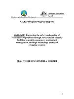
Median OS was 22.0 months (95% CI: 14.1–29.9 months)
in the TACE + sorafenib group and 18.0 months (95%
CI: 11.8–24.2 months) in the sorafenib group, with no
significant difference between groups (P = 0.223; Fig. 1
and Table 2). However, median PFS was significantly
longer in the TACE + sorafenib group (8.0 months;
95% CI: 3.4–12.6) than in the sorafenib group
Fig. 1 Comparison of survival outcomes between patients treated with sorafenib mono-therapy (sorafenib group) and those treated with
transarterial chemoembolization plus sorafenib combination therapy (TACE + sorafenib group). a Overall survival (OS, months). b Progressionfree survival (PFS, months)
Wu et al. BMC Cancer (2017) 17:645
Page 6 of 11
Table 2 Overall and progression-free survival of patients in the two treatment groups
Group
OS (months)
P
Median
95% CI
Sorafenib
18
11.797–24.203
TACE + sorafenib
22
14.095–29.905
0.223
PFS (months)
P
Median
95% CI
6
3.270–8.730
8
3.400–12.600
0.004
CI confidence interval, OS overall survival, PFS progression-free survival
(6.0 months; 95% CI: 3.3–8.7 months; P = 0.004; Fig. 1
and Table 2), indicating that combination therapy was
more effective than sorafenib mono-therapy at limiting
disease progression.
Tumor response
Data for tumor response at 6 months were available for
40 patients in the sorafenib group and 48 patients in the
TACE + sorafenib group (Table 3). There were no significant differences between treatment groups in the CR
rate (P = 1.000), PR rate (P = 0.502), SD rate (P = 0.574),
PD rate (P = 0.906) and OR rate (P = 0.425). Furthermore, subgroup analysis on the basis of the presence
(i.e., types I, II, III or IV) or absence of PVTT also
showed no statistical differences between the sorafenib
and TACE + sorafenib groups in the tumor response
6 months after treatments (all P > 0.05; Table 4). This
suggests that the two treatment regimens were similar
with regard to reducing tumor size.
Adverse events
There were no significant differences between the sorafenib and TACE + sorafenib groups in the incidences of
grade I, II, III and IV ADEs (all P > 0.05), and all ADEs
were tolerable. Grade III ADEs occurred in 14 patients
in the sorafenib group and 13 patients in the TACE + sorafenib group, while no Grade IV ADEs were observed
(Table 5). Symptoms in patients with grade III ADEs
disappeared or were alleviated following adjustment of
the sorafenib dose or administration of symptomatic
supportive treatments. These findings indicate that the
addition of TACE to sorafenib therapy does not result in
a notable increase in the incidence or severity of ADEs.
Table 3 Tumor response at 6 months in the two treatment
groups
Tumor response
Sorafenib group
(n = 40)
TACE + sorafenib group
(n = 48)
P
CR, n (%)
2 (5.00%)
3 (6.25%)
1.000
PR, n (%)
3 (7.50%)
6 (12.50%)
0.502
SD, n (%)
18 (45.00%)
18 (37.50%)
0.476
PD, n (%)
17 (42.50%)
21 (43.75%)
0.906
OR, n (%)
5 (12.50%)
9 (18.75%)
0.425
CR complete response, OR overall response (CR + PR), PD progressive disease,
PR partial response, SD stable disease
Clinical factors influencing OS and PFS
Multivariate Cox regression analysis identified use of
TACE + sorafenib combination therapy (HR = 0.498,
95% CI = 0.278–0.892, P = 0.019), no vascular invasion
(HR = 0.354, 95% CI = 0.183–0.685, P = 0.002) and
Child-Pugh class A (HR = 0.308, 95% CI = 0.141–0.674,
P = 0.003) as independent factors predicting better OS,
while tumor number (HR = 1.286, 95% CI = 1.031–
1.604, P = 0.026) was an independent factor predicting
poorer OS (Table 6). Similarly, use of TACE + sorafenib
combination therapy (HR = 0.461, 95% CI = 0.273–
0.780, P = 0.004) and no vascular invasion (HR = 0.557,
95% CI = 0.314–0.988, P = 0.045) were independent
factors predicting a better PFS (Table 7).
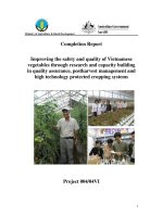
Further analyses of OS and PFS based on tumor diameter
The observation that tumor diameter was not an independent predictor of OS and PFS in the multivariate
analysis was perhaps unexpected. One possibility we
considered was that OS and PFS might only be influenced by tumor size once the tumor exceeded a certain
diameter. To explore this possibility, OS and PFS were
further analyzed based on different tumor diameters
(Table 8 and Fig. 2); the cutoff value of5 cm was based
on that used in the TNM classification, while the additional higher cutoff value of 7 cm was arbitrarily chosen.
Median OS was 44.0 months (95% CI: 21.624–66.376) in
patients with a tumor diameter < 5 cm and 17.0 months
(95% CI: 11.806–22.194) in patients with a tumor diameter ≥ 5 cm (P = 0.004; Fig. 2a); in contrast, PFS did not
differ between the two groups (8.0 months versus
7.0 months, respectively, P = 0.268; Fig. 2b). Patients with
a tumor diameter < 7 cm had a median OS of 38.0 months
(95% CI: 20.228–55.772) and a median PFS of 9.0 months
(95% CI: 6.003–11.997), while patients with a tumor
diameter ≥ 7 cm had a median OS of 14 months (95% CI:
10.409–17.591) and a median PFS of 5.0 months (95% CI:
3.007–6.993); both OS and PFS differed significantly
between the two groups (P < 0.05; Fig. 2c and d).
Discussion
The main finding of the present study was that both TACE
combined with sorafenib and sorafenib alone were safe and
effective treatments for patients with BCLC stage B/C
HCC. Although there were no significant differences
between treatment groups in OS or tumor response at
Wu et al. BMC Cancer (2017) 17:645
Page 7 of 11
Table 4 Tumor response at 6 months in the two treatment groups in patients with and without PVTT
Tumor
response
CR, n (%)
No PVTT
PVTT types I, II, III and IV
P
Sorafenib group
(n = 19)
TACE + sorafenib group
(n = 24)
2 (10.53%)
3 (12.5%)
1.000
P
Sorafenib group
(n = 21)
TACE + sorafenib group
(n = 24)
0 (0%)
0 (0%)
-
PR, n (%)
2 (10.53%)
2 (8.33%)
1.000
1 (4.76%)
4 (16.67%)
0.352
SD, n (%)
10 (52.63%)
8 (33.33%)
0.203
8 (38.1%)
10 (41.67%)
0.807
PD, n (%)
5 (26.32%)
11 (45.83%)
0.189
12 (57.14%)
10 (41.67%)
0.300
OR, n (%)
4 (21.06%)
5 (20.83%)
1.000
1 (4.76%)
4 (16.67%)
0.352
CR complete response, OR overall response (CR + PR), PD progressive disease, PR partial response, SD stable disease
6 months, patients treated with TACE/sorafenib combination therapy showed a significantly longer PFS than patients treated with sorafenib alone. Multivariate analysis
indicated that TACE/sorafenib combination therapy (versus sorafenib mono-therapy), no vascular invasion and
Child-Pugh stage A (versus B) were independent predictors
of better OS, while tumor number was a predictor of
poorer OS. Furthermore, TACE/sorafenib combination
therapy and no vascular invasion were independent predictors of better PFS. Importantly, the addition of TACE to
sorafenib therapy was not associated with a significant
increase in the occurrence of ADEs. We conclude that,
compared with sorafenib alone, TACE plus sorafenib combination therapy in patients with BCLC stage B/C HCC
may improve PFS and be associated with improved OS,
without a notable increase in adverse events.
Numerous clinical studies have reported that monotherapy with sorafenib can provide survival benefits over
placebo [29–33] or conservative management strategies
[34] in patients with advanced HCC. The median OS
and PFS in our study (18.0 and 6.0 months, respectively)
were longer than those reported in previous studies
(6.5–10.7 months and 2.8–5.5 months, respectively)
[29–34] and may reflect differences between studies in
the baseline clinical characteristics of the patients, such
as BCLC stage, Child-Pugh stage, vascular invasion and
extrahepatic spread.
TACE has also been shown to be an effective treatment option for advanced HCC [8, 9]. There have been
a number of investigations comparing the efficacy of
TACE plus sorafenib with TACE alone, and most have
suggested that combination therapy has superior efficacy
to TACE mono-therapy [35–39], although a minority
have reported no additional benefit [40]. In our study,
the median OS and PFS in patients treated with TACE/
sorafenib combination therapy were 22.0 months and
8.0 months, respectively, which are broadly in agreement
with values reported previously (12–29 months and
6.3–16.4 months, respectively) [38, 39, 41].
However, fewer studies have compared sorafenib monotherapy with TACE/sorafenib combination therapy in patients with advanced HCC. Zhang et al. [42] reported that,
compared with sorafenib alone, combination therapy
resulted in a better OS (15.0 months versus 5.0 months)
and PFS (6.0 months versus 2.5 months). Similar results
were obtained by Choi et al. [43], who found that the
addition of TACE to sorafenib yielded improvements in
OS (8.9 months versus 5.9 months) and PFS (2.5 months
Table 5 Adverse events in the two treatment groups
Adverse event
Sorafenib group (n = 48)
TACE + sorafenib group (n = 56)
Grade I
Grade II
Grade III
Grade IV
Grade I
Grade II
Grade III
Grade IV
Hand-foot skin reactions, n (%)
25 (44.64%)
8 (14.29%)
3 (5.36%)
0
24 (42.86%)
3 (5.36%)
3 (5.36%)
0
Vomiting, n (%)
19 (33.93%)
5 (8.93%)
3 (5.36%)
0
26 (46.43%)
4 (7.14%)
1 (1.79%)
0
Diarrhea, n (%)
21 (37.50%)
4 (7.14%)
2 (3.57%)
0
23 (41.07%)
1 (1.79%)
1 (1.79%)
0
Fatigue, n (%)
10 (17.86%)
2 (3.57%)
0
0
8 (14.29%)
4 (7.14%)
0
0
Hypertension, n (%)
19 (33.93%)
3 (5.36%)
0
0
10 (17.86%)
2 (3.57%)
1 (1.79%)
0
Leucopenia, n (%)
5 (8.93%)
2 (3.57%)
0
0
4 (7.14%)
0
1 (1.79%)
0
Anemia, n (%)
5 (8.93%)
2 (3.57%)
0
0
4 (7.14%)
1 (1.79%)
2 (3.57%)
0
Thrombocytopenia, n (%)
3 (5.36%)
2 (3.57%)
2 (3.57%)
0
3 (5.36%)
4 (7.14%)
1 (1.79%)
0
Alopecia, n (%)
2 (3.57%)
1 (1.79%)
0
0
3 (5.36%)
0
1 (1.79%)
0
Gastrointestinal hemorrhage, n (%)
0
0
4 (7.14%)
0
2 (3.57%)
1 (1.79%)
2 (3.57%)
0
Hepatic encephalopathy, n (%)
2 (1.79%)
1 (1.79%)
Wu et al. BMC Cancer (2017) 17:645
Page 8 of 11
Table 6 Multivariate analysis of risk factors for overall survival
Factor
Multivariate analysis
HR
95% CI
P
Male
1.423
0.481–4.210
0.524
TACE + sorafenib versus sorafenib
0.498
0.278–0.892
0.019
Age
0.984
0.963–1.005
0.140
Tumor number
1.286
1.031–1.604
0.026
Tumor diameter (cm)
1.031
0.965–1.101
0.367
No vascular invasion
0.354
0.183–0.685
0.002
Metastasis
1.365
0.784–2.375
0.271
Child-Pugh stage A
0.308
0.141–0.674
0.003
AFP < 400 ng/mL
0.648
0.373–1.124
0.123
AFP Alpha-fetoprotein, CI confidence interval, HR hazard ratio
versus 2.1 months). In agreement with these studies, we
also observed a significantly longer PFS in patients treated
with combination therapy than in those receiving sorafenib mono-therapy. Although our univariate analysis found
no significant difference between groups in OS, the multivariate analysis did identify combination therapy (versus
sorafenib alone) as a predictor of longer OS. This apparent
inconsistency may have been due to one or more confounding factors (which were accounted for in the multivariate analysis) influencing the results of the direct
comparisons of outcome measures between groups. Taken
together, these data support the use of TACE/sorafenib
combination therapy in patients with advanced HCC.
The most common ADEs noted in our study were
hand-foot skin reactions, vomiting and diarrhea, and the
majority were grade 1 adverse events, consistent with
previous research [39, 44, 45]. Importantly, no serious
ADEs were reported in patients with TACE combined
with sorafenib, indicating that this therapy is safe. Our
observations are in agreement with previous studies
Table 7 Multivariate analysis of risk factors for progression-free
survival
Factors
Multivariate Analysis
HR
95% CI
P
Male
1.364
0.613–3.035
0.447
TACE + sorafenib versus sorafenib
0.461
0.273–0.780
0.004
Age
0.995
0.976–1.014
0.581
Tumor number
1.140
0.936–1.389
0.193
Tumor diameter (cm)
1.038
0.969–1.111
0.288
No vascular invasion
0.557
0.314–0.988
0.045
Metastasis
1.334
0.834–2.133
0.229
Child-Pugh stage A
0.991
0.484–2.030
0.980
AFP < 400 ng/mL
0.695
0.437–1.106
0.125
AFP Alpha-fetoprotein, CI confidence interval, HR hazard ratio
reporting that the combination of TACE and sorafenib
is not associated with a significantly greater incidence/
severity of adverse events than TACE or sorafenib
mono-therapy [42, 46].
Our multivariate analysis indicated that Child-Pugh
class A, no vascular invasion and lower tumor number
were predictors of better OS. In addition, further analysis showed that tumor size ≥7 cm was also associated
with poorer OS and PFS. These findings are in agreement with previous investigations that have identified
Child-Pugh class, vascular invasion, tumor size, as well
as BCLC stage, Eastern Cooperative Oncology Group
(ECOG) performance status and alanine transaminase,
as independent predictors of prognosis [47–49]. Although
the tumor number and tumor size are both recognized as
being associated with prognosis [50], a recent study has
suggested that total tumor volume may be a better
predictor of outcomes [51].
In our study, the median survival of patients with
PVTT treated with sorafenib alone was 9 months,
which is longer than that reported previously for patients receiving conservative therapy (3.6–3.8 months)
or TACE (7.0–7.3 months) [25, 52]. One study demonstrated that sorafenib mono-therapy had similar efficacy
to TACE/sorafenib combination therapy in patients
with PVTT [53], while another reported that the
addition of sorafenib to TACE improved survival in patients with PVTT [20]. This may indicate that sorafenib
therapy may be superior to TACE in the management
of patients with advanced HCC and PVTT, and that sorafenib mono-therapy may be sufficient in this subset
of patients.
Our study has several limitations. First, this was a
retrospective study, hence selection and reporting bias
cannot be excluded. Although the baseline characteristics were similar between the two treatment groups,
suggesting that the degree of bias may not have been
large, it was notable that the TACE + sorafenib group
contained a significantly higher proportion of patients
with liver disease classed as Child-Pugh A. This was a
retrospective study in which the treatment regimen was
usually chosen by the doctor; since TACE is an invasive
procedure, it is more likely to have been recommended
to patients with better liver function. Although this potential selection bias may have influenced the results of
direct comparisons between groups, any potential bias
would have been accounted for by the multivariate regression analysis, which found that TACE/sorafenib
combination therapy was an independent predictor of
both OS and PFS. Second, tumor response was only
evaluated at one time point, whereas sequential monitoring over the period of the study would have provided
more detailed information regarding the efficacies of the
treatment regimens. Third, our sample size was relatively
Wu et al. BMC Cancer (2017) 17:645
Page 9 of 11
Table 8 Overall survival and progression-free survival of patients with tumors of differing diameters
Tumor
diameter
OS (months)
PFS (months)
P
Median
95% CI
< 5 cm
44
21.624–66.376
≥ 5 cm
17
11.806–22.194
< 7 cm
38
20.228–55.772
≥ 7 cm
14
10.409–17.591
Median
P
95% CI
Group 1
0.004
8
5.633–10.367
7
4.915–9.085
9
6.003–11.997
5
3.007–6.993
0.268
Group 2
0.002
0.012
CI confidence interval, OS overall survival, PFS progressive free survival
small, so the study may have been underpowered to detect
real differences for some comparisons. Fourth, this was
a single-center study, so the findings may not be
generalizable to other regions of China or other countries.
Therefore, multi-center, prospective, randomized, controlled trials are required to confirm and extend our
observations.
Conclusion
In conclusion, both TACE combined with sorafenib and
sorafenib alone were safe and effective treatments for patients with BCLC stage B/C HCC.TACE/sorafenib combination therapy may have advantages over sorafenib
mono-therapy in terms of progression-free survival and
possibly OS, without a notable increase in adverse events.
Fig. 2 Comparison of survival outcomes between patients with different tumor diameters. a Overall survival (OS, months) in patients with
a tumor diameter < 5 cm and those with a tumor diameter ≥ 5 cm. b Progression-free survival (PFS, months)in patients with a tumor
diameter < 5 cm and those with a tumor diameter ≥ 5 cm. c Overall survival (OS, months) in patients with a tumor diameter < 7 cm
and those with a tumor diameter ≥ 7 cm. d Progression-free survival (PFS, months)in patients with a tumor diameter < 7 cm and those
with a tumor diameter ≥ 7 cm
Wu et al. BMC Cancer (2017) 17:645
Additional file
Additional file 1: HCC Organized Data. The data organized from
original data and used for data analysis. (XLS 55 kb)
Page 10 of 11
2.
3.
4.
Abbreviations
AEs: Adverse events; BCLC: Barcelona Clinic Liver Cancer; CIs: Confidence
intervals; CR: Complete response; ECOG: Eastern Cooperative Oncology
Group; HCC: Hepatocellular carcinoma; HRs: Hazard ratios; mRECIST: Modified
Response Evaluation Criteria in Solid Tumors; ORR: Overall response rate;
OS: Overall survival; PD: Progressive disease; PDGFR: Platelet-derived growth
factor receptor; PFS: Progression-free survival; PR: Partial response; PVH: Portal
vein hypertension; PVTT: Portal vein tumor thrombus; SD: Stable disease;
TACE: Transarterial chemoembolization; TT: Tumor thrombus; VEGFRs: VEGF
receptors
Acknowledgements
None.
Funding
The study was supported by Key Laboratory of Early Prevention and
Treatment for Regional High Frequency Tumor, Ministry of Education, China
(GKZ201604); Scientific Research Fund of Ministry of Health of Guangxi
Province (S201513); Key project of Guangxi science and technology
department (GuiKe AB16380242).
Availability of data and materials
The dataset(s) supporting the conclusions of this article is(are) included
within the article and supplementary files.
Authors’ contributions
FXW, JC and TB contributed to study design, manuscript preparation and
drafting the manuscript. SLZ, TBY, LNQ, LZ, ZHL, JZY and LQL participated in
data collection, data analysis, follow-up and revising the manuscript for
important contents. All authors have read and approved the manuscript.
Ethics approval and consent to participate
This study was approved by the Institutional Review Board of Guangxi
Medical University and was conducted in accordance with the Declaration of
Helsinki and internationally accepted ethical guidelines. During their
admission for surgery, the patients enrolled in this study provided written
informed consent for their information to be stored in hospital databases
and used for research.
Consent for publication
Not applicable.
5.
6.
7.
8.
9.
10.
11.
12.
13.
14.
15.
Competing interests
The authors declare that they have no competing interests.
16.
Publisher’s Note
Springer Nature remains neutral with regard to jurisdictional claims in
published maps and institutional affiliations.
Author details
1
Department of Hepatobiliary Surgery, Affiliated Tumor Hospital of Guangxi
Medical University, He Di Rd #71, Nanning 530021, People’s Republic of
China. 2Guangxi Liver Cancer Diagnosis and Treatment Engineering and
Technology Research Center, Nanning, China. 3Key Laboratory of Early
Prevention and Treatment for Regional High Frequency Tumor, Ministry of
Education, Nanning, China.
17.
18.
19.
Received: 20 July 2016 Accepted: 14 August 2017
20.
References
1. Torre LA, Bray F, Siegel RL, Ferlay J, Lortet-Tieulent J, Jemal A. Global cancer
statistics, 2012. CA Cancer J Clin. 2015;65(2):87–108. doi:10.3322/caac.21262.
Llovet JM, Burroughs A, Bruix J. Hepatocellular carcinoma. Lancet. 2003;
362(9399):1907–17. doi:10.1016/S0140-6736(03)14964-1.
Llovet JM, Fuster J, Bruix J. Barcelona-clinic liver cancer G. The Barcelona
approach: diagnosis, staging, and treatment of hepatocellular carcinoma. Liver
Transpl. 2004;10(2 Suppl 1):S115–20. doi:10.1002/lt.20034.
Forner A, Llovet JM, Bruix J. Hepatocellular carcinoma. Lancet. 2012;
379(9822):1245–55. doi:10.1016/S0140-6736(11)61347-0.
Bruix J, Gores GJ, Mazzaferro V. Hepatocellular carcinoma: clinical
frontiers and perspectives. Gut. 2014;63(5):844–55. doi:10.1136/gutjnl2013-306627.
Poon RT, Fan ST, Lo CM, Liu CL, Wong J. Long-term survival and pattern of
recurrence after resection of small hepatocellular carcinoma in patients with
preserved liver function: implications for a strategy of salvage
transplantation. Ann Surg. 2002;235(3):373–82.
Chen MF, Hwang TL, Jeng LB, Wang CS, Jan YY, Chen SC. Postoperative
recurrence of hepatocellular carcinoma. Two hundred five consecutive
patients who underwent hepatic resection in 15 years. Arch Surg. 1994;
129(7):738–42.
Lo CM, Ngan H, Tso WK, Liu CL, Lam CM, Poon RT, et al. Randomized
controlled trial of transarterial lipiodol chemoembolization for unresectable
hepatocellular carcinoma. Hepatology. 2002;35(5):1164–71. doi:10.1053/jhep.
2002.33156.
Llovet JM, Real MI, Montana X, Planas R, Coll S, Aponte J, et al.
Arterial embolisation or chemoembolisation versus symptomatic
treatment in patients with unresectable hepatocellular carcinoma: a
randomised controlled trial. Lancet. 2002;359(9319):1734–9.
doi:10.1016/S0140-6736(02)08649-X.
Bruix J, Llovet JM, Castells A, Montana X, Bru C, Ayuso MC, et al. Transarterial
embolization versus symptomatic treatment in patients with advanced
hepatocellular carcinoma: results of a randomized, controlled trial in a single
institution. Hepatology. 1998;27(6):1578–83. doi:10.1002/hep.510270617.
Xie ZB, Ma L, Wang XB, Bai T, Ye JZ, Zhong JH, et al. Transarterial
embolization with or without chemotherapy for advanced
hepatocellular carcinoma: a systematic review. Tumour Biol. 2014;35(9):
8451–9. doi:10.1007/s13277-014-2340-z.
Xie ZB, Wang XB, Peng YC, Zhu SL, Ma L, Xiang BD, et al. Systematic review
comparing the safety and efficacy of conventional and drug-eluting bead
transarterial chemoembolization for inoperable hepatocellular carcinoma.
Hepatol Res. 2015;45(2):190–200. doi:10.1111/hepr.12450.
Bao Y, Feng WM, Tang CW, Zheng YY, Gong HB, Hou EG. Endostatin inhibits
angiogenesis in hepatocellular carcinoma after transarterial
chemoembolization. Hepato-Gastroenterology. 2012;59(117):1566–8.
doi:10.5754/hge12138.
Poon RT, Lau C, Yu WC, Fan ST, Wong J. High serum levels of vascular
endothelial growth factor predict poor response to transarterial
chemoembolization in hepatocellular carcinoma: a prospective study. Oncol
Rep. 2004;11(5):1077–84.
Jia L, Kiryu S, Watadani T, Akai H, Yamashita H, Akahane M, et al. Prognosis
of hepatocellular carcinoma with portal vein tumor thrombus: assessment
based on clinical and computer tomography characteristics. Acta Med
Okayama. 2012;66(2):131–41.
Wang B, Xu H, Gao ZQ, Ning HF, Sun YQ, Cao GW. Increased expression of
vascular endothelial growth factor in hepatocellular carcinoma after
transcatheter arterial chemoembolization. Acta Radiol. 2008;49(5):523–9.
doi:10.1080/02841850801958890.
Strebel BM, Dufour JF. Combined approach to hepatocellular carcinoma: a
new treatment concept for nonresectable disease. Expert Rev Anticancer
Ther. 2008;8(11):1743–9. doi:10.1586/14737140.8.11.1743.
Hu H, Duan Z, Long X, Hertzanu Y, Shi H, Liu S, et al. Sorafenib
combined with transarterial chemoembolization versus transarterial
chemoembolization alone for advanced-stage hepatocellular carcinoma:
a propensity score matching study. PLoS One. 2014;9(5):e96620.
doi:10.1371/journal.pone.0096620.
Pan T, Li XS, Xie QK, Wang JP, Li W, Wu PH, et al. Safety and efficacy of
transarterial chemoembolization plus sorafenib for hepatocellular carcinoma
with portal venous tumour thrombus. Clin Radiol. 2014;69(12):e553–61.
doi:10.1016/j.crad.2014.09.007.
Zhu K, Chen J, Lai L, Meng X, Zhou B, Huang W, et al. Hepatocellular
carcinoma with portal vein tumor thrombus: treatment with transarterial
chemoembolization combined with sorafenib–a retrospective controlled
study. Radiology. 2014;272(1):284–93. doi:10.1148/radiol.14131946.
Wu et al. BMC Cancer (2017) 17:645
21. Zhong JH, Li H, Xiao N, Ye XP, Ke Y, Wang YY, et al. Hepatic resection is safe
and effective for patients with hepatocellular carcinoma and portal
hypertension. PLoS One. 2014;9(9):e108755. doi:10.1371/journal.pone.0108755.
22. European Association For The Study Of The L, European Organisation
For R, Treatment Of C. EASL-EORTC clinical practice guidelines:
management of hepatocellular carcinoma. J Hepatol. 2012;56(4):908–43.
doi:10.1016/j.jhep.2011.12.001.
23. Zhong JH, Ke Y, Gong WF, Xiang BD, Ma L, Ye XP, et al. Hepatic resection
associated with good survival for selected patients with intermediate and
advanced-stage hepatocellular carcinoma. Ann Surg. 2014;260(2):329–40.
doi:10.1097/SLA.0000000000000236.
24. Quirk M, Kim YH, Saab S, Lee EW. Management of hepatocellular carcinoma
with portal vein thrombosis. World J Gastroenterol. 2015;21(12):3462–71.
doi:10.3748/wjg.v21.i12.3462.
25. Ye JZ, Zhang YQ, Ye HH, Bai T, Ma L, Xiang BD, et al. Appropriate treatment
strategies improve survival of hepatocellular carcinoma patients with portal
vein tumor thrombus. World J Gastroenterol. 2014;20(45):17141–7.
doi:10.3748/wjg.v20.i45.17141.
26. National Cancer Institute. Common terminology criteria for adverse events,
version 3.0. 2006. Accessed 18
June 2009.
27. Therasse P, Arbuck SG, Eisenhauer EA, Wanders J, Kaplan RS, Rubinstein L, et
al. New guidelines to evaluate the response to treatment in solid tumors.
European Organization for Research and Treatment of cancer, National
Cancer Institute of the United States, National Cancer Institute of Canada. J
Natl Cancer Inst. 2000;92(3):205–16.
28. Eisenhauer EA, Therasse P, Bogaerts J, Schwartz LH, Sargent D, Ford R, et al.
New response evaluation criteria in solid tumours: revised RECIST guideline
(version 1.1). Eur J Cancer. 2009;45(2):228–47. doi:10.1016/j.ejca.2008.10.026.
29. Bruix J, Raoul JL, Sherman M, Mazzaferro V, Bolondi L, Craxi A, et al. Efficacy
and safety of sorafenib in patients with advanced hepatocellular carcinoma:
subanalyses of a phase III trial. J Hepatol. 2012;57(4):821–9. doi:10.1016/j.
jhep.2012.06.014.
30. Cheng AL, Guan Z, Chen Z, Tsao CJ, Qin S, Kim JS, et al. Efficacy and safety
of sorafenib in patients with advanced hepatocellular carcinoma according
to baseline status: subset analyses of the phase III Sorafenib Asia-Pacific trial.
Eur J Cancer. 2012;48(10):1452–65. doi:10.1016/j.ejca.2011.12.006.
31. Rimassa L, Santoro A. Sorafenib therapy in advanced hepatocellular
carcinoma: the SHARP trial. Expert Rev Anticancer Ther. 2009;9(6):739–45.
doi:10.1586/era.09.41.
32. Cheng AL, Kang YK, Chen Z, Tsao CJ, Qin S, Kim JS, et al. Efficacy and safety of
sorafenib in patients in the Asia-Pacific region with advanced hepatocellular
carcinoma: a phase III randomised, double-blind, placebo-controlled trial.
Lancet Oncol. 2009;10(1):25–34. doi:10.1016/S1470-2045(08)70285-7.
33. Llovet JM, Ricci S, Mazzaferro V, Hilgard P, Gane E, Blanc JF, et al. Sorafenib
in advanced hepatocellular carcinoma. N Engl J Med. 2008;359(4):378–90.
doi:10.1056/NEJMoa0708857.
34. Kane RC, Farrell AT, Madabushi R, Booth B, Chattopadhyay S, Sridhara R, et
al. Sorafenib for the treatment of unresectable hepatocellular carcinoma.
Oncologist. 2009;14(1):95–100. doi:10.1634/theoncologist.2008-0185.
35. Yang M, Yuan JQ, Bai M, Han GH. Transarterial chemoembolization
combined with sorafenib for unresectable hepatocellular carcinoma: a
systematic review and meta-analysis. Mol Biol Rep. 2014;41(10):6575–82.
doi:10.1007/s11033-014-3541-7.
36. Zhang L, Hu P, Chen X, Bie P. Transarterial chemoembolization (TACE)
plus sorafenib versus TACE for intermediate or advanced stage
hepatocellular carcinoma: a meta-analysis. PLoS One. 2014;9(6):e100305.
doi:10.1371/journal.pone.0100305.
37. Fu QH, Zhang Q, Bai XL, Hu QD, Su W, Chen YW, et al. Sorafenib enhances
effects of transarterial chemoembolization for hepatocellular carcinoma: a
systematic review and meta-analysis. J Cancer Res Clin Oncol. 2014;140(8):
1429–40. doi:10.1007/s00432-014-1684-5.
38. Yao X, Yan D, Zeng H, Liu D, Li H. Concurrent sorafenib therapy extends the
interval to subsequent TACE for patients with unresectable hepatocellular
carcinoma. J Surg Oncol. 2016;113(6):672–7. doi:10.1002/jso.24215.
39. Abdel-Rahman O, Elsayed ZA. Combination trans arterial
chemoembolization (TACE) plus sorafenib for the management of
unresectable hepatocellular carcinoma: a systematic review of the literature.
Dig Dis Sci. 2013;58(12):3389–96. doi:10.1007/s10620-013-2872-x.
Page 11 of 11
40. Muhammad A, Dhamija M, Vidyarthi G, Amodeo D, Boyd W, Miladinovic B,
et al. Comparative effectiveness of traditional chemoembolization with or
without sorafenib for hepatocellular carcinoma. World J Hepatol. 2013;5(7):
364–71. doi:10.4254/wjh.v5.i7.364.
41. Erhardt A, Kolligs F, Dollinger M, Schott E, Wege H, Bitzer M, et al. TACE plus
sorafenib for the treatment of hepatocellular carcinoma: results of the
multicenter, phase II SOCRATES trial. Cancer Chemother Pharmacol. 2014;
74(5):947–54. doi:10.1007/s00280-014-2568-8.
42. Zhang YQ, Yang JY, Wang Y, Huang YH, Fan WZ, Li JP. The analysis of the
efficacy and safety of combined transarterial chemoembolization with
sorafenib in patients with large hepatocellular carcinoma. Zhonghua Yi Xue
Za Zhi. 2013;93(13):987–91.
43. Choi GH, Shim JH, Kim MJ, Ryu MH, Ryoo BY, Kang YK, et al. Sorafenib alone
versus sorafenib combined with transarterial chemoembolization for
advanced-stage hepatocellular carcinoma: results of propensity score
analyses. Radiology. 2013;269(2):603–11. doi:10.1148/radiol.13130150.
44. Chao Y, Chung YH, Han G, Yoon JH, Yang J, Wang J, et al. The combination
of transcatheter arterial chemoembolization and sorafenib is well tolerated
and effective in Asian patients with hepatocellular carcinoma: final results of
the START trial. Int J Cancer. 2015;136(6):1458–67. doi:10.1002/ijc.29126.
45. Cabrera R, Pannu DS, Caridi J, Firpi RJ, Soldevila-Pico C, Morelli G, et al. The
combination of sorafenib with transarterial chemoembolisation for
hepatocellular carcinoma. Aliment Pharmacol Ther. 2011;34(2):205–13.
doi:10.1111/j.1365-2036.2011.04697.x.
46. Geschwind JF, Gholam PM, Goldenberg A, Mantry P, Martin RC, Piperdi B, et
al. Use of Transarterial Chemoembolization (TACE) and Sorafenib in patients
with Unresectable Hepatocellular carcinoma: US regional analysis of the
GIDEON registry. Liver Cancer. 2016;5(1):37–46. doi:10.1159/000367757.
47. Zheng J, Shao G, Luo J. Analysis of survival factors in patients with
intermediate-advanced hepatocellular carcinoma treated with transcatheter
arterial chemoembolization combined with sorafenib. Clin Transl Oncol.
2014;16(11):1012–7. doi:10.1007/s12094-014-1189-3.
48. Uchino K, Obi S, Tateishi R, Sato S, Kanda M, Sato T, et al. Systemic
combination therapy of intravenous continuous 5-fluorouracil and
subcutaneous pegylated interferon alfa-2a for advanced hepatocellular
carcinoma. J Gastroenterol. 2012;47(10):1152–9. doi:10.1007/s00535-012-0574-3.
49. Ohki T, Sato K, Yamagami M, Ito D, Yamada T, Kawanishi K, et al.
Erratum to: efficacy of Transcatheter arterial Chemoembolization
followed by Sorafenib for intermediate/advanced Hepatocellular
carcinoma in patients in Japan: a retrospective analysis. Clin Drug
Investig. 2016;36(1):93–6. doi:10.1007/s40261-015-0363-x.
50. Martins A, Cortez-Pinto H, Marques-Vidal P, Mendes N, Silva S, Fatela N, et al.
Treatment and prognostic factors in patients with hepatocellular carcinoma.
Liver Int. 2006;26(6):680–7.
51. Lee YH, Hsia CY, Hsu CY, Huang YH, Lin HC, Huo TI. Total tumor volume is a
better marker of tumor burden in hepatocellular carcinoma defined by the
Milan criteria. World J Surg. 2013;37(6):1348–55.
52. Fan J, Zhou J, Wu ZQ, Qiu SJ, Wang XY, Shi YH, et al. Efficacy of different
treatment strategies for hepatocellular carcinoma with portal vein tumor
thrombosis. World J Gastroenterol. 2005;11(8):1215–9.
53. Zhang Y, Fan W, Wang Y, Lu L, Fu S, Yang J, et al. Sorafenib with and
without Transarterial Chemoembolization for advanced Hepatocellular
carcinoma with main portal vein tumor thrombosis: a retrospective analysis.
Oncologist. 2015;20(12):1417–24. doi:10.1634/theoncologist.2015-0196.
Submit your next manuscript to BioMed Central
and we will help you at every step:
• We accept pre-submission inquiries
• Our selector tool helps you to find the most relevant journal
• We provide round the clock customer support
• Convenient online submission
• Thorough peer review
• Inclusion in PubMed and all major indexing services
• Maximum visibility for your research
Submit your manuscript at
www.biomedcentral.com/submit