EMX2 gene expression predicts liver metastasis and survival in colorectal cancer
Bạn đang xem bản rút gọn của tài liệu. Xem và tải ngay bản đầy đủ của tài liệu tại đây (2.82 MB, 8 trang )
Aykut et al. BMC Cancer (2017) 17:555
DOI 10.1186/s12885-017-3556-2
RESEARCH ARTICLE
Open Access
EMX2 gene expression predicts liver
metastasis and survival in colorectal cancer
Berk Aykut1, Markus Ochs1, Praveen Radhakrishnan1, Adrian Brill1, Hermine Höcker1, Sandra Schwarz1,
Daniel Weissinger1, Roland Kehm2, Yakup Kulu1, Alexis Ulrich1 and Martin Schneider1*
Abstract
Background: The Empty Spiracles Homeobox (EMX-) 2 gene has been associated with regulation of growth and
differentiation in neuronal development. While recent studies provide evidence that EMX2 regulates tumorigenesis
of various solid tumors, its role in colorectal cancer remains unknown. We aimed to assess the prognostic
significance of EMX2 expression in stage III colorectal adenocarcinoma.
Methods: Expression levels of EMX2 in human colorectal cancer and adjacent mucosa were assessed by qRT-PCR
technology, and results were correlated with clinical and survival data. siRNA-mediated knockdown and adenoviral
delivery-mediated overexpression of EMX2 were performed in order to investigate its effects on the migration of
colorectal cancer cells in vitro.
Results: Compared to corresponding healthy mucosa, colorectal tumor samples had decreased EMX2 expression
levels. Furthermore, EMX2 down-regulation in colorectal cancer tissue was associated with distant metastasis (M1)
and impaired overall patient survival. In vitro knockdown of EMX2 resulted in increased tumor cell migration.
Conversely, overexpression of EMX2 led to an inhibition of tumor cell migration.
Conclusions: EMX2 is frequently down-regulated in human colorectal cancer, and down-regulation of EMX2 is a
prognostic marker for disease-free and overall survival. EMX2 might thus represent a promising therapeutic target
in colorectal cancer.
Keywords: Colorectal cancer, Homeobox gene, Adenoviral therapy, Univariate analyses, Metastasis
Background
Colorectal cancer is the third most common cancer in
men and women and the third leading cause of cancerrelated deaths in the western world [1]. While screening
programs have led to a reduction in colorectal cancer
mortality, there is considerable room for improvement
in identifying prognostic markers that predict which
patients are at risk for metastatic disease [2, 3]. International Union Against Cancer (UICC) stage III colorectal cancer is characterized by cancer spread to nearby
lymph nodes and patients with stage III disease are generally at risk for recurrent disease or distant metastasis
[4]. Therefore, most national guidelines recommend
* Correspondence:
1
Department of General, Visceral and Transplantation Surgery, University of
Heidelberg, Heidelberg University Hospital, Im Neuenheimer Feld 110, 69120
Heidelberg, Germany
Full list of author information is available at the end of the article
perioperative radiochemotherapy for management of
patients with stage III colorectal cancer [5, 6].
The homeodomain-containing transcription factor
EMX2 (Empty Spiracles Homeobox 2) belongs to the
Homeobox gene family which encodes transcriptional
regulatory proteins that are essential for growth and differentiation [7–9]. EMX2 plays a pivotal role during
brain development and homozygous EMX2 mutations in
mice are associated with ectopic Wnt expression resulting in cortical dysplasia [10–12]. Aberrant signaling of
homeobox genes has been shown in many types of cancer [13]. Accordingly, recent studies suggest a possible
involvement of EMX2 in several human cancers including lung, endometrial and gastric cancer [14–17]. Moreover, EMX2 has been shown to be a predictive marker
for survival in lung cancer [18]. However, to our knowledge, no study has evaluated the role of EMX2 in
colorectal cancer thus far. In this study, we analyzed the
© The Author(s). 2017 Open Access This article is distributed under the terms of the Creative Commons Attribution 4.0
International License ( which permits unrestricted use, distribution, and
reproduction in any medium, provided you give appropriate credit to the original author(s) and the source, provide a link to
the Creative Commons license, and indicate if changes were made. The Creative Commons Public Domain Dedication waiver
( applies to the data made available in this article, unless otherwise stated.
Aykut et al. BMC Cancer (2017) 17:555
specific expression of EMX2 transcripts in colorectal
cancer and corresponding healthy mucosa from 31 patients and investigated putative clinical correlations.
Moreover, we applied over- and underexpression of
EMX2 in vitro in order to assess its functional significance in colorectal cancer spread.
Methods
Patients
Tissue samples of primary colorectal adenocarcinoma
and corresponding healthy mucosa from a series of 31
patients suffering International Union Against Cancer
(UICC) stage III colorectal cancer were included in this
study. Samples from 29 separate patients suffering stage
IV colorectal cancer were used for additional expression
analyses in liver metastases. All patients underwent surgery at the Department of General, Visceral and Transplantation Surgery, University of Heidelberg, Germany,
between November 2005 and March 2013. Written informed consent was obtained from all patients. The
study was approved by the local ethics committee.
Clinical characteristics like gender, age at surgery, tumor
location, histopathologic diagnosis including tumor,
node, metastasis classification system and International
Union Against Cancer (UICC) stage, R classification,
perioperative radiochemotherapy and overall survival
(time from operation up to death or last follow-up) were
obtained from each patient. Exclusion criteria for tissue
samples were a histopathological type that was not
adenocarcinoma, patients who had synchronous metastasis or patients who had a histopathology-negative
lymph node status.
qRT-PCR
Total RNA was extracted using RNeasy Mini Kit
(Qiagen, Hilden, Germany). cDNA synthesis and realtime PCR were performed with a first strand cDNA
Synthesis Kit (Thermo Fisher, Karlsruhe, Germany) and
LightCycler 480 SYBR Green I Master (Roche Diagnostic
GmbH, Germany) using specific primers. Gene expression levels were normalized to the housekeeping gene
GUS for each sample. Expression of transcript levels in
human cancers were calculated as the level of gene
expression in each sample relative to the level of gene
expression in the adjacent normal mucosa using the
comparative 2-ΔΔCt method [19], whereby “overexpression” indicates overexpression relative to the adjacent
normal mucosa, whereas “underexpression” indicates
underexpression relative to the adjacent normal mucosa.
Primer sequences were as follows: GUS (forward) 5’GATCCACCTCTGATGTTCACTG-3′; GUS (reverse)
3′-TTTATTCCCCAGCACTCTCG-5′; EMX2 (forward)
5′-GCTTCTAAGGCTGGAACACG-3′; EMX2 (reverse)
3′-CCAGCTTCTGCCTTTTGAAC-5′.
Page 2 of 8
Cell culture, adenoviral infection and siRNA transfection
Human colorectal cancer cell lines DLD1 (ATCC®
CCL-221™) and CaCo2 (ATCC® HTB-37™) were obtained from ATCC (Manassas, VA, USA) and maintained in basal medium supplemented with 10% FBS
and 1% penicillin/streptomycin at 37 °C and 5% CO2.
Cell lines were routinely tested for mycoplasma. All
cell lines were free of contaminants. Adenovirus was
used for restoring expression of EMX2. Adenovirus
used to express EMX2 (Ad-EMX2) or control (Ad-Null)
was purchased from Vector Biolabs (PA, USA). Conversely, for knockdown of EMX2, transfection of cells was
performed with EMX2 siRNA using HiPerFect (both
Qiagen, Hilden, Germany).
Migration experiments
For migration assays, a modified Boyden chamber assay
(Greiner Bio-one, Germany) was used. Migration inserts
were coated with Matrigel (250 μg/mL; BD Biosciences)
and FBS was used as a chemoattractant. Migrated cells
were quantified after 24 h by dissolving cell-bound crystal violet in 10% acetic acid. Results were normalized to
cell proliferation, which was determined in parallel using
the cell proliferation reagent WST1 (Roche Diagnostic
GmbH, Germany) according to the manufacturer’s
instructions.
Western blot
Whole cell lysates were prepared in RIPA lysis buffer
(Merck Millipore, Germany). Anti-EMX2 antibody
(ab174897, Abcam, 1:500) and anti-Vinculin antibody
(ab18058, Abcam, 1:2000) was used to detect EMX2
and Vinculin, respectively. Horseradish peroxidaseconjugated goat anti-rabbit secondary antibody (sc2004, Santa Cruz, 1:1000) and goat anti-mouse
antibody (sc-2005, Santa Cruz, 1:2000), respectively,
was used to label the primary antibodies. SuperSignal
TM West Dura Extended Duration Substrate was used
as the chemiluminescence substrate. Chemiluminescence images of the western blots were recorded using
an ultra-sensitive camera detection platform from
Fusion systems (Vilber Lourmat Deutschland GmbH).
Semi-quantitative analyses of the resulting images were
performed applying ImageJ software (National Institutes of Health, Bethesda, USA).
Immunohistochemistry
For histological assessment of EMX2 expression,
paraffin-embedded tissues were sectioned at 6 μm thickness, deparaffinized with xylene and rehydrated in a
graded series of alcohols. For immunohistochemical
staining, sections were blocked, and incubated overnight
using a rabbit polyclonal anti-EMX2 primary antibody
(Thermo Fisher, 1:200) at 4 °C in a humidified chamber.
Aykut et al. BMC Cancer (2017) 17:555
The slides were then treated with biotinylated horse
anti-rabbit HRP conjugated secondary antibody (Vector
Laboratories, 1:200). The sections were counterstained
with hematoxylin. Images were captured using a Zeiss
Axiostar Plus microscope equipped with an Axiocam
MRC camera (Zeiss, Jena, Germany).
Statistical analysis
Statistical analyses were conducted with Excel 2010
(Microsoft, Redmont, WA, USA) and SPSS version 21
(IBM, Armonk, NY, USA). Expressional changes were
assessed by the two-tailed student’s t-test. Univariate
analysis was performed using the log-rank test and
Fisher’s exact test. The Kaplan-Meier method was used
to estimate cancer-related survival. Differences between
survival curves were evaluated by log-rank test. The Cox
proportional hazard model was used to calculate survival
related hazard ratios. Results were considered significant
at a P value ≤0.05.
Results
Patient cohort
A total of 31 patients with International Union Against
Cancer (UICC) stage III adenocarcinoma of the colon or
rectum were included in the study. Patients with synchronous metastasis or who had a histopathologynegative lymph node status were excluded. Median age
at the time of operation was 67 years. The median
follow-up time was 1539 ± 155 days (range 452–
3732 days). Eleven patients died of metastatic disease
during follow-up. Mean overall survival of all patients
was 54.4 months. Of the 31 cases, 15 patients presented
with disease in the rectum at initial diagnosis; the
remaining patients had a primary lesion in the colon.
Each patient underwent surgical resection according to
the localization of the tumor: 8 patients had low anterior
resection, 7 high anterior resection, 2 abdominoperineal
resection, 9 right colectomy, 2 left colectomy, 3 sigmoid
colectomy. Table 1 lists patient and disease characteristics. Down-regulation of EMX2 was significantly associated with higher T and N-stage as well as metachronous
liver metastases. Patients who suffered from liver metastasis (M1) had a significantly reduced overall survival.
Expression of EMX2 in colorectal cancer and
corresponding healthy tissue
The expression of EMX2 transcripts was measured in
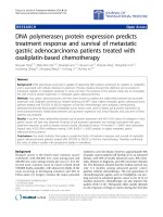
paired normal mucosa and tumor tissue samples, revealing that EMX2 expression was significantly lower in
colorectal tumor samples compared to their corresponding healthy mucosa (Fig. 1a). Next, we sought to investigate whether EMX2 was also down-regulated in
colorectal liver metastases. For this purpose, EMX2 transcript expression levels were assessed in 29 colorectal
Page 3 of 8
liver metastases from our tissue biobank, and EMX2
expression in metastases was compared to expression
levels in primary colorectal tumors. Indeed, EMX2
expression levels were further down-regulated in colorectal cancer liver metastases compared to primary
tumor tissue from patients suffering stage III colorectal
cancer (Fig. 1b). Given that EMX2 transcript levels were
downregulated in primary colorectal cancer as well as
colorectal liver metastases, we sought to determine protein levels of EMX2 by means of immunohistochemistry
and Western blotting. In line with the transcript expression data outlined above, we could detect expression of
EMX2 in healthy mucosa, but not in primary colorectal
cancer samples or colorectal liver metastases (Fig. 1c,d).
Taken together, these data suggest that down-regulation
of EMX2 expression occurs in primary and, even more,
in metastatic colorectal cancer.
Down-regulation of EMX2 is associated with decreased
survival in stage III patients
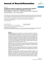
To investigate putative effects of down-regulated EMX2
expression on patient prognosis, we analyzed whether
EMX2 transcript expression was associated with progression to metastatic disease. To measure expressional
changes in our patient collective, we normalized EMX2
expression levels of primary tumors to those of their adjacent normal mucosa tissue. A comparison of patients
harboring EMX2 over-expressing tumors to those carrying EMX2 under-expressing tumors was performed by
Kaplan-Meier analysis using Cox proportional hazards
modeling, and demonstrated a significant association between down-regulated tumoral EMX2 expression and
the occurrence of colorectal liver metastases. Consistently, both disease free and overall survival were
significantly decreased in patients displaying downregulated EMX2 expression levels in their primary
tumors (Fig. 2a,b). Taken together, these results indicate that decreased EMX2 expression predicts metastatic progression and unfavorable outcome in stage
III colorectal cancer.
Adenoviral delivery of EMX2 attenuates the migration of
colorectal cancer cells
In an attempt to further elucidate whether enhanced
metastatic progression of colorectal tumors could indeed
be attributable to expressional changes of EMX2, we examined the effects of EMX2 on the migratory potential
of colorectal tumor cells in vitro. In search for a suitable
in vitro model, imitating up- or down-regulation of
EMX2, we screened various colorectal cancer cell lines
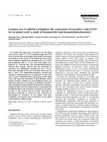
for their EMX2 expression levels. While DLD1 cells
were found to lack EMX2, CaCo2 cells displayed sustained expression of EMX2 (Fig. 3a). We therefore performed siRNA-mediated EMX2 knockdown in CaCo2
Aykut et al. BMC Cancer (2017) 17:555
Page 4 of 8
Table 1 Correlation of clinical parameters of 31 patients with expression of EMX2 and overall survival
n
EXM2high(%)
EXM2low(%)
P-value
(Fisher’s exact test)
Male
23
11(47,83%)
12(52,17%)
0,1552
Female
8
3(37,50%)
5(62,50%)
< median = 67
17
7(41,18%)
10(58,82%)
< median = 67
14
7(50,00%)
7(50,00%)
Colon
16
6(37,50%)
10(62,50%)
Rectum
15
8(53,33%)
7(46,67%)
T1
0
0
0
T2
4
3(75,00%)
1(25,00%)
78,5
T3
22
11(50,00%)
11(50,00%)
51,9
T4
5
0(0,00%)
5(100%)
46,4
N1
17
9(52,94%)
8(47,06%)
N2
14
5(35,71%)
9(64,29%)
R0
31
14(45,16%)
17(54,84%)
R1
0
0
0
N.A.
R2
0
0
0
N.A.
Rx
0
0
0
N.A.
Characteristic
Mean overall
survival (months)
P-value
(Log-rank test)
55,2
0,7989
Gender
52,1
Age at operation
0,2559
51,4
0,5648
57,7
Localisation
0,0649
54,9
0,9256
53,9
T stage
0,0413
N.A.
0,2123
N stage
0,0226
54,8
0,9444
54,0
Resection margin
N.A.
54,4
N.A
Occurrence of liver metastasis
Yes
14
3(21,43%)
11(78,57%)
No
17
11(64,71%)
6(35,29%)
0.0001
43,0
0,0415
63,8
Perioperative chemotherapy
Yes
27
12(44,44%)
15(55,56%)
No
4
2(50,00%)
2(50,00%)
0,4788
52,6
0,3842
66,7
NA not applicable
cells, which significantly reduced EMX2 expression on
the transcript level (EMX2 mRNA, norm. to GUS:
678.0 ± 43.41 in control-transfected cells versus 438.0
± 26.53 in siEMX2-transfected cells; P < 0.05; n = 3),
as well as on the protein level (Fig. 3b). Intriguingly,
tumor cell migration was significantly increased upon
knockdown of EMX2 in this cell line (Fig. 3d). We
likewise examined the significance of EMX2 in cell
migration by infection of DLD1 cells (which display
low EMX2 expression levels at normal baseline
conditions) with an adenovirus expressing EMX2 (AdEMX2), or with an empty vector control (Ad-Null).
Transfection with Ad-EMX2 caused robust and stable
over-expression of EMX2 in DLD1 cells on the transcript level (EMX2 mRNA, norm. to GUS: 3.9 ± 0.3 in
Ad-Null-transfected cells versus 3466.654 ± 840.82 in
Ad-EMX-2-transfected cells; P < 0.001; n = 3), as well
as on the protein level (Fig. 3c). Consistent with our
results obtained from CaCo2 cells, adenoviral overexpression of EMX2 resulted in significantly decreased
migration of DLD1 tumor cells (Fig. 3e). Collectively,
these in vitro data confirm the notion that EMX2
down-regulation in colorectal cancer cells affects their
migratory potential, thus contributing to an increased
rate of distant metastasis and unfavorable outcome.
Discussion
The homeodomain-containing transcription factor
EMX2 was first described as an important mediator
in embryonic development as EMX2 has been shown
to play a role in neuroblast proliferation, migration
and differentiation [20–22]. While transcriptional
Aykut et al. BMC Cancer (2017) 17:555
Page 5 of 8
Fig. 1 qRT-PCR depicting relative EMX2 mRNA expression in a primary stage III colorectal cancer samples and adjacent normal tissue. EMX2 levels
in b liver metastases from CRC were further down-regulated compared to primary tumors. Bars indicate mean ± SEM (* P < 0.05, n = 31; ** P < 0.01,
n = 31 primary tumors; 29 colorectal liver metastases). c Immunohistochemical staining, revealing EMX2 protein expression (arrowheads) in normal
mucosa, but not in corresponding primary tumors or in liver metastases of each 2 representative patients. d Western Blot revealing EMX2 expression in
primary tumor and normal mucosa from 2 representative patients
targets of EMX2 remain largely unidentified, loss of
EMX2 function is associated with impaired development of the cortex [23–25]. Moreover, homozygously
EMX2-deficient mice have been shown to display ectopic Wnt expression [12]. Wnt is an oncogene and
its signaling represents an early event in a majority of
colorectal cancers [26]. Wnt pathway signaling has
also been associated with metastatic spread and stemness in CRC [27–29].
Recently, the role of EMX2 has been explored in various
solid tumors. Several lines of evidence suggest that EMX2
is down-regulated in lung cancer [14, 30, 31]. While the
mode of EMX2 down-regulation in non small-cell lung
cancer (NSCLC) has been identified as epigenetic silencing, restoration of EMX2 has been shown to antagonize
Wnt and to restore sensitivity to cisplatin [14]. More recently, loss of EMX2 expression has been demonstrated in
gastric cancer cell lines and primary gastric cancer. EMX2
Aykut et al. BMC Cancer (2017) 17:555
Page 6 of 8
Fig. 2 Correlation of EMX2 mRNA expression levels (tumor/mucosa) and a disease-free survival UICC III patents in univariate analysis (* P < 0.05, n = 31;
hazard ratio 3.254; 95% confidence interval 1.137; 9.312). b Overall survival of UICC III assessed by Kaplan-Meier plot (** P < 0.001, n = 31; hazard ratio
6.619; 95% confidence interval 2.021; 21.68)
Fig. 3 a qRT-PCR revealing EMX2 transcript expression in two different colorectal cancer cell lines (DLD1, CaCo2). b Semiquantitative analysis of
immunoblotting revealing residual EMX2 expression after control-transfection versus siRNA-mediated EMX2 knockdown in CaCo2 cells and c Ad-Null
versus Ad-EMX2-transfected DLD1 cells. Bars indicate mean ± SEM. d Transwell migration assay revealing enhanced migratory potential of CaCo2 cells
upon siRNA-mediated knockdown of EMX2 compared to control-transfected (scrambled) cells. Restoration of EMX2 expression in e DLD1 cells using an
adenoviral delivery system resulted in attenuated migration. Ad-Null and Ad-EMX2 represent the empty adenoviral vector control and the adenoviral
vector containing EMX2 cDNA, respectively. Bars indicate mean ± SEM of three independent experiments (* P < 0.05)
Aykut et al. BMC Cancer (2017) 17:555
down-regulation was associated with promoter hypermethylation and the adenoviral delivery of EMX2 in a mouse
model of gastric cancer significantly suppressed tumor
growth [16]. Thus, these previous observations support
our present finding that down-regulation of EMX2 has
protumorigenic effects in colorectal cancer.
To our knowledge, this is the first investigation of the
functional role of EMX2 in colorectal cancer. In our
patient collective, EMX2 was frequently down-regulated
in tumor tissue in comparison to matched normal mucosa samples. Down-regulation of EMX2 was associated
with metastatic tumor progression and decreased overall
survival. These results differ from a previous study,
where loss of EMX2 was found in only 2–5% of colorectal cancers [32]. In contrast to the latter study, we found
a down-regulation of EMX2 in 54% of our patient
collective. These observed differences can in part be
explained by the different methods that were applied to
assess EMX2 expression. While Kim et al. used immunohistochemical analysis, we applied qRT-PCR to detect
EMX2 expression levels [32]. We then normalized expression levels of EMX2 in tumors to expression levels
in adjacent normal mucosa. We believe that qRT-PCR is
a robust and sensitive tool for quantification of gene
expression levels.
Nevertheless, our study has several limitations. First,
the power to make statistical inferences was limited by a
modest sample size. The main reason for this is the limited number of cases where both tumor and normal mucosa samples were available for RNA extraction, along
with appropriate follow-up data allowing for the detection of metastatic disease and assessment of survival.
Second, although colorectal liver metastases showed a
further decrease in EMX2 expression levels when compared to primary colorectal cancer samples, it remains
unknown whether this is due to an evolutionary
decrease of EMX2 expression from primary tumor to
metastasis and therefore a possible driver or prerequisite
for metastatic outgrowth or whether this is a mere coincidence. Metastatic progression of cancer is a complex
process involving a multi-step process, where migration
represents a key element in the process of the metastasic
cascade [33]. The functional assays in this study assessing tumor cell migration suggest a potential role for
EMX2 in metastatic disease progression since EMX2
knockdown resulted in increased migration while restoration of EMX2 using an adenoviral vector led to
decreased migration. We used a recombinant human
adenovirus type 5 as delivery system since this is the
vector of choice for functional genomics research [34].
While there are still many challenges that need to be
overcome before adenoviral vectors can be safely used in
cancer patients, adenoviral vector-based therapeutic
strategies represent a promising tool for cancer gene
Page 7 of 8
therapy [35, 36]. Altogether the role of EMX2 expression
in metastatic spread would be a valuable area of future
research in a larger patient cohort.
In summary, our data encourage a significant role of
EMX2 in the progression and metastasis of colorectal
cancer. Our study demonstrates that a low EMX2 expression level is an independent prognostic factor and
correlates with dismal prognosis, decreased overall survival and the development of metastatic disease in stage
III colorectal cancer patients. Thus, the study at hand
provides a first evidence for the role of EMX2 as a suppressor of metastasis in colorectal cancer. Further, we
provide evidence that EMX2 has predictive value as a
prognostic factor in stage III colorectal cancer as well as
a possible functional role in metastatic spread. Therefore, restoration of EMX2 via gene therapy may represent a promising therapeutic strategy for tailored
anticancer therapy.
Conclusions
EMX2 is frequently down-regulated in both primary
colorectal cancer and colorectal cancer liver metastases.
Down-regulated EMX2 is a strong predictor for shortened disease-free and overall survival in stage III colorectal cancer. In vitro knockdown of EMX2 leads to
increased tumor cell migration while adenoviral restoration of EMX2 is associated with decreased migration.
EMX2 might represent a promising molecular target for
colorectal cancer therapy.
Abbreviations
CRC: colorectal cancer; EMX2: Empty Spiracles Homeobox 2; GUS: βGlucuronidase; NSCLC: Non-small cellular lung cancer; qRT-PCR: Real-Time
Quantitative Reverse Transcription PCR; SEM: Standard error of the mean;
UICC: International Union Against Cancer
Acknowledgements
We thank Mareen Dupovac, Melanie Höfler, Vishnu Mani and Nicholas Rüdinger
for their technical assistance. We also thank Ulf Hinz for his statistical support.
Funding
This study was funded by the Heidelberger Stiftung Chirurgie (to B. Aykut)
and by the Deutsche Forschungsgemeinschaft, Germany (to M. Schneider,
grant SCHN 947/4–2 in the framework of the Clinical Research Group KFO
227). The funders have no role in the study design and data collection,
analysis, and interpretation as well as manuscript writing.
Availability of data and materials
The datasets used and/or analyzed during the current study are available
from the corresponding author upon reasonable request.
Authors’ contributions
BA and MS designed the study; BA performed the experiments; BA, AU and
MS wrote the manuscript; MO, PR, DW, HH and SS performed part of the
experiments and composition of the manuscript. AB was responsible for data
collection and analysis. RK and YK helped performing the adenoviral
experiments. All authors have read and approved the final manuscript.
Authors’ information
Not applicable.
Aykut et al. BMC Cancer (2017) 17:555
Ethics approval and consent to participate
This study was approved by the ethics committee of the Medical Faculty of
Heidelberg University (S-649/2012). Written informed consent was obtained
from all patients involved in this study.
Consent for publication
Not applicable.
Competing interests
The authors declare that they have no competing interests.
Publisher’s Note
Springer Nature remains neutral with regard to jurisdictional claims in
published maps and institutional affiliations.
Author details
1
Department of General, Visceral and Transplantation Surgery, University of
Heidelberg, Heidelberg University Hospital, Im Neuenheimer Feld 110, 69120
Heidelberg, Germany. 2Department of Biotechnology, University of
Heidelberg, Heidelberg, Germany.
Received: 24 May 2016 Accepted: 15 August 2017
References
1. Siegel R, Desantis C, Jemal A. Colorectal cancer statistics, 2014. CA Cancer J
Clin. 2014;64(2):104–17.
2. Pan J, Xin L, Ma YF, Hu LH, Li ZS. Colonoscopy reduces colorectal cancer
incidence and mortality in patients with non-malignant findings: a metaanalysis. Am J Gastroenterol. 2016;111(3):355–65.
3. Kohne CH, Lenz HJ. Chemotherapy with targeted agents for the treatment
of metastatic colorectal cancer. Oncologist. 2009;14(5):478–88.
4. Chang W, Wei Y, Ren L, et al. Randomized controlled trial of Intraportal
chemotherapy combined with adjuvant chemotherapy (mFOLFOX6) for
stage II and III colon cancer. Ann Surg. 2016;263(3):434–9.
5. Manchon-Walsh P, Borras JM, Espinas JA, Aliste L. Catalonian rectal cancer G.
Assessing the effectiveness of a guideline recommendation for pre-operative
radiochemotherapy in rectal cancer. Radiother Oncol. 2011;99(2):142–7.
6. Schmoll HJ, Van Cutsem E, Stein A, et al. ESMO consensus guidelines for
management of patients with colon and rectal cancer. A personalized
approach to clinical decision making. Ann Oncol. 2012;23(10):2479–516.
7. Cillo C, Faiella A, Cantile M, Boncinelli E. Homeobox genes and cancer. Exp
Cell Res. 1999;248(1):1–9.
8. Gangemi RM, Daga A, Muzio L, et al. Effects of Emx2 inactivation on the gene
expression profile of neural precursors. Eur J Neurosci. 2006;23(2):325–34.
9. Abate-Shen C. Deregulated homeobox gene expression in cancer: cause or
consequence? Nat Rev Cancer. 2002;2(10):777–85.
10. Dalton D, Chadwick R, McGinnis W. Expression and embryonic function of
empty spiracles: a drosophila homeo box gene with two patterning
functions on the anterior-posterior axis of the embryo. Genes Dev. 1989;
3(12A):1940–56.
11. Galli R, Fiocco R, De Filippis L, et al. Emx2 regulates the proliferation of stem
cells of the adult mammalian central nervous system. Development. 2002;
129(7):1633–44.
12. Ligon KL, Echelard Y, Assimacopoulos S, et al. Loss of Emx2 function leads
to ectopic expression of Wnt1 in the developing telencephalon and cortical
dysplasia. Development. 2003;130(10):2275–87.
13. Shah N, Sukumar S. The Hox genes and their roles in oncogenesis. Nat Rev
Cancer. 2010;10(5):361–71.
14. Okamoto J, Hirata T, Chen Z, et al. EMX2 is epigenetically silenced and
suppresses growth in human lung cancer. Oncogene. 2010;29(44):5969–75.
15. Noonan FC, Mutch DG, Ann Mallon M, Goodfellow PJ. Characterization of the
homeodomain gene EMX2: sequence conservation, expression analysis, and a
search for mutations in endometrial cancers. Genomics. 2001;76(1–3):37–44.
16. Li J, Mo M, Chen Z, et al. Adenoviral delivery of the EMX2 gene suppresses
growth in human gastric cancer. PLoS One. 2012;7(9):e45970.
17. Taylor HS, Fei X. Emx2 regulates mammalian reproduction by altering
endometrial cell proliferation. Mol Endocrinol. 2005;19(11):2839–46.
18. Yue D, Li H, Che J, et al. EMX2 is a predictive marker for adjuvant
chemotherapy in lung Squamous cell carcinomas. PLoS One. 2015;10(7):
e0132134.
Page 8 of 8
19. Schmittgen TD, Livak KJ. Analyzing real-time PCR data by the comparative
C(T) method. Nat Protoc. 2008;3(6):1101–8.
20. Cecchi C. Emx2: a gene responsible for cortical development,
regionalization and area specification. Gene. 2002;291(1–2):1–9.
21. Mariani J, Favaro R, Lancini C, et al. Emx2 is a dose-dependent negative
regulator of Sox2 telencephalic enhancers. Nucleic Acids Res. 2012;40(14):
6461–76.
22. Shinozaki K, Yoshida M, Nakamura M, Aizawa S, Suda Y. Emx1 and Emx2
cooperate in initial phase of archipallium development. Mech Dev. 2004;
121(5):475–89.
23. Mallamaci A, Muzio L, Chan CH, Parnavelas J, Boncinelli E. Area identity
shifts in the early cerebral cortex of Emx2−/− mutant mice. Nat Neurosci.
2000;3(7):679–86.
24. Shinozaki K, Miyagi T, Yoshida M, et al. Absence of Cajal-Retzius cells and
subplate neurons associated with defects of tangential cell migration from
ganglionic eminence in Emx1/2 double mutant cerebral cortex.
Development. 2002;129(14):3479–92.
25. Brunelli S, Faiella A, Capra V, et al. Germline mutations in the homeobox gene
EMX2 in patients with severe schizencephaly. Nat Genet. 1996;12(1):94–6.
26. Suzuki H, Watkins DN, Jair KW, et al. Epigenetic inactivation of SFRP genes
allows constitutive WNT signaling in colorectal cancer. Nat Genet. 2004;
36(4):417–22.
27. Vermeulen L, De Sousa EMF, van der Heijden M, et al. Wnt activity defines
colon cancer stem cells and is regulated by the microenvironment. Nat Cell
Biol. 2010;12(5):468–76.
28. Ormanns S, Neumann J, Horst D, Kirchner T, Jung A. WNT signaling and distant
metastasis in colon cancer through transcriptional activity of nuclear betacatenin depend on active PI3K signaling. Oncotarget. 2014;5(10):2999–3011.
29. Clevers H. Wnt/beta-catenin signaling in development and disease. Cell.
2006;127(3):469–80.
30. Okamoto J, Kratz JR, Hirata T, et al. Downregulation of EMX2 is associated
with clinical outcomes in lung adenocarcinoma patients. Clin Lung Cancer.
2011;12(4):237–44.
31. Shen DW, Pouliot LM, Hall MD, Gottesman MM. Cisplatin resistance: a
cellular self-defense mechanism resulting from multiple epigenetic and
genetic changes. Pharmacol Rev. 2012;64(3):706–21.
32. Kim MS, An CH, Yoo NJ, Lee SH. Rare somatic mutation and loss of expression
of EMX2 gene in common solid cancers. APMIS. 2011;119(10):733–4.
33. Rahbari NN, Bork U, Scholch S, et al. Metastatic spread emerging from liver
metastases of colorectal cancer: does the seed leave the soil again? Ann
Surg. 2016;263(2):345–52.
34. Sharma A, Tandon M, Bangari DS, Mittal SK. Adenoviral vector-based
strategies for cancer therapy. Curr Drug ther. 2009;4(2):117–38.
35. Sheridan C. Gene therapy finds its niche. Nat Biotechnol. 2011;29(2):121–8.
36. Waehler R, Russell SJ, Curiel DT. Engineering targeted viral vectors for gene
therapy. Nat Rev Genet. 2007;8(8):573–87.
Submit your next manuscript to BioMed Central
and we will help you at every step:
• We accept pre-submission inquiries
• Our selector tool helps you to find the most relevant journal
• We provide round the clock customer support
• Convenient online submission
• Thorough peer review
• Inclusion in PubMed and all major indexing services
• Maximum visibility for your research
Submit your manuscript at
www.biomedcentral.com/submit