Skeletal muscle depletion during chemotherapy has a large impact on physical function in elderly Japanese patients with advanced non–small-cell lung cancer
Bạn đang xem bản rút gọn của tài liệu. Xem và tải ngay bản đầy đủ của tài liệu tại đây (1.04 MB, 9 trang )
Naito et al. BMC Cancer (2017) 17:571
DOI 10.1186/s12885-017-3562-4
RESEARCH ARTICLE
Open Access
Skeletal muscle depletion during
chemotherapy has a large impact on
physical function in elderly Japanese
patients with advanced non–small-cell lung
cancer
Tateaki Naito1* , Taro Okayama2, Takashi Aoyama3, Takuya Ohashi2,4, Yoshiyuki Masuda2, Madoka Kimura1,5,
Hitomi Shiozaki3, Haruyasu Murakami1, Hirotsugu Kenmotsu1, Tetsuhiko Taira1, Akira Ono1, Kazushige Wakuda1,
Hisao Imai1,6, Takuya Oyakawa1,7, Takeshi Ishii2, Shota Omori1, Kazuhisa Nakashima1, Masahiro Endo8,
Katsuhiro Omae9, Keita Mori9, Nobuyuki Yamamoto10, Akira Tanuma2 and Toshiaki Takahashi1
Abstract
Background: Elderly patient with advanced cancer is one of the most vulnerable populations. Skeletal muscle
depletion during chemotherapy may have substantial impact on their physical function. However, there is little
information about a direct relationship between quantity of muscle and physical function. We sought to explore
the quantitative association between skeletal muscle depletion, and muscle strength and walking capacity in elderly
patients with advanced non–small cell lung cancer (NSCLC).
Methods: Thirty patients aged ≥70 years with advanced NSCLC (stage III-IV) scheduled to initiate first-line
chemotherapy were prospectively enrolled between January 2013 and November 2014. Lumbar skeletal muscle
index (LSMI, cm2/m2), incremental shuttle walking distance (ISWD, m), and hand-grip strength (HGS, kg) were
assessed at baseline, and 6 ± 2 weeks (T2) and 12 ± 4 weeks (T3) after study enrollment. Associations were
analyzed using linear regression.
Results: Altogether, 11 women and 19 men with a median age of 74 (range, 70–82) years were included in the
study; 24 received cytotoxic chemotherapy and 6, gefitinib. Mean ± standard deviation of LSMI, ISWD and HGS
were 41.2 ± 7.8 cm2/m2, 326.0 ± 127.9 m, and 29.3 ± 8.5 kg, respectively. LSMI and ISWD significantly declined from
baseline to T2 and T3. HGS significantly declined from baseline to T2 and T3 only in men. Change in LSMI was
significantly associated with change in HGS (β = 0.3 ± 0.1, p = 0.0127) and ISWD (β = 8.8 ± 2.4, p = 0.0005).
Conclusions: Skeletal muscle depletion accompanied with physical functional decline started in the early phase of
the chemotherapy in elderly patients with advanced NSCLC. Our results suggest that there may be a need for early
supportive care in these patients to prevent functional decline during chemotherapy.
Trial registration: Trial registration number: UMIN000009768
Name of registry: UMIN (University hospital Medical Information Network).
URL of registry: Date of registration: 14 January 2013.
(Continued on next page)
* Correspondence:
1
Division of Thoracic Oncology, Shizuoka Cancer Center, 1007,
Shimonagakubo, Nagaizumi-cho, Sunto-gun, Shizuoka 411-8777, Japan
Full list of author information is available at the end of the article
© The Author(s). 2017 Open Access This article is distributed under the terms of the Creative Commons Attribution 4.0
International License ( which permits unrestricted use, distribution, and
reproduction in any medium, provided you give appropriate credit to the original author(s) and the source, provide a link to
the Creative Commons license, and indicate if changes were made. The Creative Commons Public Domain Dedication waiver
( applies to the data made available in this article, unless otherwise stated.
Naito et al. BMC Cancer (2017) 17:571
Page 2 of 9
(Continued from previous page)
Date of enrolment of the first participant to the trial: 23 January 2013.
Keywords: Non–small cell lung cancer, Incremental shuttle walking distance, Hand-grip strength, Skeletal muscle
mass, Sarcopenia, Cancer cachexia
Background
The number of elderly people living with advanced lung
cancer is increasing worldwide, owing to the aging population and advances in cancer treatment [1]. In Japan,
65% of lung cancer morbidity cases and 73% of annual
lung cancer deaths were attributed to elderly individuals
aged ≥70 years in 2012 [2]. Elderly patient with advanced
cancer is one of the most vulnerable populations [3]. Patients with advanced non-small-cell lung cancer (NSCLC)
frequently have cancer cachexia [4, 5] and skeletal muscle
depletion [5, 6]. In addition, cancer treatment including
radiotherapy [7], chemotherapy [8], and supportive care
such as hospitalization [9] or the use of corticosteroids
may cause muscle dysfunction [10]. Consequently, skeletal
muscle depletion may cause physical dysfunction [11–14]
and develop disability [15–17] before and during cancer
treatment in NSCLC. Currently however, limited information exists on the quantitative association between loss of
skeletal muscle mass and physical dysfunction in elderly
patients with advanced NSCLC.
Accordingly, we sought to quantify impact of skeletal
muscle mass depletion on muscle strength and walking
capacity in elderly patients with advanced NSCLC receiving chemotherapy.
Methods
Patient selection
This prospective longitudinal observational study was performed at the Shizuoka cancer center, Japan, from January
2013 to January 2014. Shizuoka cancer center is a 615-bed
prefectural hospital designated as an advanced treatment
hospital by the Japanese Ministry of Health, Labor and
Welfare. The eligibility criteria were as follows: (1) histologically and/or cytologically proven stage III or IV NSCLC including postoperative recurrence; (2) age ≥ 70 years, with
planned first-line systemic chemotherapy; (3) no previous
systemic chemotherapy or thoracic radiotherapy (adjuvant
chemotherapy was not counted as a prior chemotherapy);
(4) Eastern Cooperative Oncology Group performance status of 0–2; (5) ability to ambulate, read, and respond to
questions without assistance; and (6) expected survival of >12 weeks. Patients were excluded if they had a
severe psychiatric disorder, active infectious disease, unstable cardiac disease, or untreated symptomatic brain or
bone metastases that prevented safe assessment.
All patients provided written informed consent. The
study was approved by the institutional review board and
registered on the clinical trials site of the University
Hospital Medical Information Network Clinical Trials
Registry in Japan (registration number: UMIN000009768).
Patient enrollment and timing of data collection
The first patient was enrolled on January 23, 2013, and
the last on November 7, 2013. The last physical assessment was performed on January 27, 2014. Lumbar skeletal muscle index (LSMI, cm2/m2), incremental shuttle
walking distance (ISWD, m), and hand-grip strength
(HGS, kg) were assessed at baseline (T1), and 6 ± 2 weeks
(T2) and 12 ± 4 weeks (T3) after study enrollment. Baseline study assessments were performed by the attending
physicians, physiotherapists, and national registered dietitians at the time between study entry and initiation of
the first chemotherapy.
Patient assessment
Body weight (kg) was measured to the nearest 0.1 kg
and the body mass index (BMI; kg/m2) was subsequently
calculated. The ISWD and HGS on the dominant side
were measured by physiotherapists (T.O., T.O., Y.M.,
and T.I.). The incremental shuttle walking test was conducted according to the recent guideline [18] and original protocol described by Singh et al. [19]. The 10-m
course was established in the corridor of our hospital.
Walking speed was dictated by a timed signal played on
a CD-recorder provided by the manufacturer (Japanese
version, produced by the Graduate School of Biomedical
Sciences, Nagasaki University, Japan, 2000). All patients
were tested once under standardized conditions and
were carefully observed during the test, so that they
would not exceed their exercise limit. The instructor
stayed alongside the course and provided no encouragement. The end of the test was determined by either (1)
the patient, when he or she was too breathless to keep
the required walking speed; (2) the instructor, if the
patient could not complete a shuttle within the time
allotted (ie, > 0.5 m away from the cone when the bleep
sounded); or (3) attainment of 85% or higher of the
predicted maximal heart rate derived from the formula
[210 - (0.65 x age)]. The maximal walking distance was
described as ISWD. Loss of 40 m was defined to be a
clinically significant reduction in ISWD in this study
[20]. HGS was measured using a grip strength dynamometer (GRIP-D, Takei Scientific Instruments Co.,
LTD, Niigata, Japan). Patient was in an upright position
Naito et al. BMC Cancer (2017) 17:571
and held the dynamometer in one hand with the grip
range adjusted so that the second joint of the forefinger
was bent 90°. The instrument was then held down at the
patient’s side without letting the arm touch the body,
with the arm fully extended. Patient was then asked to
exert full force with his or her hand for about 3 s to obtain the maximum kilogram-force, during which the instructor provided verbal encouragement. One trial was
performed for each hand, and the result from the strongest hand was used for this analysis. Lumbar skeletal
muscle mass was measured by analyzing electronically
stored computed tomography images using SYNAPSE
VINCENT version 3 (FUJIFILM Medical Systems,
Japan). Conditions of CT image included contrast enhanced or unenhanced, 5-mm slice thickness. Two consecutive CT images at the third lumbar vertebra (L3)
were chosen to measure the cross-sectional area of the
skeletal muscle that was identified based on Hounsfield
unit thresholds of −29 to +150. The sum of the crosssectional areas (cm2) of the muscles in the L3 region
was computed for each image. The mean value of 2 images was normalized for height in meters squared and
reported as LSMI (cm2/m2) [21]. The disease stage was
determined according to the TNM classification, and the
best response to chemotherapy was evaluated according
to the Response Evaluation Criteria in Solid Tumors.
Diagnosis of muscle depletion and cancer cachexia
Skeletal muscle depletion was defined based on the cutoff point of the LSMI of 43 cm2/m2 for men with a
BMI < 25.0, 53 cm2/m2 for men with a BMI ≥ 25.0, and
41 cm2/m2 for women [22].
Cancer cachexia was defined as unintentional weight
loss >5% during the past 6 months or >2% in patients with
a BMI <20 kg/m2, or the presence of muscle depletion according to the consensus criteria [23]. The patient’s weight
6 months before study entry was obtained by interviewing
the patient and their family members at study entry.
Statistical analysis
Chi-square or Fisher’s exact tests were used to compare
categorical variables. Wilcoxon signed-rank test was used
for the pairwise comparison of measurement changes between study visits, whereas the Wilcoxon rank-sum test
was used for comparisons between 2 independent groups.
For all analyses, p-values <0.05 were considered significant. All statistical analyses were performed using JMP
version 12.0 for Windows (SAS Institute Inc., USA).
Results
Patients
Among 31 patients screened, 30 patients with a median
age of 74 years (range, 70–82 years) were enrolled into
this study; 11 patients (36.7%) were women (Table 1).
Page 3 of 9
Common comorbidities included chronic obstructive
pulmonary disease, cardiovascular disease, and type 2
diabetes. There was a higher percentage never smokers
among women than men (81.2 vs. 0%, p < 0.05).
Cancer treatment during the study period
All patients received first-line chemotherapy within
1 week after the baseline assessment. All patients initially received a standard dose of chemotherapy with a
standard schedule. Ten patients received single-agent
chemotherapy, including docetaxel (60 mg/m2, every
3 weeks, n = 8) and vinorelbine (25 mg/m2, day 1 and 8
every 3 weeks, n = 2), until disease progression or unacceptable toxicity. Median treatment cycle (range) was
5 (2–12) cycles. Two patients required dose reduction
due to febrile neutropenia and moderate nausea. One
patient discontinued chemotherapy after 2 cycles of docetaxel due to performance status deterioration and bacterial pneumonia. Fourteen patients received platinumbased chemotherapy, including 7 patients who received
carboplatin (target area under the curve of 6, every
3 weeks) + paclitaxel (200 mg/m2, every 3 weeks), 5 who
received cisplatin (75 mg/m2, every 3 weeks) + pemetrexed
(500 mg/m2, every 3 weeks), 1 who received cisplatin
(80 mg/m2, every 3 weeks) + gemcitabine (1000 mg/m2,
day1 and 8 every 3 weeks), and 1 patient cisplatin
(80 mg/m2, every 3 weeks) + vinorelbine (25 mg/m2,
day1 and 8 in every 3 weeks). Treatment was planned
for 4 to 6 cycles unless there was evidence of unacceptable toxicity or disease progression. Median cycle (range)
was 4 (2–6) cycles. Two patients required dose reduction due to elevated serum creatinine level and severe
weight loss. One patient changed his regimen after 2 cycles of carboplatin + paclitaxel due to a severe allergic
reaction. Six patients with epidermal growth factor receptor gene mutations received gefitinib (250 mg once
daily). The median treatment period was 10.2 months.
One patient required a dose reduction due to moderate
liver dysfunction. None of the patients discontinued
treatment due to adverse events. An objective tumor response was seen in 12 patients (40.0%).
Evaluable patient data
Patient flow and the number of evaluable data at each time
point are summarized in the flow diagram (Fig. 1). Among
30 patients enrolled, 30 and 28 patients were alive and eligible for evaluation at T2 and T3, respectively. One man
died from disease progression and one woman was transferred to another hospital until T3 point. At baseline, the
HGS test was refused by a patient. At T2 point, the shuttle
walking test was refused by one patient and abandoned by
the physiotherapist in 2 patients for safety reason; and
computed tomography data in 2 patients were not obtained during the designed period. At T3 point, the shuttle
Naito et al. BMC Cancer (2017) 17:571
Page 4 of 9
Table 1 Baseline characteristics
Variables
All (N = 30)
Men (N = 19)
Women(N = 11)
Age, median (range)
74 (70–82)
74 (70–82)
76 (70–80)
0
11 (36.7)
7 (36.8)
4 (36.4)
1
18 (60.0)
12 (63.2)
6 (54.5)
2
1 (3.3)
0 (0.0)
1 (9.1)
Reference value (men/women)
ECOG-PS, n (%)
Stage, n (%)
IIIA or IIIB
1 (3.3)
1 (5.3)
0
IV or postoperative reccurence
29 (96.7)
18 (94.7)
10 (100)
Adenocarcinoma
21 (70.0)
13 (68.4)
8 (72.7)
Other non-small-cell lung cancer
9 (30)
6 (31.6)
0
Cytotoxic regimen
24 (80.0)
17 (89.5)
7 (63.6)
Targeted regimen
6 (20.0)
2 (10.5)
4 (36.4)
9 (30.0)
0*
9 (81.2)
Chronic obstructive pulmonary disease
10 (33.3)
7 (36.8)
3 (27.3)
Type 2 diabetes
6 (20.0)
5 (26.3)
1 (9.1)
Cerebrovascular disease
4 (13.3)
4 (21.1)
0
Cardiovascular disease
1 (3.3)
0
1 (9.1)
21.1 ± 3.4
21.6 ± 3.5
20.2 ± 3.1
Lumbar skeletal muscle index (cm /m )
41.2 ± 7.8
44.5 ± 7.6*
35.4 ± 4.1
Skeletal muscle depletiona, n (%)
20 (66.7)
10 (52.6)*
10 (90.9)
b
19 (63.3)
11 (57.9)
8 (72.7)
Hand grip strength (dominant side, kg)
29.3 ± 8.5
33.9 ± 7.1*
21.7 ± 4.1
32/ 20 [24]
Shuttle walk distance (m)
326.0 ± 127.9
338.4 ± 143.0
304.5 ± 99.2
360–400 [25]
Tumor Histology, n (%)
Chemotherapeutic regimen, n (%)
Never smoke, n (%)
Comorbidities, n (%)
Body composition
Body-mass index (kg/m2)
2
2
Cancer cachexia , n (%)
17.2/ 19.9 [26]
Physical function
*Significant gender difference (P < 0.05) tested by Chi-square test, Fisher exact test, or Wilcoxon test. askeletal muscle mass depletion was defined as lumbar skeletal
muscle mass index of <43.0 cm2/m2 for men with a BMI <25.0 kg/m2, <53.0 cm2/m2 for men with a BMI ≥25.0, and <41.0 cm2/m2 in women bDiagnosis was based on
the international consensus criteria for cancer cachexia. ECOG-PS: Eastern cooperative oncology group performance status
walking test was refused by 2 patients and abandoned
by the physiotherapist in one patient for safety reason; the HGS test was abandoned by the physiotherapist in 2 patients for safety reason; and computed
tomography data in 2 patients were not obtained during the designed period.
Body mass, muscle mass, and physical function at baseline
At baseline, mean ± standard deviation of BMI was
21.5 ± 3.4 kg/m2 in men and 20.1 ± 3.1 kg/m2 in women
(Table 1). Mean LSMI was 44.5 ± 7.6 cm2/m2 in men
and 35.4 ± 4.1 cm2/m2 in women with a significant difference between the sexes (P < 0.05). Skeletal muscle depletion was diagnosed in 20 (66.7%) patients and higher
proportion of women were diagnosed with skeletal
muscle depletion than men (90.9 vs. 52.6%, p < 0.05).
Cancer cachexia was diagnosed in 19 (63.3%) patients.
In regard to the physical function, mean HGS was
33.8 ± 7.0 kg in men and 21.7 ± 4.0 kg in women with a
significant difference between the sexes (P < 0.05). These
values were almost comparable to the reference value in
the Japanese community-dwelling elderly population
[24] (shown in Table 1). Mean ISWD was 338.4 ± 142.9
in men and 304.5 ± 99.2 in women without gender difference (P = 0.54). The values were relatively low in
comparison with the reference values in the Japanese
community-dwelling elderly population [25].
Longitudinal changes in muscle mass and physical function
Statistically significant reductions between baseline
values, and T2 and T3, were seen for weight, BMI,
LSMI, and ISWD. A clinically significant reduction in
Naito et al. BMC Cancer (2017) 17:571
Page 5 of 9
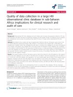
Fig. 1 Flow diagram. The number of patients and evaluable data at the T1 (baseline), T2 (6 ± 2 weeks), and T3 (12 ± 2 weeks) point is shown.
The number of evaluable data for each variable is described in the box. The reasons for a missing value are described in the right side of each
box. HGS, hand-grip strength; ISWD, incremental shuttle walking distance; LSMI, lumbar skeletal muscle index
ISWD was also seen in 11 patients (40.7%) at T2 and in
13 patients (52.0%) at T3. No statistically significant reductions were observed between T2 and T3 for weight,
BMI, LSMI, HGS, and ISWD (Table 2 and Fig. 2). Men
had a significant reduction in HGS at T2 (p < 0.05) and
T3 (p < 0.05), whereas women had no reduction in either time point (p = 0.45 and p = 0.78, respectively).
Association between changes in skeletal muscle mass and
physical function
There was a statistically significant linear association between changes in LSMI and HGS (β = 0.3 ± 0.1,
p = 0.0127, Fig. 3a). There was also a positive linear association between LSMI and changes in HGS (β = 8.8 ± 2.4,
p = 0.0005, Fig. 3b).
Table 2 Longitudinal changes in physical parameters
Variables
Mean difference from baseline (±SE)
Mean difference between T2 and T3 (±SE)
6 ± 2wks
12 ± 4wks
N
30
28
25
Body weight (kg)
−0.9 ± 0.4*
−1.1 ± 0.6*
−0.2 ± 0.4
Body-mass index (kg/m )
−0.3 ± 0.1*
−0.4 ± 0.1*
−0.1 ± 0.1
L3 muscle index (cm2/m2)
−1.8 ± 0.4*
−1.8 ± 0.7*
−0.1 ± 0.4
Hand grip strength (non-dominant, kg)
−0.7 ± 0.6
−0.7 ± 0.6
−0.5 ± 0.3
Shuttle walk distance (m)
−40.0 ± 12.6*
−46.4 ± 15.8*
−10.8 ± 11.3
11 (40.7)
13 (52.0)
5 (20.0)
2
b
Clinically significant decline , n (%)
*p < 0.05 in Wilcoxon signed-rank test compared with baseline value
b
Clinically significant decline is defined as losing ≥40 m of shuttle walk distance from baseline
Naito et al. BMC Cancer (2017) 17:571
Page 6 of 9
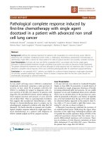
Fig. 2 Longitudinal changes in body-mass, muscle mass, and physical function. Mean changes ± standard error of physical parameters from baseline
value is shown. P-value of Wilcoxon signed-rank test was shown
Subset analysis for changes in skeletal muscle mass at T2
point
In subset analysis in LSMI at T2 point, depletion in
LSMI was observed in most of the subsets classified by
gender, smoking status, performance status, presence of
cancer cachexia, response to chemotherapy, and treatment regimens (Fig. 4). Smokers had a larger reduction
in LSMI than never-smokers (P < 0.05). Similarly, patient with tumor progression at T2 had larger reduction
in LSMI than patients without tumor progression
a
(P < 0.05). There was no statistical association between
treatment modification (dose reduction or discontinuation) and reduction in LSMI.
Discussion
To our knowledge, this is the first prospective study to
show longitudinal changes in skeletal muscle mass
associated with physical function in elderly patients with
advanced NSCLC receiving chemotherapy. First, we
showed that majority of this patient population had
b
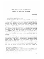
Fig. 3 Association between changes in skeletal muscle mass and physical function. The association between change in muscle mass, and hang-grip
strength (a) and shuttle walking distance (b) at all time points are plotted. Dotted line indicates the 95% confidence interval. Circle, triangle, and square
mark represents change at T2 from baseline, T3 from baseline, and T3 from T2, respectively
Naito et al. BMC Cancer (2017) 17:571
Page 7 of 9
Fig. 4 Subset analysis for change in skeletal muscle mass at T2 point. Median change of skeletal muscle mass at T2 point in each subset was
shown. The number of patients in each subset is indicated in parenthesis. White line indicates the median. The top and bottom of each box
represent the upper and lower quartiles of the values for the sample. Bars extend above and below each box to the maximal and minimal values
in the sample. P-value of Wilcoxon rank-sum test was shown. PS, Eastern Cooperative Oncology Group performance status; PD, progressive
disease assessed by the Response Evaluation Criteria in Solid Tumors at T2 point
skeletal muscle depletion, cancer cachexia, and decreased walking capacity at baseline. Second, we found
that they rapidly lost their body mass, skeletal muscle
mass, muscle strength, and walking capacity in the early
course of systemic chemotherapy. Third, we found positive associations between changes in skeletal muscle
mass and muscle strength or walking capacity.
Dewys WD et al. [4] reported that two-thirds of incurable chemotherapy-naïve patients with NSCLC experienced weight loss, especially in those with advanced
disease. In our previous research, we reported that
45.6% of chemotherapy-naïve patients with advanced
NSCLC had cancer cachexia at baseline [5]. The incidence of cancer cachexia in the present study (63.3%)
was somewhat higher. The possible reasons for this difference is that this study only included elderly patients
(median age, 74 years vs. 66 years in our previous study)
and more patients with metastatic disease (97% vs. 88%).
High incidences of sarcopenia have been reported in patients with advanced lung cancer [5, 6]. Consistently, our
patients had relatively high incidence of skeletal muscle
depletion (52.6% in men and 90.9% in women), compared
with those of community-dwelling Japanese elderly population (17.2% in men and 19.9% in women [26]). In
addition, patients with advanced lung cancer have been
reported to have poorer physical function, compared with
community-dwelling elderly in regards to muscle strength
and endurance performance measured by the 6-min walking test [12, 13, 27]. In this study, the baseline values of
the incremental shuttle walking distance tended to be
lower, compared with the reference values of communitydwelling elderly populations [25].
Weight loss during cancer treatment is commonly observed in patients with lung cancer receiving chemotherapy
[5, 28] or thoracic radiotherapy [7], and is accompanied by
a marked decrease in skeletal muscle mass [5, 8]. Similarly,
our patients had a significant decrease in body mass and
skeletal muscle mass during the first 6–12 weeks of cancer
treatment. Stene GB et al. [8] reported that patients with
disease progression following chemotherapy tended to have
a larger reduction in skeletal muscle mass, compared with
patients with disease control following chemotherapy. Our
data also showed that patients with tumor progression had
greater muscle loss in the subset analysis.
Change in walking capacity during chemotherapy or
radiotherapy has rarely been described in patients with
advanced lung cancer. Kasymjanova et al. reported that
6-min walking distance significantly declined after 2 cycles of systemic chemotherapy with or without radiotherapy in patients with advanced NSCLC. They
reported a 30% dropout rate during follow-up evaluation
mainly due to patients being too ill to complete the test,
or because they had died [13]. However, 29% of patients
who completed the study had a clinically significant
(>54 m) decline in walking distance. In our study, 3 patients (10.0%) at T2 and 5 patients (16.6%) at T3
dropped out of follow-up assessment of ISWD mainly
due to disease progression. Among those who completed
Naito et al. BMC Cancer (2017) 17:571
the study, 40.7% patients at T2 and 52.0% at T3 showed
clinically significant reduction in ISWD (≥40 m). Older
age and worse disease burden may elevate the proportion of deterioration in walking capacity.
Our study has several limitations. First, this was a
small study that included only Japanese patients treated
at a single institution. Second, our study population was
heterogeneous in regard to the treatment regimens received. This may have affected the physical or nutritional
changes analyses. Patients who receive platinum-based
chemotherapy and are treated with a steroid antiemetic
in hospital may be much more vulnerable to treatmentrelated muscle dysfunction, compared with patients
receiving oral targeted treatment (e.g. gefitinib) on an
outpatient basis. However, this had little impact on the
comparison of endpoints in this study.
There is only a limited evidence about an early nutritional and exercise intervention for the patients with advanced cancer who are receiving chemotherapy [29, 30].
One of the reasons for this is a lack of information about
the longitudinal changes in body composition and its
impact on physical function during chemotherapy for
specific cancer types. Recently, Kaasa S et al. [31] reported the results of a randomized phase II study comparing a multimodal intervention (exercise, nutritional
intervention, and anti-inflammatories) versus standard
cancer care in patients with advanced NSCLC and
pancreatic cancer (Pre-MENAC study, Clinical Trials
Registry No. NCT01419145). They showed that the
intervention was feasible and was associated with statistically significant weight gain. However, there was no significant improvement in muscle mass and physical activity.
The MENAC study, a phase III randomized, open-label
trial of this multimodal intervention plus standard care vs.
standard care alone to prevent cachexia in advanced cancer
patients undergoing chemotherapy, is now underway
(Clinical Trials Registry No. NCT02330926). Based on
the results of our study, we further narrow the study
population to the elderly patients and are now conducting a prospective multicenter study of early exercise and nutritional intervention for advanced NSCLC
and pancreatic cancer in Japan (Clinical Trials Registry No.UMIN000023207).
Conclusion
Skeletal muscle depletion accompanied with physical
functional decline started in the early phase of the chemotherapy in elderly patients with advanced NSCLC. Our results suggest that there may be a need for early supportive
care in these patients to prevent functional decline during
chemotherapy. Further randomized control study is
needed to determine whether early exercise and nutritional intervention may be useful to prevent muscle depletion and functional decline in this population.
Page 8 of 9
Abbreviations
BMI: Body mass index; HGS: Hand grip strength; ISWD: Incremental shuttle
walking distance; LSMI: Lumbar skeletal muscle index; NSCLC: Non–small cell
lung cancer
Acknowledgements
Not applicable.
Funding
This work was supported by the 35th grant-in-aid from the Japanese Foundation
for the Multidisciplinary Treatment of Cancer in 2014. They have no role in
designing of the study, collecting data, and analyzing data. They supported the
interpretation of data in the annual research conference and research fund was
used in writing the manuscript and proofreading.
Availability of data and materials
The datasets generated and analyzed during the current study are available
from the corresponding author on reasonable request.
Authors’ contributions
TN, the principal and corresponding author, designed the clinical trial and
prepared the draft of manuscript. TOk, MK, HM, HK, HI, TOy, NY, AT, and TTak,
the member of protocol committee, designed the clinical trial and revised the
draft of the manuscript. ME, a diagnostic radiologist and the instructor of
muscle mass analysis using computed tomography. TA and HS, the registered
dietitian, collected nutritional data and revised the draft of the manuscript. TOh,
YM, and TI, the physiotherapist, collected physical function data and revised the
draft of the manuscript. SO, TTair, AO, KW and KN, the oncologist, recruited the
patients, collected clinical data, and revised the draft of the manuscript. KO and
KM, the biostatistician, designed the statistical methodology and analyzed the
data. All authors have read and approved the manuscript.
Ethics approval and consent to participate
This clinical trial was approved by the institutional review board of Shizuoka
Cancer Center (study number: T24–30–24-1-3) at January 11, 2013 and was
conducted in accordance with the ethical principles in the Declaration of Helsinki.
Written informed consent was obtained from all participants in this study.
Consent for publication
Not applicable.
Competing interests
The authors declare that they have no competing interests.
Publisher’s Note
Springer Nature remains neutral with regard to jurisdictional claims in
published maps and institutional affiliations.
Author details
1
Division of Thoracic Oncology, Shizuoka Cancer Center, 1007,
Shimonagakubo, Nagaizumi-cho, Sunto-gun, Shizuoka 411-8777, Japan.
2
Division of Rehabilitation Medicine, 1007, Shimonagakubo, Nagaizumi-cho,
Sunto-gun, Shizuoka 411-8777, Japan. 3Division of Nutrition, 1007,
Shimonagakubo, Nagaizumi-cho, Sunto-gun, Shizuoka 411-8777, Japan.
4
Division of Physical Medicine and Rehabilitation, Shizuoka General Hospital,
4-27-1 Kita Ando Aoi-ku, Shizuoka City 420-8527, Japan. 5Department of
Clinical Oncology, Osaka Medical Center for Cancer and Cardiovascular
Diseases, 1-3-3 Nakamichi, Tosei-ku, Osaka 537-8511, Japan. 6Division of
Respiratory Medicine, Gunma Prefectural Cancer Center, 617-1
Takabayashi-nishi-machi, Ohta-shi, Gunma 373-8550, Japan. 7Division of
Cardiology, 1007, Shimonagakubo, Nagaizumi-cho, Sunto-gun, Shizuoka
411-8777, Japan. 8Division of Diagnostic Radiology, 1007, Shimonagakubo,
Nagaizumi-cho, Sunto-gun, Shizuoka 411-8777, Japan. 9Division of Clinical
Research Center, Cancer Center, 1007, Shimonagakubo, Nagaizumi-cho,
Sunto-gun, Shizuoka, Shizuoka 411-8777, Japan. 10Third Department of
Internal Medicine, Wakayama Medical University, 811-1, Kimiidera,
Wakayama-city, Japan.
Naito et al. BMC Cancer (2017) 17:571
Page 9 of 9
Received: 8 January 2017 Accepted: 17 August 2017
19.
References
1. Miller KD, Siegel RL, Lin CC, Mariotto AB, Kramer JL, Rowland JH, Stein KD,
Alteri R, Jemal A. Cancer treatment and survivorship statistics, 2016. CA
Cancer J Clin. 2016;66:271–89.
2. Hori M, Matsuda T, Shibata A, Katanoda K, Sobue T, Nishimoto H. Cancer
incidence and incidence rates in Japan in 2009: a study of 32 populationbased cancer registries for the monitoring of cancer incidence in Japan
(MCIJ) project. Jpn J Clin Oncol. 2015;45:884–91.
3. Kanesvaran R, Roy Chowdhury A, Krishna L. Practice pearls in the
management of lung cancer in the elderly. J Geriatr Oncol. 2016;7:362–7.
4. Dewys WD, Begg C, Lavin PT, Band PR, Bennett JM, Bertino JR, Cohen MH,
Douglass HO, Engstrom PF, Ezdinli EZ, Horton J, Johnson GJ, Moertel CG, Oken
MM, Perlia C, Rosenbaum C, Silverstein MN, Skeel RT, Sponzo RW, Tormey DC.
Prognostic effect of weight loss prior to chemotherapy in cancer patients.
Eastern cooperative oncology group. Am J Med. 1980;69:491–7.
5. Kimura M, Naito T, Kenmotsu H, Taira T, Wakuda K, Oyakawa T, Hisamatsu Y,
Tokito T, Imai H, Akamatsu H, Ono A, Kaira K, Murakami H, Endo M, Mori K,
Takahashi T, Yamamoto N. Prognostic impact of cancer cachexia in patients with
advanced non-small cell lung cancer. Support Care Cancer. 2015;23:1699–708.
6. Baracos VE, Reiman T, Mourtzakis M, Gioulbasanis I, Antoun S. Body
composition in patients with non-small cell lung cancer: a contemporary
view of cancer cachexia with the use of computed tomography image
analysis. Am J Clin Nutr. 2010;91:1133S–7S.
7. Op den Kamp CM, De Ruysscher DK, van den Heuvel M, Elferink M, Houben
RM, Oberije CJ, Bootsma GP, Geraedts WH, Pitz CC, Langen RC, Wouters EF,
Schols AM, Dingemans AM. Early body weight loss during concurrent
chemo-radiotherapy for non-small cell lung cancer. J Cachexia Sarcopenia
Muscle. 2014;5:127–37.
8. Stene GB, Helbostad JL, Amundsen T, Sørhaug S, Hjelde H, Kaasa S, Grønberg
BH. Changes in skeletal muscle mass during palliative chemotherapy in
patients with advanced lung cancer. Acta Oncol. 2015;54:340–8.
9. Kortebein P, Ferrando A, Lombeida J, Wolfe R, Evans WJ. Effect of 10 days of
bed rest on skeletal muscle in healthy older adults. JAMA. 2007;297:1772–4.
10. Prado CM, Antoun S, Sawyer MB, Baracos VE. Two faces of drug therapy in
cancer: drug-related lean tissue loss and its adverse consequences to
survival and toxicity. Curr Opin Clin Nutr Metab Care. 2011;14:250–4.
11. Jones LW, Eves ND, Mackey JR, Peddle CJ, Haykowsky M, Joy AA, Courneya KS,
Tankel K, Spratlin J, Reiman T. Safety and feasibility of cardiopulmonary exercise
testing in patients with advanced cancer. Lung Cancer. 2007;55:225–32.
12. Jones LW, Hornsby WE, Goetzinger A, Forbes LM, Sherrard EL, Quist M, Lane
AT, West M, Eves ND, Gradison M, Coan A, Herndon JE, Abernethy AP.
Prognostic significance of functional capacity and exercise behavior in patients
with metastatic non-small cell lung cancer. Lung Cancer. 2012;76:248–52.
13. Kasymjanova G, Correa JA, Kreisman H, Dajczman E, Pepe C, Dobson S,
Lajeunesse L, Sharma R, Small D. Prognostic value of the six-minute walk in
advanced non-small cell lung cancer. J Thorac Oncol. 2009;4:602–7.
14. LeBlanc TW, Nipp RD, Rushing CN, Samsa GP, Locke SC, Kamal AH, Cella DF,
Abernethy AP. Correlation between the international consensus definition
of the cancer anorexia-Cachexia syndrome (CACS) and patient-centered
outcomes in advanced non-small cell lung cancer. J Pain Symptom Manag.
2015;49:680–9.
15. Arthur ST, Van Doren BA, Roy D, Noone JM, Zacherle E, Blanchette CM.
Cachexia among US cancer patients. J Med Econ. 2016;19:874–80.
16. Global Burden of Disease Cancer Collaboration, Fitzmaurice C, Dicker D,
Pain A, Hamavid H, Moradi-Lakeh M, MF MI, et al. The global burden of
cancer 2013. JAMA Oncol. 2015;1:505–27.
17. Tsilidis KK, Papadimitriou N, Capothanassi D, Bamia C, Benetou V, Jenab M,
Freisling H, Kee F, Nelen A, O'Doherty MG, Scott A, Soerjomataram I,
Tjønneland A, May AM, Ramón Quirós J, Pettersson-Kymmer U, Brenner H,
Schöttker B, Ordóñez-Mena JM, Karina Dieffenbach A, Eriksson S, Bøgeberg
Mathiesen E, Njølstad I, Siganos G, Wilsgaard T, Boffetta P, Trichopoulos D,
Trichopoulou A. Burden of Cancer in a Large Consortium of Prospective
Cohorts in Europe. J Natl Cancer Inst. 2016;108(10).
18. Singh SJ, Puhan MA, Andrianopoulos V, Hernandes NA, Mitchell KE, Hill CJ,
Lee AL, Camillo CA, Troosters T, Spruit MA, Carlin BW, Wanger J, Pepin V,
Saey D, Pitta F, Kaminsky DA, McCormack MC, MacIntyre N, Culver BH,
Sciurba FC, Revill SM, Delafosse V, Holland AE. An official systematic review
of the European Respiratory Society/American Thoracic Society:
20.
21.
22.
23.
24.
25.
26.
27.
28.
29.
30.
31.
measurement properties of field walking tests in chronic respiratory disease.
Eur Respir J. 2014;44:1447–78.
Singh SJ, Morgan MD, Scott S, Walters D, Hardman AE. Development of a
shuttle walking test of disability in patients with chronic airways
obstruction. Thorax. 1992;47:1019–24.
Dyer CA, Singh SJ, Stockley RA, Sinclair AJ, Hill SL. The incremental shuttle
walking test in elderly people with chronic airflow limitation. Thorax. 2002;
57:34–8.
Mourtzakis M, Prado CM, Lieffers JR, Reiman T, McCargar LJ, Baracos VE. A
practical and precise approach to quantification of body composition in
cancer patients using computed tomography images acquired during
routine care. Appl Physiol Nutr Metab. 2008;33:997–1006.
Martin L, Birdsell L, Macdonald N, Reiman T, Clandinin MT, McCargar LJ,
Murphy R, Ghosh S, Sawyer MB, Baracos VE. Cancer cachexia in the age of
obesity: skeletal muscle depletion is a powerful prognostic factor,
independent of body mass index. J Clin Oncol. 2013;31:1539–47.
Fearon K, Strasser F, Anker SD, Bosaeus I, Bruera E, Fainsinger RL, Jatoi A,
Loprinzi C, MacDonald N, Mantovani G, Davis M, Muscaritoli M, Ottery F,
Radbruch L, Ravasco P, Walsh D, Wilcock A, Kaasa S, Baracos VE. Definition
and classification of cancer cachexia: an international consensus. Lancet
Oncol. 2011;12:489–95.
Doba N, Tokuda Y, Goldstein NE, Kushiro T, Hinohara S. A pilot trial to
predict frailty syndrome: the Japanese Health Research volunteer study. Exp
Gerontol. 2012;47:638–43.
Sampaio RA, Sewo Sampaio PY, Yamada M, Yukutake T, Uchida MC,
Tsuboyama T, Arai H. Arterial stiffness is associated with low skeletal muscle
mass in Japanese community-dwelling older adults. Geriatr Gerontol Int.
2014;14(Suppl 1):109–14.
Tanimoto Y, Watanabe M, Sun W, Hirota C, Sugiura Y, Kono R, Saito M, Kono
K. Association between muscle mass and disability in performing
instrumental activities of daily living (IADL) in community-dwelling elderly
in Japan. Arch Gerontol Geriatr. 2012;54:e230–3.
Hummler S, Thomas M, Hoffmann B, Gartner P, Zoz M, Huber G, Ulrich CM,
Wiskemann J. Physical performance and psychosocial status in lung cancer
patients: results from a pilot study. Oncol Res Treat. 2014;37:36–41.
Ross PJ, Ashley S, Norton A, Priest K, Waters JS, Eisen T, Smith IE, O'Brien ME.
Do patients with weight loss have a worse outcome when undergoing
chemotherapy for lung cancers? Br J Cancer. 2004;90:1905–11.
Aapro M, Arends J, Bozzetti F, Fearon K, Grunberg SM, Herrstedt J,
Hopkinson J, Jacquelin-Ravel N, Jatoi A, Kaasa S. Strasser F; ESMO (European
School of Medical Oncology). Early recognition of malnutrition and cachexia
in the cancer patient: a position paper of a European School of Oncology
Task Force Ann Oncol. 2014;25:1492–9.
Arends J, Bachmann P, Baracos V, Barthelemy N, Bertz H, Bozzetti F, Fearon
K, Hutterer E, Isenring E, Kaasa S, Krznaric Z, Laird B, Larsson M, Laviano A,
Muhlebach S, Muscaritoli M, Oldervoll L, Ravasco P, Solheim T, Strasser F, de
van der Schueren M, Preiser JC. ESPEN guidelines on nutrition in cancer
patients. Clin Nutr. 2017;36:11–48.
Kaasa S, Solheim T, Laird B, Balstad T, Stene G, Bye A, Fallon M, Fayers P,
Fearon K. A randomised, open-label trial of a multimodal intervention
(exercise, nutrition and anti-infl ammatory medication) plus standard care
versus standard care alone to prevent / attenuate cachexia in advanced cancer
patients undergoing chemotherapy. J Clin Oncol. 2015;33 Suppl:abstr 9628.
Submit your next manuscript to BioMed Central
and we will help you at every step:
• We accept pre-submission inquiries
• Our selector tool helps you to find the most relevant journal
• We provide round the clock customer support
• Convenient online submission
• Thorough peer review
• Inclusion in PubMed and all major indexing services
• Maximum visibility for your research
Submit your manuscript at
www.biomedcentral.com/submit