Is neuron-specific enolase useful for diagnosing malignant pleural effusions? evidence from a validation study and meta-analysis
Bạn đang xem bản rút gọn của tài liệu. Xem và tải ngay bản đầy đủ của tài liệu tại đây (719.15 KB, 8 trang )
Zhu et al. BMC Cancer (2017) 17:590
DOI 10.1186/s12885-017-3572-2
RESEARCH ARTICLE
Open Access
Is neuron-specific enolase useful for
diagnosing malignant pleural effusions?
evidence from a validation study and
meta-analysis
Jing Zhu†, Mei Feng†, Liqun Liang†, Ni Zeng, Chun Wan, Ting Yang, Yongchun Shen*
and Fuqiang Wen
Abstract
Background: Neuron-Specific enolase (NSE) has been used as a typical tumor marker and shows a potential to
diagnose malignant pleural effusion (MPE). The ability of NSE in diagnosing MPE has been investigated in many
studies, but with inconsistent conclusions. This study sought to investigate the diagnostic accuracy of NSE for MPE
through a clinical study and together with a meta-analysis.
Methods: Pleural effusion samples from 136 patients with MPE and 102 patients with benign pleural effusion (BPE)
were collected, and NSE levels were measured by electrochemiluminescence immunoassay. Receiver operating
characteristic (ROC) curve analysis was performed to assess the ability of NSE to differentiate MPE from BPE.
Literature search was conducted to identify suitable publications, data were extracted and diagnostic indexes
including sensitivity, specificity, positive/negative likelihood ratio (PLR/NLR), and diagnostic odds ratio (DOR) were
pooled. Summary ROC curve was generated to determine the overall diagnostic accuracy of NSE for MPE.
Results: Levels of NSE were significantly increased in pleural effusion from patients with MPE than that from BPE
(18.53 ± 27.30 vs. 6.41 ± 6.95 ng/ml, p < 0.001). With a cut-off value of 8.92 ng/ml, pleural NSE had a sensitivity of
59.56% and a specificity of 83.33% in diagnosing MPE. A total of 14 studies with 1896 subjects were included for
meta-analysis. The diagnostic parameters of NSE were listed as follows: sensitivity, 0.53 (95% CI: 0.38–0.67);
specificity, 0.85 (95% CI: 0.75–0.91); PLR, 3.54 (95% CI: 2.33–5.39); NLR, 0.56 (95% CI: 0.42–0.73); and DOR, 6.39 (95%
CI: 3.72–10.96). The area under the summary ROC curve was 0.78.
Conclusions: The role of pleural NSE measurement in diagnosing MPE is limited and with a low sensitivity. The
clinical utility of NSE assay should be combined with the results of other tumor markers examination and the detail
clinical information of patient. Further studies are needed to confirm the role of NSE in diagnosing MPE.
Keywords: Neuron-specific enolase, Malignant pleural effusion, Diagnosis, Meta-analysis
Background
Neuron-specific enolase (NSE), which localized predominately in the cytoplasm of neurons, is a cell specific isoenzyme of the glycolytic enzyme enolase [1]. During normal
condition, NSE is not secreted. While NSE is up-regulated
to maintain homeostasis when axons are injured, thus, NSE
* Correspondence:
†
Equal contributors
Department of Respiratory and Critical Care Medicine, West China Hospital of
Sichuan University and Division of Pulmonary Diseases, State Key Laboratory
of Biotherapy of China, Chengdu 610041, China
is a classical biomarker that directly evaluates functional
damage to neurons [2], and lots of studies have found that
NSE is a biomarker of neurological disorders [3]. Considering NSE as a specific biomarker for neurons and peripheral
neuroendocrine tissues, the increased expression of NSE in
both tissues and circulations may be presented with malignant proliferation of neuroendocrine tissues, and thus could
be of potential value in the diagnosing, staging and guiding
treatment of such cancers [1, 4].
Small-cell lung cancer (SCLC), a malignant disease associated with neuroendocrine differentiation, is characterized
© The Author(s). 2017 Open Access This article is distributed under the terms of the Creative Commons Attribution 4.0
International License ( which permits unrestricted use, distribution, and
reproduction in any medium, provided you give appropriate credit to the original author(s) and the source, provide a link to
the Creative Commons license, and indicate if changes were made. The Creative Commons Public Domain Dedication waiver
( applies to the data made available in this article, unless otherwise stated.
Zhu et al. BMC Cancer (2017) 17:590
by its rapid doubling time, high growth fraction, and early
propensity for metastases [5, 6]. Non-small-cell lung cancer
(NSCLC) also presented with neuroendocrine properties,
since both SCLC and NSCLC originate from a common
cell lineage and differentiated lately for oncogenetic
development, studies reported that about 11.7–28% of patients with NSCLC presented with increased serum NSE
levels [7, 8]. Thus, neuroendocrine marker like NSE has
been proved to be useful in immunohistochemically differentiating NSCLC and SCLC, which released into the blood
and body fluid, can be used as tumor marker [1].
Malignant pleural effusion (MPE) is caused by lung cancer and other malignant diseases. The presence of pleural
effusion also suggests metastases of tumor, indicating an
unoptimistic prognosis [9]. Thus, to diagnose MPE early
and accurately may benefit patient with timely and effective treatments [10]. Many studies have reported that NSE
levels increased significantly in MPE, NSE may be a biomarker for MPE [11, 12]. However, the results of these
studies are so different, and there is no definite conclusion
on the diagnostic value of NSE for MPE. The present
study sought to validate the diagnostic accuracy of NSE
for MPE in Chinese patients, and summarize the overall
diagnostic accuracy of NSE for MPE through a metaanalysis based on current available literatures.
Method
Patient inclusion
Ethics Committee of West China Hospital of Sichuan
University approved this study protocol. This study was
performed based on the principles expressed in the Declaration of Helsinki. Written informed consents were
collected from all patients for the collection of clinical
samples and subsequent analysis at admission.
From February 2011 to August 2013, 238 patients with
undiagnosed pleural effusion admitted to our hospital
for further investigation were included this retrospective
clinical study. Among them, 136 patients were diagnosed
as MPE, which was diagnosed by experienced pathologists based on identification of malignant cells in pleural
fluid as detected using cytological tests or biopsy analysis
on pleura or lung tissues. 102 patients with benign
pleural effusion (BPE) were also recruited as controls.
Sample collection and measurement
All include patients underwent a standard thoracocentesis
before the treatment, during which pleural effusion samples
were collected. When multiple thoracenteses were performed on the same patient, only the first sample was analyzed. For serum sample collection, after fast overnight from
21:00, venous blood samples from patients were collected
and serum was separated immediately. Both pleural effusion
and serum samples were collected and sent for biochemical
analysis in the department of laboratory medicine. Serum
Page 2 of 8
and pleural NSE levels were measured by an electrochemiluminescence immunoassay (Roche Cobas 8000 modular analyser series; Roche Diagnostics, USA). Pleural glucose, total
protein, lactate dehydrogenase levels were examined simultaneously. Technicians processing pleural effusion samples
for NSE measurement and biochemical assays were blinded
to patient details.
Statistical analysis
Data were presented as the Means ±standard deviation. Difference in MPE and BPE groups was analyzed by the nonparametric Mann-Whitney U-test. Differences among
multiple groups were detected with analysis of variance
(ANOVA). Receiver operating characteristic (ROC) curves
were constructed, and areas under the curve (AUC) were
measured to quantify the accuracy of NSE to discriminate
MPE from BPE. The optimal cut-off value was set to obtain
the best sensitivity and specificity for diagnosing MPE. Statistical analysis was performed using SPSS 18.0 software
(Chicago, IL, USA). A value of p < 0.05 was set as significant.
Meta-analysis
This meta-analysis was carried out based on the standard method that recommend for meta-analysis of diagnostic studies and the guidelines of the Preferred
Reporting Items for Systematic Reviews [13].
We searched in PubMed and EMBASE for eligible articles published up to March 2016, the following search
terms were used as Medical Headings and/or text words:
“Neuron specific enolase or NSE” AND “Malignant pleural
effusion or malignant pleural fluid or malignant hydrothorax” AND “sensitivity or specificity or accuracy”. Potential related studies were also checked from the reference
lists of the included original and review articles. Studies
were included if: they measured the accuracy of pleural
NSE for differentiating MPE and BPE in humans; they presented sufficient data to calculate true positive (TP), false
positive (FP), false negative (FN), and true negative (TN)
rates, and they were published in English. Data were retrieved and formed a 2 × 2 table of diagnostic performance.
A 14-items Quality Assessment of Diagnostic Accuracy
Studies (QUADAS) list was used to evaluate the quality of
included studies [14].
The meta-analysis was carried out using a bivariate regression model [15, 16], with which we calculated pooled
sensitivity, specificity, positive/negative likelihood ratios
(PLR/NLR), and diagnostic odds ratios (DOR). We also
generated summary receiver operating characteristic
(SROC) curves to summarize the diagnostic accuracy performance of NSE [17]. Heterogeneity was evaluated using
the I2 inconsistency test, I2 > 50% suggested substantial
heterogeneity. Potential publication bias was detected by
Deeks’s funnel plot test [18]. All statistical analysis was
conducted using STATA 12.0 (Stata Corp., College Station,
Zhu et al. BMC Cancer (2017) 17:590
Page 3 of 8
TX). All statistical analysis was two-sided, a p value <0.05
was set as statistically significant.
Results
General clinical data of patients
There were 136 patients with MPE, including 74 males
and 62 females with mean ages of 58 years. In MPE patients, Cytology examinations were positive in 56 cases,
corresponding to a positive rate of 41.17%. Among patients with MPE, 101 had NSCLC (90, lung adenocarcinoma; 11, lung squamous cell carcinoma); 11, small cell
lung carcinoma; 18, metastatic carcinoma; 5, lymphoma,
and 1, malignant mesothelioma.
There were 102 patients with BPE as controls, including 68 males and 34 females, with mean ages of 56 years.
These patients had been diagnosed with the following
conditions: tuberculous pleurisy, 49; parapneumonic effusion, 26; heart failure, 25; liver cirrhosis, 1; and chylothorax, 1. The MPE and BPE groups didn’t differ
significantly on age or gender. The clinical information
and pleural fluid characteristics of both MPE and BPE
group are listed in Table 1.
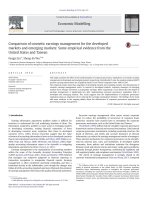
Levels of NSE
The levels of NSE in both serum and pleural effusion were
significantly increased in patients with MPE than in patients with BPE (serum 19.51 ± 16.54 vs. 13.77 ± 13.33 ng/
ml, p = 0.004; pleural effusion 18.53 ± 27.30 vs.
6.41 ± 6.95 ng/ml, p < 0.001) (Table 1). In patients with
MPE, the SCLC patients showed the highest levels of NSE
in both serum and pleural effusion when compared with
other causes of MPE (both P < 0.001), as shown in Fig. 1.
After adjusted by pleural protein, the patients with MPE
remained have a higher levels of NSE in serum and pleural
effusion than patients with BPE (Additional file 1: Fig. S1).
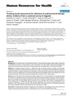
Diagnostic accuracy of NSE
Next, we evaluated the diagnostic accuracy of NSE for
MPE with ROC curves. At a cut off value of 8.92 ng/ml,
the diagnostic sensitivity and specificity of pleural NSE
for MPE were 59.56% and 83.33%, respectively, and the
AUC was 0.76. At a cut off value of 12.29 ng/ml, serum
levels of NSE play a role in diagnosing MPE with the
sensitivity and specificity of 66.91% and 62.75%, respectively, but the AUC was only 0.65, as shown in Fig. 2.
Meanwhile, the AUC of pleural/serum NSE ratio in diagnosing MPE was 0.68 (Fig. 2).
We also noticed that in 11 SCLC patients, the serum/
pleural levels of NSE were the highest among all causes of
MPE. When compared with BPE patients, at a cut-off value
of 17.42 ng/ml, pleural NSE plays a valuable role in diagnosing MPE with the sensitivity and specificity of 100% and
92.16%, respectively, and the AUC was 0.99. The diagnostic
summary of serum and pleural levels of NSE for MPE and
SCLC related MPE was listed in Table 2.
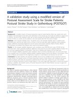
Meta-analysis
This meta-analysis included 14 studies (including present
study), consisting 1093 cases of MPE and 803 BPE controls [19–31]. All the MPEs were diagnosed based on cytology and histology examinations, which were widely
accepted as the gold standard for MPE diagnosis. There
were 11 studies with QUADAS score ≥ 9, indicating the
reliability of statistical results. The clinical summary of individual study and QUADAS score were listed in Table 3.
The pooled parameters for pleural NSE in diagnosing MPE over all 14 studies were listed as follows:
sensitivity, 0.53 (95% CI: 0.38–0.67); specificity, 0.85
(95% CI: 0.75–0.91); PLR, 3.54 (95% CI: 2.33–5.39);
NLR, 0.56 (95% CI: 0.42–0.73); and DOR, 6.39 (95%
CI: 3.72–10.96). Figure 3 showed the corresponding
SROC curve, which yield an AUC of 0.78.
Table 1 The demographics characteristics and biochemical results of patients
p value
Benign pleural effusion
Malignant pleural effusion
No. of Patient
102
136
Sex(male/female)
68/34
74/62
0.056
Age (years)
56 ± 19
58 ± 13
0.167
Pleural NSE (ng/ml)
6.41 ± 6.95
18.53 ± 27.30
<0.001
Pleural NSE (ng/mg of pleural protein)
0.15 ± 0.16
0.51 ± 0.88
<0.001
Pleural protein(g/l)
42.02 ± 13.04
41.84 ± 10.62
0.906
Pleural glucose (mmol/l)
5.84 ± 1.71
5.48 ± 2.51
0.216
Pleural LDH (U/l)
256.57 ± 181.22
502.99 ± 414.15
<0.001
Pleural LDH (U/g of pleural protein)
5.79 ± 3.95
13.68 ± 14.96
<0.001
Serum NSE (ng/ml)
13.77 ± 13.33
19.51 ± 16.54
0.004
Serum NSE (ng/mg of pleural protein)
0.39 ± 0.63
0.54 ± 0.61
0.067
Pleural/serum NSE ratio
0.56 ± 0.55
1.08 ± 1.54
0.001
LDH Lactic Dehydrogenase, NSE Neuron-specific enolase
Zhu et al. BMC Cancer (2017) 17:590
Page 4 of 8
All diagnostic indices revealed high I2 values: sensitivity, 93.69; specificity, 91.55; PLR, 78.44; NLR, 91.50; and
DOR, 99.85(p < 0.05 in all cases), indicating significant
heterogeneity across all studies. Deeks’s funnel plot
asymmetry test was used to evaluate the likelihood of
publication bias among all 14 studies, and Deeks’s test
identified low likelihood of publication bias, and with
the p value of slope coefficient was 0.56 (Fig. 4).
Fig. 1 Serum and pleural levels of neuron specific enolase in patients.
NSE: Neuron specific enolase; BPE: Benign pleural effusion; MPE: Malignant
pleural effusion; LAC-MPE: Lung adenocarcinoma-malignant pleural
effusion; LSCC-MPE: Lung squamous cell carcinoma- malignant pleural
effusion; SCLC-MPE: Small cell lung cancer- malignant pleural effusion
Discussion
To diagnose MPE accurately remains a clinical challenge, and the searching for useful biomarkers for MPE
is still on the way. NSE is typical marker for cancers with
neuroendocrine characteristic, especially for SCLC.
Growing studies suggested that NSE is increased in
MPE, and it may be a biomarker for MPE [32]. However,
these studies gave different results. This study validated
the diagnostic accuracy of NSE for MPE in 238 patients,
which included the second largest patients that evaluated the diagnostic utility of NSE for MPE. In addition,
we next performed a meta-analysis with 1896 subjects to
make a full judgment of NSE for diagnosing MPE based
on current available publications.
In this study, we enrolled 136 MPE patients, and we
observed that both serum and pleural levels of NSE were
higher in patients with MPE than in patients with BPE,
even after adjustment by pleural protein. Pleural NSE
Fig. 2 Receiver operating characteristic curve for neuron specific enolase in serum and pleural effusion for distinguishing between malignant and
benign pleural effusions. NSE: Neuron specific enolase; ROC: Receiver operating characteristic
Zhu et al. BMC Cancer (2017) 17:590
Page 5 of 8
Table 2 Diagnostic summary of NSE for malignant pleural effusion
MPE
Cut-off
SCLC-MPE
Serum NSE
Pleural NSE
Pleural/serum NSE ratio
Serum NSE
Pleural NSE
Pleural/serum NSE ratio
12.29 ng/ml
8.92 ng/ml
0.39
12.29 ng/ml
17.42 ng/ml
0.60
Sensitivity
66.91%
59.56%
79.41%
100%
100%
100%
Specificity
62.75%
83.33%
57.84%
62.75%
92.16%
69.60%
AUC
0.65
0.76
0.68
0.86
0.99
0.92
AUC Area under the curve, MPE Malignant pleural effusion, NSE Neuron-specific enolase, SCLC Small cell lung cancer
shows a better diagnostic performance than serum NSE
and pleural/serum NSE ratio, and its sensitivity and specificity were 59.56% and 83.33%, respectively. Pleural
NSE showed a low sensitivity and a high rate of missed
diagnoses, which may be due to only a limited proportion of NSCLC patients with neuroendocrine characteristic [7, 8]. Thus, the clinical value of NSE alone in
screening MPE is limited. It may be more appropriate to
use the combination of NSE and other tumor markers
for diagnosing MPE [22–26].
The diagnostic performance of a serial of tumor
markers for MPE, such as carcino-embryonic antigen,
carbohydrate antigen 19–9, carbohydrate antigen 15–3,
has been summarized by two meta-analysis, and studies
suggested that one tumor marker alone doesn’t have not
enough sensitivity to diagnose MPE, the combination of
two or more tumor markers may increase the sensitivity
and play more role in MPE diagnosis and management
[33, 34]. In clinical utility, the results of NSE test should
be used in conjugation with other tumor markers tests,
and clinical information of patients, such as previous
medical history, radiological findings.
For MPE with multiple causes (Lung adenocarcinoma,
lung squamous cell carcinoma, SCLC, other etiologies),
we noticed that both serum and pleural levels of NSE were
highest in patients with SCLC, and NSE show a high diagnostic accuracy for SCLC-related MPE. Both serum and
pleural NSE reach a sensitivity of 100% for SCLC-related
MPE. Such results were also supported by Miédougé’s report [23]. These findings suggest the diagnostic performance of NSE may be tumor-subtype specific. Based on
above findings, the NSE may not be used for screening
MPE at the first choice. But for patients who were highly
suspected for SCLC or neuroendocrine tumors, the examination of NSE may provide more valuable information.
To make a systemic assessment of the diagnostic performance of NSE for MPE, we performed an updated
meta-analysis. A recent published meta-analysis has discussed the diagnostic role of NSE for MPE [35], however,
it included only seven studies, and missed several studies.
Thus, we made a more systemic literature search and updated this meta-analysis. In our meta-analysis, there were
1896 cases of patients, and the pooled sensitivity and specificity of NSE were 0.53 and 0.85, respective, confirmed
Table 3 Clinical summary of included studies
First author
Year
Pettersson T
1988 Finland
Country
Cases/controls Standard
31/22
Method
Cytology, Histology Radioimmunoassay
Cut-off value TP FP FN
TN
QUADAS
12.5μg/L
18
7
10 4
21
Shimokata K
1989 Japan
59/39
Cytology, Histology EIA
26 ng/ml
11 2
48
37
8
Menard O
1993 France
24/18
Histology
8 ng/ml
13 2
11
16
8
San Jose ME
1997 Spain
88/183
Cytology, Histology EIA
8.8μg/L
26 21 62
215/121
Cytology, Histology EIA
18.1 ng/ml
39 3
Miédougé M 1999 France
Radioimmunoassay
162 9
176 118 10
Kuralay F
2000 Turkey
21/40
Histology
ELISA
8.7 ng/ml
21 2
0
38
9
Lee JH
2005 Korea
34/16
Histology
ELISA
20 ng/ml
12 1
22
15
10
Ghayumi SM 2005 Iran
5.21μg/ml
27 9
13
28
10
Topolcan O
2007 Czech Republic 80/78
40/37
Cytology, Histology Immuno-radiometric assay 13.1 ng/ml
Cytology, Histology ELISA
34 4
46
74
9
Wu GP
2007 China
74/34
Cytology, Histology Immunoassay
5.2μg/L
51 15 23
19
9
Korczynski P
2009 Poland
36/38
Cytology, Histology ECLIA
0.22 ng/ml
34 24 2
14
10
Wang Y
2013 China
160/40
Cytology, Histology ECLIA
NA
95 15 65
25
11
Gu Y
2016 China
95/35
Histology
ECLIA
9.71 ng/ml
50 8
45
27
11
Zhu J
2016 China
136/102
Cytology, Histology ECLIA
8.92 ng/ml
81 17 55
85
10
EIA Enzyme immunoassay, ECLIA Electrochemiluminescence immunoassay, ELISA Enzyme linked immunosorbent assay, FN false negative, FP false positive, NA Not
available, QUADAS Quality Assessment of Diagnostic Accuracy Studies, TN true negative, TP true positive
Zhu et al. BMC Cancer (2017) 17:590
Page 6 of 8
Fig. 3 Summary receiver operating characteristic (SROC) curve for pleural neuron specific enolase tests. AUC: Area under the curve
our findings that NSE plays a role in confirming the diagnosis of MPE, rather than to screen MPE. The AUC was
only 0.78, suggesting the diagnostic role of NSE for MPE
is limited. Likelihood ratios are another indices of diagnostic accuracy, and PLR >10 or NLR <0.1 suggested high accuracy. In our meta-analysis, the PLR was 3.54, suggesting
patients with MPE have about 3.5-fold higher possibility
being pleural NSE measurement-positive. However, the
NLR was as high as 0.56, which means that if the pleural
NSE assay was negative, the chance that this patient has
MPE was still as high as 56%, suggesting lack of differential ability. Anyway, the results of meta-analysis indicate
that pleural NSE examination alone plays a limited role in
diagnosing MPE.
Fig. 4 The Deek’s funnel plot for the assessment of potential publication bias
Zhu et al. BMC Cancer (2017) 17:590
Our study had several limitations. First, we only recruited 238 patients, and our meta-analysis only included 1896 patients, such limited number of patients
may be not adequate for building final conclusions on
the ability of NSE in diagnosing MPE. Second, only articles published in English were included, and there may
be language bias exist, we may also miss the studies that
not in the searched databases, both may bias the results.
Further studies should include more patients from different centers to confirm the diagnostic role of NSE for
MPE. The current NSE assay is with low sensitivity, it
may be helpful to develop a novel method to examine
NSE and increase the diagnostic accuracy. In addition,
we found substantial heterogeneity among included
studies. However, we didn’t investigate potential covariates with meta-regression analysis due to limited included studies.
Conclusions
Taken together, the role of pleural NSE examination in
diagnosing MPE is limited with low sensitivity. Our study
suggests that the interpretation of NSE results should be
in combination with the results of other tumor markers,
and clinical data of patients. Further studies are needed to
confirm our findings.
Additional file
Additional file 1: Fig. S1. Serum and pleural levels of neuron specific
enolase in patients standardized by pleural protein levels. After
standardized by pleural protein levels, both serum and pleural levels of
neuron specific enolase were higher than that in patient with benign
pleural effusion. NSE: Neuron specific enolase; BPE: Benign pleural
effusion; MPE: Malignant pleural effusion; LAC-MPE: Lung
adenocarcinoma-malignant pleural effusion; LSCC-MPE: Lung squamous
cell carcinoma- malignant pleural effusion; SCLC-MPE: Small cell lung
cancer- malignant pleural effusion (TIFF 2523 kb)
Abbreviations
ANOVA: Analysis of variance; AUC: Areas under the curve; BPE: Benign pleural
effusion; DOR: Diagnostic odds ratios; FN: False negative; FP: False positive;
MPE: Malignant pleural effusion; NLR: Negative likelihood ratios; NSCLC: Non-smallcell lung cancer; NSE: Neuron-specific enolase; PLR: Positive likelihood ratios;
QUADAS: Quality Assessment of Diagnostic Accuracy Studies; ROC: Receiver
operating characteristic; SCLC: Small-cell lung cancer; SROC: Summary receiver
operating characteristic; TN: True negative; TP: True positive
Acknowledgements
Not Applicable.
Availability of data materials
The data of this article is available at reasonable request from the
corresponding author.
Funding
This work was supported by grants from the National Natural Science
Foundation of China (81300032), and Projects in the Science and Technology
Pillar Program from the Department of science and technology of Sichuan
province (2015SZ0151). The funders had no role in design, data collection and
analysis for this study, decision to publish, or preparation of the manuscript.
Page 7 of 8
Authors’ contributions
JZ, MF, and LL developed the study design and proposal, performed data analysis,
interpretation and drafting of the manuscript. NZ, CW, TY, and FW contributed to
study design, data collection, statistical analysis, and critical revision of this
manuscript. YS developed the study design and proposal, revised final manuscript,
and is responsible for fielding correspondence. All authors read and approved the
final version of this manuscript and agreed to be accountable for all aspects of
this work in ensuring that questions related to the accuracy or integrity of any
part of the work are appropriately investigated and resolved.
Ethics approval and consent to participate
This study protocol was approved by the Ethics Committee of West China
Hospital of Sichuan University. Written informed consents were collected
from all patients for the collection of clinical samples and subsequent
analysis at admission.
Consent for publication
Not applicable because no personal data were presented in this manuscript.
Competing interests
The authors declare that they have no competing interests.
Publisher’s Note
Springer Nature remains neutral with regard to jurisdictional claims in
published maps and institutional affiliations.
Received: 15 August 2016 Accepted: 21 August 2017
References
1. Isgrò MA, Bottoni P, Scatena R. Neuron-specific enolase as a biomarker:
biochemical and clinical aspects. Adv Exp Med Biol. 2015;867:125–43.
2. Wu HM, Huang SC, Hattori N, Glenn TC, Vespa PM, Yu CL, et al. Selective
metabolic reduction in gray matter acutely following human traumatic
brain injury. J Neurotrauma. 2004;21(2):149–61.
3. Cheng F, Yuan Q, Yang J, Wang W, Liu H. The prognostic value of serum
neuron-specific enolase in traumatic brain injury: systematic review and
meta-analysis. PLoS One. 2014;9(9):e106680.
4. Tapia FJ, Polak JM, Barbosa AJ, Bloom SR, Marangos PJ, Dermody C, et al.
Neuron-specific enolase is produced by neuroendocrine tumours. Lancet.
1981;1(8224):808–11.
5. Jackman DM, Johnson BE. Small-cell lung cancer. Lancet. 2005;366(9494):
1385–96.
6. Karachaliou N, Pilotto S, Lazzari C, Bria E, de Marinis F, Rosell R. Cellular and
molecular biology of small cell lung cancer: an overview. Transl Lung
Cancer Res. 2016;5(1):2–15.
7. Giovanella L, Piantanida R, Ceriani L, Bandera M, Novario R, Bianchi L, et al.
Immunoassay of neuron-specific enolase (NSE) and serum fragments of
cytokeratin 19 (CYFRA 21.1) as tumor markers in small cell lung cancer: clinical
evaluation and biological hypothesis. Int J Biol Markers. 1997;12(1):22–6.
8. Chen Y, Nowak I, Huang J, Keng PC, Sun H, Xu H, et al. Erk/MAP kinase
signaling pathway and neuroendocrine differentiation of non-small-cell
lung cancer. J Thorac Oncol. 2014;9(1):50–8.
9. Clive AO, Kahan BC, Hooper CE, Bhatnagar R, Morley AJ, Zahan-Evans N, et
al. Predicting survival in malignant pleural effusion: development and
validation of the LENT prognostic score. Thorax. 2014;69(12):1098–104.
10. Kastelik JA. Management of malignant pleural effusion. Lung. 2013;191(2):
165–75.
11. Alataş F, Alataş O, Metintaş M, Colak O, Harmanci E, Demir S. Diagnostic
value of CEA, CA 15-3, CA 19-9, CYFRA 21-1, NSE and TSA assay in pleural
effusions. Lung Cancer. 2001;31(1):9–16.
12. Li CS, Cheng BC, Ge W, Gao JF. Clinical value of CYFRA21-1, NSE, CA15-3,
CA19-9 and CA125 assay in the elderly patients with pleural effusions. Int J
Clin Pract. 2007;61(3):444–8.
13. Leeflang MM, Deeks JJ, Gatsonis C, Bossuyt PM. Cochrane diagnostic test
accuracy working group. Systematic reviews of diagnostic test accuracy.
Ann Intern Med. 2008;149(12):889–97.
14. Whiting PF, Weswood ME, Rutjes AW, Reitsma JB, Bossuyt PN, Kleijnen J.
Evaluation of QUADAS, a tool for the quality assessment of diagnostic
accuracy studies. BMC Med Res Methodol. 2006;6:9.
Zhu et al. BMC Cancer (2017) 17:590
15. Shen Y, Zhu H, Wan C, et al. Can cholesterol be used to distinguish pleural
exudates from transudates? Evidence from a bivariate meta-analysis. BMC
Pulm Med. 2014;14:61.
16. Riley RD, Abrams KR, Sutton AJ, Lambert PC, Thompson JR. Bivariate
random-effects meta-analysis and the estimation of between-study
correlation. BMC Med Res Methodol. 2007;7:3.
17. Jones CM, Athanasiou T. Summary receiver operating characteristic curve
analysis techniques in the evaluation of diagnostic tests. Ann Thorac Surg.
2005;79(1):16–20.
18. Deeks JJ, Macaskill P, Irwig L. The performance of tests of publication bias
and other sample size effects in systematic reviews of diagnostic test
accuracy was assessed. J Clin Epidemiol. 2005;58(9):882–93.
19. Pettersson T, Klockars M, Fröseth B. Neuron-specific enolase in the diagnosis
of small-cell lung cancer with pleural effusion: a negative report. Eur Respir
J. 1988;1(8):698–700.
20. Shimokata K, Niwa Y, Yamamoto M, Sasou H, Morishita M. Pleural fluid
neuron-specific enolase. A useful diagnostic marker for small cell lung
cancer pleurisy. Chest. 1989;95(3):602–3.
21. Menard O, Dousset B, Jacob C, Martinet Y. Improvement of the diagnosis of
the cause of pleural effusion in patients with lung cancer by simultaneous
quantification of carcinoembryonic antigen (CEA) and neuron-specific
enolase (NSE) pleural levels. Eur J Cancer. 1993;29A(13):1806–9.
22. San Jose ME, Alvarez D, Valdes L, Sarandeses A, Valle JM, Penela P. Utility of
tumour markers in the diagnosis of neoplastic pleural effusion. Clin Chim
Acta. 1997;265(2):193–205.
23. Miédougé M, Rouzaud P, Salama G, Pujazon MC, Vincent C, Mauduyt MA, et
al. Evaluation of seven tumour markers in pleural fluid for the diagnosis of
malignant effusions. Br J Cancer. 1999;81(6):1059–65.
24. Kuralay F, Tokgöz Z, Cömlekci A. Diagnostic usefulness of tumour marker
levels in pleural effusions of malignant and benign origin. Clin Chim Acta.
2000;300(1–2):43–55.
25. Lee JH, Chang JH. Diagnostic utility of serum and pleural fluid
carcinoembryonic antigen, neuron-specific enolase, and cytokeratin 19
fragments in patients with effusions from primary lung cancer. Chest. 2005
Oct;128(4):2298–303.
26. Ghayumi SM, Mehrabi S, Doroudchi M, Ghaderi A. Diagnostic value of
tumor markers for differentiating malignant and benign pleural effusions of
Iranian patients. Pathol Oncol Res. 2005;11(4):236–41.
27. Topolcan O, Holubec L, Polivkova V, Svobodova S, Pesek M, Treska V, et al.
Tumor markers in pleural effusions. Anticancer Res. 2007;27(4A):1921–4.
28. Wu GP, Ba J, Zhao YJ, Wang EH. Diagnostic value of CEA, CYFRA 21-1, NSE
and CA 125 assay in serum and pleural effusion of patients with lung
cancer. Acta Cytol. 2007;51(4):679–80.
29. Korczynski P, Krenke R, Safianowska A, Gorska K, Abou Chaz MB, MaskeyWarzechowska M, et al. Diagnostic utility of pleural fluid and serum markers
in differentiation between malignant and non-malignant pleural effusions.
Eur J Med Res. 2009;14(Suppl 4):128–33.
30. Wang Y, Chen Z, Chen J, Pan J, Zhang W, Pan Q, et al. The diagnostic value
of apolipoprotein E in malignant pleural effusion associated with non-small
cell lung cancer. Clin Chim Acta. 2013;421:230–5.
31. Gu Y, Zhai K, Shi HZ. Clinical value of tumor markers for determining cause
of pleural effusion. Chin Med J. 2016;129(3):253–8.
32. Lyubimova NV, Yag'ya TN, Chuchalin AG, Kushlinskii NE. Diagnostic value of
tumor markers Cyfra 21-1 and neuron-specific enolase in analysis of pleural
fluid. Bull Exp Biol Med. 2002;133(5):478–80.
33. Shi HZ, Liang QL, Jiang J, Qin XJ, Yang HB. Diagnostic value of
carcinoembryonic antigen in malignant pleural effusion: a meta-analysis.
Respirology. 2008;13(4):518–27.
34. Liang QL, Shi HZ, Qin XJ, Liang XD, Jiang J, Yang HB. Diagnostic accuracy of
tumour markers for malignant pleural effusion: a meta-analysis. Thorax.
2008;63(1):35–41.
35. Nguyen AH, Miller EJ, Wichman CS, Berim IG, Agrawal DK. Diagnostic value
of tumor antigens in malignant pleural effusion: a meta-analysis. Transl Res.
2015;166(5):432–9.
Page 8 of 8
Submit your next manuscript to BioMed Central
and we will help you at every step:
• We accept pre-submission inquiries
• Our selector tool helps you to find the most relevant journal
• We provide round the clock customer support
• Convenient online submission
• Thorough peer review
• Inclusion in PubMed and all major indexing services
• Maximum visibility for your research
Submit your manuscript at
www.biomedcentral.com/submit