Distinct preoperative clinical features predict four histopathological subtypes of high-grade serous carcinoma of the ovary, fallopian tube, and peritoneum
Bạn đang xem bản rút gọn của tài liệu. Xem và tải ngay bản đầy đủ của tài liệu tại đây (1.12 MB, 8 trang )
Ohsuga et al. BMC Cancer (2017) 17:580
DOI 10.1186/s12885-017-3573-1
RESEARCH ARTICLE
Open Access
Distinct preoperative clinical features
predict four histopathological subtypes of
high-grade serous carcinoma of the ovary,
fallopian tube, and peritoneum
Takuma Ohsuga1, Ken Yamaguchi1* , Aki Kido2, Ryusuke Murakami1, Kaoru Abiko1, Junzo Hamanishi1,
Eiji Kondoh1, Tsukasa Baba1, Ikuo Konishi1,3 and Noriomi Matsumura1
Abstract
Background: The Cancer Genome Atlas Research Network reported that high-grade serous carcinoma (HGSC) can
be classified based on gene expression profiles into four subtypes, termed “immunoreactive,” “differentiated,”
“proliferative,” and “mesenchymal.” We previously established a novel histopathological classification of HGSC,
corresponding to the gene expression subtypes: immune reactive (IR), papillo-glandular (PG), solid and proliferative
(SP), and mesenchymal transition (MT). The purpose of this study is to identify distinct clinical findings among the
four pathological subtypes of HGSC, as well as to predict pathological subtype based on preoperative images.
Methods: We retrospectively assessed 65 HGSC cases (IR: 17, PG: 7, SP: 14, MT: 27) and analyzed preoperative images.
Results: All IR cases originated from either the ovary or fallopian tube (P = 0.0269). Significantly more IR cases were
diagnosed at earlier stages (P = 0.0013), and IR cases displayed lower levels of ascites (P = 0.0014), fewer peritoneal
lesions (P = 0.0080), a sporadic pattern of peritoneal lesions (P = 0.0016), a lower incidence of omental cake (P = 0.0416)
, and fewer distant metastases (P = 0.0146) compared with the other subtypes. MT cases were more likely to be of
peritoneal origin (P = 0.0202), presented at advanced stages with higher levels of ascites (P = 0.0008, 0.0052,
respectively), and more frequently had a diffuse pattern of peritoneal lesions (P = 0.0059), omental cake (P = 0.0179),
and distant metastasis (P = 0.0053). A decision tree analysis estimated the histopathological subtypes based on
preoperative images, with a sensitivity of 67.3%.
Conclusions: Pathological subtypes of HGSC have distinct clinical behaviors, and preoperative images enable better
prediction of pathological subtype. These findings may lead to individualized treatment plans if the effect of treatment
based on the HGSC subtype is elucidated.
Keywords: High-grade serous carcinoma, Ovarian cancer, Subtype, MRI
Background
Ovarian carcinoma is the most common cause of gynecologic cancer death and the fifth leading cause of cancer
deaths in women in the United States [1]. High-grade
serous carcinoma (HGSC), accounting for 68% of ovarian
carcinoma cases, is usually diagnosed at an advanced stage
and has a poor prognosis [2]. Chemotherapy with a
* Correspondence:
1
Department of Gynecology and Obstetrics, Kyoto University, 54
Kawahara-cho, Shogoin, Sakyo-ku, Kyoto 606-8507, Japan
Full list of author information is available at the end of the article
taxane- and platinum-based regimen is typically provided
after debulking surgery, and 75% of high-grade serous
ovarian carcinoma cases respond to initial treatment [3].
However, many patients experience recurrence and ultimately succumb to the disease.
Serous carcinoma is the most common histological subtype of primary peritoneal cancer and fallopian tube cancer, as well as epithelial ovarian cancer [4]. Traditionally,
serous epithelial tumors in the ovaries, primary fallopian
tubes, and peritoneum have all been approached as primary epithelial ovarian tumors in clinical and research
© The Author(s). 2017 Open Access This article is distributed under the terms of the Creative Commons Attribution 4.0
International License ( which permits unrestricted use, distribution, and
reproduction in any medium, provided you give appropriate credit to the original author(s) and the source, provide a link to
the Creative Commons license, and indicate if changes were made. The Creative Commons Public Domain Dedication waiver
( applies to the data made available in this article, unless otherwise stated.
Ohsuga et al. BMC Cancer (2017) 17:580
settings because of their shared clinical behavior and treatment [5]. However, in practice, HGSC seems to be a heterogeneous disease, because cases have a diversity of
clinical features, therapeutic responses, and prognoses.
Analysis of gene expression microarray data from The
Cancer Genome Atlas (TCGA) project revealed that
HGSC could be classified as one of four gene expression
subtypes: “immunoreactive,” “differentiated,” “proliferative,” or “mesenchymal” [6, 7]. These sub-classifications
display distinct prognoses and sensitivities to chemotherapy [7–9]. We recently established four histopathological
classifications of HGSC that correlate with the TCGA
gene expression subtypes and prognoses: immune reactive
(IR), which is defined by lymphocytes surrounding and infiltrating the malignant tissue; papillo-glandular (PG),
which is defined by a papillary architecture; solid and proliferative (SP), which is defined by a solid growth pattern;
and mesenchymal transition (MT), which is defined by a
remarkable desmoplastic reaction [10]. Of these histopathological sub-classifications, the MT subtype has the
worst prognosis, and the IR subtype has the most favorable prognosis. Therefore, the exact diagnosis of MT and
IR subtypes is important for the clinical management of
HGSC. However, diagnosis of these subtypes requires
biopsy of the tumor, which is located in the abdominal
cavity and can be difficult to access.
Although the mesenchymal gene expression subtype of
ovarian tumors is accompanied by mesenteric infiltration
and diffuse peritoneal disease on computed tomography
(CT) imaging, it was previously not possible to define
conclusive association of the four pathological subtypes
with distinct clinical features [11]. The first purpose of
this study is the identification of distinct clinical features
among the four pathological subtypes of HGSC. The
second aim is to predict pathological subtype using preoperative factors, including images. Prediction of pathological subtypes of HGSC will enable clinicians to
estimate chemosensitivity and prognosis, allowing the
administration of individualized therapies without performing exploratory laparotomy or laparoscopy.
Methods
Eligibility criteria
This retrospective study was approved by the institutional ethics committee. Patients were included if they:
(a) underwent primary debulking surgery or exploratory
laparoscopy and were newly diagnosed histologically
with HGSC of the ovary, fallopian tube, or peritoneum
between 2005 and 2014 at the Kyoto University Hospital,
and (b) underwent magnetic resonance (MR) imaging of
the pelvis and CT of the neck, chest, abdomen and pelvis prior to initial treatment. Sixty-five patients, all of
whom provided informed consent, satisfied the eligibility
criteria and were included in this study.
Page 2 of 8
Patient characteristics and pathological review
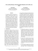
We classified HGSC into four subtypes using our previously described algorithm (Fig. 1) [10]. At least four
blinded pathologists and gynecologists determined the
pathological subtypes of HGSC cases. We used the
International Federation of Gynecology and Obstetrics
classification for ovary, fallopian tube, and primary peritoneal carcinoma [12].
Image analysis
Pretreatment imaging was obtained within one month before starting initial treatment. Firstly, a gynecologist and a
radiologist specializing in gynecological diagnostic imaging who were blinded to the patients’ histopathological
subtypes independently evaluated all MR and CT images.
When their image interpretations differed, the final decision was settled by discussion. Secondly, another radiologist also specializing in gynecological diagnostic imaging
evaluated all of the images. In cases of differing interpretations, consensus was reached by discussion.
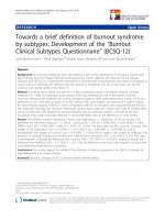
The following features were evaluated by CT and MR
imaging:
1: the location of the main tumor as ovary/fallopian
tube or peritoneum (peritoneum included any cases
without an ovarian or fallopian tube mass) (Fig. 2a)
2: the morphology of the main tumor as solid (more
than 50% of the main tumor was solid) or cystic (more
than 50% of the main tumor was cystic) (Fig. 2b)
3: the amount of ascites as small (within the pelvis) or
large (beyond the pelvis) on MR imaging (Fig. 2c)
4: the presence/absence of peritoneal dissemination
5: the morphological pattern of peritoneal
dissemination as sporadic (single or multiple nodules
scattered sporadically in the peritoneum) or diffuse
(numerous nodules or masses spread diffusely along
the peritoneum) on CT or MR imaging, according
to the criteria defined previously [11] (Fig. 2d)
6: the presence/absence of omental cake, defined as an
abnormally thickened omentum (Fig. 2e)
7: the presence/absence of lymph node metastasis on
CT (lymph nodes more than 10 mm in the short
axis were considered metastatic)
8: the presence/absence of distant metastasis on CT
The decision tree was generated using Weka, a publically
available data mining software program ( />Statistical methods
All statistical analyses were performed with EZR
(Saitama Medical Center, Jichi Medical University,
Saitama, Japan), which is a graphical user interface for R
(The R Foundation for Statistical Computing, Vienna,
Ohsuga et al. BMC Cancer (2017) 17:580
Page 3 of 8
Fig. 1 Four pathological subtypes of HGSC. a) Immune reactive (IR): infiltration by numerous lymphocytes with a smooth invasive front. b)
Papillo-Glandular (PG): papillary architecture. c) Solid and Proliferative (SP): a solid growth pattern. d) Mesenchymal Transition (MT): a remarkable
desmoplastic reaction and a scattered invasion or labyrinthine pattern. Left figures are loupe images, and right images are at 200× magnification
Austria). More precisely, it is a modified version of the R
commander designed to add statistical functions frequently used in biostatistics [13]. Fisher’s exact probability test was used to examine the relationships between
the clinical parameters stated above and the histopathological subtypes of HGSC. P-values <0.05 were considered significant.
Results
Distinct clinical findings among the four pathological
subtypes of HGSC
The 65 cases included 17 IR, 7 PG, 14 SP, and 27 MT
histopathological subtypes. Table 1 shows the association
between clinical findings and the four histopathological
subtypes. Further details of these findings are shown in
Additional file 1: Table S1. More IR cases were diagnosed at earlier stages compared with the other subtypes
(8/17 vs. 4/48, P = 0.0013), and more MT cases were diagnosed at advanced stages (27/27 vs. 26/38,
P = 0.0008). As for the location of the main tumor on
MR imaging (Fig. 2a), all of the IR cases had a main
tumor in the ovaries or fallopian tubes, and this was less
common in the other subtypes (17/17 vs. 36/48,
P = 0.0269), while significantly more MT cases had a
main peritoneal lesion compared with the other subtypes
(9/27 vs. 3/38, P = 0.0202). Although the morphology of
Ohsuga et al. BMC Cancer (2017) 17:580
a-1
a-2
b-1
b-2
c-1
c-2
d-1
d-2
e
Page 4 of 8
Fig. 2 Representative magnetic resonance (MR) imaging and computed
tomography (CT) findings. a) Location of the main tumor by MR
imaging. a-1) Ovary or fallopian tube (arrow). a-2) Peritoneum (arrow). b)
Morphology of the main tumor by MR imaging. b-1) Solid: more than
50% of the main tumor is solid (arrow). b-2) Cystic: more than 50% of
the main tumor is cystic (arrow). c) The amount of ascites by MR
imaging. c-1) Small amount: ascites within the pelvis. c-2) Large amount:
ascites beyond the pelvis. d) Pattern of peritoneal dissemination by MR
imaging. d-1) Sporadic pattern: single or multiple nodules scattered
sporadically in the peritoneum (arrow). d-2) Diffuse pattern: numerous
nodules or masses spread diffusely along the peritoneum (arrow). e)
Omental cake by CT: abnormally thickened greater omentum (arrow)
the main tumor was not significantly associated with
histopathological subtype, PG cases tended to exhibit
cystic tumors (Fig. 2b). The amount of ascites on MR
imaging (Fig. 2c) was significantly lower in IR cases
compared with the other subtypes (14/17 vs. 17 /48,
P = 0.0014) and higher in MT cases (20/27 vs. 14/38,
P = 0.0052). The frequency of peritoneal lesions on MR
imaging was significantly lower in IR cases compared
with the other subtypes (8/17 vs. 40/48, P = 0.0080) and
higher in MT cases (26/27 vs. 22/38, P = 0.0004). Peritoneal lesions on MR imaging (Fig. 2d) were more
frequently sporadic in IR cases than in other subtypes
(6/8 vs. 6/40, P = 0.0016) and more frequently diffuse in
MT cases than in other subtypes (24/26 vs. 12/22,
P = 0.0059). Omental cakes were observed on CT (Fig.
2e) significantly less frequently in IR cases compared
with other subtypes (7/17 vs. 34/48, P = 0.0416) and
more frequently in MT cases (22/27 vs. 19/38,
P = 0.0179). Lymphadenopathy on CT was detected significantly more frequently in SP cases compared with
the other subtypes (9/14 vs. 14/51, P = 0.0243), while no
radiographic lymphadenopathy was detected in PG cases
(0/7 vs. 23/58, P = 0.0452). IR cases showed no distant
metastases on CT (0/17 vs. 13/48, P = 0.0146). Additionally, none of the 7 PG cases showed distant metastases
on CT. On the other hand, MT cases showed significantly more distant metastases on CT (10/27 vs. 3/38,
P = 0.0053). These findings are summarized in Table 2.
Prediction of pathological subtypes using pre-treatment
clinical findings
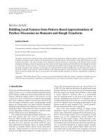
We conducted a decision tree analysis for HGSC cases
suggestive of stage III and IV to predict pathological
subtype preoperatively using suggestive origin (location
of main tumor), morphology of the primary tumor,
amount of ascites, presence of omental cake, presence
and pattern of peritoneal disease, radiographically enlarged lymph nodes, and presence of distant metastasis
(Fig. 3). Thirteen cases that were suggestive of stage I
and II because of the small amount of ascites and absence of omental cake, peritoneal dissemination, radiographically enlarged lymph nodes, and distant metastasis
Ohsuga et al. BMC Cancer (2017) 17:580
Page 5 of 8
Table 1 Clinical features among the four pathological subtypes
of HGSC
Pathological subtype
IR
PG
SP
MT
8
2
2
0
FIGO stage
Early (Stage I & II)
Advanced (Stage III & IV)
9
5
12
27
P value
0.0013*
0.6043
1.0000
0.0008*
17
5
13
18
Location of main tumor in MRI
Ovary or Fallopian tube
Peritoneum
0
2
1
9
P value
0.0269*
0.6043
0.4364
0.0202*
4
11
21
Morphology of main tumor in MRI
Solid
11
Cystic
6
3
3
6
P value
0.5299
0.3853
0.7402
0.5748
14
5
5
7
The amount of ascites in MRI
Small
Large
3
2
9
20
P value
0.0014*
0.2440
0.3750
0.0052*
9
4
3
1
Peritoneal lesion in MRI
Presence
Negative
Positive
8
3
11
26
0.0080*
0.0700
0.7448
0.0004*
Sporadic
6
0
4
2
Diffuse
2
3
7
24
0.0016*
0.5629
0.4304
0.0059*
P value
Pattern
P value
Omental cake in CT
Negative
10
4
5
5
Positive
7
3
9
22
P value
0.0416*
0.4080
1.0000
0.0179*
radiographically enlarged lymph nodes in CT
Negative
9
7
5
21
Positive
8
0
9
6
P value
0.2554
0.0452*
0.0243*
0.0717
Distant metastasis in CT
Negative
17
7
11
17
Positive
0
0
3
10
P value
0.0146*
0.3287
1.0000
0.0053*
P value: Fisher’s exact test, comparing between each subtype and the other
subtypes, * p < 0.05
were excluded from the decision tree analysis. A decision tree with the 52 remaining samples (IR: 10, PG: 3,
SP: 13, and MT: 26 cases) that were suggestive of stage
III and IV indicated that the pattern of peritoneal dissemination, radiographically enlarged lymph nodes, and
presence of omental cake were useful identifiers for subclassification of HGSC. Of 52 patients, 24 out of 36
cases with diffuse pattern of peritoneal lesion were the
MT subtype. Of the other 16 cases, a sporadic pattern of
peritoneal dissemination identified 2 MT, 6 IR, and 4 SP
cases. Of the 12 cases with a sporadic pattern of dissemination, 4 did not exhibit radiographically enlarged
lymph nodes, two of which were the SP subtype. Of the
8 cases with a sporadic pattern of peritoneal dissemination and radiographically enlarged lymph nodes, 5 cases
belonged to the IR subtype. Of the 4 cases without peritoneal lesions, 2 cases without omental cake were the SP
subtype and the other two cases with omental cake
showed the IR subtype. This algorithm in total had a
sensitivity of 67.3%. The sensitivities of diagnosis were
70.0% (7/10), 0% (0/3), 30.8% (4/13) and 92.3% (24/26),
for the IR, PG, SP, and MT subtypes, respectively.
Discussion
This investigation clearly shows that the pathological
subtypes of HGSC exhibit distinct clinical behaviors
(Table 2). Our previous study revealed that these pathological subtypes are statistically correlated with the previously defined TCGA gene expression subtypes, and
that the MT subtype is a poor prognostic factor, while
the IR subtype is a favorable prognostic factor [10]. Although, in general, high-grade serous ovarian, fallopian
tube, and peritoneal cancer are considered a single clinical entity because of their shared clinical behavior and
treatment, some studies have found a significantly
poorer survival or a non-significant trend of poorer survival for primary peritoneal tumors compared with ovarian tumors [5]. Vargas et al. suggested that mesenteric
infiltration and diffuse peritoneal disease on CT are associated with the mesenchymal gene expression subtype
and shorter progression-free survival [11]. These reports
are compatible with our findings that the MT subtype,
which had the poorest outcome, included significantly
more diseases of peritoneal origin and omental cake.
Feigenberg et al. suggested that advanced stage HGSC
cases presenting with low-volume ascites are associated
with the upregulation of immune-related genes, more
immune cells infiltrating the tumor, and better clinical
outcomes [14]. In addition, Baek et al. reported that patients with stage III disease solely by lymph node metastasis showed even better outcomes than did those with
stage III disease with peritoneal dissemination [15]. Our
study indicated that the IR subtype tends to show lower
volume ascites, less peritoneal dissemination, and more
radiographic lymphadenopathy on CT than other subtypes. Our decision tree analysis also showed that IR
cases with advanced stage exhibit radiographically enlarged lymph nodes and a small amount of ascites on
CT. The IR subtype has a favorable prognosis [6, 7, 9,
16], which may be owing to these advanced cases having
a lower volume of ascites, less peritoneal dissemination,
Ohsuga et al. BMC Cancer (2017) 17:580
Page 6 of 8
Table 2 Summary of clinical features among the four histopathological subtypes of HGSC
Pathological subtype
IR
PG
SP
MT
FIGO stage
Early
Advanced
Advanced
Advanced
Location of main tumor
Ovary or Fallopian tube
Ovary or Fallopian tube
Ovary or Fallopian tube
Peritoneum
Ascites
↓↓
↑
↑↑
Peritoneal lesion
↓↓
↑
↑↑
diffuse
diffuse
↑
↑↑
sporadic
diffuse
Omental cake
↓↓
LN swelling
↑
↓↓
↑↑
Distant metastasis
↓↓
↓
↓
Prognosis
favorable
↑↑
poor
Abbreviation: ↑↑: significantly more ↑: more ↓: less ↓↓: significantly less
and radiographically smaller lymph nodes. Vargas et al.
reported that ovarian mass morphology in high-grade
serous ovarian cancer on CT imaging was not definitely
associated with gene expression subtype [11]. In our
study, morphology of the main tumor in MR imaging
was not specifically associated with any of the four
pathological subtypes.
Additionally, this study may be useful in determining
the initial chemotherapeutic regimen for individualized
treatment. Studies indicate that the four pathological
subtypes show different patterns of anticancer drug sensitivity. Recently, Symeonides et al. suggested that distinct molecular subgroups of high-grade serous ovarian
cancer respond very differently to bevacizumab [8]. In
this analysis, the two proangiogenic subgroups, which
represent non-IR subtypes in this study, had worse overall survival but included all the patients who benefited
from bevacizumab. The immune subgroup had a
Fig. 3 Algorithm for pretreatment prediction of the four
histopathological subtypes. Algorithm for pretreatment prediction of
the four histopathological subtypes using decision tree analysis. MT:
mesenchymal transition, IR: immune reactive, SP: solid and
proliferative, PG: papillo-glandular
superior prognosis, but bevacizumab had a detrimental
effect in these patients. Therefore, it is likely that the
pathological subtypes may be biomarkers for bevacizumab benefit and resistance. Our previous study suggested that the mesenchymal subtype may be
particularly sensitive to taxanes and resistant to carboplatin [17]. Because dose-dense paclitaxel and carboplatin (TC) therapy, which gives more weight to paclitaxel
than conventional TC chemotherapy, confers a more favorable prognosis compared to conventional TC for
HGSC, chemotherapy with dose-dense TC may be beneficial for patients with the MT subtype. With regard to
the molecular features, George et al. suggested that
breast cancer susceptibility gene 1 (BRCA1) disruptions
are associated with the immunoreactive molecular subtype of HGSC [18]. Furthermore, Soslow et al. implied
that there is a positive association between tumorinfiltrating lymphocytes and BRCA1 loss in HGSC [19].
These findings indicate that the IR subtype has a likelihood of benefiting from poly(ADP-ribose) polymerase
inhibitor use [20].
Recently, neo-adjuvant chemotherapy (NAC) has been
shown to be a valuable alternative treatment for patients
with advanced epithelial ovarian cancer who are not
amenable to primary optimum surgery. NAC provides a
higher rate of optimal cytoreduction and equivalent survival with less invasive surgery and reduced morbidity
compared to conventional therapy [21, 22]. Exploratory
laparotomy or laparoscopy is necessary to select the
anti-cancer agents for NAC based on the pathological
and TCGA gene expression subtypes. However, exploratory surgery is contraindicated in some advanced cases,
such as in the presence of a large amount of ascites or
pleural effusion. Preoperative estimation of the pathological subtype using imaging possibly allows the
patients for whom the surgery is contraindicated to start
NAC immediately using the optimal regimen.
There are several limitations to this study. The limited
number of cases may have caused statistically
Ohsuga et al. BMC Cancer (2017) 17:580
Page 7 of 8
insignificant results, particularly in the PG subtype. A
decision tree using pattern of peritoneal dissemination,
the existence of radiographic lymphadenopathy, and
omental cake did not predict the PG subtype, whereas
the IR and MT subtypes were identified with 70.0% and
92.3% sensitivity, respectively. Additionally, our findings
were not validated using external datasets. However, it is
meaningful that this decision tree can diagnose the IR
and MT subtypes, which show the most favorable and
poorest prognosis, respectively. Further studies should
be performed with a larger sample size, and the accuracy
of this algorithm on the effect of treatment needs to be
validated in a prospective manner.
Competing interests
None of the authors of this paper has any financial or other conflict of
interests in relation to this article.
Conclusions
We revealed four histopathological subtypes of HGSC of
the ovary, fallopian tube, and peritoneum, demonstrating
distinct clinical features and pretreatment images that
enable estimation of the histopathological subtypes.
These findings have the potential to help in determining
an initial treatment strategy for individualized treatment.
References
1. Siegel R, Ma J, Zou Z, Jemal A. Cancer statistics, 2014. CA Cancer J Clin.
2014;64:9–29.
2. Seidman JD, Horkayne-Szakaly I, Haiba M, Boice CR, Kurman RJ, Ronnett BM.
The histologic type and stage distribution of ovarian carcinomas of surface
epithelial origin. Int J Gynecol Pathol. 2004;23:41–4.
3. Miller DS, Blessing JA, Krasner CN, Mannel RS, Hanjani P, Pearl ML, et al.
Phase II evaluation of pemetrexed in the treatment of recurrent or
persistent platinum-resistant ovarian or primary peritoneal carcinoma: a
study of the gynecologic oncology group. J Clin Oncol. 2009;27:2686–91.
4. Goodman MT, Shvetsov YB. Incidence of ovarian, peritoneal, and fallopian
tube carcinomas in the United States, 1995-2004. Cancer Epidemiol Biomark
Prev. 2009;18:132–9.
5. Sørensen RD, Schnack TH, Karlsen MA, Høgdall CK. Serous ovarian, fallopian
tube and primary peritoneal cancers: a common disease or separate entities
- a systematic review. Gynecol Oncol. 2015;136:571–81.
6. Cancer Genome Atlas Research Network. Integrated genomic analyses of
ovarian carcinoma. Nature. 2011;474:609–15.
7. Verhaak RG, Tamayo P, Yang JY, Hubbard D, Zhang H, Creighton CJ, et al.
Prognostically relevant gene signatures of high-grade serous ovarian
carcinoma. J Clin Invest. 2013;123:517–25.
8. Symeonides S, Gourley C. Ovarian cancer molecular stratification and tumor
heterogeneity: a necessity and a challenge. Front Oncol. 2015;5:229.
9. Konecny GE, Wang C, Hamidi H, Winterhoff B, Kalli KR, Dering J, et al.
Prognostic and therapeutic relevance of molecular subtypes in high-grade
serous ovarian cancer. J Natl Cancer Inst 2014;106:pii:dju249.
10. Murakami R, Matsumura N, Mandai M, Yoshihara K, Tanabe H, Nakai H, et al.
Establishment of a novel histopathological classification of high-grade
serous ovarian carcinoma correlated with Prognostically distinct gene
expression subtypes. Am J Pathol. 2016;186:1103–13.
11. Vargas HA, Micco M, Hong SI, Goldman DA, Dao F, Weigelt B, et al.
Association between morphologic CT imaging traits and prognostically
relevant gene signatures in women with high-grade serous ovarian cancer:
a hypothesis-generating study. Radiology. 2015;274:742–51.
12. Prat J, FIGO Committee on Gynecologic Oncology. Staging classification for
cancer of the ovary, fallopian tube, and peritoneum. Int J Gynaecol Obstet
2014;124:1–5.
13. Kanda Y. Investigation of the freely available easy-to-use software 'EZR' for
medical statistics. Bone Marrow Transplant. 2013;48:452–8.
14. Feigenberg T, Clarke B, Virtanen C, Plotkin A, Letarte M, Rosen B, et al.
Molecular profiling and clinical outcome of high-grade serous ovarian
cancer presenting with low- versus high-volume ascites. Biomed Res Int.
2014;2014:367103.
15. Baek SJ, Park JY, Kim DY, Kim JH, Kim YM, Kim YT, et al. Stage IIIC epithelial
ovarian cancer classified solely by lymph node metastasis has a more
favorable prognosis than other types of stage IIIC epithelial ovarian cancer. J
Gynecol Oncol. 2008;19:223–8.
16. Tothill RW, Tinker AV, George J, Brown R, Fox SB, Lade S, et al. Novel
molecular subtypes of serous and endometrioid ovarian cancer linked to
clinical outcome. Clin Cancer Res. 2008;14:5198–208.
17. Murakami R, Matsumura N, Brown JB, Wang Z, Yamaguchi K, Abiko K, Yet al.
Prediction of taxane and platinum sensitivity in ovarian cancer based on
gene expression profiles. Gynecol Oncol 2016;141:49–56.
Additional file
Additional file 1: Table S1. Clinical findings of individual cases.
Individual findings include pathological subtype, stage, suggestive origin
(location of primary tumor), morphology of the primary tumor, amount
of ascites, presence of omental cake, presence and pattern of peritoneal
disease, radiographically enlarged lymph nodes, and presence of distant
metastasis. (XLSX 13 kb)
Abbreviations
BRCA1: breast cancer susceptibility gene 1; CT: computed tomography;
HGSC: high-grade serous carcinoma; IR: immune reactive; MR: magnetic
resonance; MT: mesenchymal transition; NAC: neo-adjuvant chemotherapy;
PG: papillo-glandular; SP: solid and proliferative; TC: paclitaxel and
carboplatin; TCGA: The Cancer Genome Atlas
Acknowledgements
The authors thank Ryo Kuwahara for helping in image evaluation.
Funding
No specific funding was received for this study.
Availability of data and materials
All data analyzed during this study are included in this published article and
the Additional file 1: Table S1.
Consent for publication
Not applicable.
Authors’ contributions
KY proposed the conception, designed this study, and analyzed the data. TO
and AK contributed to acquisition of data, analysis, and interpretation of
data. RM, KA, JH, EK, TB, IK, and NM analyzed and interpreted the data. All
authors have read and approved the final version of this manuscript.
Ethics approval and consent to participate
This study has been performed in accordance with the Declaration of
Helsinki and has been approved as the ethics committee reference number,
G531, by the Kyoto University Graduate School and Faculty of Medicine,
Ethics Committee. All study participants provided informed consent with a
written form.
Publisher’s Note
Springer Nature remains neutral with regard to jurisdictional claims in
published maps and institutional affiliations.
Author details
1
Department of Gynecology and Obstetrics, Kyoto University, 54
Kawahara-cho, Shogoin, Sakyo-ku, Kyoto 606-8507, Japan. 2Department of
Diagnostic Imaging and Nuclear Medicine, Kyoto University, Kyoto, Japan.
3
National Hospital Organization Kyoto Medical Center, Kyoto, Japan.
Received: 14 April 2017 Accepted: 21 August 2017
Ohsuga et al. BMC Cancer (2017) 17:580
Page 8 of 8
18. George J, Alsop K, Etemadmoghadam D, Hondow H, Mikeska T, Dobrovic A,
et al. Nonequivalent gene expression and copy number alterations in highgrade serous ovarian cancers with BRCA1 and BRCA2 mutations. Clin
Cancer Res. 2013;19:3474–84.
19. Soslow RA, Han G, Park KJ, Garg K, Olvera N, Spriggs DR, et al. Morphologic
patterns associated with BRCA1 and BRCA2 genotype in ovarian carcinoma.
Mod Pathol. 2012;25:625–36.
20. Ledermann J, Harter P, Gourley C, Friedlander M, Vergote I, Rustin G, et al.
Olaparib maintenance therapy in patients with platinum-sensitive relapsed
serous ovarian cancer: a preplanned retrospective analysis of outcomes by
BRCA status in a randomised phase 2 trial. Lancet Oncol. 2014;15:852–61.
21. Lee SJ, Kim BG, Lee JW, Park CS, Lee JH, Bae DS. Preliminary results of
neoadjuvant chemotherapy with paclitaxel and cisplatin in patients with
advanced epithelial ovarian cancer who are inadequate for optimum
primary surgery. J Obstet Gynaecol Res. 2006;32:99–106.
22. Wright AA, Bohlke K, Armstrong DK, Bookman MA, Cliby WA, Coleman RL,
et al. Neoadjuvant chemotherapy for newly diagnosed, advanced ovarian
cancer: Society of Gynecologic Oncology and American Society of clinical
oncology clinical practice guideline. J Clin Oncol. 2016;34:3460–73.
Submit your next manuscript to BioMed Central
and we will help you at every step:
• We accept pre-submission inquiries
• Our selector tool helps you to find the most relevant journal
• We provide round the clock customer support
• Convenient online submission
• Thorough peer review
• Inclusion in PubMed and all major indexing services
• Maximum visibility for your research
Submit your manuscript at
www.biomedcentral.com/submit