High expression of RABL6 promotes cell proliferation and predicts poor prognosis in esophageal squamous cell carcinoma
Bạn đang xem bản rút gọn của tài liệu. Xem và tải ngay bản đầy đủ của tài liệu tại đây (1.58 MB, 10 trang )
Feng et al. BMC Cancer
(2020) 20:602
/>
RESEARCH ARTICLE
Open Access
High expression of RABL6 promotes cell
proliferation and predicts poor prognosis in
esophageal squamous cell carcinoma
Yanfen Feng1,2†, Shumei Yan1,2†, Yuhua Huang1,2†, Qitao Huang1,2, Fang Wang1,3* and Yiyan Lei4*
Abstract
Background: Esophageal squamous cell carcinoma (ESCC) is a common malignant carcinoma of digestive system
with high mortality. RAB, member RAS oncogene family like 6 (RABL6), a member of the RAS subfamily, has been
reported as an important molecule in several cancers. However, its potential role in ESCC still remains unclear.
Methods: RABL6 mRNA expression was detected in 93 frozen ESCC samples using quantitative reverse
transcription-polymerase chain reaction (qRT-PCR). Immunohistochemistry was applied to evaluate the RABL6
expression in tissue microarray containing 171 pairs of ESCC tissues and paired para-cancerous tissues. We
evaluated RABL6 expression and its correlation with clinicopathological characteristics and survival. Subsequently,
the impact of RABL6 knockdown on the ability of cell proliferation, apoptosis, migration and epithelial-mesenchymal
transition (EMT) of ESCC cells was investigated by MTS, Focus formation, flow cytometry, Transwell assays, qRT-PCR,
western blot, inverted microscope observation and phalloidin staining, respectively.
Results: Compared to paired para-cancerous tissues, RABL6 was highly expressed in ESCC. The RABL6 highexpression was associated with worse prognosis. We also revealed silencing of RABL6 caused inhibition of cell
proliferation, invasion and migration. Further experiments demonstrated that knockdown of RABL6 suppressed the
aggressive biological activities of ESCC by suppressing EMT in ESCC cells.
Conclusions: RABL6 functions as a tumor oncogene in ESCC. It would be a potential biomarker predicting
prognosis, and a novelty target for ESCC therapy.
Keywords: Esophageal squamous cell carcinoma, RABL6, Proliferation, Prognosis
Background
Esophageal cancer (EC) is one of the most common malignant carcinomas worldwide. It ranks the seventh in
morbidity and sixth in mortality respectively overall
* Correspondence: ;
†
Yanfen Feng, Shumei Yan and Yuhua Huang contributed equally to this
work.
1
State Key Laboratory of Oncology in South China, Collaborative Innovation
Center for Cancer Medicine, Sun Yat-sen University Cancer Center,
Guangzhou 510060, Guangdong, China
4
Department of Thoracic Surgery, the First Affiliated Hospital, Sun Yat-sen
University, Guangzhou, Guangdong 510080, People’s Republic of China
Full list of author information is available at the end of the article
global [1]. It is estimated that 4,779,000 new cases were
diagnosed in China in 2015, while 176, 650 new cases in
USA in 2019 [2, 3]. EC is also the fifth leading causes of
deaths due to cancers in China [2]. For histological subtype, over 90% of EC cases are squamous cell carcinoma
(ESCC). Although advances have been made in multiple
therapeutic approaches, including surgery, radiotherapy,
chemotherapy and combination therapy, the prognosis is
still dissatisfactory. Currently, the exact oncogenic molecular mechanisms of ESCC remain unclarified. And effective prognostic biomarkers haven’t been found yet.
© The Author(s). 2020 Open Access This article is licensed under a Creative Commons Attribution 4.0 International License,
which permits use, sharing, adaptation, distribution and reproduction in any medium or format, as long as you give
appropriate credit to the original author(s) and the source, provide a link to the Creative Commons licence, and indicate if
changes were made. The images or other third party material in this article are included in the article's Creative Commons
licence, unless indicated otherwise in a credit line to the material. If material is not included in the article's Creative Commons
licence and your intended use is not permitted by statutory regulation or exceeds the permitted use, you will need to obtain
permission directly from the copyright holder. To view a copy of this licence, visit />The Creative Commons Public Domain Dedication waiver ( applies to the
data made available in this article, unless otherwise stated in a credit line to the data.
Feng et al. BMC Cancer
(2020) 20:602
Herein, discovering new prognostic predicting marker is
important, especially in personalized treatment era.
RABL6 is a novel gene, also known as chromosome 9
open reading frame 86 (C9orf86), or Rab-like protein 1
(RBEL1), or partner of alternative reading frame protein
(PARF). Data from the National Center for Biotechnology Information (NCBI) shows that it located at 9q34.3
[4]. It is reported that RABL6 is a member of the Ras
subfamily which are made up of small GTPases. The
GTPases have important effect in various cellular functions, such as cell proliferation, differentiation, survival,
and so on [5]. Recently, non-small cell lung cancer
(NSCLC), breast cancer and pancreatic ductal adenocarcinoma have been showed to overexpress RABL6, and
that was closely correlated with poor prognosis [6–9]. In
breast cancer cell lines SK-BR-3 and MCF-7, silencing of
RABL6 by siRNA suppressed cell proliferation and invasion capabilities in vitro [7]. Similarly, knocking down of
RABL6 in osteosarcoma cells also impaired cell colony
formation and proliferation [10]. These data implicated
that RABL6 is probably a potential oncogene and therapeutic target in cancers.
However, the role of RABL6 in ESCC has not been
studied so far. Herein, we carried out this study, to explore the correlation of RABL6 expression and the clinicopathological characteristic as well as prognosis.
Furthermore, the function of RABL6 in the tumorigenesis of ESCC was studied by silencing of RABL6 in vitro.
Methods
Patients and sample collection
In this study, 171 ESCC cases, who received treatment
of esophagectomy in the department of thoracic surgery,
Sun Yat-sen university cancer center (SYSUCC)
(Guangzhou, China) from November 2000 to November
2007, were enrolled. Histological diagnosis was confirmed by pathologists. Patients who have accepted preoperative chemotherapy or radiation, or had other
malignant tumors were excluded. We collected patients’
information about their clinical data and pathological
characteristics from patients’ medical records. We defined Overall survival (OS) as the date on which patients
underwent surgery to the date on which patients been
last followed, or death due to any cause. The ethics committee of SYSUCC approved this study.
Immunohistochemistry (IHC)
IHC staining was performed with a tissue microarray
which contained two cancer tissues and one matched
esophageal normal tissue of 171 cases of ESCC, to measure the expression of RABL6. Primary antibody was by
use of a mouse monoclonal antibody against RABL6
(No.400055684-A01, with a dilution of 1:1200, Abnova).
Positive control was using a slide with known immuno-
Page 2 of 10
reactivity with RABL6, while negative control was using
normal rabbit serum. Two pathologists reviewed the
IHC slides, counted positive tumor cell percentage in
five representative fields to evaluate the expression of
RABL6 expression and determine the IHC scores independently. The patients’ clinical characteristics was not
displayed to the pathologists. Staining extent was scored
basing on the proportion of cells with immuno-reactivity
in a microscope field: 0, 0–10% of cells stained; 1, 10–
25% of cells stained; 2, 26–50% of cells stained; 3, 51–
75% of cells stained; 4, 76–100% of cells stained. And
staining intensity varying from weak to strong marked as
different scores: negative marked 0, light yellow marked
1, heavy yellow marked 2, and brown marked 3. Any disagreement of immunohistochemical result analysis was
discussed and achieved consensus by these two pathologists. If consensus was still not achieved, a third senior
pathologist made the decision. The overall score was the
result of staining extent score multiplied by staining intensity score. High expression was defined as scores
higher than or equal to median score, and low expression was defined as scores less than median score.
Cell culture and transfection
The NE1 immortalized esophageal epithelial cell line
was obtained from Professor Libing Song from SYSUCC,
while the Het-1A cell line was applied by the American
Type Culture Collection (ATCC, Manassas, VA, United
States). ESCC cell line EC9706 were obtained from
TOKU-E Company (Bellingham, WA, United States);
Leibniz-Institut DSMZ (Braunschweig, Germany) supplied cell lines KYSE30, KYSE150, KYSE180 and
KYSE510; TE-1, TE-9, TE-2, TE-11 and TE-5 were purchased from the RIKEN BRC Cell Bank; Deutsche
Sammlung von Mikroorganismen und Zellkulturen
(DSMZ, Braunschweig, Germany) supplied cell lines
KYSE520 and HK1. The NE1 and Het-1A cells were
stored in mixed serum-free medium and EpiLife
medium (Invitrogen, Carlsbad, CA, United States); while
all cell lines were authenticated before used according to
STR fingerprinting as described previously [11]. RABL6
knockdown was accomplished by use of small interfering
RNA (siRNA). RNA interference siRNA oligonucleotides
and non-targeting siRNA were purchased from OBiO
Company (Shanghai, China). We seeded ESCC cells in
dishes with 6 wells. And 2 × 10⁵ cells were seeded per
well. Twenty-four hours after seeding, knockdown experiments were done. Cells were transfected by using Lipofectamine 2000, supplied by Invitrogen (Carlsbad, CA,
United States). Following the instructions of manufacturer, we transfected cells with 50 nanomoles negative
control siRNA (NC) or two RABL6 siRNA (RABL6siRNA) duplex oligonucleotides, RABL6-siRNA#1:
GGCCTAAAGTACCTTCATA; and RABL6-siRNA#2:
Feng et al. BMC Cancer
(2020) 20:602
GTCATGATGTTCGACATTA. Based on the WB results of RABL6 expression by ESCC cell lines, TE-2 and
YES-2 were not the highest expression cell lines, however we observed the most significant morphology
changes after RABL6 knockdown in these two cell lines
among all cell lines we tested. So, we chose YES2 and
TE2 cell lines to perform downstream experiments.
Cell proliferation
MTS assay (Promega, Madison, WI, United States) was
used to evaluate cell proliferation. We carried out the
assay according to the experimental protocol described
previously [11]. Briefly, 1500 cells were seeded into a
plate with 96 wells plates with 200 μL media, and cultured to the specified days. Then 20 μL MTS solution
was added in the plates incubated for another 2 h. Finally, by use of an enzymatic-reader (Thermo Scientific,
Waltham, MA, United States), each well’s optical density
was measured at 490 nm. We repeated independent experiments for 3 times. The data were expressed as
Mean ± Standard Error of Mean (SEM).
Focus formation
Focus formation was performed as described previously
[11]. Briefly, in a plate with 6 wells, we plated 500 cells
inside. After 10 days, we fixed surviving colonies, stained
them by using crystal violet staining and counted cell
colonies. Independent experiments were done triple
times, and the data were showed in the form of Mean ±
SEM.
Transwell migration and invasion assays
Transwell chambers inserts for 24-well plates were supplied by Corning Incorporated (New York, United
States). Briefly, in the upper chamber, 1 × 10⁵ cells/ well
in 200 μL medium were seeded. And in the lower chamber, we added 600 μL of medium (with 80% FBS) to conduct a chemoattractant. Cells were incubated for 24 h at
37 °C. And then a cotton swab was used to remove the
remaining cells on the surface of the upper. Cells migrated to the bottom of the filters were counted under
microscope. Before counting, the cells were fixed by 4%
formaldehyde and stained with 0.5% crystal violet. And
then we counted the cells in five photographed fields.
Apoptotic analysis
Annexin V-FITC/PI Apoptosis Detection Kit (BD Biosciences, Franklin Lakes, United States) was used to detect
apoptotic cells according to instruction of manufacturer.
After transfection, the cells were treated by with Annixin
V and PI, and examined by the Flow cytometry (BD Biosciences, San Diego, United States).
Page 3 of 10
RNA extraction and qRT-PCR
The methods of RNA extraction and qRT-PCR were
conducted as what was reported previously in the literature [11]. We extracted total RNA and conducted reverse transcription by using Trizol Reagent and
Superscript III Reverse Transcriptase (Invitrogen, Carlsbad, CA, United States) respectively. We listed the primer sequences in the Supporting Materials. And the
reactions were performed thrice with ABI PRISM 7900
Sequence Detector, with a SYBR Green PCR Kit (Supplied by Biosystems, Carlsbad, CA, United States). The
relative expression levels were quantified and analyzed
by the SDS 2.3 software (Applied Biosystems, Foster
City, CA, United States). GADPH was applied as an endogenous reference.
Western blot analysis
The protocol of western blot assay were performed as
reported previous [11]. The dilution of RABL6
(No.400055684-A01), E-cadherin (ab15148), β-catenin
(ab16051), Vimentin (ab8978) and slug (ab106077) used
for western blot was 1:1000, while the dilution of atubulin and GADPH was 1:5000. β–catenin, slug, atubulin (ab7291), GADPH (ab181602), E-cadherin and
Vimentin was applied by Cell Signaling Technology
(Beverly, MA, United States). GADPH and a-tubulin was
probed on the membranes as an internal control antibody, for the sake of confirming equal loading.
Morphological observation and phalloidin staining
Cell morphology changes of ESCC with or without
knocking down of RABL6 were observed by inverted
microscope, and representative images were taken. For
phalloidin staining, cells growing on the glass slide were
fixed in 4% formaldehyde at room temperature for half
an hour, and then we rinsed the slides 3 times with PBS.
After that, phalloidin-conjugate working solution (Phalloidin-iFluor 555 Reagent, ab176756) was added on the
slides and incubated at room temperature for an hour.
And we rinsed all slides 3 times with PBS. DAPI (blue)
was applied to stain nuclear DNA. Finally, we observed
cell morphology change under the fluorescence microscope, and took representative images.
Statistical analysis
The data were recorded as Mean ± SEM and analyzed
through Graphpad prism software (San Diego, CA,
United States). One-way analysis of variance (ANOVA)
and Newman Keul’s multiple comparison tests were applied to analyze the significant differences of more than
two groups. The expression of RABL6 between tumor
samples and matched non-cancerous samples was analyzed by paired t-test. Survival data were analyzed
through Kaplan–Meier survival curves. Independent
Feng et al. BMC Cancer
(2020) 20:602
prognostic factors were indentified by cox proportional
hazards regression model. P value < 0.05 was set as statistically significant.
Results
RABL6 was overexpressed by ESCC tissues and predicted
worse prognosis
The expression levels of RABL6were compared between
normal esophageal epithelial cell line NE-1 and Het-1A
and all tested cancer cell lines by qRT-PCR. The average
fold change of RABL6 mRNA was obviously higher in all
tested cancer cell lines compared with NE-1 and Het1A, except KYSE150 (Fig. 1a). Compared with Het-1A,
western blot analysis showed that RABL6 expression was
higher in cancer cell lines (Fig. 1b). To compare the expression of RABL6, two independent sets of human samples were used. Fresh tissues of 93 ESCC cases and
paired non-cancerous tissues were tested by qRT-PCR;
while an ESCC tissue microarray (including 171 informative pairs of cancerous and corresponding noncancerous tissues) were tested by IHC staining with a
monoclonal RABL6 antibody, respectively. QRT-PCR results demonstrated that in cancerous tissues, the average
fold change of RABL6 mRNA was obviously higher than
those in paired non-cancerous and prognosis of ESCC
Page 4 of 10
patients was analyzed statistically. For the IHC staining
results, the median score was set at 5. According to the
final scores, we divided all cases into two groups: the
high RABL6 expression group, with the scores ≥5, and
the low RABL6 expression group, with the scores < 5.
The median overall survival rate (OS) and median
disease-free survival rate (DFS) of all cases with RABL6
high-expression was significantly lower than those with
low-expression (44.4 months vs. 70.4 months, P = 0.001
and 39.7 months vs. 94.0 months, P = 0.001, respectively)
by Kaplan-Meier analysis. Multivariate analysis indicated
that RABL6 was an independent prognostic factor in this
cohort of 171 resected ESCC patients (HR = 1.631; 95%
CI, 1.03–2.59; P = 0.038, Table 1).
RABL6 expression was obviously correlated with lymphvascular invasion
Clinical parameters of the 171 ESCC patients, including
age, gender, tumor location, lymph-vascular invasion,
peri-neural invasion, histological differentiation, pathological stage and lymph node metastatic status are summarized in Table 2. The location of tumor was defined
as upper esophagus, middle esophagus, and lower
esophagus, according to where the tumors located.
Pathological stage was defined according to the criteria
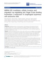
Fig. 1 RABL6 is highly-expressed by ESCC and is associated with worse prognosis. a RABL6 expression was tested and compared between normal
esophageal epithelial cell line NE-1 and Het-1 and all tested ESCC cell lines via qRT-PCR. b Expression of RABL6 in different ESCC cell lines and in
normal esophageal epithelial cell line Het-1A was compared by Western blot analysis; the original full-length gels are presented in Supplementary
Figure S1. c-d The scores of RABL6 expression in 171 ESCC cancerous samples were compared with matched non-cancerous samples by
immunohistochemistry staining. c Representative images of RABL6 IHC staining in 2 pairs of ESCC cases (original magnification: 100x). d Results
are expressed in the form of mean+/−SEM. e- f Kaplan-Meier analysis showed the overall survival rate and disease-free survival rate of ESCC
patients stratified by RABL6 expression. * indicates P < 0.05, ** indicates P < 0.01, *** indicates P < 0.01 for statistical results
Feng et al. BMC Cancer
(2020) 20:602
Page 5 of 10
Table 1 Univariate and multivariate Cox Regression analyzes for overall survival (OS) in ESCC patients
Factors
HR (95% CI)
P value
HR (95% CI)
Age (≤ 55 vs. > 55)
0.956 (0.63–1.45)
0.833
–
P value
–
Gender
0.468 (0.27–0.83)
0.009
0.608 (0.34–1.08)
0.092
Tumor location
0.813 (0.55–1.19)
0.293
–
–
Lymph-vascular invasion
0.479 (0.31–0.73)
0.001
0.479 (0.31–0.73)
0.001
Perineural invasion
0.652 (0.43–0.99)
0.044
0.766 (0.49–1.19)
0.243
Histological differentiation
1.666 (1.22–2.27)
0.001
1.396 (1.02–1.90)
0.035
Pathological Stage
3.458 (2.26–5.29)
0.000
2.233 (1.09–4.58)
0.028
Lymph node metastasis
3.650 (2.33–5.71)
0.000
1.566 (0.74–3.31)
0.240
RABL6 expression
(High vs. low)
2.119 (1.35–3.32)
0.001
1.631 (1.03–2.59)
0.038
HR Hazard ratio, CI confidential interval
of the AJCC (2017 version). Patients’ median age in this
study was 55.0 years (range: 30.0–75.0 years). There were
129 (75.5%) males and 42 (24.6%) females (male to
female ratio, 3.07:1). RABL6 expression was associated
with patients’ age and lymph-vascular invasion (P =
0.040 and P < 0.000, respectively). High expression of
Table 2 Baseline characteristics of ESCC patients and the correlation with RABL6 expression
Characteristics
P value
Total [cases (%)]
RABL6 expression [cases (%)]
N = 171
High
N = 96
Low
N = 75
≤ 55
79 (46.2)
45 (26.3)
34 (19.9)
> 55
92 (53.8)
51 (29.8)
41 (24.0)
Age (years)
0.040
Gender
0.320
Male
129 (75.4)
74 (43.2)
55 (32.2)
Female
42 (24.6)
22 (12.9)
20 (11.7)
Upper
13 (7.6)
8 (4.7)
5 (2.9)
Middle
114 (66.7)
69 (40.4)
45 (26.3)
Low
44 (25.7)
19 (11.1)
25 (14.6)
Yes
79 (46.2)
56 (32.7)
23 (13.5)
No
92 (53.8)
40 (23.4)
52 (30.4)
Tumor location
0.132
Lymph-vascular invasion
0.000
Perineural invasion
0.139
Yes
63 (36.8)
40 (23.4)
23 (13.5)
No
108 (63.2)
56 (32.7)
52 (30.4)
39 (22.8)
22 (12.9)
17 (9.9)
Histological differentiation
Well
0.282
Moderate
93 (54.4)
48 (28.1)
45 (26.3)
Poor
39 (22.8)
26 (15.2)
13 (7.6)
I & II
103 (60.2)
55 (32.2)
48 (28.1)
III & IV
68 (39.8)
41 (23.9)
27 (15.8)
Pathological stage
0.374
Lymph node metastasis
0.162
No
90 (52.6)
46 (26.9)
44 (25.7)
Yes
81 (47.4)
50 (56.1)
31 (18.1)
Feng et al. BMC Cancer
(2020) 20:602
RABL6 was more common in patients older than 55
years, and in patients with lymph-vascular invasion. No
statistical association was found between expression of
RABL6 and other clinicopathological characteristics such
as gender, tumor location, peri-neural invasion, histological differentiation, pathological stage, and lymph
node metastatic status in our study (Table 2).
Knockdown of RABL6 inhibited proliferation of cancer
cells
Owing to RABL6 high-expression predicted poorer
prognosis in ESCC patients, we raised a hypothesis
that RABL6 might play oncogenic roles in ESCC,
and RABL6 expression might promote cancer cell
growth. So, the role of RABL6 on cell growth was
invested via knockdown of RABL6. We used small
interfering RNA (siRNA) to knock down RABL6 in
YES-2 and TE-2 cells stably. The expression of
RABL6 in RABL6-knocked-down cells was examined
via qRT-PCR method (Fig. 2a). MTS assay displayed
that cell growth rates in RABL6-knocked-down YES2 and TE-2 cells were obviously lower than the
negative control cells (NC) (Fig. 2b, c). The result of
focus formation assay showed that the RABL6knocked-down YES-2 and TE-2 cells formed lower
Page 6 of 10
number and smaller colonies than the control cells
(Fig. 2d, e). All the results indicated that knockdown
of RABL6 inhibited cell proliferation and growth in
YES-2 and TE-2 cells.
Knockdown of RABL6 had no influence on apoptosis in
ESCC cells in vitro
The effect of RABL6 in cell apoptosis was evaluated by
flow cytometry analysis. Results revealed that RABL6 silencing couldn’t induce apoptosis in YES-2 and TE1 cells
(Fig. 3a, b).
Knockdown of RABL6 inhibited migrating and invading of
ESCC cells
Since the statistical results showed that RABL6 expression was closely associated with lymph-vascular invasion,
we studied the influence of RABL6 on migration and invasion in ESCC cells through Transwell assay. Results
showed that the abilities of migrating and invading of
ESCC cells were significantly decreased after silencing of
RABL6 (Fig. 4a, b, c, d). These results indicated that
knockdown of RABL6 remarkably inhibited migrating
and invading of cancer cells.
Fig. 2 Knockdown of RABL6 suppressed ESCC cell proliferation. a-e YES-2 and TE-2 cells were transfected with siRNAs specifically targeting RABL6
(si-#1, si-#2) or scrambled siRNA control (si-N). a QRT-PCR confirmed the effective knockdown of RABL6 in these ESCC cells. b-c MTS assay tested
the cell growth rate of these RABL6 knockdown cells, and the results were analyzed statistically. d-e Single cell clone assay detected the colony
formation. d Representative image of clone formation e Results are analyzed statistically. The experiments were repeated 3 times independently,
and the representative data of the experiments are presented in figures. * indicates P < 0.05, ** indicates P < 0.01 for statistical results
Feng et al. BMC Cancer
(2020) 20:602
Page 7 of 10
Fig. 3 Knockdown of RABL6 had no impact on ESCC cells. a Flow cytometry assay was performed to analyze apoptosis of YES-2 and TE-2 cells
with RABL6 knockdown. b Results are statistically analyzed. Values represented the mean ± SD data in triplicate, ns: no significance
Knockdown of RABL6 inhibited epithelial-mesenchymal
transition (EMT) in ESCC cells
Whether RABL6 promoted tumor cell migration via inducing EMT remains unclear. In order to analyze the
function of RABL6 on EMT, we studied the expression
of EMT markers and EMT-related transcription factors
through qRT-PCR and western blot, and also observed
cell morphology changes by inverted microscope in
RABL6 knockdown cells and control cells. QRT-PCR results showed that the expression of epithelial markers Ecadherin and β-catenin was obviously increased, and the
mesenchymal marker Vimentin and slug was significantly decreased in knocked-down RABL6 cells compared to control groups (Fig. 5a, b). The results of
Fig. 4 Knockdown of RABL6 inhibited invading and migrating of ESCC cells. a-d Transwell assay was conducted to compare migration and
invasion between cells treated with siRNAs (si-#1, si-#2) and si-N. a-b Representative images of invasive or migrated cells are provided. c-d Results
are analyzed statistically. The experiments were repeated 3 times independently, and the representative data of the experiments are presented in
figures. * indicates P < 0.05, ** indicates P < 0.01 for statistical results
Feng et al. BMC Cancer
(2020) 20:602
Page 8 of 10
Fig. 5 Knockdown of RABL6 inhibited epithelial-mesenchymal transition in ESCC cells (a-c). a-b Relative expressions of E-cadherin, a-catenin, bcatenin, Vimentin, and Slug in YES-2 and TE-2 cells were compared by qRT-PCR between siRNA RABL6-silenced cells (si#-1, si-#2) and their control
cells (si-N). c Western blots comparing siRNA RABL6-silenced cells (si#-1, si-#2) with their control cells (si-N) are showed with the expression of
vimentin, slug, β-catenin and E-cadherin. A-tubulin was taken as control; the original full-length gels are presented in Supplementary Figure S2. d
Representative bright-field images of ESCC cells and ESCC cells stained with fluorescent phalloidin to show actin structures, with or without
silencing of RABL6. DAPI (blue) was applied to stain nuclear DNA. (original magnification of bright-field: 400x; original magnification of fluorescent
phalloidin: 1000x). Data are expressed in the form of the mean ± SEM. * indicates P < 0.05, ** indicates P < 0.01 versus the control
western blot were consistent with the results of qRTPCR (Fig. 5c). We observed by bright field that RABL6
knockdown ESCC cells were less spindle and became
cobble stone like shape compared to their negative control cells in shape. Phalloidin staining showed circumferential actin belts in RABL6 knockdown ESCC cells,
while the actin structures were spindle-shaped in the
control cells (Fig. 5d). This indicated the occurrence of
EMT. All the results together revealed that silencing of
RABL6 inhibited EMT in ESCC cells.
Discussion
Esophageal cancer is one of the most common malignant carcinomas worldwide, especially in China. Surgical
treatment is a main therapeutic modality for early stage
and locally advanced ESCC. For locally advanced cases,
neoadjuvant or adjuvant chemotherapies are now applied. For unresectable or late-staged cases, the concurrent chemoradiotherapy maybe the recommended
therapy [12]. Nevertheless, the treatment outcome for
ESCC is still under satisfactory with these traditional
therapeutic methods. Recently, targeted therapy has led
to significant breakthroughs in cancer therapy, such as
metastatic melanoma, NSCLC, gastrointestinal stromal
tumor, and so on [13]. For ESCC, some studies reveal
that mTOR, PTEN, and Forkhead box M1 (FOXM1)
maybe the prognostic predictors and therapeutic targets
[14, 15]. However, effective targeted therapy for ESCC is
Feng et al. BMC Cancer
(2020) 20:602
still needed to be developed, due to the exact cellular
and molecular mechanisms of oncogenesis and progression for ESCC remains unclear.
RABL6 is a novel Ras superfamily protein. The Ras
superfamily of GTPases comprises several subfamilies of
small GTP-binding proteins which play pivotal roles in
tumorigenesis, as their functions included cell proliferating, differentiating, and apoptosis [16, 17]. Several studies found elevated RABL6 expression in various human
cancers, including pancreatic ductal adenocarcinomas,
pancreatic neuroendocrine tumors and breast tumors.
And RABL6 overexpression was associated with poor
survival in those cancers [7–9]. However, its role in
ESCC is yet to be discovered. Here we firstly investigated
RABL6 in ESCC. We found that RABL6 was overexpressed in ESCC tissues and cell lines. And patients with
high RABL6 expression had statistical poorer prognosis
than those with low RABL6 expression. High RABL6 expression was an independent prognostic factor in ESCC.
And patients with high expression of RABL6 had obviously higher rate of lymph-vascular invasion compared
to those with low expression of RABL6. Studies showed
that metastasis and recurrence are key factors contributing to poor prognosis of cancer, and the presence of
lymph-vascular invasion strongly associated with high
risk of metastasis and recurrence in endometrial cancer,
ESCC, and so on [18, 19]. These may be one of the reasons to explain RABL6 high expression associated with
poor prognosis, and implied that RABL6 played a vital
role in the malignant progress of ESCC.
To investigate the role of RABL6, a series of functional
studies were carried out. In vitro studies showed that
knockdown of RABL6 inhibited tumor cell growth, proliferation, invasion and migration. Tang et al. found that
silencing of RABL6 gene in U2-OS and SAOS2 osteosarcoma cell lines suppressed cell colony formation and
proliferation in vitro [10]. They also demonstrated that
RABL6 modulated G1-S transition in cell cycle analysis.
RABL6 regulated retinoblastoma 1 (Rb1) activity in
osteosarcoma cells, which was the major player in cell
cycle control [10, 20, 21]. Li Y et al. also reported that silencing of RABL6 promoted breast cancer cell growth
was via promoting apoptosis [7]. Besides cell cycle and
apoptosis, Montalbano et al. reported that RABL6
knockdown resulted in marked cell growth suppression,
which is associated with inhibition of extracellular
signal-regulated kinase phosphorylation [22]. These reports indicated that RABL6 displays an oncogenic function by regulating cell cycle or inducing apoptosis, or
influencing signal pathway. However, our study showed
that silencing of RABL6 couldn’t induce apoptosis in
ESCC cell in vitro. Whether RABL6 carried out its oncogenic function in ESCC via regulating cell cycle or signaling pathway needs to be further investigated.
Page 9 of 10
We also demonstrated that knockdown of RABL6
inhibited cell invasion and migration via EMT. It was reported that EMT inducers were important for morphogenesis and organogenesis through regulating cell
migrating. The abnormal activation ofEMT played a key
role in development and metastasis in tumors [23]. Up
regulation of mesenchymal markers, down regulation of
epithelial markers, abnormal localization of β-catenin
and nuclear expression of Vimentin are some of its characteristics [24, 25]. In the development of epithelial tumors, cancer cells obtain phenotypes of invasion and
motivation through EMT, which results in invasion or
metastasis, leading to the death of about 90% of patients
[26].
Taken all these together, we found that RABL6 plays
an important part in the tumorigenesis and progression
in ESCC.
Conclusions
Conclusively, we indicated firstly that RABL6 is highly
expressed by ESCC and associated with poor prognosis.
Its expression is associated with lymph-vascular invasion. Downregulation of RABL6 suppressed proliferation,
migration and EMT of ESCC cells. Thus, RABL6 exert
an important function on the progression of ESCC, and
may be promising prognostic marker and a potential
therapeutic target for ESCC. Its underlying mechanism
and clinical application need to be further developed.
Supplementary information
Supplementary information accompanies this paper at />1186/s12885-020-07068-w.
Additional file 1.
Additional file 2.
Additional file 3.
Abbreviations
RABL6: RAB, member RAS oncogene family like 6; C9orf86: Chromosome 9
open reading frame 86; ESCC: Esophageal squamous cell carcinoma; qRTPCR: Quantitative reverse transcription-polymerase chain reaction;
EMT: Epithelial-mesenchymal transition; EC: Esophageal cancer; RBEL1: Rablike protein 1; PARF: Partner of alternative reading frame protein;
NCBI: National Center for Biotechnology Information; NSCLC: Non-small cell
lung cancer; OS: Overall survival; ATCC: American Type Culture Collection;
siRNA: Small interfering RNA; SDS-PAGE: Sodium dodecyl sulfatepolyacrylamide gel electrophoresis; ANOVA: One-way analysis of variance;
NC: Negative control cells; FOXM1: Forkhead box M1; Rb1: Retinoblastoma 1
Acknowledgements
Not applicable.
Authors’ contributions
YFF, FW and YYL designed the study. YFF, SMY, YHH and QTH performed the
experiments. YFF, FW and YYL analyzed, interpreted the data and wrote the
article. All authors have approved this submission.
Funding
Not applicable.
Feng et al. BMC Cancer
(2020) 20:602
Availability of data and materials
The data in this study are available from the corresponding author on
request.
Ethics approval and consent to participate
All the patients in this study signed consents for the approval of use of their
tissues and information in research. The institutional ethics committee of Sun
Yat-sen University cancer center approved this study.
Consent for publication
Not applicable.
Competing interests
The authors declare that they have no conflict of interest.
Author details
1
State Key Laboratory of Oncology in South China, Collaborative Innovation
Center for Cancer Medicine, Sun Yat-sen University Cancer Center,
Guangzhou 510060, Guangdong, China. 2Department of Pathology, Sun
Yat-sen University Cancer Center, Guangzhou 510060, Guangdong, China.
3
Department of Molecular Diagnostics, Sun Yat-sen University Cancer Center,
Guangzhou 510060, China. 4Department of Thoracic Surgery, the First
Affiliated Hospital, Sun Yat-sen University, Guangzhou, Guangdong 510080,
People’s Republic of China.
Received: 19 September 2019 Accepted: 15 June 2020
References
1. Bray F, Ferlay J, Soerjomataram I, Siegel RL, Torre LA, Jemal A. Global cancer
statistics 2018: GLOBOCAN estimates of incidence and mortality worldwide
for 36 cancers in 185 countries. Ca-a Cancer J Clin. 2018;68(6):394–424.
2. Chen W, Zheng R, Baade P, Zhang S, Zeng H, Bray F, Jemal A, Yu X, He J.
Cancer statistics in China, 2015. CA Cancer J Clin. 2016;66(2):115–32.
3. Siegel RL, Miller KD. Cancer statistics, 2019. CA Cancer J Clin. 2019;69(1):7–
34.
4. Zhang XF, Hagen J, Muniz VP, Smith T, Coombs GS, Eischen CM, Mackie DI,
Roman DL, Van Rheeden R, Darbro B, Tompkins VS, Quelle DE. RABL6A, a
novel RAB-like protein, controls centrosome amplification and chromosome
instability in primary fibroblasts. PLoS One. 2013;8(11):e80228.
5. Wennerberg K, Rossman KL, Der CJ. The Ras superfamily at a glance. J Cell
Sci. 2005;118(Pt 5):843–6.
6. Cheng KW, Lahad JP, Kuo WL, Lapuk A, Yamada K, Auersperg N, Liu J,
Smith-McCune K, Lu KH, Fishman D, et al. The RAB25 small GTPase
determines aggressiveness of ovarian and breast cancers. Nat Med. 2004;
10(11):1251–6.
7. Li YY, Fu S, Wang XP, Wang HY, Zeng MS, Shao JY. Down-regulation of
c9orf86 in human breast cancer cells inhibits cell proliferation, invasion and
tumor growth and correlates with survival of breast cancer patients. PLoS
One. 2013;8(8):e71764.
8. Muniz VP, Askeland RW, Zhang X, Reed SM, Tompkins VS, Hagen J,
McDowell BD, Button A, Smith BJ, Weydert JA, et al. RABL6A promotes
Oxaliplatin resistance in tumor cells and is a new marker of survival for
resected pancreatic ductal adenocarcinoma patients. Genes Cancer. 2013;
4(7–8):273–84.
9. Yoshimura K, Osman M, Inoue Y, Suda T, Sugimura H. A novel prognostic
marker of non-small cell lung cancer: chromosome 9 open reading frame
86 (C9orf86). J Thorac Dis. 2016;8(9):2284–6.
10. Tang H, Ji F, Sun J, Xie Y, Xu Y, Yue H. RBEL1 is required for osteosarcoma
cell proliferation via inhibiting retinoblastoma 1. Mol Med Rep. 2016;13(2):
1275–80.
11. Feng YF, Lei YY, Lu JB, Xi SY, Zhang Y, Huang QT, Wu QL, Wang F. RIT1
suppresses esophageal squamous cell carcinoma growth and metastasis
and predicts good prognosis. Cell Death Dis. 2018;9(11):1085.
12. Kato H, Nakajima M. Treatments for esophageal cancer: a review. Gen
Thorac Cardiovasc Surg. 2013;61(6):330–5.
13. Patel S. Long-term efficacy of imatinib for treatment of metastatic GIST.
Cancer Chemother Pharmacol. 2013;72(2):277–86.
14. Lu J, Pan Y, Xia X, Gu Y, Lei Y. Prognostic significance of mTOR and PTEN in
patients with esophageal squamous cell carcinoma. Biomed Res Int. 2015;
2015:417210.
Page 10 of 10
15. Song L, Wang X, Feng Z. Overexpression of FOXM1 as a target for
malignant progression of esophageal squamous cell carcinoma. Oncol Lett.
2018;15(4):5910–4.
16. Hernandez-Alcoceba R, del Peso L, Lacal JC. The Ras family of GTPases in
cancer cell invasion. Cell Mol Life Sciences. 2000;57(1):65–76.
17. Peng GL, Tao YL, Wu QN, Zhang Y, He JX. Positive expression of protein
chromosome 9 open reading frame 86 (C9orf86) correlated with poor
prognosis in non-small cell lung cancer patients. J Thorac Dis. 2016;8(7):
1449–59.
18. Loizzi V, Cormio G, Lorusso M, Latorre D, Falagario M, Demitri P, Scardigno
D, Selvaggi LE. The impact of lymph vascular space invasion on recurrence
and survival in patients with early stage endometrial cancer. Eur J Cancer
Care. 2014;23(3):380–4.
19. Qu Y, Lin H, Zhang C, Li K, Zhang H. Cribriform pattern in lung invasive
adenocarcinoma correlates with poor prognosis in a Chinese cohort. Pathol
Res Pract. 2019;215(2):347–53.
20. Burkhart DL, Sage J. Cellular mechanisms of tumour suppression by the
retinoblastoma gene. Nat Rev Cancer. 2008;8(9):671–82.
21. Classon M, Harlow E. The retinoblastoma tumour suppressor in
development and cancer. Nat Rev Cancer. 2002;2(12):910–7.
22. Montalbano J, Lui K, Sheikh MS, Huang Y. Identification and characterization
of RBEL1 subfamily of GTPases in the Ras superfamily involved in cell
growth regulation. J Biol Chem. 2009;284(27):18129–42.
23. Ansieau S, Courtois-Cox S, Morel AP, Puisieux A. Failsafe program escape
and EMT: a deleterious partnership. Semin Cancer Biol. 2011;21(6):392–6.
24. Kanai Y, Oda T, Tsuda H, Ochiai A, Hirohashi S. Point mutation of the Ecadherin gene in invasive lobular carcinoma of the breast. Japanese J
Cancer Res. 1994;85(10):1035–9.
25. Gamallo C, Palacios J, Suarez A, Pizarro A, Navarro P, Quintanilla M, Cano A.
Correlation of E-cadherin expression with differentiation grade and
histological type in breast carcinoma. Am J Pathol. 1993;142(4):987–93.
26. Pradella D, Naro C, Sette C, Ghigna C. EMT and stemness: flexible processes
tuned by alternative splicing in development and cancer progression. Mol
Cancer. 2017;16(1):8.
Publisher’s Note
Springer Nature remains neutral with regard to jurisdictional claims in
published maps and institutional affiliations.