Sterile, abscess-like cerebral lesion during trastuzumab therapy after HER2 status switch in a triple negative breast cancer patient: A case report and literature review
Bạn đang xem bản rút gọn của tài liệu. Xem và tải ngay bản đầy đủ của tài liệu tại đây (5.57 MB, 8 trang )
Mezei et al. BMC Cancer
(2020) 20:615
/>
CASE REPORT
Open Access
Sterile, abscess-like cerebral lesion during
trastuzumab therapy after HER2 status
switch in a triple negative breast cancer
patient: a case report and literature review
Tamás Mezei1,2*, Melinda Hajdu3, Gábor Czigléczki1,2, Gábor Lotz4, Judit Kocsis5, Janina Kulka4 and Anna Horváth5
Abstract
Background: Breast cancer is a global health problem – it is the most common malignancy among women. Triple
negative breast cancers (TNBC) account for 10–20% of female breast cancer. Most TNBC cases confer poor
prognosis. Brain metastasis appears in more than 15% in the triple negative breast cancer population, which causes
serious decrease in survival. Changes of immunophenotype are not uncommon in breast cancer, offering new
therapeutic options in cases where targetable proteins or pathways are being identified.
Case presentation: After five lines of chemotherapy and 82 months following the first diagnosis, our patient with
brain metastatic triple negative breast cancer had human epidermal growth factor receptor 2 (HER2) genetic
heterogeneity in the metastatic tissue sample interpreted as HER2 status conversion. After the removal of the
metastasis, we started first line therapy for metastatic HER2 positive cancer with trastuzumab and paclitaxel. After
the first cycle of trastuzumab, on day 8, she had a seizure, and neurosurgical examination showed an abscess-like
lesion. The punctate proved to be sterile by microbiological and pathological examination, so we continued
cytostatic therapy without the anti-HER2 antibody. 3 months later, we could not identify the previous abscess-like
lesion in the control computer tomography (CT) scan, and our patient had no neurological deficits.
Conclusion: We emphasize the importance of regular tissue confirmation of predictive markers in progressive tumorous
disease even if our presented case is not unequivocally a “conversion case”. Tumor subtype is determined according to
algorithms and definitions published in guidelines, nevertheless, use of different guidelines may lead to controversial
interpretation in cases where HER2 genetic heterogeneity is present. Furthermore, we suggest that seronegative, aseptic
intracranial fluid effusion after the removal of a brain metastasis may possibly be a side effect of trastuzumab.
Keywords: Triple negative breast cancer, Brain metastasis, HER-2 immunophenotype switch, Trastuzumab, Abscess-like
lesion
* Correspondence:
1
Department of Neurosurgery, Semmelweis University, 57 Amerikai street,
Budapest, Pest 1145, Hungary
2
National Institute of Clinical Neurosciences, 57 Amerikai street, Budapest,
Pest 1145, Hungary
Full list of author information is available at the end of the article
© The Author(s). 2020 Open Access This article is licensed under a Creative Commons Attribution 4.0 International License,
which permits use, sharing, adaptation, distribution and reproduction in any medium or format, as long as you give
appropriate credit to the original author(s) and the source, provide a link to the Creative Commons licence, and indicate if
changes were made. The images or other third party material in this article are included in the article's Creative Commons
licence, unless indicated otherwise in a credit line to the material. If material is not included in the article's Creative Commons
licence and your intended use is not permitted by statutory regulation or exceeds the permitted use, you will need to obtain
permission directly from the copyright holder. To view a copy of this licence, visit />The Creative Commons Public Domain Dedication waiver ( applies to the
data made available in this article, unless otherwise stated in a credit line to the data.
Mezei et al. BMC Cancer
(2020) 20:615
Background
Breast cancer is responsible for the most common female cancer-related morbidity and mortality all over the
world [1]. The prevalence of the disease is 23% among
women living with cancer. It is the second most common cause of central nervous system (CNS) metastasis.
The risk of developing metastasis is 10–16% among
those living with breast cancer, and it may be higher in
advanced cases with poor prognosis [2, 3]. TNBC is a
subgroup of breast cancer, being negative for hormone
receptors and HER2. There is a high overlap with the
basal-like subgroup of breast cancers characterized by
gene expression profiling methods [4–6]. TNBC accounts for roughly one-sixth of all breast carcinomas [7].
A number of studies provided evidence about the instability of hormone receptor and HER2 status during
the progression of the disease, with special focus on the
relationship of primary tumor and metastases. Alterations occur most frequently in hormone receptor status
and Ki67 labeling index, and these changes usually carry
a worse prognosis: for example, an estrogen receptor
(ER) positive tumor may become negative, and the Ki67
index may increase during the progression of the disease. The triple negative phenotype, and specifically,
HER2 negative status is relatively conserved [8].
This report presents the case of a germline breast cancer
type 1 (BRCA1) mutation carrier patient with TNBC cancer without androgen receptor (AR) expression, and with
a HER2 status switch during the course of the disease.
Trastuzumab therapy produced an assumed side effect,
which is described here for the first time in the literature.
Case presentation
Our patient was 32 years old when she first presented at
the oncology unit of the 3rd Department of Internal
Medicine, Semmelweis University, in July 2010. She had
palpated nodules in her right breast. Ultrasonography
(USG) and magnetic resonance imaging (MRI) showed
Page 2 of 8
bilateral multifocal tumor, with nodules of 8 × 7 mm and
7 × 4 mm located at 6 and 8 o’clock in her right breast,
respectively, and an 8 × 5 mm tumor focus located at 5
o’clock in her left breast. Aspiration cytology confirmed
the diagnosis of invasive breast carcinoma (no special
type). There was no detectable distant metastasis. In her
medical history, there was an ovulation induction therapy by clomiphene 3 years before, at age 29, and a childbirth via normal vaginal delivery after that.
The patient underwent bilateral mastectomy with axillary sentinel lymph node biopsy. Pathological examination showed bilateral, grade 3 invasive breast carcinoma
(no special type) with ER, progesterone receptor (PR)
and HER2 negativity by immunohistochemistry (IHC)
and fluorescence in situ hybridization (FISH) (Fig. 1).
Disease stage was pmT1b N0(sn) cM0. First line adjuvant chemotherapy was initiated in August 2010, according to the 5-fluorouracil, epirubicin, cyclophosphamide
and docetaxel (FEC100-TXT) protocol (3 cycles each),
after which she had breast reconstruction surgery twice.
Chemotherapy and the other interventions were well tolerated. Chemoterapy doses see schedule (Table 1).
Surveillance MRI in December 2013 detected two lesions indicating disease recurrence: a 10 × 5 mm mass
on the outer contour of the implant on the left side, and
a paracentral 22 × 28 mm lesion on the right side, the
latter involving nearby ribs. USG-guided fine needle aspiration biopsy (FNAB) biopsy demonstrated invasive
breast carcinoma (no special type). No repeated immunocytochemical reactions were performed on the
sample. Positron emission tomography - computer tomography (PET-CT) showed enhancement in the supraclavicular and parasternal lymph nodes, such that tumor
removal was contraindicated. Thus, we started first line
bevacizumab-paclitaxel therapy for metastatic TNBC,
which was given for 12 cycles. Partial regression was
seen in PET-CT after 5 cycles, but surgical intervention
was still contraindicated due to the activity seen in the
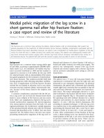
Fig. 1 Histopathology from the primary tumor. a. Primary ductal invasive breast carcinoma with prominent lymphocytic infiltration (tumor
infiltrating lymphocyte (TIL) score was 80–90%), H&E section.b. HER2 FISH with single probe: Chr 17 polysomy was diagnosed based on the
finding of 3–6 HER2 copies in many tumor cells
Mezei et al. BMC Cancer
(2020) 20:615
Page 3 of 8
Table 1 Summary of our patient therapies and its doses
Surgery
Bilateral mastectomy with axillary sentinel lymph node biopsy
pmT1b N0(sn) cM0
Adjuvant therapy
FEC100-TXT
5-Fluorouracyl 500 mg/m2
Cyclophosphamide 500 mg/m2
Epirubicin 70 mg/m2
Docetaxel 100 mg/m2
1st line
Bevacizumab+TAX
Bevacizumab 15 mg/kg
Paclitaxel 175 mg/m2
Radiotherapy
IMRT
54 Gy simultaneous boost was given to the infra-axillary
and axillary region and the operated area
2nd line
CDDP+TXT
Cisplatin 75 mg/m2
Docetaxel 75 mg/m2
3rd line
XEL
Capecitabine 2500 mg/m2
4th line
PARP inhibitor
Olaparib 300 mg
5th line
VNB
Vinorelbine 30 mg/m2 (only 2 cycle)
6th line
Trastuzumab+TAX
Trastuzumab 4 mg/kg
Paclitaxel 175 mg/m2
7th line
VNB/reinduction
Vinorelbine 30 mg/m2
8th line
CMF
5-Fluorouracyl 600 mg/m2
Cyclophosphamide 600 mg/m2
Methotrexate 40 mg/m2
lymph nodes, and chemotherapy was continued. Deoxyribonucleic acid (DNA) sequencing performed during
this time period revealed a germline mutation in the
BRCA1 gene. Progressive disease was diagnosed by PETCT at the end of the protocol. Because of the lack of
new foci, the patient underwent thoracic surgery in February 2015. Pathological examination of the lymph nodes
showed metastases with extracapsular extension and
perineural invasion (stage pN3). Predictive markers were
not investigated in the surgical specimen. Radiotherapy
followed, based on the decision of the multidisciplinary
oncoteam: intensity-modulated radiotherapy (IMRT, 54
Gy) and simultaneous boost was given to the infraaxillary and axillary region and the operated area.
In November 2015, multiple lesions (progression in the
parasternal lymph node, new pulmonary hilar lymph node
involvement and lung metastasis) were visible by PET-CT
imaging. We started second line therapy for metastatic
TNBC according to the cisplatin, docetaxel (CDDP+TXT)
protocol. Control CT imaging performed after 9 cycles
showed progression; therefore, the patient was enrolled in
a clinical trial, where she received oral capecitabine as
third line treatment, and the poly (adenosine biphosphate
- ribose) polymerase (PARP) inhibitor olaparib as fourth
line therapy.
Although clinically stable, progression was seen by imaging methods in May 2017 (multiple bone metastases and
a new lung metastasis, in addition to parasternal, mediastinal, pulmonary hilar and pelvic lymph node involvement),
and the oncological team decided on initiating fifth line
therapy (vinorelbine - VNB). Meanwhile, core biopsy was
taken from a tumorous mass (34x15mm) above the sternum, and pathological examination confirmed ER- and PRnegative breast cancer involvement, but – surprisingly –
HER2 status turned out to be positive by FISH (Fig. 2).
She developed motor aphasia in June 2017, while at
home; she suddenly felt confused, and had a grand mal
seizure. She was urgently transferred to the neurosurgery
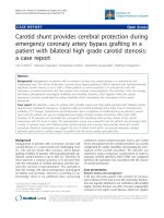
Fig. 2 Core biopsy specimen of the tumor mass in the sternum region. a. Highly atypical, pleomorphic tumor cells are seen on the H&E section.
b. HER2 immunohistochemistry c. FISH examination confirmed amplification of the HER-2 gene (red) in more than 30% of tumor cells, and
chromosome 17 polysomy (green)
Mezei et al. BMC Cancer
(2020) 20:615
department of the National Institute of Clinical Neurosiences (NICN), and had an MRI scan, which showed a
metastasis in the left frontal lobe. Neurosurgical intervention was performed, and the entire tumor was removed, which proved to be the metastasis of TNBC by
histopathology (Fig. 3).
Sixth line trastuzumab and paclitaxel treatment was
initiated at the end of July – based on the positive HER2
status of the previously sampled sternal mass –, which
was given for 2 cycles.
She had a repeated seizure in the middle of August 2017,
and she was taken to the NICN. CT and MRI scans showed
an abscess-like lesion in the cavity of the previously operated area, surrounded by large perifocal edema (Fig. 4).
Mannisole and furosemide was administered for the reduction of intracranial pressure. Stereotactic biopsy was taken
on August 09, 2018, and stereotactic drainage was performed on August 29, 2018. During sampling, pus-like content was gained, therefore she received antibiotic therapy
(ceftriaxone, vancomycin and metronidazole).
Aerobic and anaerobic cultures were negative for bacteria, fungi and parasites as well, and histopathology also
excluded the possibility of a true abscess (Fig. 5). After a
30 day pause, she received subcutaneous trastuzumab for
the second time, without any side effect.
After seventh line chemotherapy (5 cycles of VNB),
control cranial CT showed a new metastasis in the
contralateral frontal lobe; the previous abscess-like lesion
was not present. The new, right-sided frontal metastasis
was treated by stereotactic irradiation.
To be able to decide on further therapy, FISH examination was performed from the intracranial tumor metastasis. It showed HER2 non-amplified status again, and we
started eighth line intravenous cytostatic therapy according to the CMF protocol. When she arrived for the 3rd
cycle of cytostatic therapy, her performance status
dropped (to ECOG 3), and gastric hemorrhage was diagnosed as the cause of weakness. A nasogastric tube was introduced, and the stomach was flushed with acepramine.
Page 4 of 8
She received blood transfusion and had a gastroscopy,
which identified a gastric ulcer (post-mortem examination
later on confirmed the metastatic involvement of the gastric wall). After supportive care was concluded, the patient
was placed under hospice care.
She came back to the hospital in very poor general
condition (ECOG 4) in December 2017, with symptoms
of dehydration, respiratory failure and cardiac decompensation. Intensive therapy was avoided because of her
advanced disease. Basic life support and pain relief was
provided, and she died without alarming symptoms.
Discussion and conclusion
The survival curve of TNBC patients shows a decrease between the 3rd and 5th years after first diagnosis; later progression is relatively uncommon [7]. This drop in survival
is due to poor prognostic clinicopathological features,
such as advanced stage at presentation, unfavorable histopathology, high tumor grade, high Ki67 index, and a
higher rate of metastasis [9]. Hazard ratio for relapse in
the TNBC group is three times that of the non-TNBC
group during this period [7]. Our patient had a similar disease course, with the first relapse occurring 3.5 years after
the surgical intervention and adjuvant therapy. Liedtke
et al. reported 64% 5 years overall survival in the examined
TNBC population [10]. Agarwal et al. reported an 81.8
months median overall survival for TNBC patients [11].
The overall survival of a metastatic TNBC patient is 18
months from the diagnosis of the metastasis [4, 12, 13].
Our patient lived 89 months after her first diagnosis of
TNBC (25 months with stage IV disease).
The therapy of TNBC remains a challenge for oncologists treating breast cancer patients [4, 14]. Therapeutic
choices include surgical intervention, radiation therapy,
systemic chemotherapy and targeted therapy. Our patient underwent bilateral mastectomy as the first step, in
accordance with published recommendations for multifocal breast cancer [4]. Adjuvant cytotoxic regimen consisted of anthracyclines and taxanes in her case. The
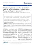
Fig. 3 Histopathology of the brain metastasis. a. Poorly differentiated tumor cells, some showing very bizarre nuclei, H&E section.b. HER2
immunohistochemical reaction heterogeneous positive membrane reaction of tumor cells (20% of tumor cells showed complete, moderately
intense membrane reaction).c. HER2 FISH: In four different tumor areas 80 tumor cells were counted, the mean HER2 gene copy number was 4.0/
tumor cell, and 1,62/Chr 17. However, 43% of tumor cells showed HER2 gene amplification with a mean HER2 gene copy number of 4.6/tumor
cell and 2.4/Chr 17. Furthermore, polysomy was identified in 36% of tumor cells with a mean of 3,6 Chr 17/tumor cell. The final conclusion was
HER2 negative status of the metastatic tumor
Mezei et al. BMC Cancer
(2020) 20:615
Fig. 4 T1-weighted contrast-enhanced horizontal (a) and sagittal (b)
MRI image of the abscess-like cerebral lesion. Ring-enhancing lesion
with a central low intensity content and peripheral low intensity, the
latter of which is due to the surrounding extensive vasogenic edema
presence of germline BRCA1 mutation allowed for the
incorporation of platinum agents into the treatment
protocol [4, 15–17], which was utilized as second line
therapy. Bevacizumab – an anti-vascular endothelial
growth factor (VEGF) antibody inhibiting angiogenesis –
was also utilized as part of personalized therapy in the
metastatic setting [4]. PARP inhibitors are pharmacological inhibitors of poly-ADP ribose polymerase, an
Page 5 of 8
enzyme responsible for repairing single-stranded DNA
breaks [18–20]; as they may be given in BRCA mutant
cancer, the PARP inhibitor olaparib was included as
fourth line therapy. Approximately 25–45% of TNBC express AR, which may be utilized as a therapeutic target,
and it is associated with a better prognosis [4, 16, 21];
however, AR status was negative in our case. We used
eight lines of systemic therapy and tried every possibility
in order to achieve the best quality of life and a longer
progression free and overall survival for our patient.
Breast cancer is a heterogeneous disease, and immunophenotypic changes may occur during progression. As the
disease progresses, hormone receptor (especially PR) positivity is usually lost and mitotic activity increases [22, 23].
HER2 conversion is also known in the literature, but it is
less frequent than the other phenotypic changes [24]. If
phenotypic changes in metastases are not searched for,
the patient may not receive adequate treatment in the
metastatic setting; therefore, the biopsy of the metastatic
lesions is recommended [24–27], such as in our case.
Changes in tumor subtype during progression of
breast cancer has gained major attention in the recent
decade. The largest meta-analysis to date found an overall change in ER receptor status in 19.3%, in PR receptor
status in 30.9% and changes in HER2 status in 10.3%
[28]. In our case, however, the HER2 status change
might not have occurred purely as a biological
phenomenon (e.g. clonal selection, or accumulating further mutations, etc.) during disease progression. As
Table 2. shows, interpretation guidelines for HER2 IHC
and FISH changed considerably during the course of the
patient’s disease [29, 30]. Furthermore, in our case, both
the primary tumor and its two investigated metastases
showed HER2 heterogeneity, both at protein expression
level and at the genetic level. Interpretation guidelines for
HER2 genetic heterogeneity are not fully unequivocal. The
last guideline dealing especially with this problematic field
dates from 2009 [31], and defines HER2 (scattered) genetic heterogeneity as 5–50% of tumor cells showing HER2
amplification. In such cases the mean HER2 copy numbers and the mean HER2/chromosome enumeration
probe 17 (CEP17) ratio (counted in at least 60 tumor cells)
would define the final HER2 status. The tumor can be
interpreted as HER2 positive – and thus eligible for targeted therapy – only if the ratio of amplified tumor cells
exceeds 50%. The situation is different when there is a
well identifiable separate clone present as a cluster of
HER2 amplified tumor cells. The rule for interpretation in
such cases is that if the cohesive cluster of HER2 amplified
cells exceeds 10% of the tumor area investigated, HER2
positive status is to be reported. However, different ideas
were published by opinion leaders in the following years:
Hanna and co-workers [32] suggested an algorithm for
the interpretation of scattered HER2 genetic heterogeneity
Mezei et al. BMC Cancer
(2020) 20:615
Page 6 of 8
Fig. 5 Histopathology from the sampling of the frontal abscess-like lesion. (H&E) Reactive (a) and necrotic tissues (b) without bacteria or tumor
cells, which corresponds to the healing surgical area
different from that of Vance and members [31]: according
to their suggestion, even in cases of scattered amplified
tumor cells, HER2 amplified status can be assigned in
cases where the ratio of amplified cells exceeds 10%. In
our case, HER2 status of the primary tumor was interpreted as negative according to the 2007 American Society
of Clinical Oncology/College of Amercian Pathologists
(ASCO/CAP) guideline. The first investigator of the metastasis that occurred in 2017 in the sternum region, interpreted HER2 status as positive using Hanna’s criteria,
while the HER2 status of the brain metastasis occurring in
the same year - and analyzed in another pathology
department - was interpreted as negative using Vance’s
and CAP’s criteria.
Brain metastases occur more frequently in younger
women, in the case of poor prognostic markers, such as
TNBC and high-grade tumors. The most common symptoms are headache, nausea, vomiting, hemiparesis and visual disturbances; less frequently, seizures. More than half
of the metastases are supratentorial, and approximately
25% are localized in the frontal lobe [3]. The appropriate
treatment option for a patient with a solitary metastasis
and good performance status is the surgical approach
[33]. The rate of postoperative complications does not
Table 2 Interpretation guidelines for HER2 IHC and FISH changes in our case
YEAR LESION/
MATERIAL
EXAMINED
HER2 IHC
HER2 FISH
Respective ASCO/ Respective ASCO/CAP GUIDELINE
definition for HER2 positive ISH result
CAP GUIDELINE
definition for HER2
positive IHC result
2010 Primary
tumor
surgical
resection
specimen
20% of tumor cells
showed complete, intense
circumferential membrane
reaction:
2+
(SP3 antibody)
Polysomy of Chr17 suggested: many
tumor cells showed 3–6 HER2
copies/cell
Interpreted as HER2 negative
(single probe ISH assay)
2007 ASCO/CAP
guideline
> 30% of tumor
cells show
complete, intense
circumferential
reaction
2017 Metastasis
(sternum
region)
20% of tumor cells
showed complete, weak
or moderate,
circumferential membrane
reaction:
2+
(4B5 antibody)
30% of tumor cells showed
polysomy-co-amplification
(3–6 CEP17 signals and 6–10 HER2
signals/cells; dual probe ISH assay)
Interpreted as HER2 positive
2013 ASCO/CAP
2013 ASCO/CAP guideline
guideline
See: Wolff AC et al. Arch Pathol Lab
> 10% of tumor
Med. 2014 Feb; 138 (2): 241–256.
cells show
complete, in tense
circumferential
reaction
2017 Metastasis
(brain –
analysis
following
second
opinion
request)
15–20% of the tumor cells
showed complete, weak
or moderate,
circumferential membrane
reaction:
2+
(4B5 antibody)
80 tumor cells were counted: mean
HER2 copy number/cell was 4.0,
mean HER2/CEP17 ratio was 1.62.
However,
scattered, heterogeneous
amplification was present: In 43% of
the tumor cells 4.6 HER2/cell was
found and the HER2/CEP17 ratio
was 2.4.
(dual probe ISH assay)
Interpreted as heterogeneous
amplification, HER2 negative
2013 ASCO/CAP
guideline
> 10% of tumor
cells show
complete, intense
circumferential
reaction
2007 ASCO/CAP guideline
> 6 HER2copy/cell (single probe ISH)
> 2.2 HER2/CEN17 ratio/cell (dual probe
ISH)
2013 ASCO/CAP guideline
and
2009 CAP guidelines for genetic
heterogeneity in HER2 testing:
„HER2 genetic heterogeneity (GH)
exists if there are more than 5% but
less than 50% of infiltrating tumor cells
with a ratio higher than 2.2 …. If more
than 50% of the infiltrating tumor cells
have a ratio higher than 2.2, then the
tumor is considered HER2 amplified.”
Mezei et al. BMC Cancer
(2020) 20:615
increase with en bloc resection compared to piecemeal resection; on the contrary, Patel et al. published their overall
complication rates: 13% with en bloc resection and 19%
with piecemeal resection (the probability of infections is
under 1%) [34]. The overall survival after brain metastasis
develops in a TNBC patient is really poor, but it can be
prolonged with personalized systemic therapy. Our patient
survived for 7 months after the detection of the first cerebral lesion, and seizures were the initial symptoms of the
metastasis.
Remarkably, 82 months after the first diagnosis of
TNBC, the biopsy taken from a metastatic sternal mass
was interpreted as HER2 positive, which provided the
opportunity for starting biological therapy for HER2
positive breast cancer following surgical removal of the
brain metastasis (our choice of immunotherapy was trastuzumab, because at that time, pertuzumab treatment
was only defrayed by the Hungarian National Health Insurance Fund in the first-line therapy of the breast cancer patients). Trastuzumab treatment was well tolerated,
but the patient presented with repeated seizures after 6
weeks, and MRI scan showed an abscess-like cerebral lesion. However, no new metastasis was detected, and we
assumed that the abscess-like sterile effusion in the operated area could be a side effect of trastuzumab. After
30 days she got subcutaneous trastuzumab for the second time, without any side effect. Although we suggest
that seronegative, aseptic intracranial fluid effusion after
the removal of a brain metastasis may possibly be a hitherto undescribed side effect of iv. trastuzumab, we also
assume that subcutaneous administration may be safe,
as was in our case.
General complications of trastuzumab therapy are well
known. It may cause a flu-like syndrome (similarly to other
immunological therapies), which is relatively rare because
of the humanized nature of the monoclonal antibody. The
major problem with trastuzumab therapy is cardiotoxicity.
Cardiac dysfunction is primarily characterized by cardiomyopathy – most often as an asymptomatic decrease in left
ventricular ejection fraction, and less frequently as congestive heart failure [35]. Trastuzumab combined with taxanes
or vinorelbine has also been reported to cause general fluid
retention and pleural effusion, which may also represent
symptoms of heart failure [36].
Interestingly, despite the HER2 positive, rapidly proliferating metastatic sternal mass, the brain metastasis
appearing later had an ER, PR and HER2 negative status;
therefore, the possibility of subsequent lapatinib therapy
was ruled out.
Taken together, our case teaches us humility: even in
the era of advanced molecular genetic diagnostic methods
and major breakthroughs in targeted therapy, imperfections in HER2 diagnostic/interpretation methods may be
painfully tangible in certain cases. Unequivocal guidelines
Page 7 of 8
(and their unanimous use in practice) for the interpretation of HER2 genetic heterogeneity are mandatory.
Abbreviations
AR: Androgen receptor; ASCO: American Society of Clinical Oncology;
BRCA1: Breast cancer type 1; CAP: College of Amercian Pathologists;
CDDP: Cisplatin; CEP17: Chromosome enumeration probe 17;
CMF: Cyclophosphamide, methotrexate and 5-fluorouracil; CNS: central
nervous system; CT: Computer tomography; DNA: Deoxyribonucleic acid;
ER: Estrogen receptor; FEC100: 5-fluorouracil, epirubicin and
cyclophosphamide; FISH: Fluorescent in situ hybridization; FNAB: Fine needle
aspiration biopsy; HE: Hematoxylin-eosin; HER-2: Human epidermal growth
factor receptor 2; IHC: Immunohistochemistry; IMRT: Intensity-modulated
radiotherapy; MRI: Magnetic resonance imaging; NICN: National Institute of
Clinical Neurosciences; PARP: Poly (adenosine biphosphate - ribose)
polymerase; PET-CT: Positron emission tomography - computer tomography;
PR: Progesterone receptor; TAX: Paclitaxel; TIL: tumor infiltrating lymphocyte;
TNBC: Triple negative breast cancer; TXT: Docetaxel; USG: Ultrasonography;
VEGF: Vascular endothelial growth factor; VNB: Vinorelbine; XEL: Capecitabine
Acknowledgements
We thank for the patient and for her family to contribute this article.
Authors’ contributions
T.M.: design the work, collection and interpretation of the data, writing and
editing the manuscript, approved the submitted version. M.H.: evaluation of
histological samples, collection of the materials, major contributor in writing
the manuscript, approved the submitted version. G.C.: development of the
neurosurgical aspects of the manuscript, data collection, approved the
submitted version. G.L.: evaluation of the histological samples, approved the
submitted version. J.Ko.: responsible for the diagnosis and treatment of the
patient, assisted in the collection of data, approved the submitted version.
J.K.: data collection, significant work on the interpretation of HER-2 data, approved the submitted version. A.H.: the patient’s doctor, organizing the preparation of the manuscript, significant contribution to writing the manuscript,
approved the submitted version. The authors read and approved the final
manuscript.
Funding
Not applicable.
Availability of data and materials
The datasets used and/or analysed during the current study are available
from the corresponding author on reasonable request.
Ethics approval and consent to participate
The patient of our case report died on 13th of December 2017.
In her testament, she offered her body for scientific purposes to the
Semmelweis University, and we used samples and pictures based on this
knowledge. The data were handled anonymously, and no individual
description of the patient was seen in the used materials.
Consent for publication
The patient’s next of kin signed a consent for publication form. A copy of
this form has been made available to the Editor of this journal.
Competing interests
The authors declare that they have no competing interests.
Author details
1
Department of Neurosurgery, Semmelweis University, 57 Amerikai street,
Budapest, Pest 1145, Hungary. 2National Institute of Clinical Neurosciences,
57 Amerikai street, Budapest, Pest 1145, Hungary. 31st Department of
Pathology and Experimental Cancer Research, Semmelweis University, 26
Üllői street, Budapest, Pest 1085, Hungary. 42nd Department of Pathology,
Semmelweis University, 93 Üllői street, Budapest, Pest 1091, Hungary. 53rd
Department of Internal Medicine, Semmelweis University, 4 Kútvölgyi street,
Budapest, Pest 1125, Hungary.
Mezei et al. BMC Cancer
(2020) 20:615
Received: 20 August 2019 Accepted: 25 June 2020
References
1. Torre LA, Siegel RL, Ward EM, Jemal A. Global cancer incidence and
mortality rates and trends—an update. Cancer Epidemiol Prev Biomarkers.
2016;25(1):16–27.
2. Matsuo S, Watanabe J, Mitsuya K, Hayashi N, Nakasu Y, Hayashi M. Brain
metastasis in patients with metastatic breast cancer in the real world: a
single-institution, retrospective review of 12-year follow-up. Breast Cancer
Res Treat. 2017;162(1):169–79.
3. Rostami R, Mittal S, Rostami P, Tavassoli F, Jabbari B. Brain metastasis in
breast cancer: a comprehensive literature review. J Neuro-Oncol. 2016;
127(3):407–14.
4. Kumar P, Aggarwal R. An overview of triple-negative breast cancer. Arch
Gynecol Obstet. 2016;293(2):247–69.
5. Shah SP, Roth A, Goya R, Oloumi A, Ha G, Zhao Y, et al. The clonal and
mutational evolution spectrum of primary triple-negative breast cancers.
Nature. 2012;486(7403):395–9.
6. Lehmann BD, Bauer JA, Chen X, Sanders ME, Chakravarthy AB, Shyr Y, et al.
Identification of human triple-negative breast cancer subtypes and
preclinical models for selection of targeted therapies. J Clin Invest. 2011;
121(7):2750–67.
7. Foulkes WD, Smith IE, Reis-Filho JS. Triple-negative breast cancer. N Engl J
Med. 2010;363(20):1938–48.
8. Kim C, Lee J, Lee W, Kim A. Changes in intrinsic subtype of breast cancer
during tumor progression in the same patient. Int J Clin Exp Pathol. 2015;
8(11):15184–90.
9. Yadav BS, Chanana P, Jhamb S. Biomarkers in triple negative breast cancer:
A review. World J Clin Oncol. 2015;6(6):252–63.
10. Liedtke C, Mazouni C, Hess KR, Andre F, Tordai A, Mejia JA, et al. Response
to neoadjuvant therapy and long-term survival in patients with triplenegative breast cancer. J Clin Oncol. 2008;26(8):1275–81.
11. Agarwal G, Nanda G, Lal P, Mishra A, Agarwal A, Agrawal V, et al. Outcomes
of triple-negative breast cancers (TNBC) compared with non-TNBC: does the
survival vary for all stages? World J Surg. 2016;40(6):1362–72.
12. Cardoso F, Costa A, Senkus E, Aapro M, Andre F, Barrios CH, et al. 3rd ESOESMO international consensus guidelines for advanced breast Cancer (ABC
3). Ann Oncol. 2017;28(12):3111.
13. Thientosapol ES, Tran TT, Della-Fiorentina SA, Adams DH, Chantrill L,
Stockler MR, et al. Survival times of women with metastatic breast cancer
starting first-line chemotherapy in routine clinical practice versus
contemporary randomised trials. Intern Med J. 2013;43(8):883–8.
14. Mersin H, Yildirim E, Berberoglu U, Gulben K. The prognostic importance of
triple negative breast carcinoma. Breast. 2008;17(4):341–6.
15. Oualla K, El-Zawahry HM, Arun B, Reuben JM, Woodward WA, Gamal El-Din
H, et al. Novel therapeutic strategies in the treatment of triple-negative
breast cancer. Ther Adv Med Oncol. 2017;9(7):493–511.
16. Collignon J, Lousberg L, Schroeder H, Jerusalem G. Triple-negative breast
cancer: treatment challenges and solutions. Breast Cancer (Dove Med Press).
2016;8:93–107.
17. Zeichner SB, Terawaki H, Gogineni K. A review of systemic treatment in
metastatic triple-negative breast Cancer. Breast Cancer (Auckl). 2016;10:25–
36.
18. Jamdade VS, Sethi N, Mundhe NA, Kumar P, Lahkar M, Sinha N. Therapeutic
targets of triple-negative breast cancer: a review. Br J Pharmacol. 2015;
172(17):4228–37.
19. Bartsch R, Bergen E. ASCO 2017: highlights in breast cancer. Memo. 2017;
10(4):228–32.
20. Park Y, Moriyama A, Kitahara T, Yoshida Y, Urita T, Kato R. Triple-negative
breast cancer and poly (ADP-ribose) polymerase inhibitors. Anti Cancer
Agents Med Chem. 2012;12(6):672–7.
21. Gucalp A, Traina TA. Targeting the androgen receptor in triple-negative
breast cancer. Curr Probl Cancer. 2016;40(2–4):141–50.
22. Hoefnagel LD, van de Vijver MJ, van Slooten HJ, Wesseling P, Wesseling J,
Westenend PJ, et al. Receptor conversion in distant breast cancer
metastases. Breast Cancer Res. 2010;12(5):R75.
23. Liedtke C, Broglio K, Moulder S, Hsu L, Kau SW, Symmans WF, et al.
Prognostic impact of discordance between triple-receptor measurements in
primary and recurrent breast cancer. Ann Oncol. 2009;20(12):1953–8.
Page 8 of 8
24. Guarneri V, Giovannelli S, Ficarra G, Bettelli S, Maiorana A, Piacentini F, et al.
Comparison of HER-2 and hormone receptor expression in primary breast
cancers and asynchronous paired metastases: impact on patient
management. Oncologist. 2008;13(8):838–44.
25. Qu Q, Zong Y, Fei XC, Chen XS, Xu C, Lou GY, et al. The importance of
biopsy in clinically diagnosed metastatic lesions in patients with breast
cancer. World J Surg Oncol. 2014;12:93.
26. Amir E, Miller N, Geddie W, Freedman O, Kassam F, Simmons C, et al.
Prospective study evaluating the impact of tissue confirmation of metastatic
disease in patients with breast cancer. J Clin Oncol. 2012;30(6):587–92.
27. Ding J, Hu P, Chen J, Wu X, Cao Y. The importance of tissue confirmation of
metastatic disease in patients with breast cancer: lesson from a brain
metastasis case. Oncoscience. 2016;3(9–10):268–74.
28. Schrijver W, Suijkerbuijk KPM, van Gils CH, van der Wall E, Moelans CB, van
Diest PJ. Receptor conversion in distant breast Cancer metastases: A
systematic review and meta-analysis. J Natl Cancer Inst. 2018;110(6):568–80.
29. Wolff AC, Hammond ME, Schwartz JN, Hagerty KL, Allred DC, Cote RJ, et al.
American Society of Clinical Oncology/College of American Pathologists
guideline recommendations for human epidermal growth factor receptor 2
testing in breast cancer. J Clin Oncol. 2007;25(1):118–45.
30. Wolff AC, Hammond ME, Hicks DG, Dowsett M, McShane LM, Allison KH,
et al. Recommendations for human epidermal growth factor receptor 2
testing in breast cancer: American Society of Clinical Oncology/College of
American Pathologists clinical practice guideline update. J Clin Oncol. 2013;
31(31):3997–4013.
31. Vance GH, Barry TS, Bloom KJ, Fitzgibbons PL, Hicks DG, Jenkins RB, et al.
Genetic heterogeneity in HER2 testing in breast cancer: panel summary and
guidelines. Arch Pathol Lab Med. 2009;133(4):611–2.
32. Hanna WM, Ruschoff J, Bilous M, Coudry RA, Dowsett M, Osamura RY, et al.
HER2 in situ hybridization in breast cancer: clinical implications of polysomy
17 and genetic heterogeneity. Mod Pathol. 2014;27(1):4–18.
33. Ferguson SD, Wagner KM, Prabhu SS, McAleer MF, McCutcheon IE, Sawaya
R. Neurosurgical management of brain metastases. Clin Exp Metastasis.
2017;34(6–7):377–89.
34. Patel AJ, Suki D, Hatiboglu MA, Rao VY, Fox BD, Sawaya R. Impact of surgical
methodology on the complication rate and functional outcome of patients
with a single brain metastasis. J Neurosurg. 2015;122(5):1132–43.
35. Widakowich C, de Castro G Jr, de Azambuja E, Dinh P, Awada A. Review:
side effects of approved molecular targeted therapies in solid cancers.
Oncologist. 2007;12(12):1443–55.
36. Burstein HJ, Keshaviah A, Baron AD, Hart RD, Lambert-Falls R, Marcom PK,
et al. Trastuzumab plus vinorelbine or taxane chemotherapy for HER2overexpressing metastatic breast cancer: the trastuzumab and vinorelbine
or taxane study. Cancer. 2007;110(5):965–72.
Publisher’s Note
Springer Nature remains neutral with regard to jurisdictional claims in
published maps and institutional affiliations.