Distribution pattern and prognosis of metastatic lymph nodes in cervical posterior to level V in nasopharyngeal carcinoma patients
Bạn đang xem bản rút gọn của tài liệu. Xem và tải ngay bản đầy đủ của tài liệu tại đây (1.71 MB, 9 trang )
Jiang et al. BMC Cancer
(2020) 20:667
/>
RESEARCH ARTICLE
Open Access
Distribution pattern and prognosis of
metastatic lymph nodes in cervical
posterior to level V in nasopharyngeal
carcinoma patients
Chaoyang Jiang1, Hui Gao1, Ling Zhang1, Hua Li1, Tao Zhang1, Ji Ma2* and Bisheng Liu3*
Abstract
Background: Lymph node metastasis in the cervical region posterior to level V (PLV) can occurs in patients with
nasopharyngeal carcinoma (NPC), but the significance of lymph node metastasis in this region and the delineation
of the radiotherapy target area have not been reported. We aimed to explore the distribution pattern and
prognosis of metastatic lymph nodes in the PLV region in patients with NPC.
Methods: We retrospectively studied 605 cases of NPC diagnosed by pathological detection from December 2011
to November 2017. The nodal distribution at each level was assessed in accordance with the Radiation Therapy
Oncology Group (RTOG) guidelines proposed in 2013. The central points of the metastatic lymph nodes of the PLV
region in the patients were recreated proportionally on the CT images of a standard patient with N0 NPC in
reference to the normal anatomy of the PLV area. The correlation between the PLV region and the other levels, the
nodal location, and the characteristics and prognosis of the PLV region were analyzed.
Results: Lymph node metastasis occurred in 557 (92.06%) of 605 patients. There were 30 patients (4.95%) with
lymph node metastasis in the PLV region. A total of 49 metastatic lymph nodes from the PLV region were counted,
and the mean vertical distance of the central point of each lymph node from the anterior surface of the trapezius
muscle was 14 mm. Linear regression correlation analysis suggested that lymph node metastasis in the PLV region
was associated with ipsilateral level IVa (P = 0.018), level Va, level Vb, and level Vc lymph node metastasis (all
P < 0.001). The 5-year OS, PFS, LRFS, and DMFS of 29 patients with lymph node metastasis in the PLV region were
41.6, 27.7, 89.1, and 47.3%, respectively. Multivariate analysis showed that lymph node metastasis in the PLV region
was an independent prognostic factor for DMFS (P < 0.05).
Conclusion: NPC patients with lymph node metastasis in the PLV region had a poor prognosis and a high risk of
distant metastasis. We recommend that the margin of the PLV region may be a new cervical lymph node segment
for NPC.
Keywords: Nasopharyngeal carcinoma, Lymph node metastasis, Posterior to level V, Radiotherapy, Prognosis
* Correspondence: ;
2
Department of Medical Oncology, West China Hospital, Sichuan University,
Chengdu, Sichuan Province 610041, PR China
3
Department of Radiation Oncology, Sichuan Cancer Hospital and Research
Institute, University of Electronic Science and Technology of China, Chengdu,
Sichuan Province 610041, PR China
Full list of author information is available at the end of the article
© The Author(s). 2020 Open Access This article is licensed under a Creative Commons Attribution 4.0 International License,
which permits use, sharing, adaptation, distribution and reproduction in any medium or format, as long as you give
appropriate credit to the original author(s) and the source, provide a link to the Creative Commons licence, and indicate if
changes were made. The images or other third party material in this article are included in the article's Creative Commons
licence, unless indicated otherwise in a credit line to the material. If material is not included in the article's Creative Commons
licence and your intended use is not permitted by statutory regulation or exceeds the permitted use, you will need to obtain
permission directly from the copyright holder. To view a copy of this licence, visit />The Creative Commons Public Domain Dedication waiver ( applies to the
data made available in this article, unless otherwise stated in a credit line to the data.
Jiang et al. BMC Cancer
(2020) 20:667
Background
Nasopharyngeal carcinoma (NPC) is a malignant tumor
of the head and neck. Approximately 80% of NPCs occur
in Southeast Asia and South China, including Guangdong, Guangxi, and Hunan Provinces [1]. Since the early
symptoms of NPC are not obvious, many patients reach
the advanced stage of the disease, and the clinical treatment effect is very poor [2]. Despite improvements in its
detection, surgical resection, and radiotherapy, the mortality of NPC is still very high. Currently, radiotherapy is
the main treatment for NPC. Radiotherapy combined
with chemotherapy or surgery can effectively improve
the survival rate of patients with NPC [3]. Importantly,
accurate delineation of the radiotherapy target area is
key to delivering precise treatment and reducing the side
effects for patients with NPC.
The lymph node metastasis rate of NPC is approximately 80%, which not only affects the clinical stage and
radiotherapy plan of NPC but is also one of the main influencing factors of prognosis [4, 5]. In 2013, the new
European version of the “National Head and Neck Cancer Cervical Lymph Node Division Guide” (referred to
as the 2013 Guideline) not only elaborated the boundaries of each subarea but also further standardized the delineation of radiotherapy target areas of head and neck
tumors [6]. In the 2013 Guideline, level V nodes were
refined into levels Va, Vb, and Vc, where the anterior
border of the trapezius and 1 cm anterior to the serratus
anterior muscles were defined as the posterior border of
levels Va, Vb and Vc [6]. However, the 2013 Guideline
did not describe the cervical region posterior to level V
(PLV) (the region between the trapezius muscle and the
scapular levator). There are some patients with lymph
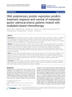
node metastasis in the PLV region (Fig. 1). Existing studies do not suggest a reference for the delineation of the
radiotherapy target area of the PLV region for NPC patients, and the prognosis of NPC patients with lymph
node metastasis in the PLV region is unclear [7–10].
In this study, we retrospectively studied the clinical
data of 605 patients with NPC, analyzed the extent of
cervical lymph node metastasis, and explored the
Page 2 of 9
distribution pattern and prognosis of metastatic lymph
nodes in the PLV region. Our study provides a useful
reference for the delineation of the radiotherapy target
area of the PLV region as well as a further revision of
the head and neck lymph node division and N stage.
Methods
Patient population
This study was approved by the Ethics Committee of
The General Hospital of Western Theater Command
and Sichuan Cancer Hospital and Institute. We retrospectively reviewed the records of 605 patients with
NPC from December 2011 to November 2017 in two
hospitals. All patients had been pathologically confirmed
as having NPC. Tumor stages and disease grades were
classified according to the 2017 edition for the staging of
NPC in China [11]. All patients underwent a CT crosssectional enhanced scan before treatment. The scan
range was cranial apex to 2 cm below the sternoclavicular joints, and the thickness of each slice was 3 mm.
Diagnostic criteria for cervical lymph node metastasis
All patients’ CT and MRI images were reviewed and
interpreted by two experienced radiological experts. The
criteria for neck lymph node levels are based on the
2013 updated consensus guidelines for neck lymph node
levels. The diagnostic criteria for cervical lymph node
metastasis were as follows: 1) minimal diameter of
lymph nodes on the largest cross-sectional image ≥10
mm [12]; 2) central necrosis or ring enhancement; 3) ≥ 3
lymph nodes in one high-risk area, and a minimum
diameter of the largest cross-section of ≥8 mm; 4) lymph
node extracapsular invasion, including irregular enhancement of the edge of the lymph node, partial or
total disappearance of the peripheral fat space, and
lymph node fusion; 5) a minimum diameter of the largest cross-section of the retropharyngeal lymph node of
≥5 mm; 6) a shrunken lymph node after radiotherapy
and chemotherapy. One of the above criteria can be
judged as an eligibility criterion.
Fig. 1 a. The PLV region in the CT scan. The PLV region is between the trapezius muscle and the scapular levator. The yellow line presents the
trapezius, and the red line presents the levator scapulae. b. Metastatic lymph nodes in the PLV region. A typical picture of the metastatic lymph
nodes in the PLV region is shown. The white arrows indicate the metastatic lymph nodes
Jiang et al. BMC Cancer
(2020) 20:667
Delineation of the center point of the lymph node at the
PLV
A case of N0 NPC was selected as the standard for a CT
simulation scan. According to its anatomical structure
and proportion, the central points of the metastatic
lymph nodes in the PLV region were outlined on a CT
image from the standard case. The central point is defined as the geometric center of the metastatic lymph
node. When an individual fused lymph node could not
be distinguished, the common geometric center of the
observed fused lymph nodes was drawn. The epicenter
of every node was contoured by marking the geometric
center with a pen tip with a diameter of 5 mm. A horizontal line was drawn on the anterior surface of the trapezius to measure the vertical distance between the
center point of each metastatic lymph node in the PLV
region and the horizontal line, and the vertical distance
of the fusion lymph nodes in the PLV region was measured between the common geometric center of the
lymph nodes and the horizontal line.
Treatment strategy and follow-up
Patients with stage I cancer received radiotherapy alone.
Patients with stage II ~ IVa cancer received radiotherapy
and chemotherapy with a cisplatin-based regimen. The
primary nasopharyngeal tumor and metastatic retropharyngeal lymph nodes were defined as GTVnx. Cervical metastatic lymph nodes were defined as GTVnd.
The clinical target volume (CTV1) was defined as a
high-risk area that included the GTVnx with a 5–10 mm
margin, the whole nasopharynx, GTVnd and the level II
and III cervical lymphatic drainage regions. CTV2 was
defined as a low-risk area that encompassed CTV1 and
the retropharyngeal lymph nodal regions, the base of
skull, the anterior half of clivus, the parapharyngeal
space, the pterygoid fossa, the inferior sphenoid sinus,
the posterior edge of the nasal cavity, the maxillary sinuses and the lower neck. CTV1 and CTV2 volumes
underwent a volumetric expansion of 3–5 mm to create
PTV1 and PTV2. The prescribed radiation doses of each
target volume were as follows: 66–72 Gy for GTVnx,
64–70 Gy for GTVnd, 56–64 Gy for PTV1, and 50–56
Gy for PTV2. All patients received IMRT and irradiated
with 1 fraction per day, 5 days per week, for a total of
30–33 fractions. The radiation dose limits for critical
structures were within the tolerance recommended by
the RTOG 0225 protocol. All patients were followed up
by hospitalization, outpatient visits, and telephone enquiries until December 2018. Follow-up examinations
included the following: nasopharyngeal and cervical
MRI, fiber nasopharyngoscopy, abdominal ultrasound,
and chest CT.
Page 3 of 9
Statistical analysis
All data were analyzed using the SPSS 20.0 software.
Linear regression was performed to identify the correlation between the PLV region and the remaining lymph
node levels. The Kaplan-Meier method was employed to
calculate the survival rate, and the log-rank method was
used to compare survival curves between groups. A Cox
hazard model with enter method was used to perform
multivariate analysis. Overall survival (OS), progressionfree survival (PFS), local recurrence-free survival (LRFS),
and distant metastasis-free survival (DMFS) were analyzed. P values of less than 0.05 were considered statistically significant.
Results
Patient characteristics and prognosis
Of the 605 patients with pathologically confirmed NPC,
433 were males and 172 were females. The median age
of the patients was 48 years old (12–81 years old),
97.52% of the pathological types were WHO II-III, and
2.47% of the pathological types were WHO type I. The
counts and percentages of patients with T1, T2, T3, and
T4 NPC were 156 (25.78%), 120 (19.83%), 161 (26.61%),
and 168 (27.76%), respectively; and the counts and percentages of patients with N0, N1, N2, and N3 NPC were
48 (7.93%) and 165 (27.27%), 303 (50.08%), 89 (14.71%),
respectively. The number and percentages of patients in
stages I, II, III, IVa and IVb were 22 (3.63%), 86
(14.21%), 250 (41.32%), 237 (39.17%) and 10 (1.65%), respectively (Table 1). In all patients, 10 patients were in
stage IVb, and 4 patients gave up treatment during
radiotherapy. A total of 591 patients were followed up
for 8–81 months with a median of 37 months, and the 5year OS, PFS, LRFS, and DMFS were 80.1, 69.4, 88.4,
and 83.9%, respectively. Seventy-four patients died, and
the main cause of death was distant metastasis, followed
by local recurrence and hemorrhage of the nasopharynx.
Forty-two cases had local recurrence, mainly in the
nasopharyngeal cavity, skull base bone, carotid sheath
area, intracranial cavernous sinus area, etc. Seventy-five
cases had distant metastasis, most commonly in the
liver, lungs and bones. Patients with a single metastasis
site were rare, and most patients had two or three sites
with simultaneous metastasis.
Cervical lymph node metastasis
In the 605 patients, 557 patients (92.06%) had cervical
lymph node metastasis (Supplementary Table 1). The
top four levels with the highest probability of lymph
node metastasis were IIb (77.85%), VIIa (73.05%), IIa
(60.0%), and III (41.48%). The levels with less than a 5%
probability of the lymph node metastasis was IVb
(1.98%), Vc (1.48%), VIIb (0.82%), and VIII (0.49%), and
no lymph node metastasis was found in levels Ia, VI, IX
Jiang et al. BMC Cancer
(2020) 20:667
Page 4 of 9
Table 1 Patient characteristics
Characteristic
Number of patients (n)
Gender
Male
433 (71.57%)
Female
172 (28.42%)
Age (years)
< 45
209 (34.54%)
≥ 45
396 (65.45%)
Histology
WHO I
15 (2.47)
WHO II- III
590 (97.52)
T stage
T1
156 (25.78%)
T2
120 (19.83%)
T3
161 (26.61%)
T4
168 (27.76%)
short diameter of less than 10 mm, 22 metastatic lymph
nodes with a short diameter of 11–20 mm, and 4 metastatic lymph nodes with a short diameter of 21–30 mm.
The median vertical distance of the center point of each
metastatic lymph node from the anterior surface of the
trapezius muscle in the standard NPC patient was 14
mm (3–37 mm). There were 25 lymph nodes with a vertical distance of less than 10 mm, 14 lymph nodes with a
vertical distance between 11 and 20 mm, 7 lymph nodes
with a vertical distance between 21 and 30 mm, and 3
lymph nodes with a vertical distance of more than 31
mm. The centers of 93.87% (46/49) of the metastatic
lymph nodes in the PLV region were located less than
25 mm from the anterior surface of the trapezius muscle.
The distribution of the metastatic lymph nodes in the
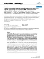
PLV region is shown in Supplementary Table 3. The location of the corresponding CT layer of the standard
NPC patient is shown in Fig. 2.
N stage
N0
48 (7.93%)
N1
165 (27.27%)
N2
303 (50.08%)
N3
89 (14.71%)
TNM stage
I
22 (3.63%)
II
86 (14.21%)
III
250 (41.32%)
IVa
237 (39.17%)
IVb
10 (1.65%)
and X. There were 12 patients with lymph node metastasis in level IVb, and these patients also had lymph
node metastasis in the level IVa. Nine patients had
lymph node metastasis in level Vc, and these patients
were also accompanied by lymph node metastasis in
level Vb. Three patients with lymph node metastasis in
level VIII were associated with lymph node metastasis in
levels Ib, II, and III, and one patient with lymph node
metastasis in level II showed local necrosis and lymph
node fusion.
Distribution characteristics of metastatic lymph nodes in
the PLV region
In the whole group of 605 patients, 30 patients (4.95%)
showed lymph node metastasis in the PLV region
(Supplementary Table 2). There was a total of 49 metastatic lymph nodes, including 25 metastatic lymph nodes
in the left neck and 24 metastatic lymph nodes in the
right neck. In one patient, lymph node metastasis in the
PLV region occurred simultaneously on both sides of
the neck. There were 23 metastatic lymph nodes with a
Correlation analysis of lymph node metastasis in the PLV
region
To analyze the relationship between lymph node metastasis in the PLV region and other cervical lymph node
metastasis, linear regression analysis was used. The
lymph node metastasis in the PLV region was used as
the dependent variable, and the remaining lymph node
regions were included as independent variables in the
analysis. The results showed that the lymph node metastasis of the PLV region was associated with the ipsilateral IVa (P = 0.018), Va, Vb and Vc levels (all P < 0.001),
and no correlations were found for the other variables
(Table 2).
Prognosis of patients with lymph node metastasis in the
PLV region
The number of patients with lymph node metastasis in
PLV region was 30, but 1 patient with stage IVB was excluded and we followed up 29 patients. A total of 29 patients with lymph node metastasis in PLV were followed
up for a median of 21 (4 to 60) months. Fourteen patients had distant metastasis, 11 patients died during the
follow-up period (death overlapped with distant metastasis), and 2 patients relapsed. The 5-year OS, PFS, LRFS,
and DMFS were 41.6, 27.7, 89.1, and 47.3%, respectively.
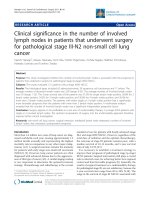
Prognosis of patients with N3 NPC with or without lymph
node metastasis in the PLV region
The number of patients with N3 was 89, but 3 patients
with N3 in stage IVB were excluded, so the cases of patients with N3 NPC with or without lymph node metastasis in the PLV region were 26 and 60, respectively. The
5-year OS, PFS, LRFS, and DMFS of the two groups
were 41.8 and 67.3% (P = 0.007), 27.8 and 48.5% (P =
Jiang et al. BMC Cancer
(2020) 20:667
Page 5 of 9
Fig. 2 Schematic diagram of the distribution of metastatic lymph nodes in the PLV region. The blue line is the horizontal line through the
anterior surface of the trapezius, which was used to calculate the vertical distance between the central points of the metastatic lymph nodes in
the PLV region and the horizontal line. The light blue line is the delineation of the PLV region with the ipsilateral metastatic lymph nodes of
levels Va, Vb, and Vc. The green circle indicates some metastatic lymph nodes of the PLV region that were recreated proportionally on the CT
images of a standard patient to further understand the location of their distribution
0.005), 92.3 and 80.5% (P = 0.521), and 40.6 and 78.4%
(P < 0.001), respectively (Table 3, Fig. 3).
Univariate and multivariate analysis
The univariate results showed gender and age were
prognostic factors for 5-year OS (all P < 0.05), T-stage
was a prognostic factor for 5-year LRFS(P = 0.003), The
N-stage and TNM stage were prognostic factors for 5year OS, PFS, LRFS, and DMFS (all P < 0.05), Involvement of lower neck was a prognostic factor for 5-year
OS, PFS, and DMFS (all P < 0.001) (Table 4). Involvement of lower neck was refined into levels IVa, IVb, Vb,
Table 2 The data of linear-regression analysis for neck node
Variable
B
Std.Error
t
p
95% CI for B
Ib
0.014
0.036
0.374
0.708
−0.057-0.085
IIa
−0.004
0.016
−0.228
0.820
−0.035-0.028
IIb
− 0.004
0.019
− 0.224
0.823
− 0.042-0.034
III
−0.012
0.018
0.669
0.504
−0.047-0.023
IVa
0.062
0.026
2.366
0.018
0.011–0.114
IVb
0.106
0.059
1.776
0.076
−0.011-0.222
Va
0.118
0.021
5.729
0.000
0.078–0.159
Vb
0.243
0.038
6.391
0.000
0.168–0.318
Vc
0.435
0.071
6.112
0.000
0.295–0.574
VIIa
0.017
0.016
1.041
0.298
−0.015-0.048
VIIb
0.001
0.077
0.017
0.987
−0.151-0.153
VIII
−0.162
0.101
−1.597
0.111
−0.360-0.037
Abbreviations: B regression coefficient, t t test, Dependent Variable: PLV
and Vc and the PLV region in multivariate analysis, and
the parameters were designed as two categorical variables (Table 5). Analysis showed that lymph node metastasis in the PLV region was an independent prognostic
factor for DMFS (P = 0.044) rather than for OS and PFS
(Table 5, Supplementary Table 4 and 5).
Discussion
Nasopharyngeal carcinoma (NPC) is very prone to
lymph node metastasis [13]. Some studies have reported
that approximately 40% of patients with NPC have a first
symptom of cervical lymphadenopathy [14]. Approximately 60 to 90% of patients with newly diagnosed NPC
have lymph node metastasis [15]. Therefore, the segmentation and metastatic characteristics of cervical
lymph nodes are of great importance for the delineation
of target areas of radiotherapy. In the 2013 Guideline,
level V of cervical lymph nodes is refined into levels Va,
Vb, and Vc [6]. However, the 2013 Guideline did not describe the PLV gap (the gap between the trapezius
muscle and the scapular levator). In the PLV region,
Table 3 The survival data of N3 patients with or without node
metastasis of PLV
Variable
5-year OS
%
P
5-year PFS
%
P
5-year LRFS
%
PLV (+) (n = 26) 41.8 0.007 27.8 0.005 92.3
PLV (−) (n = 60) 67.3
48.5
80.5
P
5-year DMFS
%
P
0.521 40.6 < 0.001
78.4
Abbreviations: PLV (+) node metastasis with posterior to level V, PLV (−) node
metastasis without posterior to level V
Jiang et al. BMC Cancer
(2020) 20:667
Page 6 of 9
Fig. 3 The 5-year survival curve of patients with N3 NPC with or without lymph node metastasis in the PLV region. a OS (P = 0.007), b PFS (P = 0.005),
c LRFS (P = 0.521), d DMFS (P < 0.001)
metastatic lymph nodes can be found. Unfortunately,
this PLV region is often overlooked. The delineation of
the radiotherapy target area for the PLV region and the
prognosis of NPC patients with lymph node metastasis
in the PLV region are still unknown.
Previous studies have shown that the rate of lymph
node metastasis is not high in the PLV region; the metastasis rates in the studies [16–18] were 1.1, 2.4 and
2.5%, respectively. In this study, we found that in the
whole group of 605 patients, there were 30 patients with
lymph node metastasis in the PLV region, and the metastasis rate was 4.95%, slightly higher than in the abovementioned studies, which may be related to the
relatively low level of awareness of the disease in patients
in western China. In this study, we also found 49 metastatic lymph nodes in the PLV region. Most of the central points of these metastatic lymph nodes were located
25 mm from the anterior surface of the trapezius muscle,
and metastatic lymph nodes were located in both the
trapezius and scapular levator muscles. Further analysis
showed that 132 patients had lymph node metastasis in
level Va, including 30 patients with lymph node metastasis in the PLV region. At the same time, we also found
that all lymph node metastasis in the PLV region were
associated with ipsilateral lymph node metastasis in level
Va. Correlation analysis suggested that the lymph node
metastasis in the PLV region was also associated with
lymph node metastasis in levels IVa, Va, Vb and Vc.
Therefore, when NPC patients present with lymph node
metastasis especially in level Va, Vb, and Vc but not in
the PLV region, it is recommended that the target delineation of posterior boundary of the ipsilateral V region
be appropriately moved back to 25 mm behind the anterior surface of the trapezius muscle to prevent lymph
node metastasis (Fig. 2); when NPC patients present
without lymph node metastasis in these areas, the posterior border of the level V region can be outlined to the
anterior surface of the trapezius muscle.
A previous study showed that 5-year OS, PFS, LRFS,
and DMFS rates in NPC patients were 77.1, 69.6, 89.8,
Jiang et al. BMC Cancer
(2020) 20:667
Page 7 of 9
Table 4 Univariate analysis of prognosis in 591 NPC patients
Variable
N
5-year OS (%)
Male
423
78.0
Female
168
85.6
< 45
205
86.3
≥ 45
386
76.8
T1 + T2
271
82.4
T3 + T4
320
77.2
N0 + N1
211
86.6
N2 + N3
380
73.9
I + II
108
89.6
III + IVa
483
76.7
p
p
5-year PFS (%)
5-year LRFS (%)
p
5-year DMFS (%)
89.0
0.619
82.2
p
Gender
0.04
67.5
0.194
74.1
87.2
0.328
87.8
Age (years)
0.039
72.0
0.463
67.9
87.3
0.744
84.9
89.0
0.846
83.2
T stage
0.28
72.8
0.096
62.4
93.1
0.003
85.2
82.9
0.802
81.7
N stage
0.004
79.8
< 0.001
58.8
91.7
0.025
90.4
86.0
0.002
78.9
TNM stage
0.006
84.4
< 0.001
64.6
94.9
0.014
92.3
86.3
0.015
81.1
Involvement of lower neck levels
Yes
90
57.6
No
501
83.5
< 0.001
41.7
< 0.001
74.2
82.3
0.07
68.7
89.3
and 74.1%, respectively [19]. Another study showed 5year OS, PFS, LRFS, and DMFS rates in NPC patients of
83.3, 76.3, 92.7, 85.5%, respectively [20]. This finding is
similar to that of our study, which also showed that the
5-year OS, PFS, LRFS, and DMFS of patients with lymph
node metastasis in the PLV were 41.6, 27.7, 89.1, and
47.3%, respectively. Moreover, patients with N3 NPC accompanied by lymph node metastasis in the PLV region
have a worse prognosis. Multivariate analysis showed
that lymph node metastasis in the PLV region was an independent prognostic factor for DMFS. This further indicates that the prognosis of NPC patients with PLVregion lymph node metastasis is poor, and lymph node
metastasis in this region indicates a high risk of distant
metastasis. For patients with PLV metastasis, on the one
hand, the delineation scope of the radiotherapy target
< 0.001
86.7
should be expanded, on the other hand, because of the
increased risk of distant metastasis (Table 3), it is necessary to strengthen the systemic treatment.
This study has the following limitations: first, a pathological diagnosis of metastatic lymph nodes is lacking,
especially in the deep fat gap of the PLV region, and performing histopathology in this region is difficult. Second,
since the measurement of the vertical distance was performed on a standard patient, the position of the frontal
area of the trapezius muscle may be different depending
on the weight, age, and body type of different patients.
To minimize these variations, we tried to recreate the
position of the central point of each lymph node on the
CT images of the standard patient. Third, there is a difference in the fat space between the trapezius muscle
and the levator scapulae of in different patients. To
Table 5 Multivariate analysis of DMFS in 591 NPC patients
B
SE
P
HR
95%CI
level IVa
0.241
0.358
0.501
1.273
0.631–2.568
level IVb
0.909
0.564
0.107
2.481
0.821–7.500
level Vb
0.660
0.460
0.151
1.934
0.786–4.763
level Vc
0.102
0.650
0.875
1.107
0.310–3.956
PLV
0.925
0.460
0.044
2.522
1.023–6.214
N stage (N0 + 1 vs. N2 + 3)
−0.296
0.360
0.410
0.744
0.367–1.506
TNM stage (I + II vs. III + IVa)
−0.390
0.475
0.411
0.677
0.267–1.716
Variable
Involvement of lower neck levels (yes vs. no)
Jiang et al. BMC Cancer
(2020) 20:667
clearly show the fat gap between the trapezius muscle
and the scapular levator muscle, we selected a patient
with a wide gap as the standard patient.
Conclusion
To the best of our knowledge, in this study, we report
for the first time the distribution and metastasis of
lymph nodes in the PLV region of NPC patients and
provide a reference for the delineation of the lymph
node area in the PLV region. In addition, we found that
the metastasis rate of lymph nodes in the PLV region
was 4.95%, which was close to or even exceeded the metastasis rate in the Ib, IVb, Vb, and Vc regions. However,
the 2013 Guideline did not fully consider the PLV region, which may be a missing neck segment. Therefore,
we propose to use the PLV region as a new cervical
lymph node level as follows: cranial boundary: the caudal
edge of the cricoid cartilage; caudal boundary, the level
of the clavicle or serratus anterior muscle; the anterior
boundary: the anterior surface of the trapezius muscle;
posterior boundary: the intersection of the trapezius
muscle and the scapula levator; medial boundary: the
lateral edge of the scapular levator; and lateral boundary:
the medial edge of the trapezius muscle.
Supplementary information
Supplementary information accompanies this paper at />1186/s12885-020-07146-z.
Additional files 1: Supplementary Table 1. Patterns of cervical nodal
metastasis of nasopharyngeal carcinoma
Additional files 2: Supplementary Table 2. Baseline characteristics of
patients with metastasis of posterior to level V
Additional files 3 Supplementary Table 3. Patterns of cervical nodal
metastasis of posterior to level V
Additional files 4: Supplementary Table 4. Multivariate analysis for
OS in 591 NPC patients
Additional files 5: Supplementary Table 5. Multivariate analysis for
PFS in 591 NPC patients
Abbreviations
PLV: Posterior to level V; NPC: Nasopharyngeal carcinoma; RTOG: Radiation
Therapy Oncology Group; CTV: Clinical target volume; OS: Overall survival;
PFS: Progression-free survival; LRFS: Local recurrence-free survival;
DMFS: Distant metastasis-free survival
Acknowledgements
None.
Authors’ contributions
CYJ, JM and BSL, study concept and design, acquisition of data, analysis and
interpretation of data, statistical analysis and project funding. JM, drafting
and revising of the manuscript. HG, LZ, TZ and HL, acquisition of data and
material support. All authors read and approved the final manuscript.
Funding
This research was supported by China Postdoctoral Science Foundation
(2017 M613430 and 2018 T111158 to JM) and Key Research and
Development Project of Sichuan Province (2020YFS0273 to JM). The funder
had role in designing of the study and drafting of the manuscript.
Page 8 of 9
Availability of data and materials
The datasets used and/or analysed during the current study are available
from the corresponding author on reasonable request.
Ethics approval and consent to participate
This study was approved by the Ethics Committee of The General Hospital of
Western Theatre Command and Sichuan Cancer Hospital and Institute. The
written informed consent was obtained from all participants.
Consent for publication
Not Applicable.
Competing interests
The authors declare that they have no competing interests.
Author details
Department of Radiation Oncology, The General Hospital of Western
Theater Command, Chengdu, Sichuan Province 610083, PR China.
2
Department of Medical Oncology, West China Hospital, Sichuan University,
Chengdu, Sichuan Province 610041, PR China. 3Department of Radiation
Oncology, Sichuan Cancer Hospital and Research Institute, University of
Electronic Science and Technology of China, Chengdu, Sichuan Province
610041, PR China.
1
Received: 3 March 2020 Accepted: 7 July 2020
References
1. Chen YP, Chan A, Le QT, Blanchard P, Sun Y, Ma J. nasopharyngeal
carcinoma. Lancet. 2019;394:64–80.
2. Chua MLK, Wee JTS, Hui EP, Chan ATC. Nasopharyngeal carcinoma. Lancet.
2016;387:1012–24.
3. Sun XS, Li XY, Chen QY, Tang LQ, Mai HQ. Future of radiotherapy in
nasopharyngeal carcinoma. Br J Radiol. 2019;92:20190209.
4. Guo R, Mao YP, Tang LL, Chen L, Sun Y, Ma J. The evolution of
nasopharyngeal carcinoma staging. Br J Radiol. 2019;92:20190244.
5. Lee AW, Ma BB, Ng WT, Chan AT. Management of Nasopharyngeal
Carcinoma: current practice and future perspective. J Clin Oncol. 2015;33:
3356–64.
6. Grégoire V, Ang K, Budach W, et al. Delineation of the neck node levels for
head and neck tumors: a 2013 update. DAHANCA, EORTC, HKNPCSG, NCIC
CTG, NCRI, RTOG, TROG consensus guidelines. Radiother Oncol. 2014;110:
172–81.
7. Grégoire V, Levendag P, Ang KK, et al. CT-based delineation of lymph node
levels and related CTVs in the node-negative neck: DAHANCA, EORTC,
GORTEC, NCIC, RTOG consensus guidelines. Radiother Oncol. 2003;69:227–36.
8. Grégoire V, Eisbruch A, Hamoir M, Levendag P. Proposal for the delineation
of the nodal CTV in the node-positive and the post-operative neck.
Radiother Oncol. 2006;79:15–20.
9. Lee AW, Ng WT, Pan JJ, et al. International guideline for the delineation of
the clinical target volumes (CTV) for nasopharyngeal carcinoma. Radiother
Oncol. 2018;126:25–36.
10. Vorwerk H, Hess CF. Guidelines for delineation of lymphatic clinical target
volumes for high conformal radiotherapy: head and neck region. Radiat
Oncol. 2011;6:97.
11. Chinese Committee for Staging of Nasopharyngeal Carcinoma. The 2017
edition for staging of nasopharyngeal carcinoma in China (the Chinese
2008 expert). Chin J Radiat Oncol. 2017;26:1119–25.
12. van den Brekel MW, Stel HV, Castelijns JA, et al. Cervical lymph node
metastasis: assessment of radiologic criteria. Radiology. 1990;177:379–84.
13. Ho FC, Tham IW, Earnest A, Lee KM, Lu JJ. Patterns of regional lymph node
metastasis of nasopharyngeal carcinoma: a meta-analysis of clinical
evidence. BMC Cancer. 2012;12:98.
14. Lee HM, Okuda KS, González FE, Patel V. Current perspectives on
nasopharyngeal carcinoma. Adv Exp Med Biol. 2019;1164:11–34.
15. Sharma M, Bartlett E, Yu E. Metastatic retropharyngeal lymph nodes in
nasopharyngeal carcinoma: imaging criteria. Expert Rev Anticancer Ther.
2010;10:1703–6.
16. Wang X, Hu C, Ying H, et al. Patterns of lymph node metastasis from
nasopharyngeal carcinoma based on the 2013 updated consensus
guidelines for neck node levels. Radiother Oncol. 2015;115:41–5.
Jiang et al. BMC Cancer
(2020) 20:667
17. Ding ZX, Liang BL, Shen J, et al. Value of radiation therapy oncology group
guidelines base on MR imaging in diagnosing lymph node metastasis of
nasopharyngeal carcinoma. Chin J of Cancer. 2009;28:533–7.
18. Sun Y, Ma J, Lu TX, et al. Regulation for distribution of metastatic cervical
lymph nodes of 512 cases of nasopharyngeal carcinoma. Chin J of cancer.
2004;23:1523–7.
19. Feng M, Fan ZX, Li J, et al. Long-term results and prognostic factors in 582
nasopharyngeal carcinoma treated by intensity-modulated radiotherapy.
Chin J Radiat Oncol. 2011;20:369–73.
20. Zhao C, Xiao WW, Han F, et al. Long-term outcome and prognostic factors
of patients with nasopharyngeal carcinoma treated with intensitymodulated radiation therapy. Chin J Radiat Oncol. 2010;19:191–6.
Publisher’s Note
Springer Nature remains neutral with regard to jurisdictional claims in
published maps and institutional affiliations.
Page 9 of 9