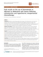
Use of the QIAGEN GeneReader NGS system for detection of KRAS mutations, validated by the QIAGEN Therascreen PCR kit and alternative NGS platform
Bạn đang xem bản rút gọn của tài liệu. Xem và tải ngay bản đầy đủ của tài liệu tại đây (1.25 MB, 8 trang )
Darwanto et al. BMC Cancer (2017) 17:358
DOI 10.1186/s12885-017-3328-z
RESEARCH ARTICLE
Open Access
Use of the QIAGEN GeneReader NGS
system for detection of KRAS mutations,
validated by the QIAGEN Therascreen PCR
kit and alternative NGS platform
Agus Darwanto1,7 , Anne-Mette Hein2, Sascha Strauss3, Yi Kong4, Andrew Sheridan1, Dan Richards4, Eric Lader5,
Monika Ngowe1,8, Timothy Pelletier1, Danielle Adams1,9, Austin Ricker1, Nishit Patel1, Andreas Kühne3,
Simon Hughes6, Dan Shiffman4, Dirk Zimmermann3, Kai te Kaat3 and Thomas Rothmann3*
Abstract
Background: The detection of somatic mutations in primary tumors is critical for the understanding of cancer
evolution and targeting therapy. Multiple technologies have been developed to enable the detection of such
mutations. Next generation sequencing (NGS) is a new platform that is gradually becoming the technology of
choice for genotyping cancer samples, owing to its ability to simultaneously interrogate many genomic loci at
massively high efficiency and increasingly lower cost. However, multiple barriers still exist for its broader adoption
in clinical research practice, such as fragmented workflow and complex bioinformatics analysis and interpretation.
Methods: We performed validation of the QIAGEN GeneReader NGS System using the QIAact Actionable Insights Tumor
Panel, focusing on clinically meaningful mutations by using DNA extracted from formalin-fixed paraffin-embedded (FFPE)
colorectal tissue with known KRAS mutations. The performance of the GeneReader was evaluated and compared to data
generated from alternative technologies (PCR and pyrosequencing) as well as an alternative NGS platform. The results
were further confirmed with Sanger sequencing.
Results: The data generated from the GeneReader achieved 100% concordance with reference technologies.
Furthermore, the GeneReader workflow provides a truly integrated workflow, eliminating artifacts resulting from
routine sample preparation; and providing up-to-date interpretation of test results.
Conclusion: The GeneReader NGS system offers an effective and efficient method to identify somatic (KRAS) cancer
mutations.
Keywords: GeneReader, Kras, Mutation, Cancer, Ngs
Background
Somatic mutations in the KRAS oncogene are common in
human cancers. They are found in 70-90% of pancreatic
cancers [1, 2], 30-50% of colorectal cancers [3–5] and 1030% of Non-Small Cell Lung Cancers (NSCLC) [6–8].
Several methods have been developed for the detection of
KRAS mutations, each with specific advantages and
limitations [5, 9, 10].
* Correspondence:
3
QIAGEN GmbH, QIAGEN Strasse 1, 40724 Hilden, Nordrhein-Westfalen,
Germany
Full list of author information is available at the end of the article
Sanger sequencing has been the ‘gold standard’ for
mutation analysis in cancer detection since the 1970s
[11]. However, limited by its low sensitivity (10-20%
mutant allele frequency (MAF)) and low throughput
[10], Sanger sequencing is no longer sufficient for the
needs of today’s cancer molecular diagnostics.
The therascreen KRAS RGQ PCR kit is a real-time
qPCR-based assay used to detect the most common KRAS
mutations including those in codons 12 and 13. It has
greatly improved sensitivity over Sanger sequencing, and
has been approved by the Food and Drug Administration
(FDA) [9] for colorectal cancer patient stratification.
© The Author(s). 2017 Open Access This article is distributed under the terms of the Creative Commons Attribution 4.0
International License ( which permits unrestricted use, distribution, and
reproduction in any medium, provided you give appropriate credit to the original author(s) and the source, provide a link to
the Creative Commons license, and indicate if changes were made. The Creative Commons Public Domain Dedication waiver
( applies to the data made available in this article, unless otherwise stated.
Darwanto et al. BMC Cancer (2017) 17:358
Pyrosequencing also offers an attractive alternative to
Sanger due to its fast turnaround time (TAT) and lower
sensitivity threshold, even in tissues with low tumor cell
content [5].
Next-generation sequencing (NGS) differs radically
from the above mentioned methods. Coupled with
amplicon-based targeting technology, NGS has the capability to simultaneously sequence in a massively parallel
way multiple genetic loci with minimal amounts of nucleic acid input and limited time and expense [12–15].
This technology has revolutionized the speed of genetic
and genomic discovery, and advanced our understanding
of molecular mechanisms of diseases. In recent years,
NGS has played an important role in advancing personalized healthcare and precision medicine by enabling the
identification of mutations associated with therapeutic
response or resistance. As more clinically significant
genetic biomarkers and targeted therapies become available, the profiling of such genetic variations is becoming
increasingly more critical. Several NGS platforms are
already commercially available for sequencing and identification of genetic alterations associated with diseases,
such as point mutations, deletions, insertions and copy
number variants [16]. However, QIAGEN’s GeneReader
System presented here includes all upstream sample
processing steps starting from nucleic acid extraction,
together with an integrated downstream bioinformatics
solution that enables a direct access to real-time updates
from the rapidly evolving literature, and clinical knowledge
and evidence.
To this end, we recently evaluated the QIAGEN
GeneReader System workflow from DNA extraction and
purification from FFPE tissue samples, to library preparation, sequencing and data analysis and interpretation.
Herein we show that the GeneReader presents a unified
workflow that provides accurate results and a simple
solution for any laboratory to use in clinical research.
Methods
Sample and DNA isolation
FFPE Tumor material from colorectal cancer tumors
(Origene Technologies, MD, USA and Asterand Biosciences, MI, USA) was used to prepare 56 DNA samples
with known KRAS mutation status, previously determined
using therascreen assay (Pyrosequencing and PCR) and
Sanger sequencing according to methods further described
below. Tissue sections of 10 μm in thickness, ranging from
3 to 20 years of age were used for DNA extraction utilizing
either: i) the QIAamp DNA FFPE Tissue Kit (QIAGEN,
Hilden, Germany) or ii) the GeneRead FFPE DNA Kit
(QIAGEN, Hilden, Germany) according to manufacturer’s
instructions. DNA concentration was determined using
the Nanodrop System (Thermo Fisher Scientific, MA,
USA) and Qubit dsDNA HS assay (Life Technologies,
Page 2 of 8
Gaithersburg, USA). The DNA was assessed using the
GeneRead DNA QuantiMIZE System (QIAGEN, Hilden,
Germany) which utilizes a qPCR-based approach to determine the quality of sample DNA prior to NGS. Furthermore, both NA12878 (Coriell Institute for Medical
Research) (for which the Genome in the Bottle (GIAB)
consortium has published a set of high confident variants [17]) and AcroMetrix (Thermo Fisher Scientific,
MA, USA) samples were used as a gold standard set of
variant calls.
GeneReader sample preparation and sequencing run
In total, 40 ng of DNA measured by Qubit (Thermo
Fisher Scientific, MA, USA) was used as template to
generate libraries for sequencing. Libraries were prepared using the QIAGEN Library Kit v2.0 and the
GeneRead QIAact Actionable Insight Tumor Panel
(QIAGEN, Hilden, Germany), which amplifies 330
amplicons covering 16.7 kb, containing 773 unique
variant positions in 12 genes (KRAS, NRAS, KIT, BRAF,
PDGFRA, ALK, EGFR, ERBB2, PIK3CA, ERBB3, ESR1
and RAF1). All steps of library preparation were performed according to the manufacturer’s protocol. The
libraries were then quantified using a Qubit dsDNA HS
Assay Kit (Life Technologies, MA, USA) and QIAxcel
(QIAGEN, Hilden, Germany). Ten individual libraries
were pooled prior to emulsion PCR and bead enrichment steps that were carried out using an automated
protocol on the GeneRead QIAcube (QIAGEN, Hilden,
Germany) using the GeneRead Clonal Amp Q Kit
(QIAGEN, Hilden, Germany), according to the manufacturer’s protocol. Following bead enrichment, the
pooled libraries were sequenced using the GeneReader
platform (QIAGEN, Hilden, Germany).
GeneReader data processing
QIAGEN Clinical Insight (QCI™) Analyze software
(QIAGEN, Hilden, Germany) was used to QC, align the
read data to the hg19 reference genome sequence, call
sequence variants, and generate an interactive report
for visualization of the sequencing results, as well as a
summary of the data. QCI Analyze software reports a
set of high- and low-confidence variants based on the
coverage of variant positions. Users have an option to
analytically confirm if a variant listed should be valid or
invalid before uploading to QCI Interpret software for
the clinical interpretation. For each sample the report
was used to assess the quality of the overall sequencing
run and to identify/call the individual variants. After
review, variants confirmed as analytically valid were
uploaded to QCI Interpret for creation of a report for
each sample based on detected variants and curated
content, with a summary of findings and direct links to
evidence sources.
Darwanto et al. BMC Cancer (2017) 17:358
Illumina MiSeq
The Actionable Insight Tumor Panel (QIAGEN, Hilden,
Germany) was used for a MiSeq (Illumina, CA, USA)
sequencing run. The Kapa “with bead” PCR free protocol
(KAPABiosystems, MA, USA) was used in further Illumina
library preparation steps. Samples were then paired-end
sequenced on a MiSeq instrument (Illumina, CA, USA)
according to Illumina guidelines. The resulting reads were
mapped to the hg19 reference genome sequence using
BWA mem software followed by GATK (best practices) to
recalibrate base quality scores. Variants were called using
MuTect. Variants were then filtered using GATK (best
practice) and annotated using SnpEff. Variants at hotspot
positions were selected using GATK.
Pyrosequencing and Sanger analyses
The sample DNA obtained with the QIAamp FFPE
DNA Kit (QIAGEN, Hilden, Germany) was subjected to
Pyrosequencing analysis and Sanger sequencing. For
Pyrosequencing the samples were analyzed using the
therascreen RAS Extension Pyro Kit (QIAGEN, Hilden,
Germany) which covers mutations in KRAS codons 59,
61, 117 and 146 as well as NRAS codons 59, 117 and
146. Samples with mutations in KRAS or NRAS codons
12 and 13 were further analyzed with the therascreen
KRAS or NRAS Pyro Kit (QIAGEN, Hilden, Germany)
according to manufacturer’s instructions. In addition,
samples that failed the initial PyroMark KRAS analysis
were subjected to a second round of analysis. Samples
with an initial “check” status, or with an indicated mutation signal of LOD + 3% (“Potential low level mutation”)
were subjected to a second round of analysis performed
in duplicate. Sanger sequencing was performed using
Big Dye Terminator Technology and an ABI 3730xl
sequencer (Thermo Fisher Scientific, MA, USA). Mutations were detected by analyzing the sequence trace files
and the quantity of a base at a certain position was
calculated from the area under the curve (AUC) at the
mutation specific position in the electropherogram.
Therascreen qPCR
The therascreen KRAS RGQ PCR Kit (QIAGEN, Hilden,
UK) is an allele-specific PCR-based technology with specific primers for the seven most common KRAS codon
12 and 13 mutations. The assay screens for the following
mutations: 12 GCT (Ala), 12 GAT (Asp), 12 CGT (Arg),
12 TGT (Cys), 12 AGT (Ser), 12 GTT (Val), and 13
GAC (Asp). Mutation analysis was performed according
to manufacturer’s instructions, using the RotorGene
real-time PCR instrument (QIAGEN, Hilden, UK). Analysis of results was performed following the recommendations in the manual, e.g. samples with a control assay
with a cycle threshold (Ct) of 35 or higher were deemed
invalid and excluded from the analysis. Samples were
Page 3 of 8
called mutation positive based on the delta Ct values
reported in the handbook. Values over 40 cycles were
scored as negative (wild-type).
Results
Evaluation of DNA quality by QuantiMIZE
FFPE samples with ages ranging from 3 to 20 years were
used for this study. The quality of the extracted DNA was
measured by the GeneRead DNA QuantiMIZE QC assay
(QIAGEN, Hilden, UK). Thirteen out of 56 samples failed
quality checks and were excluded from further analysis
(Additional file 1: Table S1). For the remaining 43 samples, 3 to 9 PCR cycles were added (depending on the
QuantiMIZE quality scores) to compensate for differences
in DNA quality during enrichment PCR. The additional
cycles ensured that poor quality (highly fragmented) DNA
samples yielded enough material for downstream library
preparation. The quality of DNA purified from formalin
fixated tissue decreases over the sample storage period
time [18–20], but also depends on how tissues were
treated, handled and processed before and during sample
fixation [19, 21, 22].
GeneReader sequencing performance
The QIAact Actionable Insights Tumor Panel (QIAGEN,
Hilden, UK) contains 773 unique variant positions in 12
genes (Table 1). An analysis of the reads mapped to the
reference showed coverage levels that met the industrystandard 5% sensitivity criteria, even with aged FFPE
samples. A 200× minimum read coverage cutoff was
used for calling a variant at any position in the panel.
For the 43 FFPE samples analyzed, an average amplicon
coverage of 97.2% was observed, and an average variant
insight coverage (hotspot coverage) of 99.8% was observed at read depths ≥200× (Table 1). For NA12878
samples, an average amplicon coverage of 98.5% was
observed and an average variant insight coverage of
99.9% was observed at read depths of ≥200× (Table 1).
Table 1 Parameter and sequencing coverage of Actionable
Insight Tumor Panel
Parameter
Details
Panel size
12 genes/16.7 kb
Insight size
773 unique variant positions
Amplicons
330
Variant allele fraction detection limit
5%
Frequency cut-off and amplicon coverage
>500×: 96.4% (A), 92.0% (B)
>200×: 98.5% (A), 97.2% (B)
Frequency cut-off and variant insight
coverage
>500×: 99.8% (A), 98.6% (B)
>200×: 99.9% (A), 99.8% (B)
Positive samples included into the study have all been confirmed with Sanger
sequencing and passed QuantiMIZE (<0.4). (A) An average of 12 NA12878
samples, (B) average of 43 colorectal cancers FFPE samples (ages 3-20 years)
Darwanto et al. BMC Cancer (2017) 17:358
Page 4 of 8
No false negatives (FN; where an expected variant was
not detected) were observed.
Performance comparison between the QIAamp and
GeneRead DNA FFPE kits for DNA purification using the
GeneReader
Two DNA purification kits were used to isolate DNA from
FFPE samples. Table 2 demonstrates the superior performance of the GeneRead DNA FFPE Kit (QIAGEN, Hilden,
UK) over the QIAamp DNA FFPE Tissue Kit (QIAGEN,
Hilden, UK) in terms of true positives at lower variant calling sensitivity. Fourteen true positive KRAS variants were
detected using an allele fraction cut-off of >5% for DNA
isolated by GeneRead DNA FFPE Kit (QIAGEN, Hilden,
UK). For the QIAamp DNA FFPE Tissue Kit (QIAGEN,
Hilden, UK), 15 KRAS variants were detected using an
allele fraction cut-off of >5%. Of the 15 KRAS variants
detected, 14 were true positive variants and 1 was a false
positive (Table 2) as confirmed by several independent
methods. Decreasing the allele fraction cutoff to >2.5% resulted in identification of the same 14 KRAS true positive
samples for GeneRead DNA FFPE Kit (QIAGEN, Hilden,
UK) extractions. However, for QIAamp DNA FFPE Tissue
Kit (QIAGEN, Hilden, UK) extracted samples at >2.5%
allele fraction cut-off, 11 additional false positive KRAS
mutations (25 variants in total) were detected. The
additional mutations were mostly C to T transitions. It is
known that FFPE fixation deaminates certain bases, most
prominently cytosine deamination to uracil [23–25]. The
GeneRead DNA FFPE Kit (QIAGEN, Hilden, UK) contains
an integrated uracil DNA glycosylase (UDG) step which
removes uracil from the DNA before the final purification
step, yielding high-quality DNA with minimal artifacts.
Table 3 KRAS agreement study between GeneReader and
Pyrosequencing and Therascreen PCR Assays
>5% KRAS variant allele
frequency cut off
Pyrosequencing and
Therascreen PCR Assays(a)
GeneReader NGS System(b)
(MT)
(WT)
Total
(MT)
14
0
14
(WT)
0
29
29
Total
14
29
43
(a)
If KRAS is mutant by Therascreen KRAS RGQ PCR assay or Therascreen RAS
extension Pyrosequencing assay, the condition is recorded as a mutant (MT)
(b)
For Actionable Insights Tumor Panel, a 5% allelic frequency cut off was used
to call variants for codon 12, 13, 59, 61, 117 and 146, which are addressed by
established QIAGENTherascreen PCR assays
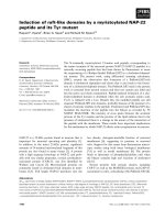
The use of the NA12878 control (Fig. 1, Additional
file 2: Table S2) and AcroMetrix (Fig. 1, Additional
file 3: Tables S3) reference standard materials demonstrated the good performance of the GeneReader
platform on high frequency and low frequency variants,
respectively. NA12878 has been used extensively as a reference standard material for verifying NGS platforms [17]
and acts as a useful control in establishing background
error. Besides its use as a GeneReader platform performance standard, AcroMetrix has also been used previously
as a control for variant calls [26].
Discussion
A major advantage of NGS over traditional mutation
detection methods is the ability to sequence multiple
genes and variants simultaneously. Other advantages
include minimal DNA input, faster turnaround time;
Table 4 The concordance study between GeneReader, MiSeq,
Pyrosequencingand Therascreen PCR assays
Sample
no.
KRAS AA
change
KRAS variant allele fraction (%)
Therascreen PCR/Pyro
GeneReadera
MiSeqa
1
G12D
+
21
7
2
G12D
+
39
12
3
A59T
19
15
14
4
G13D
+
41
15
5
G12D
+
45
11
6
Q61H
14
9
13
7
A146P
41
40
32
8
Q61H
36
35
26
9
Q61H
32
22
35
10
K117 N
24
34
39
Table 2 The GeneReader FFPE DNA sample preparation kit
successfully corrects FFPE artifacts
11
G13D
+
47
15
12
G12C
+
32
10
Type of DNA purification kit
Allele frequency cut off
13
G13D
+
39
10
>5%
>2.5%
14
Q61H
26
21
23
QIAamp FFPE DNA purification Kit
15
25
GeneRead DNA FFPE Kit
14
14
+: Variant identified by Therascreen PCR; allele fraction not available
a
: Sample processed from different FFPE section with potentially different
tumor content and variant allele fraction
Confirmation of variants by MiSeq, pyrosequencing and
therascreen qPCR assays
The GeneReader NGS System variant calls demonstrated
100% agreement with KRAS mutation status previously
determined by either pyrosequencing or therascreen
qPCR (Table 3). Of the 43 samples, 14 tested positive for
KRAS variants and 29 samples were confirmed as wild
type. The 5% allelic fraction cut-off was used to call
KRAS variants for codons 12, 13, 59, 61, 117 and 146.
The true positive variants observed by the GeneReader
NGS System share a 100% concordance with MiSeqIllumina (Table 4).
Darwanto et al. BMC Cancer (2017) 17:358
Page 5 of 8
Fig. 1 Variant calling performances of GeneReader pipeline. Each individual data point was generated from 18 data points (a) NA12878 and (b)
AcroMetrix Oncology Hotspot
lower overall cost and higher throughput and sensitivity
compared to traditional methods [12, 27–29]. NGS has
revolutionized the speed of genetic and genomic discovery, and advanced our understanding of the molecular
mechanisms of disease and potential treatment options.
However, several major hurdles remain and still prevent
NGS from being broadly adopted in clinical practice.
This is especially true for laboratories that are new to
this technology, and may lack the in-house expertise
required for processing complex bioinformatics data and
interpretation of results. Such expertise is crucial to
construct a bioinformatics pipeline and to evaluate the
software and generate quality reports. The QIAGEN
GeneReader NGS System allows users to perform
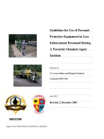
Fig. 2 QCI Analyze report showing the alignment of the reads at the variant positions along with the induced amino acid change
Darwanto et al. BMC Cancer (2017) 17:358
experiments from sample to insight, tissue sample to
decipherable report based on the interpretation of
sequence variants detected.
The QIAGEN GeneReader NGS workflow utilizes ‘QCI
Analyze’ and ‘QCI Interpret’ for bioinformatics analysis and
reporting of variants, including read mapping, variant calling and interpretation of results. It provides visualization of
the alignment of sequencing results (Fig. 2) as well as a
summary of the data. Quality assessment is also supported,
both at the overall sequencing run level and for the analytic
validity of individual variants to reduce false positive and
negative results. Using the data visualization tools within
QCI Analyze, it is possible to determine the quality of the
results and assess any variants of interest. Further analysis
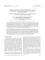
of variants using QCI Interpret provides access to the
curated information contained within the QIAGEN Knowledge Base enabling a deeper analysis and interpretation of
results for each sample (Fig. 3). With all relevant information, a report can be created with a summary of findings
and direct links to evidence sources. At the single variant
level the QCI software is able to identify an individual variant as an actionable cancer mutation, and provides links to
current clinical research insights, e.g. the KRAS G12D
somatic variant it is established to confer resistance to the
colorectal cancer drugs cetuximab and panitumumab,
based on evidence curated from their FDA drug labels and
clinical practice guidelines. Within QCI-Interpret information on active clinical trials recruiting colorectal cancer
patients with particular mutations are provided with drug,
nearest location, and trial phase information.
Page 6 of 8
The relationship between FFPE DNA quality and sequencing accuracy is a critical point for any sequencing
analysis. The GeneReader workflow starts with the GeneRead FFPE DNA Kit for DNA extraction and is specifically
designed to reduce artifacts known to commonly occur in
FFPE treated samples. As seen in Table 2, by using FFPE
samples aged from 3 to 20 years, the GeneRead FFPE
DNA Kit successfully reduced the number of low frequency false positive variants detected. These low frequency false positive variants are likely caused by cytosine
deamination and other fixation associated artifacts. Similar
phenomena were observed by Bourgon [23], where pretreatment of FFPE samples with uracil DNA glycosylase
(UDG) resulted in a dramatic reduction of false positives,
with overall reductions of 77% for C > T and 94% for
G > A changes, respectively. Biochemical removal of deaminated DNA eliminates deamination-associated false
positive results; however, for samples with very low quality
DNA such as highly fragmented FFPE treated samples,
UDG-treated may constitute an issue, as the treatment introduces possible further strand breaks leading to even
higher fragmentation and lower availability of intact template strands. Therefore, using the QuantiMIZE assay to
identify those samples suitable for sequencing, based on
an assessment of original intact and amplifiable templates,
before starting an experiment is a critical point for an
amplification based NGS technology. Previous reports observed that samples with lower amounts of amplifiable
DNA are more likely to give a markedly increased number
of false positive results [30, 31].
Fig. 3 QCI Interpret actionable report, showing summary of findings and link to the insights that can be used to guide clinical research
Darwanto et al. BMC Cancer (2017) 17:358
Conclusions
In summary, this study confirms that the GeneReader
NGS System performs consistently and accurately in the
identification of somatic mutations from FFPE samples,
with results confirmed by both alternative technologies
as well as an alternative NGS platform. With a full endto-end solution with integrated sample preparation and
bioinformatics interpretation, the GeneReader NGS
System is suitable for any laboratory interested in cancer
clinical research.
Additional files
Additional file 1: Table S1. The QC results of the extracted DNA
samples were measured using GeneRead DNA QuantiMIZE. (DOCX 28 kb)
Additional file 2: Table S2. List of NA12878 Gold Standard Variants
from 18 samples sequenced by GeneReader. (DOCX 27 kb)
Additional file 3: Table S3. List of AcroMetrix™ Oncology Hotspot
Gold Standard Variants from 18 samples sequenced by GeneReader
(DOCX 28 kb) (DOCX 27 kb)
Abbreviations
Ct: Cycle threshold; DNA: Deoxyribonucleic acid; dsDNA: Double-stranded
DNA; FDA: Food and Drug Administration; FFPE: Formalin-fixed paraffinembedded; GR: GeneReader; NGS: Next generation sequencing; NSCLC: Nonsmall cell lung cancer; PCR: Polymerase chain reaction; qPCR: Quantitative
PCR; QC: Quality control; QCI: QIAGEN clinical insight; TAT: Turn-around time;
UDG: Uracil-DNA glycosylase
Acknowledgements
We greatly appreciate Drs. Scott Steelman, Kathleen Steinmann and Robert
Lintner from Broad Technology Labs (Broad Institute of MIT and Harvard, 75
Ames Street (Rm 8021), Cambridge, MA 02141) for their support in samples
processing, GeneReader testing, and manuscript revisions. We thanks to Drs.
Vikas Gupta, Naomi Thompson Kiran Divakar and Dietrich Lueerssen from
QIAGEN for the input of the manuscript.
Funding
This work was supported by QIAGEN.
Disclaimer
The sequencing chemistry used in this manuscript is currently available outside
the US. The GeneReader NGS System is for research use only. An upgraded and
different sequencing chemistry has meanwhile been made available in the US
since April 2017 and will become available worldwide later in 2017. For the
release of the new sequencing chemistry in the US in April, we have shown
equivalency to the sequencing chemistry used in the manuscript.
Availability of data and materials
The analyzed data sets generated during the study are available from the
corresponding author on reasonable request.
Authors’ contributions
AD, AMH, SS, AS, EL, SH, TR designed the study. AD, AMH, SS, AS, DR, MN, TP,
DA, AR, NP, DS, TR performed experiments and analyzed data. AD, YK and TR
wrote the manuscript. AK, DS, DZ, KK assisted in preparing the manuscript.
All authors read and approved the final manuscript.
Competing interests
At the time of the work was being done the authors where employees of
QIAGEN, and QIAGEN funded the research and the publication costs. We
declare that our current or previous employment with QIAGEN did not
influence our interpretation of data or presentation of information.
Consent for publication
Not applicable.
Page 7 of 8
Ethics approval and consent to participate
FFPE Tumor material from colorectal cancer tumors (Origene Technologies,
MD, USA and Asterand Biosciences, MI, USA). We refer to Origene’s and
Asterand’s quality system as well as ethics and compliance processes with
informed consent for commercial clinical samples. We expect Origene and
Asterand to follow industry standard ethics approval and consent processes.
Publisher’s Note
Springer Nature remains neutral with regard to jurisdictional claims in
published maps and institutional affiliations.
Author details
1
QIAGEN Waltham, 35 Gatehouse Dr, Waltham, MA 02451, USA. 2QIAGEN
Arhus, Silkeborgvej 2, 8000 Aarhus, Denmark. 3QIAGEN GmbH, QIAGEN
Strasse 1, 40724 Hilden, Nordrhein-Westfalen, Germany. 4QIAGEN Redwood
City, 1700 Seaport Blvd, Redwood, CA 94063, USA. 5QIAGEN Frederick, 6951
Executive Way, Frederick, MD 21703, USA. 6QIAGEN Manchester, Skelton
House Lloyd Street North, Manchester M15 6SH, UK. 7Novartis Institutes for
BioMedical Research, Cambridge, MA 02139, USA. 8T2 Biosystems, Lexington,
MA 02421, USA. 9Macherey-Nigel, Bethlehem, PA 18020, USA.
Received: 28 November 2016 Accepted: 5 May 2017
References
1. Di Marco M, Astolfi A, Grassi E, Vecchiarelli S, Macchini M, Indio V, Casadei R,
Ricci C, D’Ambra M, Taffurelli G, Serra C, Ercolani G, Santini D, D’Errico A,
Pinna AD, Minni F, Durante S, Martella LR, Biasco G. Characterization of
pancreatic ductal adenocarcinoma using whole transcriptome sequencing
and copy number analysis by single-nucleotide polymorphism array. Mol
Med Rep. 2015;12:7479–84.
2. Huang J, Löhr JM, Nilsson M, Segersvärd R, Matsson H, Verbeke C, Heuchel R,
Kere J, Iafrate AJ, Zheng Z, Ye W. Variant profiling of candidate genes in
pancreatic Ductal Adenocarcinoma. Clin Chem. 2015;61:1408–16.
3. Gao J, Wu H, Wang L, Zhang H, Duan H, Lu J, Liang Z. Validation of
targeted next-generation sequencing for RAS mutation detection in FFPE
colorectal cancer tissues: comparison with Sanger sequencing and
ARMS-scorpion real-time PCR. BMJ Open. 2016;6:e009532.
4. Sakai K, Yoneshige A, Ito A, Ueda Y, Kondo S, Nobumasa H, Fujita Y, Togashi Y,
Terashima M, De Velasco MA, Tomida S, Nishio K. Performance of a novel KRAS
mutation assay for formalin-fixed paraffin embedded tissues of colorectal
cancer. Spring. 2015;4:7.
5. Sundström M, Edlund K, Lindell M, Glimelius B, Birgisson H, Micke P, Botling J.
KRAS analysis in colorectal carcinoma: analytical aspects of pyrosequencing
and allele-specific PCR in clinical practice. BMC Cancer. 2010;10:660.
6. Casadio C, Guarize J, Donghi S, Di Tonno C, Fumagalli C, Vacirca D, Dell’Orto P,
De Marinis F, Spaggiari L, Viale G, Barberis M. Molecular testing for targeted
therapy in advanced non-small cell lung cancer: suitability of Endobronchial
ultrasound Transbronchial needle aspiration. Am J Clin Pathol. 2015;144:629–34.
7. Papadopoulou E, Tsoulos N, Tsirigoti A, Apessos A, Agiannitopoulos K,
Metaxa-Mariatou V, Zarogoulidis K, Zarogoulidis P, Kasarakis D, Kakolyris S,
Dahabreh J, Vlastos F, Zoublios C, Rapti A, Papageorgiou NG, Veldekis D,
Gaga M, Aravantinos G, Karavasilis V, Karagiannidis N, Nasioulas G.
Determination of EGFR and KRAS mutational status in Greek non-small-cell
lung cancer patients. Oncol Lett. 2015;10:2176–84.
8. Lièvre A, Bachet JB, Boige V, Cayre A, Le Corre D, Buc E, Ychou M, Bouché O,
Landi B, Louvet C, André T, Bibeau F, Diebold MD, Rougier P, Ducreux M,
Tomasic G, Emile JF, Penault-Llorca F, Laurent-Puig P. KRAS mutations as an
independent prognostic factor in patients with advanced colorectal cancer
treated with cetuximab. J Clin Oncol. 2008;26:374–9.
9. Rodriguez R. Biomarker testing for treatment of metastatic colorectal cancer:
role of the pathologist in community practice. J Community Support Oncol.
2014;12:27–32.
10. Martinez DA, Nelson MA. The next generation becomes the now
generation. PLoS Genet. 2010;6:e1000906.
11. Sanger F, Nicklen S, Coulson AR. DNA sequencing with chain-terminating
inhibitors. Proc Natl Acad Sci U S A. 1997;74:5463–7.
12. D’Haene N, Le Mercier M, De Nève N, Blanchard O, Delaunoy M, El Housni H,
Dessars B, Heimann P, Remmelink M, Demetter P, Tejpar S, Salmon I. Clinical
validation of targeted next generation sequencing for Colon and Lung
cancers. PLoS One. 2015;10:e0138245.
Darwanto et al. BMC Cancer (2017) 17:358
13. Le Mercier M, D’Haene N, De Nève N, Blanchard O, Degand C, Rorive S,
Salmon I. Next-generation sequencing improves the diagnosis of thyroid FNA
specimens with indeterminate cytology. Histopathology. 2015;66:215–24.
14. Beadling C, Neff TL, Heinrich MC, Rhodes K, Thornton M, Leamon J,
Andersen M, Corless CL. Combining highly multiplexed PCR with
semiconductor-based sequencing for rapid cancer genotyping. J MolDiagn.
2013;15:171–6.
15. Metzger GJ, Dankbar SC, Henriksen J, Rizzardi AE, Rosener NK, Schmechel SC.
Development of multigene expression signature maps at the protein level
from digitized immunohistochemistry slides. PLoS One. 2012;7:e33520.
16. Metzker ML. Sequencing technologies - the next generation. Nat Rev Genet.
2010;11:31–46.
17. Zook JM, Chapman B, Wang J, Mittelman D, Hofmann O, Hide W, Salit M.
Integrating human sequence data sets provides a resource of benchmark
SNP and indel genotype calls. Nat Biotechnol. 2014;32:246–51.
18. Frickmann H, Tenner-Racz K, Eggert P, Schwarz NG, Poppert S, Tannich E,
Hagen RM. Influence of parasite density and sample storage time on the
reliability of Entamoeba Histolytica-specific PCR from formalin-fixed and
paraffin-embedded tissues. Diagn Mol Pathol. 2013;22:236–44.
19. Obersteller S, Neubauer H, Hagen RM, Frickmann H. Comparison of five
commercial nucleic acid extraction kits for the PCR-based detection of
Burkholderia Pseudomallei DNA in formalin-fixed, paraffin-embedded
tissues. Eur J Microbiol Immunol (Bp). 2016;6:244–52.
20. Kokkat TJ, Patel MS, McGarvey D, VA LV, Baloch ZW. Archived formalin-fixed
paraffin-embedded (FFPE) blocks: a valuable underexploited resource for
extraction of DNA, RNA, and protein. Biopreserv Biobank. 2013;11:101–6.
21. Chung JY, Braunschweig T, Williams R, Guerrero N, Hoffmann KM, Kwon M,
Song YK, Libutti SK, Hewitt SM. Factors in tissue handling and processing
that impact RNA obtained from formalin-fixed, paraffin-embedded tissue.
J Histochem Cytochem. 2008;56:1033–42.
22. Frickmann H, Loderstaedt U, Racz P, Tenner-Racz K, Eggert P, Haeupler A, Bialek R,
Hagen RM. Detection of tropical fungi in formalin-fixed, paraffin-embedded
tissue: still an indication for microscopy in times of sequence-based diagnosis?
Biomed Res Int. 2015;2015:938721–32.
23. Bourgon R, Lu S, Yan Y, Lackner MR, Wang W, Weigman V, Wang D, Guan Y,
Ryner L, Koeppen H, Patel R, Hampton GM, Amler LC, Wang Y. Highthroughput detection of clinically relevant mutations in archived tumor
samples by multiplexed PCR and next-generation sequencing. Clin Cancer
Res. 2014;20:2080–91.
24. Hosein AN, Song S, McCart Reed AE, Jayanthan J, Reid LE, Kutasovic JR,
Cummings MC, Waddell N, Lakhani SR, Chenevix-Trench G, Simpson PT.
Evaluating the repair of DNA derived from formalin-fixed paraffinembedded tissues prior to genomic profiling by SNP-CGH analysis. Lab
Investig. 2013;93:701–10.
25. Do H, Dobrovic A. Dramatic reduction of sequence artefacts from DNA
isolated from formalin-fixed cancer biopsies by treatment with uracil- DNA
glycosylase. Oncotarget. 2012;3:546–58.
26. Masucci GV, Cesano A, Hawtin R, Janetzki S, Zhang J, Kirsch I, Dobbin KK,
Alvarez J, Robbins PB, Selvan SR, Streicher HZ, Butterfield LH, Thurin M.
Validation of biomarkers to predict response to immunotherapy in cancer:
volume I - pre-analytical and analytical validation. J Immunother Cancer.
2016;4:76–101.
27. Castéra L, Krieger S, Rousselin A, Legros A, Baumann JJ, Bruet O, Brault B,
Fouillet R, Goardon N, Letac O, Baert-Desurmont S, Tinat J, Bera O, Dugast C,
Berthet P, Polycarpe F, Layet V, Hardouin A, Frébourg T, Vaur D. Nextgeneration sequencing for the diagnosis of hereditary breast and ovarian
cancer using genomic capture targeting multiple candidate genes. Eur J
Hum Genet. 2014;22:1305–13.
28. McCourt CM, McArt DG, Mills K, Catherwood MA, Maxwell P, Waugh DJ,
Hamilton P, O’Sullivan JM, Salto-Tellez M. Validation of next generation
sequencing technologies in comparison to current diagnostic gold
standards for BRAF, EGFR and KRAS mutational analysis. PLoS One.
2013;8:e69604.
29. Tuononen K, Mäki-Nevala S, Sarhadi VK, Wirtanen A, Rönty M, Salmenkivi
K, Andrews JM, Telaranta-Keerie AI, Hannula S, Lagström S, Ellonen P,
Knuuttila A, Knuutila S. Comparison of targeted next-generation
sequencing (NGS) and real-time PCR in the detection of EGFR, KRAS, and
BRAF mutations on formalin-fixed, paraffin-embedded tumor material of
non-small cell lung carcinoma-superiority of NGS. Genes Chromosom
Cancer. 2013;52:503–11.
Page 8 of 8
30. Betge J, Kerr G, Miersch T, Leible S, Erdmann G, Galata CL, Zhan T, Gaiser T,
Post S, Ebert MP, Horisberger K, Boutros M. Amplicon sequencing of
colorectal cancer: variant calling in frozen and formalin-fixed samples. PLoS
One. 2015;10:e0127146.
31. Sah S, Chen L, Houghton J, Kemppainen J, Marko AC, Zeigler R, Latham GJ.
Functional DNA quantification guides accurate next-generation sequencing
mutation detection in formalin-fixed, paraffin-embedded tumor biopsies.
Genome Med. 2013;5:77–89.
Submit your next manuscript to BioMed Central
and we will help you at every step:
• We accept pre-submission inquiries
• Our selector tool helps you to find the most relevant journal
• We provide round the clock customer support
• Convenient online submission
• Thorough peer review
• Inclusion in PubMed and all major indexing services
• Maximum visibility for your research
Submit your manuscript at
www.biomedcentral.com/submit