Power of PgR expression as a prognostic factor for ER-positive/HER2-negative breast cancer patients at intermediate risk classified by the Ki67 labeling index
Bạn đang xem bản rút gọn của tài liệu. Xem và tải ngay bản đầy đủ của tài liệu tại đây (1.01 MB, 9 trang )
Kurozumi et al. BMC Cancer (2017) 17:354
DOI 10.1186/s12885-017-3331-4
RESEARCH ARTICLE
Open Access
Power of PgR expression as a prognostic
factor for ER-positive/HER2-negative breast
cancer patients at intermediate risk
classified by the Ki67 labeling index
Sasagu Kurozumi1,3, Hiroshi Matsumoto1, Yuji Hayashi1, Katsunori Tozuka1, Kenichi Inoue2, Jun Horiguchi3,
Izumi Takeyoshi3, Tetsunari Oyama4 and Masafumi Kurosumi5*
Abstract
Background: The Ki67 labeling index (LI) is regarded as a significant prognostic marker in ER-positive/HER2-negative
breast cancer patients. The expression of PgR has recently been identified as another prognostic marker. In the present
study, we investigated the prognostic utilities and most suitable cut-off values for Ki67 and PgR, and evaluated the
relationship between Ki67 LI and PgR expression in ER-positive/HER2-negative breast cancer.
Patients and methods: In the present study, 177 consecutive Japanese women with ER-positive/HER2-negative
invasive carcinoma of no special type who were treated between 2000 and 2001 were enrolled. Recurrence-free
survival (RFS) and cancer-specific survival (CSS) were analyzed according to Ki67 LI and PgR expression, and significant
cut-off values for selecting patients with a poor prognosis were evaluated.
Results: The cut-off values for Ki67 LI as a prognostic marker plotted against P values showed bimodal peaks at 10%
and 30%. Among the cut-off points examined for the PgR status, 20% PgR positivity was the most significant for
predicting survival differences (RFS: P = 0.0003; CSS: P < 0.0001). A multivariate analysis showed that PgR (≥20%) was an
independent prognostic marker (RFS: P = 0.0092; CSS: P = 0.00014). Furthermore, in the intermediate risk group with
Ki67 LI of 10–30%, the low PgR <20% group had a markedly poorer prognosis for RFS and CSS (RFS: P < 0.0001;
CSS: P < 0.0001).
Conclusions: The expression of PgR is a potent prognostic indicator for evaluating the long-term prognosis of ERpositive/HER2-negative breast cancer, and the most suitable cut-off value was found to be 20%. Furthermore, the PgR
status is a powerful method for selecting patients with a poor prognosis among ER-positive/HER2-negative patients at
intermediate risk, as assessed using Ki67 LI.
Keywords: ER-positive and HER2-negative breast cancer, Ki67 labeling index, Progesterone receptor, Prognosis
Background
Breast cancer has clinical and biological heterogeneity,
and research is ongoing to detect potent indicators
associated with cell growth and differentiation, which
are involved in tumor formation and the progression of
breast cancer. Breast cancer has recently been classified
into 6 intrinsic subtypes: luminal A, luminal B, human
* Correspondence:
5
Department of Pathology, Saitama Cancer Center, 780 Komuro, Ina-machi,
Kitaadachi-gun, Saitama 362-0806, Japan
Full list of author information is available at the end of the article
epidermal growth factor receptor type 2 (HER2)enriched, basal-like, claudin-low, and normal-like, using
semi-unsupervised gene expression array analyses [1–3].
In routine practice, intrinsic subtypes have been
obtained using immunohistochemical evaluations of the
estrogen receptor (ER), progesterone receptor (PgR),
HER2, and Ki67 labeling index (LI), and the following
practical classification of intrinsic subtypes was proposed
at the St. Gallen consensus meeting of breast cancer:
luminal A-like type (ER-positiveand/or PgR-positive,
HER2-negative, low proliferation, and low tumor
© The Author(s). 2017 Open Access This article is distributed under the terms of the Creative Commons Attribution 4.0
International License ( which permits unrestricted use, distribution, and
reproduction in any medium, provided you give appropriate credit to the original author(s) and the source, provide a link to
the Creative Commons license, and indicate if changes were made. The Creative Commons Public Domain Dedication waiver
( applies to the data made available in this article, unless otherwise stated.
Kurozumi et al. BMC Cancer (2017) 17:354
burden), luminal B-like type (ER-positiveand/or PgRpositive, HER2-negative, high proliferation, and high
tumor burden), hormone receptor-positive and HER2positive type, hormone receptor-negative and HER2postive type, and triple-negative (TN) type (hormone
receptor-negative and HER2-negative) [4, 5].
Ki67 has been associated with cell cycle activity and is
expressed at various levels during the G1, S, G2, and M
phases [6]. Ki67 expression was found to correlate well
with the growth fraction in various human cancers
including breast cancer [7]. In previous studies, Ki67 LI
was valued as a prognostic factor associated with ERpositive/HER2-negative breast cancer outcomes. Ki67 LI
is also regarded as a biomarker for therapeutic decisions
for ER-positive/HER2-negative breast cancer [8, 9].
However; definite cut-off values for Ki67 have not yet
been decided, and evidence to indicate that patients with
low Ki67 LI among those with ER-positive/HER2-negative breast cancer are at a lower risk of breast cancer
relapse is limited [10, 11]. Dowsett et al., on behalf of
the International Ki67 in Breast Cancer Working Group
of the Breast International Group and North American
Breast Cancer Group, provided an overview of the state
of the art of Ki67 evaluations and proposed a set of
guidelines for the analysis and reporting of Ki67 [12, 13].
They also suggested that a standardized method and
value set need to be established for the evaluation of
Ki67 [14]. Manual counting appears to be accepted, but
represents a huge task for pathologists and is not highly
reproducible. Hida et al. modified their method of a visual assessment to create a new 5-grade scale for the
evaluation of Ki67 and verified its utility [15]. On the
other hand, Perou et al. initially proposed a molecular
classification for breast cancer [1, 2], and the subsequent
expansion of this work into a larger cohort of patients
showed that luminal B tumors had a poorer prognosis
than Luminal A tumors despite treatments with hormonal therapy [16]. These discrepancies between luminal A
and luminal B may be due to the different estrogenrelated intracellular signaling pathways in breast cancer
cells. However, many questions regarding distinguishing
between the mechanisms responsible for luminal A and
luminal B breast cancer, which lead to the proliferation
and metastasis of breast cancer cells, remain unanswered
[17]. Prat et al. reported that an empiric cut-off of more
than 20% of PgR-positive tumor cells was statistically
proven to be significant for predicting survival differences
within luminal-type breast cancer defined by their molecular classification. They concluded that the new definition of the luminal A-like type was ER-positive/HER2negative/Ki67 LI less than 14%/PgR more than 20% [18].
Therefore, PgR may be a useful indicator for classifying
ER-positive/HER2-negative breast cancer between the
luminal A-like subtype and B-like subtype [19].
Page 2 of 9
However, the relationship between Ki67 LI and the
expression of PgR has not yet been examined, and the
utility of a combined evaluation method using these 2
factors for the selection of a poor prognosis group from
ER-positive/HER2-negative breast cancer patients has
not yet been established. In the present study, we investigated the prognostic utilities and most suitable cut-off
values for Ki67 LI and PgR expression, and then analyzed the relationship between Ki67 LI and PgR expression as a prognostic marker in patients with ER-positive/
HER2-negative breast cancer.
Methods
Patient backgrounds and eligibility
The paraffin-embedded samples of tumors from 272
consecutive patients with invasive breast cancer of no
special type that were larger than 5 mm and diagnosed
at Saitama Cancer Center between January 2000 and
December 2001 were initially retrieved, the status of ER,
PgR, HER2, and Ki67 LI were assessed, and the intrinsic
subtypes of these patients were decided.
After the evaluation of intrinsic subtypes, 177 patients
with ER-positive/HER2-negative breast cancer were selected and enrolled in this study. All patients underwent
breast-conserving surgery or modified radical mastectomy without neoadjuvant chemotherapy or neoadjuvant
endocrine therapy. We excluded patients with bilateral
breast cancer and male breast cancer. The medical records of these ER-positive/HER2-negative patients were
reviewed for clinicopathological characteristics including
the pathological T and N status and American Joint
Committee on Cancer (AJCC) stage, and follow-up data
for all patients were obtained with a median follow-up
period of 130 months (4–149 months).
This study was conducted in accordance with the
Declaration of Helsinki, and the protocol of the study
was approved by the Institutional Review Board of the
Saitama Cancer Center. All patients enrolled in this
study agreed to the scientific examination of tumor tissues obtained by surgery and provided written comprehensive informed consent.
Procedures to examine ER, PgR, HER2, and Ki67
Buffered formalin-fixed paraffin-embedded specimens
were cut into 4-μm-thick sections to be prepared for
immunohistochemistry for ER, PgR, HER2, and Ki67 as
well as dual HER2 in situ hybridization (DISH). The
sources of primary antibodies were as follows: ER (1D5,
DAKO, Denmark), PgR (PgR636, DAKO, Denmark),
HER2 (HercepTest, DAKO, Denmark), and Ki67 (MIB-1,
DAKO, Denmark). Immunohistochemistry for ER,
PgR, and HER2 was performed manually using the
streptavidin-biotin method. In patients with a HER2
score 2+ by immunohistochemistry, amplification of
Kurozumi et al. BMC Cancer (2017) 17:354
the HER2 gene was evaluated using the dual in situ
hybridization (DISH) method with an automated slide
processing system (BenchMark® XT, Ventana Medical
Systems, Inc., Tucson, Arizona). Furthermore, immunohistochemistry for Ki67 was performed automatically
using an automated immunohistochemistry instrument
(BenchMark® XT, Ventana Medical Systems, Inc., Tucson,
Arizona).
Evaluation of ER, PgR, and HER2 status and Ki67 LI
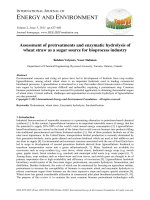
The percentages of nuclei stained for ER and PgR were
calculated (Fig. 1), and a patient was considered to be
“positive” if the breast tumor contained at least 1%
positive cells, in accordance with the American Society
of Clinical Oncology (ASCO) and College of American
Pathologists (CAP) criteria. In addition, the degrees of
staining for ER and PgR were evaluated using the Allred
score. In the Allred scoring system, proportion scores
were defined as: 0 (0% staining), 1 (<1%), 2 (1-10%), 3
(10-33%), 4 33-67%), and 5 (>67%), while intensity scores
were defined as: 0 (no staining), 1 (weak staining), 2
(intermediate staining), and 3 (strong staining). The total
score was obtained by adding the proportion score and
intensity score in order to attain final scores of 0 and 2–
8. We also added a “20%” cut-off point to evaluate PgR
staining. Since it has been reported that tumors with an
Allred score ≤ 2 are hormone non-responsive [20], we
evaluated breast cancer patients with an Allred
score ≥ 3.
An evaluation of the HER2 status using immunohistochemistry and DISH was performed using the guidelines
Page 3 of 9
of ASCO/CAP proposed in 2013. Membranous staining
for HER2 was graded as follows: scores 0, 1+, 2+, and
3+. Tumors with a score 2+ were subjected to an in
situ hybridization (ISH) assay in order to assess the
gene amplification of HER2. A HER2 score of 3+ or
2+ /DISH positive was defined as HER2-positive cancer. We excluded HER2-positive/ER-positive patients
from further examination because their prognosis is
worse and the strategy of treatment using HER2targeting agents markedly differs from that of HER2negative/ER-positive patients.
Images of Ki67 staining were captured using a digital
pathology system (NanoZoomer 2.0-HT, C9600–13,
Hamamatsu Photonics, Co., Japan) with viewer software
(NDP.view2, Hamamatsu Photonics, Co., Japan), and
photographs of the selected area were printed. Evaluations of Ki67 LI (percentage of positivity) were performed using printed photographs. We initially selected
the representative area from the whole area of Ki67stained sections. We principally observed the front line
of the invasive region, and selected warm to hot spots in
density for Ki67 labeling. The numbers of positive and
negative nuclei stained by Ki67 immunohistochemistry
were counted. At least 500 tumor cells were counted
and Ki67 LI was calculated.
Statistical analysis
Statistical analyses were conducted using SPSS v22.0
(IBM Corp., USA). The relationship between Ki67 LI
and PgR expression (Allred score) was analyzed by
Spearman’s rank correlation test. The Kaplan-Meier
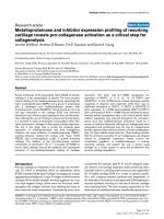
Fig. 1 Combination patterns of ER and PgR expression. Case 1: ER-positive (≥1%) and high PgR expression breast cancer (a ER expression, b PgR
expression) Case 2: ER-positive (≥1%) and low PgR expression breast cancer (c ER expression, d PgR expression)
Kurozumi et al. BMC Cancer (2017) 17:354
Page 4 of 9
method and Log-rank test were used to estimate
recurrence-free survival (RFS) and cancer-specific survival (CSS). RFS was defined as the length of time from
the period of surgery to any recurrence (including ipsilateral breast recurrence). CSS was defined as the time
from the day of surgery until the time of death due to
the progression of breast cancer. RFS and CSS were
compared between patients divided into two groups
according to the degree of PgR staining and Ki67 LI.
Significant cut-off values were obtained for the selection
of patients with the worst prognosis based on the lowest
P value derived from the survival analysis. In addition,
some clinicopathological factors such as the menopausal
status, pathological T status, pathological node status,
histological grade, and type of adjuvant therapy were
included in the multivariate survival analysis using a
Cox proportional hazards regression model, and 95%
confidence intervals were assessed for each factor. A
P value < 0.05 was defined as being significant.
Table 1 Patient and tumor characteristics at baseline
Results
Pathological stage
Total
No. of patients
Percent
177
100
Menopausal status at diagnosis
Premenopausal
77
43.5
Postmenopausal
100
56.5
T1
99
55.9
T2
58
32.8
T3
10
5.6
T4
10
5.6
N0
95
53.7
N1
46
26.0
N2
23
13.0
N3
9
5.1
Not evaluated
4
2.3
Pathological tumor size
Pathological nodal status
Patient and tumor characteristics
I
63
35.6
Patient and tumor characteristics were shown in Table 1.
The median age of the 177 patients enrolled in this
study was 54 years (age range, 26–87 years); 162 patients
(91.5%) were older than 41 years and 100 patients
(56.5%) were post-menopausal. Seventy patients (39.5%)
received adjuvant chemotherapy, while 146 (82.5%)
received adjuvant endocrine therapy. The distribution of
patients stratified by Allred scores and the proportion of
PgR was shown in Table 2. The median Ki67 LI of all
patients was 18.2% (index range, 0.8–74%), and the
distribution of patients stratified by the Ki67 LI was also
shown in Table 2. Forty-six patients (26.0%) were in the
low Ki67 (less than 10%) LI group, while 33 (18.6%) were
in the high Ki67 (more than 30%) LI group.
II A
55
31.1
II B
22
12.4
III A
14
7.9
III B
10
5.6
III C
9
5.1
Not evaluated
4
2.3
Survival analysis according to the status of PgR
The hazard ratios of RFS and CSS stratified by the PgR
status were evaluated using the Kaplan-Meier method
and Log-rank test. The cut-off values for the PgR status
and associated P values for the difference in the probability of survival between low and high PgR expression
groups stratified by the Allred score were as follows: 0
vs 2–8, cut-off point 2 (RFS: HR = 5.88, P = 0.015; CSS:
HR = 3.73, P = 0.053), 0–2 vs 3–8, cut-off point 3 (RFS:
HR = 5.88, P = 0.015; CSS: HR = 3.73, P = 0.053), 0–3
vs 4–8, cut-off point 4 (RFS: HR = 5.43, P = 0.020; CSS:
HR = 4.39, P = 0.036), 0–4 vs 5–8, cut-off point 5 (RFS:
HR = 2.95, P = 0.086; CSS: HR = 2.72, P = 0.099), 0–5
vs 6–8, cut-off point 6 (RFS: HR = 3.59, P = 0.058; CSS:
HR = 4.35, P = 0.037), 0–6 vs 7–8, cut-off point 7 (RFS:
HR = 8.68, P = 0.0032; CSS: HR = 14.75, P = 0.0001),
and 0–7 vs 8, cut-off point 8 (RFS: HR = 5.68,
Type of surgery
Breast-conserving surgery
147
83.1
Mastectomy
30
16.9
Sentinel lymph node biopsy alone
95
53.7
Axillary lymph node dissection
79
44.6
No surgery
3
1.7
1
41
23.2
2
67
37.9
3
69
39.0
Yes
70
39.5
No
107
60.5
Axillary management
Histological grade
Adjuvant Chemotherapy
Adjuvant Endocrine therapy
Yes
146
82.5
No
31
17.5
P = 0.017; CSS: HR = 4.06, P = 0.044). The most
significant cut-off point for prognosis was between
the group with a score 0–6 and the group with a
score 7–8, cut-off point 7 (Fig. 2a).
Kurozumi et al. BMC Cancer (2017) 17:354
Page 5 of 9
Table 2 Distribution of PgR expression and the Ki67 labeling
Index
No
Percent
0
21
11.8
2
0
0.0
3
4
2.3
4
12
6.8
5
17
9.6
6
28
15.8
7
42
23.7
8
53
29.9
Allred Scores of PgR
Proportion of PgR (%)
0
21
11.8
> 0 and <1
4
2.3
≥ 1 and <10
17
9.6
≥ 10 and <20
23
13.0
≥ 20 and <33
10
5.6
≥ 33 and ≤67
48
27.1
> 67
54
30.5
Ki67 labeling index (%)
≤ 10
46
26.0
> 10 and <14
20
11.3
≥ 14 and <20
33
18.6
≥ 20 an <30
45
25.4
≥ 30
33
18.6
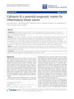
In addition, the cut-off points for the PgR status and
associated P values for the difference in the probability
of survival between the low and high PgR expression
groups stratified by the percentage of positive cells (%)
were as follows: 0% (RFS: HR = 5.88, P = 0.015; CSS:
HR = 3.73, P = 0.053), 1% (RFS: HR = 7.08, P = 0.00078;
CSS: HR = 6.47, P = 0.011), 10% (RFS: HR = 5.45,
P = 0.020; CSS: HR = 4.51, P = 0.034), 20% (RFS:
HR = 13.33, P = 0.0003; CSS: HR = 20.78, P = 0.000005),
33% (RFS: HR = 9.98, P = 0.0016; CSS: HR = 14.98,
P = 0.0001), and 67% (RFS: HR = 6.7, P = 0.014; CSS:
HR = 4.31, P = 0.038). The most significant cut-off point
for prognosis was 20% (Fig. 2b).
Survival analysis according to Ki67 LI
The hazard ratios of RFS and CSS stratified by Ki67 LI
were assessed using the Kaplan-Meier method and Logrank test. The cut-off values for Ki67 LI and associated
P values for the difference in the probability of survival
between the high Ki67 and low Ki67 groups were as follows: 10% (RFS: HR = 2.77, P = 0.096; CSS: HR = 5.21,
P = 0.022), 14% (RFS: HR = 3.57, P = 0.059; CSS:
HR = 4.77, P = 0.029), 18% (RFS: HR = 2.13, P = 0.14;
Fig. 2 Survival curves stratified by PgR expression. a Comparisons of
cancer-specific survival (CSS) between the high PgR positivity
(Allred score ≥ 7) and low PgR positivity (Allred score ≤ 6)
groups. b Comparisons of CSS between the high PgR positivity
(≥20%) and low PgR positivity (<20%) groups
CSS: HR = 1.98, P = 0.16), 20% (RFS: HR = 1.67,
P = 0.20; CSS: HR = 3.46, P = 0.063), and 30% (RFS:
HR = 2.66, P = 0.010; CSS: HR = 4.63, P = 0.031). Cutoff values for Ki67 LI as a prognostic marker plotted
against P values showed bimodal peaks at 10% and 30%.
These results allowed patients to be classified into 3
groups using the cut-off values of Ki67 as follows: a) low
Ki67 LI group, Ki67 LI: ≤10%; b) intermediate Ki67 LI
group, Ki67 LI: >10 and <30%; and c) high Ki67 LI
group, Ki67 LI: ≥30%. The survival rates of the 3 groups
were significantly different in CSS, but not in RFS (RFS:
HR = 4.28, P = 0.12; CSS: HR = 7.77, P = 0.021; Fig. 3a).
Relationship between the expression of PgR and Ki67 LI
No correlation was observed between Ki67 LI and PgR
expression (P = 0.814). The survival of the high Ki67 LI
group was significantly worse than that of the low Ki67
LI group (RFS: HR = 4.04, P = 0.044; CSS: HR = 7.76,
P = 0.0053; Fig. 3a). However, it was difficult to determine the prognosis of the intermediate Ki67 LI group, in
Kurozumi et al. BMC Cancer (2017) 17:354
Page 6 of 9
prognostic factors in this study. The menopausal status
or receiving adjuvant endocrine therapy, which we consider as important factors to treat ER-positive/HER2negative breast cancer, did not correlate with prognosis
in this study. Receiving adjuvant chemotherapy correlated with prognosis in this study (adjuvant chemotherapy no vs. yes; RFS: HR = 5.07, P = 0.024; CSS:
HR = 3.67, P = 0.055), however; a multivariate analysis
confirmed that receiving adjuvant chemotherapy did not
correlate with prognosis (adjuvant chemotherapy no vs.
yes; RFS: HR = 13.7, P = 0.35; CSS: HR = 1.25, P = 0.59).
On the other hand, a multivariate analysis (Table 3)
showed that PgR (cut-off value: 20%) was an independent prognostic marker for RFS and CSS (RFS: HR = 2.33,
P = 0.013; CSS: HR = 5.15, P = 0.00045). Based on the
results of the multivariate analysis, the pathological
lymph node status was also identified as an independent
Table 3 Results of a multivariate survival analysis using a Cox
proportional hazards regression mode on the influence of
clinicopathological variables including PgR and Ki67
RFS
Characteristics
HR
CSS
95% CI
P
HR
95% CI
P
PgR expression
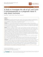
Fig. 3 Survival curves stratified by the combination tool using the
expression of PgR and Ki67. a Relationship between the Ki67
labeling index and cancer-specific survival (CSS). b Survival curves
stratified by PgR expression according to staining percentages in the
intermediate Ki67 labeling index (Ki67 > 10 and <30%) group.
Comparisons of CSS between the PgR-positive (≥20%) and
PgR-negative (<20%) groups
which as many as 98 (55.4%) ER-positive/HER2-negative
breast cancer patients were classified. In the intermediate Ki67 LI group, the low PgR group had a markedly
poorer prognosis for RFS and CSS (RFS: HR = 16.60,
P = 0.000046; CSS: HR = 18.95, P = 0.000013; Fig. 3b).
The intermediate group was clearly divided according to
Ki67 with the addition of PgR into two distinctive prognostic subgroups.
Relationships between prognosis and clinicopathological
characteristics of tumors
A univariate analysis identified the negative expression
of PgR, high Ki67 LI, high histological grade (grade 1/2
vs. 3; RFS: HR = 3.69, P = 0.055; CSS: HR = 6.44,
P = 0.011), high pathological T stage (pathological T 1/2
vs. pathological T 3/4; RFS: HR = 10.74, P = 0.0011;
CSS: HR = 8.90, P = 0.0029), and positive pathological
node status (negative vs. positive, RFS: HR = 16.94,
P = 0.000039; CSS: HR = 10.72, P = 0.0011) as worse
≥ 20%
Referent
< 20%
2.33 1.19–4.54 0.013
5.15 2.06–12.85 0.00045
Ki67 labeling index
≤ 10%
Referent
> 10 and <30% 0.52 0.18–1.53 0.24
0.28 0.05–1.44
0.13
≥ 30%
0.68 0.29–1.57
0.36
0.72 0.33–1.61
0.43
1.51 0.58–3.90
0.40
0.69 0.33–1.47 0.34
Menopausal status
Pre-
Referent
Post-
1.24 0.63–2.43 0.53
Pathological T stage
T 1–2
Referent
T 3–4
1.49 0.68–3.25 0.32
Pathological N stage
N0
Referent
N 1–3
3.16 1.51–6.58 0.0022 2.69 1.04–6.99
0.042
Histological grade
1,2
Referent
3
1.31 0.67–2.56 0.43
1.40 0.60–3.25
0.44
1.25 0.55–2.82
0.59
1.20 0.27–5.42
0.81
Adjuvant chemotherapy
No
Referent
Yes
1.37 0.71–2.64 0.35
Adjuvant endocrine therapy
No
Referent
Yes
1.15 0.39–3.37 0.80
Abbreviations: RFS recurrence-free survival, CSS cancer-specific survival,
HR hazard ratio, 95% Cl 95% Confidence interval, PgR progesterone receptor
Kurozumi et al. BMC Cancer (2017) 17:354
prognostic marker for RFS and CSS (RFS: HR = 3.16,
P = 0.0022, CSS: HR = 2.69, P = 0.042).
In addition, a multivariate analysis on the intermediate
Ki67 LI group showed that PgR (cut-off value: 20%) was
an independent potent prognostic marker for RFS and
CSS (RFS: HR = 4.67, P = 0.00052; CSS: HR = 11.66,
P = 0.00026) (Additional file 1).
Discussion
It has been known that the positive rate of ER and/or
PgR in breast cancer is approximately 70% [21], and ER
is considered to have key functions in the development
and progression of breast cancer. In addition, ER regulates many gene and protein actions within genomic and
non-genomic pathways. Furthermore, estrogen signals
mediated by ER control the genomic pathway which
works as a transcription factor for targeted genes, and
ER is activated by the signal crosstalk between estrogen
and growth factors such as epidermal growth factor and
insulin growth factor-1 via transmembrane receptor
phosphorylation [22, 23]. On the other hand, PgR induced by ER acts as a key factor in induction, progression and maintenance of the neoplastic phenotype of
ER-positive breast cancer [24]. Recent clinical findings
demonstrated that the PgR status needs to be considered
when discussing relative-risk reductions expected from
endocrine treatments in individual patients [25]. In the
present study, we revealed that the extent of PgR expression was a potent prognostic indicator for evaluating the
long-term prognosis of ER-positive/HER2-negative breast
cancer and that the most suitable cut-off value was 20%,
which was consistent with previous findings [18]. Further
research is needed in order to elucidate the biological
mechanisms underlying the relationship between PgR expression and the prognosis of ER-positive/HER2-negative
breast cancer patients.
We also classified ER-positive/HER2-negative breast
cancer more simply into the following 3 types according
to the percentages of Ki67 LI: Ki67 ≤ 10%; Ki67 > 10
and <30%; and Ki67 ≥ 30%. This Ki67 classification
correlated with the long-term survival of patients with
ER-positive/HER2-negative breast cancer. On the basis
of these results, we classified ER-positive/HER2-negative
breast cancer patients into 3 risk groups: low, intermediate, and high risk. In addition, we selected adjuvant
therapeutic options for low and high risk groups, such
as hormone therapy alone for low risk patients and
chemo-endocrine therapy for high risk patients. However, difficulties have been associated with establishing a
strategy for adjuvant therapy for the intermediate risk
group, which accounts for more than 50% of ERpositive/HER2-negative breast cancer patients.
In the St. Gallen consensus meeting of 2015, they
showed that hormone receptor-positive/HER2-negative
Page 7 of 9
breast cancer may be divided into the luminal A-like
type (high ER/PgR and clearly low Ki67), luminal-B like
type (low ER/PgR and clearly high Ki67), and intermediate type. They suggested that Ki67 scores needed to be
interpreted based on local laboratory values; if a laboratory has a median Ki67 LI of 20%, values of 30% or more
may be regarded as high, while those of 10% or less are
clearly low [5]. We also confirmed that the survival of
the Ki67 LI high (≥30%) group was significantly worse
than that of the Ki67 LI low (≤10%) group. On the other
hand, the intermediate type was defined as an uncertain
type regarding the degree of risk and responsiveness to
endocrine therapy and chemotherapy. They suggested
that in the intermediate risk type of ER-positive/HER2negative breast cancer, multi-parameter molecular tests
may be used if available. Genomic and clinical variables
both need to be included in a common algorithm in
order to yield the most accurate prediction model in ERpositive/HER2-negative breast cancer [26]. The results
of the present study indicate that the low PgR (<20%)
group has a markedly poorer prognosis among patients
with ER-positive/HER2-negative and intermediate Ki67
LI breast cancer. Maisonneuve et al. also suggested that
patients with tumors with the intermediate type (Ki67
LI: 14% to 19%) and low PgR (<20%) expression had
similar outcomes to those of patients with luminal B-like
breast cancer [27]. This combination tool using PgR and
Ki67 LI may be valuable for selecting patients with a
good prognosis in intermediate type ER-positive/HER2negative breast cancer.
For decision of appropriate cut-off values for PgR, it
might be necessary to obtain data from large-scale validation studies, but a few studies have been published on
the PgR status. Prat et al. recently reported that an empirical cut-off of more than 20% for PgR-positive tumor
cells was statistically proven to be significant for predicting survival differences among 2257 luminal-type breast
cancer patients defined by their molecular classification
[18]. Furthermore, Mohammed et al. revealed that PgR
gene loss was an independent potent prognostic marker
for survival using TCGA data [28]. However, the novel
results obtained in the present study may be limited by
the PgR cut-off values selected, and, thus, further prospective and large-scale clinical research appears to be
necessary in order to confirm the most suitable cut-off
value for PgR expression as a prognostic factor for the
Ki67-intermediate group in ER-positive/HER2-negative
breast cancer patients.
Conclusions
The extent of PgR expression as well as Ki67 LI may be
a potent prognostic indicator for evaluating the longterm prognosis of ER-positive/HER2-negative breast
cancer. The results of the present study suggest that
Kurozumi et al. BMC Cancer (2017) 17:354
Page 8 of 9
examining the extent of PgR expression allows for the
selection of patients with a poor prognosis and that the
most suitable cut-off value was 20%. Furthermore, PgR
expression and Ki67 LI represent a powerful method for
selecting patients with a poor prognosis among those
with ER-positive/HER2-negative breast cancer.
Medicine, Gunma, Japan. 4Department of Diagnostic Pathology, Gunma
University Graduate School of Medicine, Gunma, Japan. 5Department of
Pathology, Saitama Cancer Center, 780 Komuro, Ina-machi, Kitaadachi-gun,
Saitama 362-0806, Japan.
Additional file
References
1. Perou CM, Sorlie T, Eisen MB, van de Rijn M, Jeffrey SS, Rees CA, et al.
Molecular portraits of human breast tumours. Nature. 2000;406:747–52.
2. Sorlie T, Tibshirani R, Parker J, Hastie T, Marron JS, Nobel A, et al. Repeated
observation of breast tumor subtypes in independent gene expression data
sets. Proc Natl Acad Sci U S A. 2003;100:8418–23.
3. Herschkowitz JI, Simin K, Weigman VJ, Mikaelian I, Usary J, Hu Z, et al.
Identification of conserved gene expression features between murine
mammary carcinoma models and human breast tumors. Genome Biol.
2007;8:R76.
4. Goldhirsch A, Winer EP, Coates AS, Gelber RD, Piccart-Gebhart M,
Thürlimann B, et al. Personalizing the treatment of women with early breast
cancer: highlights of the St Gallen international expert consensus on the
primary therapy of early breast cancer 2013. Ann Oncol. 2013;24:2206–23.
5. Coates AS, Winer EP, Goldhirsch A, Gelber RD, Gnant M, Piccart-Gebhart M,
et al. Tailoring therapies-improving the management of early breast cancer:
St Gallen international expert consensus on the primary therapy of early
breast cancer 2015. Ann Oncol. 2015;26:1533–46.
6. Gerdes J, Lemke H, Baisch H, Wacker HH, Schwab U, Stein H. Cell cycle
analysis of a cell proliferation-associated human nuclear antigen defined by
the monoclonal antibody Ki-67. J Immunol. 1984;133:1710–5.
7. Yamamoto S, Ibusuki M, Yamamoto Y, Fu P, Fujiwara S, Murakami K, et al.
Clinical relevance of Ki67 gene expression analysis using formalin-fixed
paraffin-embedded breast cancer specimens. Breast Cancer. 2013;20:262–70.
8. Nishimura R, Osako T, Okumura Y, Hayashi M, Arima N. Clinical significance
of Ki-67 in neoadjuvant chemotherapy for primary breast cancer as a
predictor for chemosensitivity and for prognosis. Breast Cancer.
2010;17:269–75.
9. Honma N, Horii R, Iwase T, Saji S, Younes M, Ito Y, et al. Ki-67 evaluation at
the hottest spot predicts clinical outcome of patients with hormone
receptor-positive/HER2-negative breast cancer treated with adjuvant
tamoxifen monotherapy. Breast Cancer. 2015;22:71–8.
10. Andre F, Arnedos M, Goubar A, Ghouadni A, Delaloge S. Ki67–no evidence
for its use in node-positive breast cancer. Nat Rev Clin Oncol. 2015;12:296–301.
11. Varga Z, Diebold J, Dommann-Scherrer C, Frick H, Kaup D, Noske A, et al.
How reliable is Ki-67 immunohistochemistry in grade 2 breast carcinomas?
A QA study of the Swiss Working Group of Breast- and Gynecopathologists
PLoS One. 2012;7:e37379.
12. Dowsett M, Nielsen TO, A’Hern R, Bartlett J, Coombes RC, Cuzick J, et al.
Assessment of Ki67 in breast cancer: recommendations from the
international Ki67 in breast cancer working group. J Natl Cancer Inst.
2011;103:1656–64.
13. Sheri A, Dowsett M. Developments in Ki67 and other biomarkers for
treatment decision making in breast cancer. Ann Oncol 2012; 23: x219–227.
14. Polley MY, Leung SCY, McShane LM, Gao D, Hugh JC, Mastropasqua MG,
et al. An international Ki67 reproducibility study. J Natl Cancer Inst.
2013;105:1897–906.
15. Hida AI, Bando K, Sugita A, Maeda T, Ueda N, Matsukage S, et al. Visual
assessment of Ki67 using a 5-grade scale (eye-5) is easy and practical to
classify breast cancer subtypes with high reproducibility. J Clin Pathol.
2015;68:356–61.
16. Paik S, Shak S, Tang G, Kim C, Baker J, Cronin M, et al. A multigene assay to
predict recurrence of tamoxifen-treated, node-negative breast cancer.
N Engl J Med. 2004;351:2817–26.
17. Tran B, Bedard PL. Luminal-B breast cancer and novel therapeutic targets.
Breast Cancer Res. 2011;13:221.
18. Prat A, Cheang MC, Martin M, Parker JS, Carrasco E, Caballero R, et al.
Prognostic significance of progesterone receptor-positive tumor cells within
immunohistochemically defined luminal a breast cancer. J Clin Oncol.
2013;31:203–9.
19. Sato K, Miyashita M, Ishida T, Suzuki A, Tada H, Watanabe G, et al.
Prognostic significance of the progesterone receptor status in Ki67-high
Additional file 1: Results of a multivariate survival analysis on the
influence of clinicopathological variables including PgR in the intermediate
Ki67 labeling index group. (PDF 118 kb)
Abbreviations
AJCC: American Joint Committee on Cancer Staging System;
ASCO: American Society of Clinical Oncology; CAP: College of American
Pathologists; ER: Estrogen receptor; HER2: Human epidermal growth factor
receptor 2;; HR: Hazard ratio.; Ki67 LI: Ki67 labeling index; OS: Overall survival;
PgR: Progesterone receptor; RFS: Relapse-free survival
Acknowledgments
This study was presented in part at the European Breast Cancer Conference,
Glasgow, Scotland on 4 March, 2014.
Funding
This paper has been supported by a grant from the Ministry of Health,
Labour and Welfare of Japan.
Availability of data and materials
The datasets generated and/or analysed during the current study are not
publicly available because the Institutional Review Board of the Saitama
Cancer Center prohibits it, but are available from the corresponding author
on reasonable request.
Authors’ contributions
All authors participated in the study design. KS mainly performed
immunohistochemical evaluations, image acquisition, and statistical
analyses. HY, TK, IK, MH, and KM assisted in the production of the study
design and evaluating the results obtained. HY and KM assisted KS in
histological and immunohistochemical examinations and evaluating
results. HJ, TI, and OT contributed to the statistical evaluation of results
and theoretical organization of the manuscript. All authors significantly
contributed to the data interpretation and manuscript preparation.
All authors read and approved the final version of the manuscript.
Competing interests
All authors have declared no conflicts of interest.
Consent for publication
Not applicable.
Ethics approval and consent to participate
This study was conducted in accordance with the Declaration of
Helsinki, and the protocol of the study was approved by the
Institutional Review Board of the Saitama Cancer Center (Reference
number: 231 and 483). All patients enrolled in this study agreed to the
scientific examination of tumor tissues obtained by surgery and
provided written comprehensive informed consent.
Publisher’s Note
Springer Nature remains neutral with regard to jurisdictional claims in
published maps and institutional affiliations.
Author details
1
Division of Breast Surgery, Saitama Cancer Center, Saitama, Japan. 2Division
of Breast Oncology, Saitama Cancer Center, Saitama, Japan. 3Department of
Thoracic and Visceral Organ Surgery, Gunma University Graduate School of
Received: 9 January 2017 Accepted: 8 May 2017
Kurozumi et al. BMC Cancer (2017) 17:354
20.
21.
22.
23.
24.
25.
26.
27.
28.
Page 9 of 9
and -low luminal B-like HER2-negative breast cancers. Breast Cancer. 2014;
doi:10.1007/s12282-014-0575-6.
Harvey JM, Clark GM, Osborne CK, Allred DC. Estrogen receptor status by
immunohistochemistry is superior to the ligand-binding assay for predicting
response to adjuvant endocrine therapy in breast cancer. J Clin Oncol.
1999;17:1474–81.
Johnston SR, Dowsett M. Aromatase inhibitors for breast cancer: lessons
from the laboratory. Nat Rev Cancer. 2003;3:821–31.
Hayashi S, Yamaguchi Y. Estrogen signaling pathway and hormonal therapy.
Breast Cancer. 2008;15:256–61.
Song RX, Barnes CJ, Zhang Z, Bao Y, Kumar R, Santen RJ. The role of Shc
and insulin-like growth factor 1 receptor in mediating the translocation of
estrogen receptor α to the plasma membrane. Proc Natl Acad Sci U S A.
2004;101:2076–81.
Lanari C, Molinolo AA. Progesterone receptors–animal models and cell
signalling in breast cancer. Diverse activation pathways for the
progesterone receptor: possible implications for breast biology and cancer.
Breast Cancer Res. 2002;4:240–3.
Bardou VJ, Arpino G, Elledge RM, Osborne CK, Clark GM. Progesterone
receptor status significantly improves outcome prediction over estrogen
receptor status alone for adjuvant endocrine therapy in two large breast
cancer databases. J Clin Oncol. 2003;21:1973–9.
Sotiriou C, Pusztai L. Gene-expression signatures in breast cancer.
N Engl J Med. 2009;360:790–800.
Maisonneuve P, Disalvatore D, Rotmensz N, Curigliano G, Colleoni M,
Dellapasqua S, et al. Proposed new clinicopathological surrogate definitions
of luminal a and luminal B (HER2-negative) intrinsic breast cancer subtypes.
Breast Cancer Res. 2014;16:R65.
Mohammed H, Russell IA, Stark R, Rueda OM, Hichey TE, Tarulli GA, et al.
Progesterone receptor modulates estrogen receptor-α action in breast
cancer. Nature. 2016;523:313–7.
Submit your next manuscript to BioMed Central
and we will help you at every step:
• We accept pre-submission inquiries
• Our selector tool helps you to find the most relevant journal
• We provide round the clock customer support
• Convenient online submission
• Thorough peer review
• Inclusion in PubMed and all major indexing services
• Maximum visibility for your research
Submit your manuscript at
www.biomedcentral.com/submit