The repressive effect of miR-148a on Wnt/ β-catenin signaling involved in Glabridininduced anti-angiogenesis in human breast cancer cells
Bạn đang xem bản rút gọn của tài liệu. Xem và tải ngay bản đầy đủ của tài liệu tại đây (1.04 MB, 9 trang )
Mu et al. BMC Cancer (2017) 17:307
DOI 10.1186/s12885-017-3298-1
RESEARCH ARTICLE
Open Access
The repressive effect of miR-148a on Wnt/
β-catenin signaling involved in Glabridininduced anti-angiogenesis in human breast
cancer cells
Juan Mu†, Dongmei Zhu†, Zhaoxia Shen, Shilong Ning, Yun Liu, Juan Chen, Yuan Li and Zhong Li*
Abstract
Background: Glabridin (GLA), a major component extracted from licorice root, has anti-inflammatory and
antioxidant activities, but few studies report its mechanism of inhibition of angiogenesis. This study was an
extension of our previous work, which demonstrated that GLA suppressed angiogenesis in human breast cancer
(MDA-MB-231 and Hs-578T) cells. Breast cancer is one of the most common malignant diseases in females
worldwide, and the major cause of mortality is metastasis that is primarily attributed to angiogenesis. Thus, antiangiogenesis has become a strategy for the treatment of breast cancer.
Methods: Cell viability of different concentration treatment groups were detected by Cell Counting Kit-8 assay. The
expression of several related genes in the Wnt1 signaling pathway in MDA-MB-231 and Hs-578T cells treated with
GLA were measured at both the transcription and translation levels using quantitative real-time PCR analyses and
western blotting. Immunofluorescence assay analyzed the nuclear translocation of β-catenin. The microRNAinhibitor was used to knockdown microRNA-148a (miR-148a) expression. Angiogenic potentials of breast cancer
cells were analyzed by enzyme-linked immunosorbent assay (ELISA) and tube formation in vitro.
Results: GLA attenuated angiogenesis by the suppression of miR-148a-mediated Wnt/β-catenin signaling pathway
in two human breast cancer cell lines (MDA-MB-231 and Hs-578T). GLA also upregulated the expression of miR148a in a dose-dependent manner, miR-148a, which could directly target Wnt-3′-untranslated regions (UTRs), and
decreased the expression of Wnt1, leading to β-catenin accumulation in the membranes from the cytoplasm and
nucleus. Downregulation of miR-148a contributed to the reduction of GLA-induced suppression of the Wnt/βcatenin signaling pathway, the angiogenesis and vascular endothelial grow factor (VEGF) secretion.
Conclusions: Our study identified a molecular mechanism of the GLA inhibition of angiogenesis through the Wnt/
β-catenin signaling pathway via miR-148a, suggesting that GLA could serve as an adjuvant chemotherapeutic agent
for breast cancer.
Keywords: Breast cancer, Angiogenesis, Glabridin, microRNA-148a, Wnt/β-catenin signaling
* Correspondence:
†
Equal contributors
Department of Nutrition and Food Hygiene, The Key Laboratory of Modern
Toxicology, Ministry of Education, School of Public Health, Nanjing Medical
University, Nanjing 211100, China
© The Author(s). 2017 Open Access This article is distributed under the terms of the Creative Commons Attribution 4.0
International License ( which permits unrestricted use, distribution, and
reproduction in any medium, provided you give appropriate credit to the original author(s) and the source, provide a link to
the Creative Commons license, and indicate if changes were made. The Creative Commons Public Domain Dedication waiver
( applies to the data made available in this article, unless otherwise stated.
Mu et al. BMC Cancer (2017) 17:307
Background
Angiogenesis plays a crucial role in the pathogenesis of
various solid tumors. During tumorigenesis, tumor cells
induce an environment with an abundance of proangiogenic factors to facilitate the formation of blood
vessels that can infiltrate solid tumors [1]. Breast cancer
is a major health problem worldwide. The invasion and
metastasis of tumor rely on its vascular supply. The new
blood vessels play an important role in this process, in
which cancer cells pass through the basement membrane barrier to relocate to remote organs [2]. Therefore,
it is important to develop novel antiangiogenic agents to
treat patients with these tumors.
The Wnt signaling has been suggested to be involved
in cell proliferation, apoptosis, migration, stem cell
maintenance, and differentiation in different organs [3].
It also contributes to the deterioration and development
of human cancer [4]. Numerous studies have reported
that the dysregulation of the Wnt/β-catenin pathway
contributes to the regulation of VEGF expression, tumor
angiogenesis and that VEGF is a novel target of the Wnt
pathway [5, 6].
Emerging evidence has emphasized the role of microRNAs as novel molecular regulators in Wnt/β-catenin
signaling during angiogenesis [7]. As the small noncoding RNAs, the microRNAs can regulate the expression
of target genes by binding to their 3′-UTRs [8]. Take
colorectal cancer cells for instance. miR-29b downregulates Wnt signaling, then reduces tumor cell mediuminduced tube formation in endothelial cells [9]. In
addition, miR-10a plays a suppressive role in the angiogenic activity of mouse umbilical vein endothelial cells
by reducing the protein and transcriptional levels of βcatenin [10]. Recent studies report that miR-148a is a
novel microRNA that directly binds to the Wnt1 3′UTR, to inhibit the epithelial-mesenchymal transition
and cancer stem cell (CSC)-like properties of hepatocellular carcinomas (HCCs) [11]. Other studies have clarified that miR-148a is significantly decreased in breast
cancer cells associated with tumor angiogenesis, function as tumor suppressors to inhibit angiogenesis by targeting ERBB3 [12]. The potential tumor suppressors
miR-148a and miR-152 are important for breast cancer
cell proliferation, colony formation, and angiogenesis by
targeting IGF-IR and IRS1 and inhibiting their downstream PI3K/AKT and MAPK/ERK signaling pathways
[13]. Nevertheless, whether miR-148a can affect the
angiogenesis via Wnt/β-catenin signaling in breast cancer remains largely unclear.
Licorice root is a kind of traditional Chinese medicine
that has been accepted for menopausal symptoms,
coughing and fever [14, 15]. Glabridin(GLA), an isoflavane compound in licorice roots, has attracted extensive
attention because of its various biological properties,
Page 2 of 9
including the anti-proliferative [16], anti-stem cell-like
[17], and anti-cancer activities [18]. The results of a previous research had shown that GLA suppressed angiogenesis through FAK/Rho signaling in human non–small
cell lung cancer A549 cells and human breast cancer
MDA-MB-231 cells [19, 20]. Our previous data also detected that repressed NF-kB/IL-6/STAT-3 signal pathway might be responsible for the inhibition of GLA on
the angiogenic ability of human breast cancer MDAMB-231 cells [21]. Recent studies suggest GLA can upregulate miR-148a, suppress the activation of TGF-β/
SMAD2 signaling, and attenuate CSC-like functions in
HCC and breast cancer cells [17, 22]. In the following
study, we hypothesized that in breast cancer cells,
GLA partially inhibited angiogenesis through the Wnt/
β-catenin signaling pathway and that miR-148a was involved in this process. MDA-MB-231 and Hs-578 T
cells were used to verify these hypotheses and further
reveal the potentially molecular mechanisms of GLA’s
action.
Methods
Cell culture and reagents
MDA-MB-231 and HS 578 T cells were purchased from
the American Type Culture Collection (ATCC, Rockville,
MD, USA). Briefly, MDA-MB-231 and HS 578 T cells
were maintained in L-15 medium (Life Technologies/
Gibco, Grand Island, NY, USA) and Dulbecco’s modified
Eagle’s medium (DMEM; Life Technologies/Gibco), respectively, supplemented with 10% fetal bovine serum
(FBS, Life Technologies/Gibco) and 1% antibiotics (100 U/
ml penicillin and 100 mg/ml streptomycin). MDA-MB231 cells were grown at 37 °C in a humidified incubator
without CO2, while Hs-578 T cells were grown at 37 °C in
an incubator with 95% air and 5% CO2. GLA (99.0%
purity) was obtained from Sigma-Aldrich (St. Louis, MO,
USA). Only reagents of analytical grade or the highest
grade were used in the present study.
Cell vitality
Cell Counting Kit-8 (CCK8) from Dojindo Molecular
Technologies (Kumamoto, Japan) was utilized to determine the cell vitality. MDA-MB-231 cells (2 × 10³) were
seeded into a 96-well plate and grown for 24 h. Next,
cells were administrated with GLA in different concentrations (0, 10, or 20 μM) for another 24 h or 48 h. After
washing three times using sterile phosphate-buffered saline (PBS), cells were incubated with CCK-8 for 4 h. A
Bio-Rad multi-well plate reader was used for detecting
the absorbance at 450 nm.
Determination of angiogenic potentials
VEGF secretion and tube formation were used to determine the angiogenesis of those breast cancer cells. In
Mu et al. BMC Cancer (2017) 17:307
brief, after treatment as described above, we collected
the cell culture medium. After purifying by centrifugation, the supernatant samples were stored at −80 °C.
The VEGF protein secreted from cells was quantified
utilizing a standard recombinant human VEGF protein
(R&D Systems, Minneapolis, MN, USA) and the commercial human VEGF Quantikine kit (R&D Systems).
The procedure was performed based on the manufacturer’s instructions. Human umbilical vein endothelial
cells (HUVECs) were cultured for the tube formation
assay. HUVECs were grown in RPMI-1640 (Life
Technologies/Gibco) at 37 °C with 5% CO2 and were
seeded into a 48-well Multiwell™ plate (BD Biosciences,
San Jose, CA, USA) at the equal density of 5 × 10⁴ cells
per well. After culture in the conditioned media as indicated previously for 6 h, the number of tube branches
was counted in every well using an Olympus light
microscope (Tokyo, Japan).
Page 3 of 9
the membranes were blocked in 5% bovine serum albumin
(BSA) at room temperature for 1 h. The primary antibodies were prepared at a dilution of 1:500. After incubation with different primary antibodies (anti-β-catenin,
anti-Wnt, and anti- non-phospho (active) β-catenin, Cell
Signaling Technology, Beverly, MA, USA; anti-VEGF,
Beyotime) at 4 °C overnight, the membranes were
washing for 3 times. Next, the membranes were
treated with horseradish peroxidase-conjugated secondary antibodies (Beyotime, dilution: 1:1000) at room
temperature for 1 h. Then the membranes were
scanned after pretreating with enhanced chemiluminescence (Cell Signaling Technology). The densitometry
values of bands were quantified with an Eagle Eye II
imaging system (Stratagene, La Jolla, CA, USA). GAPDH
(Sigma–Aldrich) at a dilution of 1:1000 was utilized for
normalizing the protein loading.
Immunofluorescence
Quantitative real-time polymerase chain reaction
(qRT-PCR)
A standard method of TRIzol® from Invitrogen (Carlsbad,
CA, USA) was adopted for RNA extraction in human
breast cancer cells. After quantification, cDNA synthesis
was performed using total RNA (2 μg) and AMV Reverse
Transcriptase (Promega, Madison, WI, USA) for the
measurement of mRNAs. While for the determination of
miRNAs, the miRNAs-specific stem-loop RT primers, 2μg total RNA as well as MMLV reverse transcriptase
(Promega) were utilized for the reverse transcription. All
primer sequences are shown in Additional file 1: Table S1.
The amplification of cDNA was carried out in a real-time
PCR machine (ABI 7300, Applied Biosystems by Life
Technologies, Grand Island, NY, USA) with MaximaTM
SYBR Green/ROX qPCR Master Mix (Fermentas,
Waltham, MA, USA). Relative gene expression was
determined by taking the expression ratio of the gene
of interest to Glyceraldehyde-3-phosphate dehydrogenase (GAPDH). While for the detection of miR148a, U6 snRNA was regarded as an internal control
to normalize expression. Melting curve analysis was
used to evaluate the PCR reaction and a comparative
threshold cycle (Ct) method using the formula 2-(ΔΔCt)
was adopted to determine the fold changes of each
gene expression.
Methyl alcohol was used for fixing the treated cells for
10 min, then sealed with 1% sheep serum. The cells were
then washed using 1× tween-buffered phosphatebuffered saline (PBST) solution (Beyotime) and incubated for 1 h with anti-β-catenin (1:400) monoclonal
antibody at room temperature. Cells were washed five
times by 1× PBST and β-catenin was performed following incubation with Alexa Fluor 555 (1:100, Beyotime)
for 1 h at room temperature. And 4′, 6-diamidino-2phenylindole (DAPI; Sigma) was used for 15 min to stain
nuclei. An Olympus confocal scanning microscope
(Tokyo, Japan) was used for observing and photographing the immunofluorescence signal.
MicroRNA transfection
Briefly, breast cancer cells were seeded in six-well
plates at a density of 1 × 10⁵ per well. After 48 h,
anti-miR-negative control and anti-miR-148a (RiBoBio
Guangzhou, China) were transfected in cells at
50 nM using Lipofectamine® 2000 (Invitrogen) following the standard protocol. After 12 h of transfection,
the culture medium was replaced by fresh DMEM
containing 10% FBS (Gibco) for another 24 h before
further experiments. The information of miRNA inhibitors used in this study is shown in Additional file 2:
Table S2.
Western blotting
Statistical analysis
The cells were homogenized in RIPA buffer containing
1 mM PMSF protease inhibitor (Beyotime Biotechnology,
Shanghai, China). Cell lysis samples were heated to 100 °C
for 10 min and separated by sodium dodecyl sulfate
polyacrylamide gel electrophoresis (SDS-PAGE, Beyotime
Biotechnology). After transfer onto polyvinylidene
fluoride membranes (PVDF; Millipore, Billerica, USA),
All data were presented as mean ± standard deviation
(SD). Statistical analyses consisted of Student’s t-test,
and one-way analysis of variance followed by post-hoc
tests (Dunnett’s t-test) using Graphpad Prism 5
(Graphpad Software, Inc., La Jolla, CA, USA). Differences considered statistically significant only when pvalues were less than 0.05.
Mu et al. BMC Cancer (2017) 17:307
Results
GLA reduces angiogenic capacity in MDA-MB-231 cells
To assess the effects of GLA on cell viability, MDA-MB231 breast cancer cells were treated with GLA at concentrations of 10 or 20 μM for 48 h and then analyzed
by the Cell Counting Kit-8 assay. As shown (Fig. 1a),
GLA did not significantly affect the viability of MDAMB-231 cells compared with control cells. After incubation with GLA (10 μM or 20 μM) for 48 h (Fig. 1b and
Additional file 1: Table S1A), the secretion of VEGF in
breast cancer cells was markedly decreased after treatment with 20 μM GLA. Thus, this concentration was
used in the following experiments. The tube formation
assay was used to determine the angiogenic abilities of
HUVECs when they were treated by conditioned media.
MDA-MB-231 cells were administrated with GLA, after
48 h, the culture medium was substituted, and new fresh
L-15 medium was added. After 24 h, the conditioned
media from different groups were collected to treat
HUVECs. The number of tubes decreased when compared with HUVECs incubated in media collected from
controls (Fig. 1c–d). The results suggested that GLA attenuated the angiogenic ability of HUVECs incubated
Page 4 of 9
with MDA-MB-231, and this phenomenon was associated with the inhibition of VEGF secretion.
GLA blocks Wnt/β-catenin signaling in MDA-MB-231 cells
Once Wnt was activated, accumulated β-catenin in cytoplasm and membranes translocated into the nucleus,
and then modulated the transcription of the molecules
of its downstream [23]. After 48 h treatment with GLA
(10 or 20 μM), 20 μM GLA was shown to significantly
decrease the protein expression of Wnt1 and nonphospho (active) β-catenin, as well as the mRNA expression of Wnt1 (Fig. 2a and b). Nevertheless, no changes
were found in β-catenin mRNA and protein levels. So
we further studied the localization of β-catenin. In
MDA-MB-231 cells, β-catenin was mainly distributed in
the nucleus and cytoplasm. But we found it began to return to cytosolic membranes when treated with 20 μM
GLA (Fig. 2c). It has been demonstrated that the transcription factor lymphoid enhancer factor/T-cell factor 4
(LEF/TCF4) is an important downstream molecule of
Wnt signaling pathway [24]. The transcriptional factors
LEF/TCF4 can carry the upstream signal molecule βcatenin into the nucleus and activate Wnt pathways
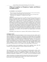
Fig. 1 GLA reduces the angiogenic capacity in MDA-MB-231 cells. a MDA-MB-231 cells were exposed to 10 or 20 μM GLA for 48 h, using a Cell
Counting Kit-8 assay of the cell vitalities, The percentage of cell viability was calculated via comparing with non-treated cells (mean ± SD, n = 3).
Cells were then exposed to 10 or 20 μM GLA for 48 h, and conditioned media was collected. b The ELISA was used to detect the effects of GLA
on VEGF secretion (mean ± SD, n = 3). MDA-MB-231 cells were pretreated with 0 or 20 μM GLA for 48 h, then the previous media was removed,
and cells were washed with 1× PBS to replace fresh media with 1% serum for 24 h. The conditioned media was collected and incubated in
(c) tube formation assays of the angiogenic capacity in MDA-MB-231 cells, HUVECs were exposed to the conditioned mediums collected as
described in (b) for 6 h. d Quantitative analyses of the tube numbers, the total number of formed tube branches in each well was counted under
the light microscope (mean ± SD, n = 5); **P < 0.01 and ***P < 0.001 compared with cells treated without GLA
Mu et al. BMC Cancer (2017) 17:307
Page 5 of 9
Fig. 2 GLA blocks Wnt/β-catenin signaling in MDA-MB-231 cells. MDA-MB-231 cells were exposed to 10 or 20 μM GLA for 48 h. a Western blot
analyses relative protein levels of Wnt1, non-phospho (active) β-catenin, and β-catenin. b Expression of Wnt1 and β-catenin (mRNAs) were
analyzed by the quantitative real-time polymerase chain reaction (qRT-PCR) (mean ± SD, n = 3). c Immunofluorescence assay analyses the nuclear
translocation of β-catenin. d Expression of TCF/LEF4 (mRNAs) analyzed by qRT-PCR (mean ± SD, n = 3). **P < 0.01 and ***P < 0.001 compared with
the control cells
downstream target genes: c-myc, cyclin D1, VEGF and so
on [25]. Here, we treated the MDA-MB-231cells with
20 μM GLA. As shown in Fig. 2d, GLA inhibited the
mRNA expression of LEF/TCF4, suggesting that GLA
could potentially suppress activation of transcriptional
factors LEF/TCF4. Collectively, these data indicated that
Wnt/β-catenin signaling can be blocked by GLA in human breast cancer cells.
knockdown, the downregulated protein level of Wnt1,
non-phospho (active) β-catenin (Fig. 3b) and mRNA expression of Wnt1, LEF/TCF4 (Fig. 3c and d) induced by
GLA were significantly further decreased, suggesting
that GLA blocked the Wnt/β-catenin signal pathway
through miR-148a.
miR-148a interferes in the Wnt/β-catenin signaling in
GLA-treated MDA-MB-231 cells
Based on our results, we hypothesized that the attenuation of Wnt/β-catenin signaling by miR-148a is involved in GLA-induced anti-angiogenesis in breast
cancer cells. To test this hypothesis, we treated miR148a knockdown MDA-MB-231 and Hs-578 T cells with
GLA to determine their angiogenic abilities. miR-148a
knockdown resulted in the decrease of GLA-induced
suppression of VEGF expression/secretion (Fig. 4a) and
tube formation (Fig. 4b and c) in these cells.
To avoid the interference of GLA and serum on
HUVECs, human cancer cells were pretreated by
GLA, then the culture medium was substituted, and
new fresh medium with 1% FBS was added. After
24 h treatment, the conditioned media was collected.
We found the up-regulation of miR-148a, the suppression of Wnt/β-catenin, and the down-regulation
A previous study suggested that miR-148a negatively
regulated the epithelial to mesenchymal transition
(EMT) and CSC-like properties of HCC by directly targeting Wnt1 [11]. In the present study, GLA increased
the expression of miR-148a in breast cancer cells when
exposed to 20 μM GLA for 48 h (Additional file 3:
Figure S1B). Subsequently, we explored whether miR148a could affect Wnt/β-catenin signaling under the
treatment of GLA. After transfection with anti-miRnegative control or anti-miR-148a for 12 h, the efficiency
of gene transfection in MDA-MB-231 or Hs-578 T cells
was assayed (Additional file 3: Figure S1C). The transfected cells were maintained for 48 in culture medium
with or without GLA (20 μM). After miR-148a
Functions of miR-148a in GLA-induced anti-angiogenesis
in breast cancer cell
Mu et al. BMC Cancer (2017) 17:307
Page 6 of 9
Fig. 3 GLA attenuates the expression/activation of Wnt/β-catenin of breast cancer cells through miR-148a. a-d MDA-MB-231 or Hs-578 T cells
were pre-transfected by anti-miR-negative control or anti-miR-148a for 12 h, and then treated with 20 μM GLA for 48 h. a qRT-PCR analyses of
miR-148a (mean ± SD, n = 3). b Western blot analysis relative protein levels of Wnt1 and non-phospho (active) β-catenin. c–d Expression of Wnt1
and TCF/LEF were analyzed by qRT-PCR (mean ± SD, n = 3). *P < 0.05 and **P < 0.01 compared with the anti-miR-negative control. #P < 0.05
compared with the cells treated with GLA and anti-miR-negative control
of VEGF. Our results further indicated that miR148a-mediated inhibition of the Wnt/β-catenin signal
pathway might be involved in GLA induced suppression of angiogenesis, and reduction of VEGF secretion
(Additional file 4: Figure S2).
Discussion
GLA, a flavonoid extracts from the phytochemical licorice, has multiple biological activities [14], such as
estrogen-like [26] and anti-inflammatory activities [27].
In addition, GLA can activate caspase-3, −8, and −9 to
induce HL-60 cell apoptosis through the regulation of
the p38 MAPK and JNK1/2 pathways [18]. GLA inhibits
migration and invasion by transcriptional inhibition of
MMP 9 through modulation of NF-κB and AP-1 activity
in human liver cancer cells. Hsieh et al. showed that
GLA inhibited the transcription factors NF-κB, activator
protein 1 signaling pathways and phosphorylation of
ERK, JNK and p38 MAPKs in human liver cancer cells
[28]. Our previous studies confirmed that GLA inhibited
the CSC-like properties through the TGF-β/SMAD signaling pathway in HCC and breast cancer cells [17, 22].
Recent studies have revealed that GLA contributes to
the inhibition of invasion, migration, and angiogenesis of
breast cancer cells and lung cancer cells via a FAK/Rho
mediated signaling pathway [19, 20]. In our study, we
addressed the role of GLA in angiogenesis and found
that GLA decreased the number of tubes in HUVECs by
decreasing the expression and secretion of VEGF in
breast cancer cells. In summary, the above results suggest that the tumor angiogenesis of breast cancer cells
can be alleviated by GLA.
In cancer cells, the aberrantly activated Wnt/β-catenin
signaling is able to regulate diverse biological processes,
such as cell motility, migration, differentiation, proliferation as well as survival [29]. Wnt signaling is activated
when it binds to the corresponding receptor, frizzled
protein. After translocation into the cell nucleus, βcatenin binds with the TCF/LEF family and forms the
complexes, modulating the transcription of various target genes, including cyclin D1, interleukin-8, and VEGF,
which is a critical proangiogenic factor [30, 31]. For
lung cancer cells, the inhibition of angiogenesis resulted from downregulation of the Wnt/β-catenin signaling axis [32]. The present study demonstrated that
GLA treatment lead to the downregulation of Wnt1 in
MDA-MB-231 and Hs-578 T cells. Besides, β-catenin
was mainly distributed in the nucleus and cytoplasm,
but we found its accumulation in membranes after
GLA treatment. These results suggest that in the
breast cancer cells, GLA can block the activation of
Wnt/β-catenin signaling.
Mu et al. BMC Cancer (2017) 17:307
Page 7 of 9
Fig. 4 Functions of miR-148a in GLA-induced anti-angiogenesis. Cells were treated as described in Fig. 3, and the conditioned media were
collected. a The ELISA was used to detect VEGF secretion (mean ± SD, n = 3). The previous media with anti-miRNAs and GLA were removed, and
the cells were washed with 1× PBS and replaced with fresh media with 1% serum for 24 h, and the conditioned media was collected. b HUVECs
were exposed to the conditioned mediums collected as described in (a) for 6 h. c Quantitative analyses of the tube numbers, the total number
of formed tube branches in each well was counted under the light microscope (mean ± SD, n = 5). *P < 0.05 and **P < 0.01 compared with the
anti-miR-negative control. #P < 0.05 compared with the cells treated with GLA and anti-miR-negative control
MiR-148a is a member of the miR-148/152 family that
is usually regulated by methylation of CpG islands [33].
Growing evidence suggests that miR-148a is poorly
expressed in various tumors, indicating that miR-148a
can serve as a biomarker for diagnosis and prognosis
[34]. miR-148a can regulated various target genes and its
corresponding pathways, which is related to cell proliferation [35], invasion and metastasis [36], and angiogenesis [12]. In breast cancer cells, miR-148a inhibits tumor
angiogenesis via targeting IGF-IR and IRS1 and suppressing their downstream AKT and MAPK/ERK signaling pathways [13]. Here, we explored the expression
changes of four target genes of miR-148a (ERBB3,
PKM2, IGF-IR and IRS1) in MDA-MB-231 cells followed
by GLA addiction and silencing of miR-148a in
Additional file 5: Figure S3. MiR-148a also modulates
angiogenesis by directly targeting the M2 isoform of
pyruvate kinase in mammary tumor cells [37]. Previous
studies have shown that Wnt1 was a direct target of
miR-148a [11]; however, little is known concerning the
association of miR-148a with Wnt/β-catenin signaling
during angiogenesis, so we aimed to uncover whether
miR-148a was involved in the blockage of Wnt/β-catenin
signaling. We determined the role of miR-148a in breast
cancer angiogenesis after transfecting anti-miR-148a into
breast cancer cells and found that the decreased expression or secretion of VEGF was reversed, indicating that
miR-148a had a negative effect on angiogenesis.
Conclusion
In conclusion, our findings suggest that GLA could be a
promising chemopreventive drug in various cancers.
Many investigators including us try to reveal its potential
molecular mechanisms. Based on the previous peoples’
studies, our results further demonstrated that GLA
could inhibit the growth and progression of tumors via
affecting multiple signaling pathways. Using breast cancer MDA-MB-231 and Hs-578 T cells, we found that
GLA decreased the formation of blood vessels by blocking the Wnt/β-catenin signaling pathway, which, in turn,
reduced the secretion of a proangiogenesis factor
(VEGF). Besides, GLA contributed to the over-expression
of miR-148a, which could directly target Wnt1, and promoted the localization in membranes in the cytoplasm
and nucleus. Downregulation of miR-148a reversed GLAinduced intervention of the Wnt/β-catenin signal pathway, the angiogenesis, and VEGF secretion. Therefore,
these results suggest a novel mechanism whereby GLA inhibits angiogenesis, which may provide promising strategies to alleviate breast cancer in the future.
Mu et al. BMC Cancer (2017) 17:307
Additional files
Additional file 1: Table S1. Primers used in this study. (DOCX 17 kb)
Additional file 2: Table S2. miRNA inhibitors used in this study.
(DOCX 16 kb)
Additional file 3: Figure S1. Hs-578 T cells were exposed to 0, 10 or
20 μM GLA for 48 h, and conditioned media was collected. (A) The ELISA
was used to detect the effects of GLA on VEGF secretion (mean ± SD,
n = 3). MDA-MB-231 or Hs-578Tcells were exposed to 0, 10 or 20 μM GLA
for 48 h, (B) qRT-PCR analyses the mRNA level of miR-148a (mean ± SD,
n = 3). The breast cancer cells transfected by anti-miR-negative control or
anti-miR-148a for 12 h, (C) the efficiency of gene transfection was analysed
by qRT-PCR (mean ± SD, n = 3); *P < 0.05, **P < 0.01 and ***P < 0.001
compared with the control cells. (DOCX 195 kb)
Additional file 4: Figure S2. MDA-MB-231 cells were pretreated with 0,
or 20 μM GLA for 48 h, then the media was removed, the cells were
washed with 1× PBS, followed by replacement with fresh media with 1%
FBS for 24 h. (A)The expression of miR-148a was analyzed by qRT-PCR
(mean ± SD, n = 3). (B-C) Western blot analyses the relative protein levels
of Wnt1, β-catenin, and non-phospho (active) β-catenin (mean ± SD,
n = 3). (D) The ELISA was used to detect the secretion of VEGF (mean ± SD,
n = 3); **P < 0.01 and ***P < 0.001 compared with the control media or
cells. (DOCX 322 kb)
Additional file 5: Figure S3. MDA-MB-231 cells were pre-transfected by
anti-miR-negative control or anti-miR-148a for 12 h, and then treated
with 20 μM GLA for 48 h. (A-D) qRT-PCR analyses in triplicate of the
mRNA level of ERBB3, PKM2, IRS1, and IGF-IR (mean ± SD, n = 3).
*P < 0.05, and **P < 0.01 compared with the anti-miR-negative control.
#P < 0.05 compared with the cells treated with GLA and anti-miRnegative control. (DOCX 255 kb)
Abbreviations
AP-1: Activator protein 1; CCK8: Cell Counting Kit-8 assay; CSC: Cancer stem
cell; DAPI: 4′, 6-diamidino-2-phenylindole; DMEM: Dulbecco’s modified
Eagle’s medium; ELISA: Enzyme-linked immunosorbent assay; FAK/Rho: Focal
adhesion kinase/Ras homolog; FBS: Fetal bovine serum; GLA: Glabridin;
HCCs: Hepatocellular carcinomas; HUVECs: Human umbilical vein endothelial
cells; IGF-IR: Insulin-like growth factor- I receptor; IL-6: Interleukin- 6;
IRS1: Insulin receptor substrate 1; JNK1/2: Jun N-terminal kinase; LEF/TCF4: T
cell factor/lymphoid enhancer binding factor 4; MAPK: Mitogen-activated
protein kinase; NF-κB: Nuclear factor-κB; PBST: Tween-buffered phosphatebuffered saline; PVDF: Polyvinylidene fluoride; qRT-PCR: Quantitative real-time
polymerase chain reaction; SDS-PAGE: Sodium dodecyl sulfate polyacrylamide
gel electrophoresis; STAT-3: Signal transducer and activator of transcription 3;
TGF-β: Transforming growth factor-β; UTRs: Untranslated regions; VEGF: Vascular
endothelial grow factor
Acknowledgements
Not applicable.
Funding
This work was supported by the National Natural Science Foundation of
China (81,171,987,81,673,205 and 81,402,667), the Major Program of Natural
Science Research of Jiangsu Higher Education Institutions (15KJA330001), the
Research Fund for the Doctoral Program of Higher Education of China
(20133234110007),and a project funded by the Priority Academic Program
Development of Jiangsu Higher Education Institutions (PAPD). The funding
agencies had no role in the study design, data collection and analysis, the
decision to publish, or the preparation of the manuscript.
Availability of data and material
All data supporting the findings in this study are included in the manuscript
and its additional files.
Authors’ contributions
Conceived and designed the experiment: YL and ZL. Performed the
experiment: JM, SLN, and DMZ. Analyzed the data: JM, DMZ, ZXS, SLN, YL, JC,
YL and ZL. Wrote the paper: JM, DMZ and ZL. Revised critically the paper:
Page 8 of 9
JM, DMZ, ZXS, SLN, YL, JC, YL and ZL. All authors has read and approved the
final manuscript.
Competing interests
The authors declare that they have no competing interests.
Consent for publication
Not applicable.
Ethics approval and consent to participate
Not applicable.
Publisher’s Note
Springer Nature remains neutral with regard to jurisdictional claims in
published maps and institutional affiliations.
Received: 16 August 2016 Accepted: 24 April 2017
References
1. Carmeliet P. Angiogenesis in health and disease. Nat Med. 2003;9:653–60.
2. Cuevas I, Layman H, Coussens L, Boudreau N. Sustained endothelial
expression of HoxA5 in vivo impairs pathological angiogenesis and tumor
progression. PLoS One. 2015;10(3):e0121720.
3. Klaus A, Birchmeier W. Wnt signalling and its impact on development and
cancer. NatRevCancer. 2008;8:387–98.
4. Reya T. Clevers H: Wnt signalling in stem cells and cancer. Nature.
2005;434:843–50.
5. Reis M, Liebner S. Wnt signaling in the vasculature. Exp Cell Res.
2013;319(9):1317–23.
6. Zhang X, Gaspard JP. Chung DC: regulation of vascular endothelial growth
factor by the Wnt and K-ras pathways in colonic neoplasia. Cancer Res.
2001;61(16):6050–4.
7. Sun X, He Y, Huang C, Ma T, Li J. Distinctive microRNA signature associated
of neoplasms with the Wnt/beta-catenin signaling pathway. Cell Signal.
2013;25(12):2805–11.
8. Reddy KB. MicroRNA (miRNA) in cancer. Cancer Cell Int. 2015;15:38.
9. Subramanian M, Rao SR, Thacker P, Chatterjee S, Karunagaran D. MiR-29b
downregulates canonical Wnt signaling by suppressing coactivators of
beta-catenin in human colorectal cancer cells. J Cell Biochem.
2014;115(11):1974–84.
10. Li J, Zhang Y, Zhao Q, Wang J, He X. MicroRNA-10a influences osteoblast
differentiation and angiogenesis by regulating beta-catenin expression. Cell
Physiol Biochem. 2015;37(6):2194–208.
11. Yan H, Dong X, Zhong X, Ye J, Zhou Y, Yang X, Shen J, Zhang J. Inhibitions
of epithelial to mesenchymal transition and cancer stem cells-like properties
are involved in miR-148a-mediated anti-metastasis of hepatocellular
carcinoma. Mol Carcinog. 2014;53(12):960–9.
12. Yu J, Li Q, Xu Q, Liu L, Jiang B. MiR-148a inhibits angiogenesis by targeting
ERBB3. J Biomed Res. 2011;25(3):170–7.
13. Xu Q, Jiang Y, Yin Y, Li Q, He J, Jing Y, Qi YT, Xu Q, Li W, Lu B, et al. A
regulatory circuit of miR-148a/152 and DNMT1 in modulating cell
transformation and tumor angiogenesis through IGF-IR and IRS1. J Mol Cell
Biol. 2013;5(1):3–13.
14. Guo B, Fang Z, Yang L, Xiao L, Xia Y, Gonzalez FJ, Zhu L, Cao Y, Ge G, Yang L,
et al. Tissue and species differences in the glucuronidation of glabridin with
UDP-glucuronosyltransferases. Chem Biol Interact. 2015;231:90–7.
15. Tantishaiyakul V, Suknuntha K, Saithong S, Pakawatchai C. Glabridin. Acta
Crystallogr Sect E Struct Rep Online. 2012;68(Pt 12):o3501.
16. Tamir S, Eizenberg M, Somjen D, Stern N, Shelach R, Kaye A, Vaya J.
Estrogenic and antiproliferative properties of glabridin from licorice in
human breast cancer cells. Cancer Res. 2000;60:5704–9.
17. Jiang F, Li Y, Mu J, Hu C, Zhou M, Wang X, Si L, Ning S, Li Z. Glabridin
inhibits cancer stem cell-like properties of human breast cancer cells: an
epigenetic regulation of miR-148a/SMAd2 signaling. Mol Carcinog.
2016;55(5):929–40.
18. Huang H, Hsieh M, Chien M, Chen H, Yang S, Hsiao P. Glabridin mediate
caspases activation and induces apoptosis through JNK1/2 and p38
MAPK pathway in human promyelocytic leukemia cells. PLoS One.
2014;9(6):e98943.
Mu et al. BMC Cancer (2017) 17:307
19. Hsu YL, Wu LY, Hou MF, Tsai EM, Lee JN, Liang HL, Jong YJ, Hung CH, Kuo PL.
Glabridin, an isoflavan from licorice root, inhibits migration, invasion and
angiogenesis of MDA-MB-231 human breast adenocarcinoma cells by
inhibiting focal adhesion kinase/rho signaling pathway. Mol Nutr Food Res.
2011;55(2):318–27.
20. Tsai YM, Yang CJ, Hsu YL, Wu LY, Tsai YC, Hung JY, Lien CT, Huang MS,
Kuo PL. Glabridin inhibits migration, invasion, and angiogenesis of
human non-small cell lung cancer A549 cells by inhibiting the FAK/rho
signaling pathway. Integr Cancer Ther. 2011;10(4):341–9.
21. Mu J, Ning S, Wang X, Si L, Jiang F, Li Y. Li Z: the repressive effect of miR520a on NF-kB/IL-6/STAT-3 signal involved in the glabridin-induced antiangiogenesis in human breast cancer cells. RSC Adv. 2015;6:24719–27.
22. Jiang F, Mu J, Wang X, Ye X, Si L, Ning S, Li Z. Li Y: the repressive effect of
miR-148a on TGF beta-SMADs signal pathway is involved in the glabridininduced inhibition of the cancer stem cells-like properties in hepatocellular
carcinoma cells. PLoS One. 2014;9(5):e96698.
23. Birdsey GM, Shah AV, Dufton N, Reynolds LE, Osuna Almagro L, Yang Y,
Aspalter IM, Khan ST, Mason JC, Dejana E, et al. The endothelial transcription
factor ERG promotes vascular stability and growth through Wnt/betacatenin signaling. Dev Cell. 2015;32(1):82–96.
24. Schuijers J, Mokry M, Hatzis P, Cuppen E, Clevers H. Wnt-induced
transcriptional activation is exclusively mediated by TCF/LEF. EMBO J.
2014;33(2):146–56.
25. Boon EM, Keller JJ, Wormhoudt TA, Giardiello FM, Offerhaus GJ, van der
Neut R, Pals ST. Sulindac targets nuclear beta-catenin accumulation and
Wnt signalling in adenomas of patients with familial adenomatous polyposis
and in human colorectal cancer cell lines. Br J Cancer. 2004;90(1):224–9.
26. Tamir S, Eizenberg M, Somjen D, Stern N, Shelach R, Kaye A, Vaya J.
Estrogenic and antiproliferative properties of glabridin from licorice in
human breast cancer cells. Cancer Res. 2000;60(20):5704–9.
27. Yehuda I, Madar Z, Leikin-Frenkel A, Tamir S. Glabridin, an isoflavan from
licorice root, downregulates iNOS expression and activity under highglucose stress and inflammation. Mol Nutr Food Res. 2015;59(6):1041–52.
28. Hsieh MJ, Lin CW, Yang SF, Chen MK, Chiou HL. Glabridin inhibits migration
and invasion by transcriptional inhibition of matrix metalloproteinase 9
through modulation of NF-kappaB and AP-1 activity in human liver cancer
cells. Br J Pharmacol. 2014;171(12):3037–50.
29. Song JL, Nigam P, Tektas SS. Selva E: microRNA regulation of Wnt signaling
pathways in development and disease. Cell Signal. 2015;27(7):1380–91.
30. Zerlin M, Julius MA, Kitajewski J. Wnt/frizzled signaling in angiogenesis.
Angiogenesis. 2008;11(1):63–9.
31. Parmalee NLKJ. Wnt signaling in angiogenesis. Curr Drug Targets.
2008;9(7):558–64.
32. Shukla S, Sinha S, Khan S, Kumar S, Singh K, Mitra K, Maurya R, Meeran SM.
Cucurbitacin B inhibits the stemness and metastatic abilities of NSCLC via
downregulation of canonical Wnt/beta-catenin signaling axis. Sci Rep.
2016;6:21860.
33. Chen Y, Song Y, Wang Z. The microRNA-148_152 family: multi-faceted
players. Mol Cancer. 2013;12:43.
34. Xia J, Guo X, Yan J, Deng K. The role of miR-148a in gastric cancer. J Cancer
Res Clin Oncol. 2014;140(9):1451–6.
35. Guo S, Peng Z, Yang X, Fan K, Ye H, Li Z, Wang Y, Xu X, Li J, Wang Y, et al.
miR-148a promoted cell proliferation by targeting p27 in gastric cancer
cells. Int J Biol Sci 2011; 7(5):567–574.
36. Zheng B, Liang L, Wang C, Huang S, Cao X, Zha R, Liu L, Jia D, Tian Q, Wu J,
et al. MicroRNA-148a suppresses tumor cell invasion and metastasis by
downregulating ROCK1 in gastric cancer. Clin Cancer Res. 2011;17(24):7574–83.
37. Xu Q, Liu LZ, Yin Y, He J, Li Q, Qian X, You Y, Lu Z, Peiper SC, Shu Y, et al.
Regulatory circuit of PKM2/NF-kappaB/miR-148a/152-modulated tumor
angiogenesis and cancer progression. Oncogene. 2015;34(43):5482–93.
Page 9 of 9
Submit your next manuscript to BioMed Central
and we will help you at every step:
• We accept pre-submission inquiries
• Our selector tool helps you to find the most relevant journal
• We provide round the clock customer support
• Convenient online submission
• Thorough peer review
• Inclusion in PubMed and all major indexing services
• Maximum visibility for your research
Submit your manuscript at
www.biomedcentral.com/submit