Presence of human papillomavirus DNA in breast cancer: A Spanish case-control study
Bạn đang xem bản rút gọn của tài liệu. Xem và tải ngay bản đầy đủ của tài liệu tại đây (687.4 KB, 11 trang )
Delgado-García et al. BMC Cancer (2017) 17:320
DOI 10.1186/s12885-017-3308-3
RESEARCH ARTICLE
Open Access
Presence of human papillomavirus DNA in
breast cancer: a Spanish case-control study
Silvia Delgado-García1* , Juan-Carlos Martínez-Escoriza1, Alfonso Alba2, Tina-Aurora Martín-Bayón1,
Hortensia Ballester-Galiana1, Gloria Peiró3, Pablo Caballero4 and Jose Ponce-Lorenzo5
Abstract
Background: Breast cancer is one of the most important neoplasia among women. It was recently suggested that
biological agents could be the etiological cause, particularly Human Papilloma Virus (HPV). The aim of this study
was to explore the presence of HPV DNA in a case-control study.
Methods: We performed our study including 251 cases (breast cancer) and 186 controls (benign breast tumors),
using three different molecular techniques with PCR (GP5/GP6, CLART® and DIRECT FLOW CHIP®).
Results: HPV DNA was evidenced in 51.8% of the cases and in 26.3% of the controls (p < 0.001). HPV-16 was the
most prevalent serotype. The odds ratio (OR) of HPV within a multivariate model, taking into account age and
breastfeeding, was 4.034.
Conclusions: Our study, with methodological rigour and a sample size not previously found in the literature,
demonstrate a significant presence of HPV DNA in breast cancer samples. A possible causal relationship, or
mediation or not as a cofactor, remains to be established by future studies.
Keywords: Breast cancer, Human papillomavirus, Prevalence, PCR
Background
Breast cancer is the most commonly diagnosed malignancy in women [1–3]. It is estimated that 1.7 million
new cases were diagnosed in 2012, representing 11.9% of
all cancers diagnosed worldwide in both genders, and
25% of those diagnosed in women [3, 4]. Breast cancer is
also the most common malignancy in Spanish women,
representing 29% of all female malignancies. Most of the
cases are diagnosed in patients between 45 and 65 years
of age [5].
Several risk factors have been cited in the literature,
including patient age, gender, hormone therapy, the
number of offspring, breastfeeding or different eating
habits. However, there are other less well known factors
that might also play an oncogenic role. Viruses are an
example in this respect [6]. A number of viruses have
been identified to date in breast cancer tissues. The
three main viruses are Epstein Barr virus (EBV), mouse
* Correspondence:
1
Department of Obstetrics and Gynecology, University General Hospital of
Alicante, c/ Pintor Baeza, 11, 03010 Alicante, Spain
Full list of author information is available at the end of the article
mammary tumor virus (MMTV) and human papillomavirus (HPV) [7–11]. All of them share a common feature
in that they can induce the initiation and progression of
cancer. Several studies [8, 9, 12–19] have attempted to
determine whether viruses in breast tissue are a casual
presence (i.e., acting as “passengers”) or they play an important role in carcinogenesis. The fact is that with the
exception of MMTV, the rest of the viruses described in
breast cancer have already been identified in other malignancies. The current published data on HPV and
breast cancer are very contradictory, since the reported
prevalence of HPV ranges from 0% [20–29] to 86.21% in
breast cancer tissue samples [8, 10, 30–35]. Furthermore,
the studies are very heterogeneous in terms of the methodology employed. A example of this is that, most of the
reviewed studies involve case studies without controls. A
few use case-control protocols, which offer greater
methodological soundness, while only a handful evaluate
statistically significant differences [9, 31, 36–43]. Moreover, the only study conducting logistic regression is that
published by Sigaroodi et al. [40], though it involves a
© The Author(s). 2017 Open Access This article is distributed under the terms of the Creative Commons Attribution 4.0
International License ( which permits unrestricted use, distribution, and
reproduction in any medium, provided you give appropriate credit to the original author(s) and the source, provide a link to
the Creative Commons license, and indicate if changes were made. The Creative Commons Public Domain Dedication waiver
( applies to the data made available in this article, unless otherwise stated.
Delgado-García et al. BMC Cancer (2017) 17:320
very wide confidence interval (1.5–130) and odds ratio
[OR] = 14, which is questionable in statistical terms.
In view of the above, the investigation of viruses as
breast cancer promoting factors remains subject to great
controversy. The present study was designed to help
clarify this issue. Specifically, we aimed to confirm the
presence of HPV in a series of samples obtained from
breast surgeries at the University General Hospital of
Alicante (Spain), estimating the strength of the association (via [OR]) between the presence of HPV in benign
breast disease and breast cancer.
Methods
A case-control study, based on a case-control ratio of
1:1, was performed to evaluate the presence of HPV infection in a subset of 250 embedded breast cancer
tissues, as cases, and 250 embedded benign breast
tissues, as controls. The estimated exposure rate (presence of HPV) was 25% and 14% in the cases and controls, respectively, with a confidence level of 95% and a
statistical power of 85% in detecting OR >2 (computed
pooling proportions of reviews or meta-analyses published until 2012) [8, 30, 32]. The samples were selected
consecutively and retrospectively from the year 2012
until the calculated sample size (n) was reached. The
following inclusion criteria were established: women
subjected to surgical treatment due to infiltrating breast
cancer and/or carcinoma in situ (cases) or benign breast
disease (controls) (period 2006–2012); patients over
18 years of age; surgical specimens embedded in paraffin
(stored in the tumor Biobank of our institution), in adequate conditions and sufficient amount of tissue for
the purposes of the study; and the obtainment of written informed consent. The following exclusion criteria
were established: males and a lack of the minimum
required quality controls in the analyzed DNA samples.
An ad-hoc case report form was created to record
demographic, histopathological and virological information. Data were anonymized in compliance with the
protection of personal data code.
Immunohistochemical and in SITU hybridization analysis
After surgical excision (either mastectomy or tumorectomy), specimens were fixed in 10% formalin solution
and subsequently embedded in paraffin. For the histological study, sections measuring 4 μm in thickness were
obtained and stained with hematoxylin-eosin. The
expressions of estrogen receptor (ER), progesterone receptor (PgR), human epidermal growth factor receptor
(HER2) and Ki-67 were determined by immunohistochemistry (IHC) using standard techniques, with commercial antibodies and conditions following the
instructions of the manufacturer on an automated basis
(Techmate-500). The following antibodies were used: ER
Page 2 of 11
(Dako, clone 1D5, dilution 1:50), PgR (Dako, clone PgR
636, dilution 1:50), Ki-67 (Dako, clone MIB-1, dilution
1:100) and HercepTest® (Dako). The study of the ER and
PR expression levels was made evaluating the percentage
of stained tumor cell nuclei and the intensity of staining
according to the guidelines of the American Society of
Clinical Oncology (2010) [44] and of the American
College of Pathologists. Positive status was considered
for >1% ER or PR. HER2 status in turn was determined
according to the recommendations of the American
Society of Clinical Oncology (2007) and guidelines of
the American College of Pathologists [45]. Immunohistochemical positive was defined as staining 3+ (uniform, membrane staining intensity >10% of the
infiltrating tumor cells), while negative was defined as
staining 0 or 1+. ERBB2 gene status was confirmed by
fluorescence in situ hybridization (FISH) (Dako pharmaDx™) or chromogenic in situ hybridization (CISH)
(Spot light™; Zymed) in equivocal cases (2+ and <10% 3+
cells). Ki67 was semiquantitatively assessed in at least
three high-magnification fields [×400] including hot-spots
areas, and classified as low (<14%) versus high (>14%) (nuclei) [44, 45].
Viral DNA sequences extraction
The search for viral DNA was carried out at the Instituto
de Estudios Celulares y Moleculares (Lugo, Spain), due to
its well demonstrated experience in molecular and genetic
studies. Sections measuring 10 μm in thickness were obtained from the tumor area of the paraffin block for the
identification of viral DNA. In order to avoid crosscontamination between samples, special care was taken in
handling and sectioning the samples. The following procedure was carried out:
1. Deparaffinization: Four paraffin-embedded tissue
sections were placed in a 1.5-ml tube, followed by
the addition of 1 ml of xylene and vortexing for 10 s.
After incubation at room temperature during
10 min, centrifugation was carried out at 13,000 rpm
for 5 min. The supernatant was discarded and 1 ml
of absolute ethanol was added, followed by centrifugation at 13,000 rpm for 2 min. The supernatant
was then again discarded. This ethanol washing step
was repeated one more time. Lastly, the sample was
incubated at 56 °C during 15 min to eliminate the
traces of ethanol.
2. DNA extraction: After completion of the
deparaffinization process we added 500 μl of lysis
buffer (10 mM Tris pH 8, 100 mM NaCl, 25 mM
EDTA, 0.5% sodium dodecylsulfate [SDS]) and 10 μl
of proteinase K (20 mg/ml), followed by vortexing
and incubation in a shaking water bath at 56 °C
overnight. Proteinase K was inactivated by
Delgado-García et al. BMC Cancer (2017) 17:320
3.
4.
5.
1. -
2. -
incubation at 95 °C during 10 min. An equivalent
volume of chloroform: isoamyl alcohol (24:1 v/v)
was added, shaking gently by inverting the tube
and then centrifuging at 10,000 rpm for 10 min.
The upper aqueous phase was transferred to a new
microcentrifugation tube, and 0.2 volumes of
ammonium acetate 10 M were added. The DNA
was precipitated by adding two volumes of absolute
ethanol, followed by vortexing for 5 s, incubation at
−20 °C during 30 min and centrifugation at
10,000 rpm for 20 min. The supernatant was
discarded and the precipitate was washed with
500 μl of cold 70% ethanol, followed by
centrifugation for 5 min at 10,000 rpm. After
discarding the supernatant again, the precipitate
was dried at room temperature during 20 min.
The DNA was finally resuspended in 50 μl of
Tris-EDTA solution.
Amount and quality of DNA: All DNA samples
were analyzed using a Nanodrop 1000 kit allowing
calculation of the concentration of DNA and the
A260/A280 and A260/A230 ratios, which indicate
the purity of the molecule.
DNA amplification capacity: The integrity of the
extracted DNA was evaluated by polymerase chain
reaction (PCR) amplification of a fragment of the
methylenetetrahydrofolate reductase (MTHFR) gene.
Detection and genotyping of HPV: The samples
were subjected to three different HPV detection
methods:
Amplification of the virus using the GP5+/GP6+
consensus primers: The presence of HPV DNA
was evaluated by PCR using the GP5+/GP6+
primers (150 bp), which act as consensus primers
for the HPV L1 gene. The PCR reaction was carried
out with 5 μl of DNA in a total reaction volume
of 50 μl containing 25 μl of DreamTaq Green PCR
Master Mix 2X (ThermoFisher Scientific), 1 μM of
each primer, 0.2 mM of DNTPs and 2 mM of
MgCl2. Amplification was performed with initial
activation of the enzyme at 95 °C during two
minutes, followed by 45 cycles under the following
conditions: 30 s at 95 °C, two minutes at 40 °C and
1.5 min at 72 °C, with a final elongation step at
72 °C during 5 min. The PCR products were
visualized in 2% agarose gel with ethidium bromide
staining using electrophoresis.
CLART® HPV2 amplification kit (Genomica): This
kit detects the presence of the 35 HPV viruses: 6, 11,
16, 18, 26, 31, 33, 35, 39, 40, 42, 43, 44, 45, 51, 52,
53, 54, 56, 58, 59, 61, 62, 66, 68, 70, 71, 72, 73, 81,
82, 83, 84, 85 and 89. Detection is carried out
through amplification of a fragment of about 450 bp
within the L1 region of the virus. Five μl of DNA of
Page 3 of 11
each sample were subjected to PCR assay using the
CLART HPV2 amplification kit (Genomica): one
cycle at 95 °C for 5 min, 40 cycles at 94 °C for 30 s/
55 °C for 60 s / 72 °C for 90 s, and one cycle at
72 °C for 8 min. The PCR products were
denaturalized at 95 °C during 10 min and placed
in a container with ice. Hybridization was performed
using 10 μl of the denaturalized PCR product in
the CLART microarray, followed by examination
according to the instructions of the manufacturer.
3. - HPV Direct Flow CHIP kit (Master Diagnostica):
The technique is based on amplification of the viral
DNA followed by membrane flow-through reverse
dot blot hybridization of the amplified products.
Types of HPV detected: High oncogenic risk
(16, 18, 26, 31, 33, 35, 39, 45, 51, 52, 53, 56, 58, 59,
66, 68, 73 and 82) and low oncogenic risk (6, 11, 40,
42, 43, 44, 54, 55, 61, 62, 67, 69, 70, 71, 72, 81, 84
and 89 (CP6108)). Six μl of purified DNA of each
sample were amplified by PCR under the following
conditions: one cycle at 98 °C for 5 min, 5 cycles at
98 °C for 5 s / 42 °C for 5 s / 72 °C for 10 s, 45 cycles
at 98 °C for 5 s / 60 °C for 5 s / 72 °C for 10 s, and
one cycle at 72 °C for one minute. The samples were
kept in refrigerated tubes (8–10 °C) until processing.
The PCR products were denaturalized by heating to
95 °C for 5 min (in a thermocycler) and then quickly
cooled in ice for two minutes.
Hybridization and interpretation of the results were
carried out following the instructions of the kit manufacturer. All samples were analyzed using the three techniques above mentioned to increase test sensitivity. A
positive result was defined when at least two of the three
methods detected the presence of HPV. If the results
proved questionable, or in the event of insufficient material, the sample was discarded to the effects of analysis.
Likewise, all the samples passed the cellular DNA test
(internal control), to avoid possible false-negative results.
As negative control we included “HPV free DNA” and as
positive control a HPV plasmid mixture with all target
types. Contradictory results were obtained in only 6
samples, and these were therefore considered lost.
The following variables were recorded: patient age, personal breast cancer history, smoker, number of children,
breastfeeding, age at menopause, history of cervical disease,
adjuvant therapy, histopathological of the tumor (tumor
size, grade, stage, number of positive lymph nodes, local/
distant metastasis), immunohistochemical characteristics
(ER, PgR, HER2, Ki67), and the detected HPV serotype.
Data analysis
A descriptive analysis was made of all the study variables. In order to analyze the homogeneity of the two
Delgado-García et al. BMC Cancer (2017) 17:320
Page 4 of 11
groups (cases and controls) with respect to those variables which the literature describes as being associated
to breast cancer, a calculation was made of the means of
the quantitative variables for both groups and comparisons were established using the Student t-test. In the
case of the qualitative variables, cross tables were generated, and associations were analyzed using the ChiSquared test. In addition, any variables (qualitative or
quantitative) found to be non-homogeneous in the cases
and controls were taken into account when explaining
the lack of homogeneity for the other variables. To this
effect, we calculated the strength of the association between the cases and controls with the variable in question, in the presence of those variables which had
already demonstrated a lack of homogeneity in both
groups. This process was carried out using a binary logistic regression model. In order to establish the association between HPV exposure and breast cancer, we
generated a cross tables between the two variables, estimating the magnitude of the association based on calculation of the raw odds ratio (OR) for the development of
breast cancer. In addition, we calculated the adjusted
OR by binary logistic regression, with the corresponding
95% confidence interval (CI). A p-value <0.05 was considered statistically significant. Statistical analysis was
carried out using the software package SPSS version 20.
Results
The final study included 437 samples: 251 cases (57.4%)
and 186 controls (42.6%). (Table 1).
The mean age of the cases (n = 251) was 56.3 years,
versus 40.1 years for the controls (n = 186) ((p < 0.001).
Of note, the two groups are not homogeneous. A statistically significant difference of 16 years was observed between the cases and controls (Tables 1 and 2). This is
justified on the basis of the natural courses of breast
cancer and benign disease, and the curves corresponding
to breast cancer and benign disease by ages described by
the World Health Organization (WHO) [4] are analogous to those of our own study. Clearly, for ethical reasons, we cannot obtain healthy tissues on a random
basis from health women without breast disease. Selection bias results if the groups are not homogeneous.
However, by using statistical tools such as binary logistic
regression analysis, we can control this bias referring to
the lack of homogeneity in terms of variables which presumably may be related to cancer. Furthermore, this
statistical tool allows us to calculate odds ratios for each
variable. After justification of the variable age, we compared the rest of the variables of the cases and controls
(Table 2), and these were found to be homogeneous after
logistic regression adjustment to age. This allows us to
establish statistical comparisons of our primary variable,
which is the presence or not of viral DNA.
Data obtained to determine the presence of HPV in
breast cancer tissue samples and establish the comparison with the samples corresponding to benign breast tissue, were analyzed using the Chi-Squared test. Using
this test, the HPV exposure rate among the cases was
significantly higher (51.8%) than the HPV exposure rate
in the controls (26.3%) (p < 0.001) (Table 3).
The raw OR was 3.0 (CI 95%: 2.0–4.5). On applying
the binary logistic regression model to control for confounding variables, the OR assigned to HPV was seen to
be 4.034 (CI 95%: 2.213–7.352) (Table 4), which means a
higher risk of suffering cancer in the presence of HPV,
taking into account patient age and breastfeeding. The
rest of the confounding variables showed no significance
in the binary logistic regression model (all p = non
significant).
The mean tumor size in HPV-positive tumors was
larger (30.53 mm) than in those HPV-negative
(28.37 mm), though the difference failed to reach statistical significance (p = 0.395). None of the analyzed
histopathological variables showed a statistically significant association with the presence of HPV (Table 5, at
the end).
Regarding the association of the different immunohistochemical subtypes with the presence or not of HPV,
the presence of HPV is related to the luminal B phenotypes (particularly HER2-negative), while in contrast the
triple negative and luminal A phenotypes are more related to the absence of HPV. However, this relationship
is not statistically significant (p = 0.055) (Table 5).
Within the global sample, 47% of the cases (n = 55)
and 61.2% of the controls (n = 30) were infected by
Table 1 Clinical characteristics of breast cancer tissues (cases) and breast benign diseases (controls)
Cases
Controls
N
Mean (CI 95%)
N
Mean (CI 95%)
Age**
251
56.32 (54.76–57.88)
186
40.08 (38.3–41.86)
Number of children
250
1.92 (1.76–2.09)
185
1.3 (1.11–1.5)
Breastfeeding duration (months)
191
8.65 (7.16–10.15)
115
9.42 (6.92–11.92)
Age at menopause
140
48.89 (48.16–49.61)
36
48 (46.47–49.53)
Tumor size (mm)
249
29.49 (26.99–31.99)
98
31.83 (29.31–34.34)
CI 95% Confidence Interval 95%, ** Statistic Signification <0.01
Delgado-García et al. BMC Cancer (2017) 17:320
Page 5 of 11
Table 2 Frequency of cases and controls by clinicopathological factors
Age (years)
Smoker
Personal BC history
Number of children
Breastfeeding (months)
Menopausial status
Age at menopause (years)
History of cervical diseases
Number
Cases n (%)
Controls n (%)
p-value
(p-value adjusting for age)
OR (CI 95%)
AOR by Age (CI 95%)
< 40
109
19 (7.6)
90 (48.4)
<0.001
1
−
40–49
119
60 (23.9)
59 (31.7)
4.8 (2.6–8.9)**
−
50–59
97
72 (28.7)
25 (13.4)
13.6 (7.0–26.7)**
−
≥ 60
112
100 (39.8)
12 (6.5)
39.5(18.2–85.8)**
−
Yes
196
93 (37.7)
103 (56.3)
0.5 (0.3–0.7)**
0.8(0.5–1.2)
No
234
154 (62.3)
80 (43.7)
1
1
Yes
13
12 (4.8)
1 (0.5)
9.3 (1.2–72.1)**
7.0(0.8–64.7)
No
424
239 (95.2)
185 (99.5)
1
1
1+
326
205 (81.7)
121 (65.1)
2.4 (1.5–3.7)**
1.0 (0.6–1.8)
None
111
46 (18.3)
65 (34.9)
1
1
12+
81
52 (27.2)
29 (25.2)
1.1 (0.6–1.9)
0.8 (0.4–1.5)
0–11
170
139 (72.8)
86 (74.8)
Yes
188
152 (62.0)
36 (19.7)
No
240
93 (38.0)
147 (80.3)
≥ 54
25
24 (15.8)
1(2.8)
42–53
149
120 (78.9)
29 (80.6)*
≤ 41
14
8(5.3)
6 (16.7)
No
302
156 (95.7)
146 (91.8)
Yes
20
7 (4.3)
13 (8.2)
<0.001
(0.446)
0.009
(0.086)
<0.001
(0.465)
0.789
(0.139)
1
1
<0.001
(0.238)
6.7 (4.2–10.4)**
0.7 (0.3–1.6)
1
1
0.012
(0.179)
18 (1.9–173)**
7.5 (0.7–82.6)
3.1 (0.9–9.6)*
1.8 (0.5–6.9)
1
1
1.9 (0.8–5.1)
1.7 (0.6–4.8)
1
1
0.171
(0.288)
OR Odd Ratio, AOR Adjusted Odd Ratio, BC Breast Cancer, CI 95% Confidence Interval 95%, * Statistic signification <0.05, ** Statistic Signification <0.01
more than one HPV serotype. In other words, coinfection by more than one viral serotype was observed
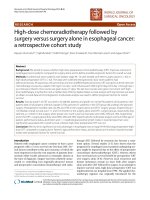
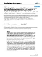
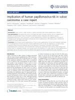
in 85 samples. With regard to the identified serotypes, in
the 179 samples (130 cases and 49 controls) with the
presence of viral DNA, we identified 16 different high
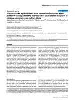
risk serotypes and 11 low risk serotypes. Figures 1 and 2
show HPV serotype 16 to be the most frequent high risk
serotype in both the cases and the controls, followed by
HPV-89 (Fig. 3).
Discussion
Band in 1991 [46] were the first to postulate that HPV
might be implicated in breast cancer. These authors suggested that HPV-16/HPV-18 could immortalize the
Table 3 Frequency of HPV-positive by cases and controls
HPV
Cases (n = 251)
Controls (n = 186)
N
%
N
%
HPV+
130
51.8%
49
26.3%
3.0 **
HPV-
121
48.2%
137
73.7%
(2.0–4.5)
** Statistic Signification <0.01, CI 95% Confidence Interval 95%
OR (CI
95%)
epithelial cells of normal mammary gland tissue through
the inhibition of apoptosis. Shortly after, in 1992, Di
Lonardo [47] by PCR techniques confirmed the presence
of HPV-16 in 29.4% of 17 breast cancer samples supporting a potential relationship between HPV and breast
carcinoma.
In the current study, the presence of HPV was shown
in 51.8% of the cases and 26.3% of the controls. Of note,
these results are higher than the prevalence described by
some meta-analyses [30], in which HPV was found to be
present in 23% of the cases and in 12.9% of the controls,
although the difference here was also statistically
Table 4 Binary logistic regression model to control for
confounding variables in a case-control study
Coef. B
Sig.
OR.
CI. 95%
HPV
1.395
<0.001
4.034
2.213–7.352
Age
0.11
<0.001
1.116
1.084–1.15
Breastfeeding
−0.032
0.022
0.969
0.943–0.996
Constant
−5.274
<0.001
0.005
Coef. B. Value of the coefficient in the logistic regression model. Sig Statistic
signification, OR Odd Ratio, CI 95% Confidence Interval 95%
Delgado-García et al. BMC Cancer (2017) 17:320
Page 6 of 11
Table 5 Frequency of HPV-positive cases (Breast cancer) by clinicopathological factors
Number
Lymph vascular invasion
Lymph node metastasis
HPV+
HPV-
n
%
n
%
Yes
43
23
17.7%
20
16.5%
No
208
107
82.3%
101
83.5%
Yes
104
52
40.0%
52
43.0%
P*
OR (CI 95%)
0.868
1.1 (0.6–2.1)
1
0.701
0.9 (0.5–1.5)
No
147
78
60.0%
69
57.0%
Metastasis
Yes
10
6
4.6%
4
3.3%
No
241
124
95.4%
117
96.7%
Neoadjuvant therapy
Yes
33
16
12.3%
17
14.0%
No
218
114
87.7%
104
86.0%
Stage
0
25
14
10.9%
11
9.3%
IA
51
30
23.4%
21
17.8%
1.1 (0.4–2.9)
IB
1
1
0.8%
0
0.0%
…
IIA
78
35
27.3%
43
36.4%
0.6 (0.3–1.6)
IIB
45
25
19.5%
20
16.9%
1.0 (0.4–2.6)
IIIA
32
15
11.7%
17
14.4%
0.7 (0.2–2.0)
IIIC
3
2
1.6%
1
0.8%
1.6 (0.1–20.0)
IV
11
6
4.7%
5
4.2%
0
39
18
14.1%
21
17.5%
ER
PgR
1
0.751
1.4 (0.4–5.1)
0.712
0.9 (0.4–1.8)
0.724
1
1
1
0.9 (0.2–3.9)
0.407
2.6 (0.2–27.0)
1–19%
4
1
0.8%
3
2.5%
1
≥20%
205
109
85.2%
96
80.0%
3.4 (0.3–33.1)
0
51
26
20.3%
25
20.8%
1–19%
37
22
17.2%
15
12.5%
0.581
0.7 (0.3–1.7)
1
≥20%
160
80
62.5%
80
66.7%
HER2
+
43
27
21.3%
16
13.8%
−
200
100
78.7%
100
86.2%
Ki-67
< 14
84
37
28.9%
47
39.5%
14–19*
71
44
34.4%
27
22.7%
2.1 (1.1–3.9)
≥ 20
91
47
36.7%
45
37.8%
1.4 (0.7–2.5)
LUMINAL A
88
37
29.4%
51
44.0%
LUMINAL B/HER2-
83
50
39.7%
33
28.4%
Immunohisto-chemical subtypes
0.7 (0.3–1.4)
0.134
1.7 (0.9–3.3)
1
0.083
0.055
1
1.2 (0.4–4.0)
2.1 (0.6–7.1)
LUMINAL B/HER2+
35
23
18.3%
12
10.3%
2.7 (0.7–10.4)
HER2+
12
5
4.0%
7
6.0%
1
TRIPLE NEGATIVE
24
11
8.7%
13
11.2%
1.2 (0.3–4.8)
ER. PgR, HER2 OR Odd Ratio, CI 95% Confidence Interval 95%, * Statistic Signification <0.05
significant. Nevertheless, it should be mentioned that
there is a broad range of HPV-positive findings in
breast cancer samples, depending on the geographical
setting involved. In fact, according to Simoes [30], the
prevalence in Europe is 13.4%, versus 42.9% in
Australia and North America. The OR calculated by
this author showed HPV-positive women to have a 5.9
fold higher risk of suffering breast cancer than HPVnegative women (95%CI: 3.36–10.67). The OR in our
study was 4.034 (95% CI: 2.213–7.352), i.e., somewhat
lower than in the above study.
Regarding the implications of the presence of HPV in
benign disease (26.3% in this study), our hypothesis is
that if we follow the pattern of cervical cancer and HPV,
and if HPV is considered to be oncogenic for breast cancer, then it should be present both in this tissue and in
some normal breasts or breasts with precancerous lesions (supposedly in lesser proportion).
In 2004, De Villiers [35] published the highest prevalence to date. She detected HPV in 86% of the cases (25/
29 breast cancers) and in 69% of the nipple tissue samples of the same breasts used as controls (20/29). Other
Delgado-García et al. BMC Cancer (2017) 17:320
Page 7 of 11
14
12
10
8
6
4
2
0
HR16 HR18 HR31 HR33 HR39 HR45 HR51 HR52 HR53 HR56 HR59 HR73 HR66 HR68 HR69 HR70
Fig. 1 Percentage of high risk (HR) viral serotypes. Percentage of high risk (HR) viral serotypes with respect to total sample size
authors [38] have also used the same cancer-affected
breast as control. However, in our opinion the use of
these controls is questionable from a methodological
perspective, since the breasts involved presented cancer
and were, therefore, not normal. Most of the published
studies do not follow a precise methodology, and the
screening criteria used are very heterogeneous. Some
studies only consider juvenile malignancies [34], while
others include inflammatory breast cancer tissues [48,
49], triple-negative tumors [50], medullary malignancies
[51], metaplastic breast cancer [52], papillary lesions [20,
53], Paget’s disease [54], or carcinoma in situ [9, 24, 25,
36, 41, 55–60]. In addition, no standards are used in
selecting the molecular technique to screen for viruses,
implying the potential detection of different viral serotypes. Therefore, it is quite likely that, discrepancies
among the studies are due to the factors mentioned
above.
The literature published to date describes the presence
of both oncological high and low risk HPV serotypes,
with a broad variety of HPV subtypes. Even cutaneous
variants have been reported, as in the studies of De
Villiers [61] or Ong [62], who found HPV-27 or −57,
and HPV-4, respectively. Our data are consistent with
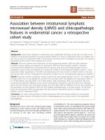
the findings in the literature, according to which HPV16 is the most frequently identified serotype. However,
in our study a low risk serotype not previously reported
was identified, and moreover was the most prevalent
among all the cases: serotype HPV-89 (Fig. 2). A possible
explanation for this observations is that we used different detection methods in order to increase the range of
our findings.
On the other hand, in all published studies which include cases and controls, the prevalence of HPV has
been found to be higher in the cases than in the controls. In contrast, Wang et al. [63] identified HPV in one
sample of 7 breast cancers and in two benign disease
samples. Obviously, this study presents clear limitations
in terms of sample size. Most of the published articles
lack a rigorous methodological design in relation to the
calculation of sample size, a fact that can weaken the results obtained. In our study, the case and control groups
were designed on a 1:1 basis, and although we finally included 251 cases and 186 controls (i.e., a precise 1:1 proportion was not achieved), the statistical power was
maintained.
12
10
8
6
4
2
0
LR6
LR11
LR40
LR42
LR43
LR44
LR54
LR61
LR62
LR72
LR89
Fig. 2 Percentage of low risk (LR) viral serotypes. Percentage of low risk (LR) viral serotypes with respect to total sample size
Delgado-García et al. BMC Cancer (2017) 17:320
Page 8 of 11
48, 12%
58, 15%
192, 50%
21, 6%
28, 7%
20, 5%
LR-89
HR-16
LR-62
19, 5%
HR-51 LR-54
HR-39
REST
Fig. 3 Proportion of viral serotypes found more frequently in
this study
To our knowledge, our study includes the larger series
of samples in which HPV has been analyzed by three different validated molecular methods. Recent studies by Li
et al. [64] (including 187 breast cancers and 92 benign
tumors) and by Fu et al. [43] (with 169 cases and 83
controls) both in China, have shown that HPV may have
a possible causal role in breast cancer pathogenesis
[65, 66] It could be concluded that demographic and
genetic characteristics may be determinant in HPVpositive breast cancer, in view of the wide range of
results obtained. This is a possible explanation because there is such a different prevalence.
In our study, the presence of HPV was associated
(but not significant p value, p = 0.055) to luminal BHER2-negative immunophenotype. This observation is
consistent with high Ki-67 levels, since luminal B
tumors present at least intermediate or high Ki-67
expression. In this respect, among HPV-positive tumors, about 40% were luminal B/HER2-negative. In this
regard, El-Shinawi et al. [49] found the expression of
Ki-67 significantly higher in both (inflammatory and
non-inflammatory) breast cancer with viral DNAs. In
contrast, Subhawong in 2009 [67] established a similarity
between the immunophenotypic characteristics of triplenegative tumors (and more specifically of basal-like
tumors) and HPV-positive squamous cell carcinomas
(functional loss of the retinoblastoma tumor suppressor,
presence of p16 or p53 overexpression). However, in that
study of 33 triple-negative breast cancers, no viral DNA
was identified by in situ hybridization techniques. Other
authors [48, 50] have also reported significant differences
with triple-negative tumors. Recently, in 2015, Fernandes
et al. [60] found no statistically significant association between the molecular subtypes and the presence of HPV;
however the sample size was very small (10 HPV-positive
samples out of a total of 24).
It is well known that luminal B tumors are ERdependent neoplasms, a condition which in turn favors
the perpetuation of cervical HPV infection. Therefore,
further studies are needed to confirm our results.
Emphasis should be placed on the importance of further studies to clarify the role of HPV in the carcinogenic mechanisms in breast cancer. First to clarify
whether causal relationship between the virus and breast
cancer actually exists. Human papillomavirus can be
transmitted by skin-to-skin contact, as well as by sexual
activity. Sexual transmission is the generally accepted
transmission route, though it is not the only route, since
transmission could occur by hand from the female perineum to the breast, wich could occur during sexual activity or even showering or bathing [8, 11, 68–70]. In an
attempt to identify the possible origin of HPV in the
breast, a number of authors [71–74] have explored the
possible relationship between presence of the virus in
the breast and cervical disease produced by HPV. Based
on their studies it is not possible to conclude that HPV
of the breast originates from the cervix. Further research
is needed. On the other hand, De Villiers et al. [61] demonstrated the presence of HPV in 69% of the nipples of
breasts with cancer. This as early as 2004 already suggested that HPV could gain access to the breast tissues
through the nipple. Based on this idea, some investigators postulated breast milk as one of the main transmission routes of the virus, with the breast epithelial cells as
the site of latent infection [9, 10].
Accordingly, breast epithelial cells that lose cell proliferation control are more susceptible to HPV infection.
This loss of control is one of the first steps in breast carcinogenesis. Human papillomavirus infection in women
takes place through contact by the hands or body fluids
(e.g. breast milk...), with microfissures in the nipple serving as entry points for HPV. Errors may occur in the
normal cell repair process, and this in combination with
other cofactors can favour cell immortalization. Some of
these immortal cells can be infected with viral DNA episomes or integrated DNA. The possible mechanisms
whereby HPV intervenes in breast carcinogenesis may
be the same as in the anogenital setting [42], through E6
and E7, though the viral load found in the breast is
much lower.
The presence of HPV might also provide a new target
allowing individualized patient treatment. The possibility
of including antiviral agents as part of the strategy for
the prevention (vaccines) [18, 75] and treatment of
breast cancer could be a reality in the future, as it is currently done in other cancers, such as hepatocellular carcinoma or Kaposi’s sarcoma.
In contrast to other viruses with known neoplastic
transformation potential, HPV can be defined as having
“indirect” oncogenic capacity. The so-called “viral transforming genes”, which synthesize proteins involved in
the inhibition and degradation of key mediators in cell
division and the control of apoptosis (p53 and Rb), promote cellular susceptibility to neoplastic transformation
Delgado-García et al. BMC Cancer (2017) 17:320
due to the impossibility of repairing DNA errors induced
by a series of intrinsic or extrinsic factors during cell
division. The oncogenic action is therefore indirect,
since there is no direct intervention as host gene promoters, regulators or inhibitors. Oncogenic papillomaviruses intervene in the cell division phase, promoting
inhibition of the cellular repair capacity. This
phenomenon, and the associated environmental
circumstances, lead to the accumulation of errors,
often acquired on a random basis (so called clastogenic effect), with a phenotype that is independent of
the initial presence of the virus. No differences would
therefore be expected in the phenotypic evolution of
tumors induced by HPV or potentially induced by
some other type of genetic-environmental event.
Conclusions
In conclusion, this study of 251 cases and 186 controls
has evidenced HPV DNA in 51.8% of the cases (breast
cancer specimens) and in 26.3% of the controls (benign
disease). Furthermore, the OR corresponding to HPV
within the multivariate model, taking age and lactation
into account, is 4.034. We have not been able to establish
a significant relationship between the presence of viral
DNA and the immunohistochemical subtypes. Nevertheless, there is a certain tendency to correlate the presence
of HPV to the HER2- luminal B subtype (p = 0.055). In
concordance with existing literature, the most prevalent
serotype was found to be HPV-16. The strongly discrepant
results in the literature are explained by the great methodological diversity found among the different studies.
Our study, with methodological rigour and a sample size
not previously found in the literature, demonstrate a significant presence of HPV DNA in the breast cancer samples. A possible causal relationship, or mediation or not as
a cofactor, remains to be established by future studies.
Abbreviations
CI: Confidence interval; ER: Estrogen receptor; FDA: Food and Drug
Administration; FISH: Fluorescence in situ hybridization; HER2: Human
epidermal growth factor receptor 2; HPV: Human papillomavirus;
PCR: Polymerase chain reaction; PgR: Progesterone receptor
Acknowledgments
The authors thank the breast cancer and benign diseases patients of the
University General Hospital of Alicante who donated their tissues for
research. Thanks to Sandra and Dra. Alenda of Alicante’s Biobank. Thanks to
Dr. Sánchez which helps us in the initial study design. Thanks to Cristina
Suárez for supervising the final version of the manuscript.
Funding
This study did not receive any extra-institutional funding.
Availability of data and materials
The dataset supporting the conclusions of this article is available at request
from the corresponding author. These datasets are in the process of further
analysis and research.
Page 9 of 11
Authors’ contributions
Conception and design: SDG, JCME. Acquisition of data: SDG, AA. Analysis of
data: SDG, JCME, PC. Interpretation of data: SDG, JCME, AA, TAMB, HBG, GP,
PC, JJPL. All authors contributed to manuscript draft and the revisions.
All authors read and approved the final manuscript.
Competing interests
The authors declare that they have no competing interests.
Consent for publication
Not applicable.
Ethics approval and consent to participate
All data linkage was performed by researchers. Patient written consent was
required for this study. The present study was carried out in strict abidance
with the basic ethical principles of the Declaration of Helsinki and Spanish
Organic Act 15/1999 referred to personal data protection. In addition, the
study was approved by both, the Clinical Research Ethics Committee and the
Managing Board of the University General Hospital of Alicante and Biobank.
Publisher’s Note
Springer Nature remains neutral with regard to jurisdictional claims in
published maps and institutional affiliations.
Author details
1
Department of Obstetrics and Gynecology, University General Hospital of
Alicante, c/ Pintor Baeza, 11, 03010 Alicante, Spain. 2Department of Genetics,
Institute of Cellular and Molecular Studies, Lugo, Spain. 3Department of
Pathology, University General Hospital of Alicante, Institute of Sanitary and
Biomedical Research of Alicante (ISABIAL), Alicante, Spain. 4Department of
Community Nursing, Preventive Medicine and Public Health and History of
Science, University of Alicante, Alicante, Spain. 5Department of Medical
Oncology, University General Hospital of Alicante, Alicante, Spain.
Received: 13 June 2016 Accepted: 1 May 2017
References
1. Organización Mundial de la Salud. Cáncer de mama: Prevención y Control.
2015; Available at: />index1.html;.
2. Ferlay J, Soerjomataram I, Ervik M, Dikshit R, Eser S, Mathers C, Rebelo M, Parkin
DM, Forman D, Bray F. Cancer incidence and mortality patterns in Europe:
estimates for 40 countries in 2012. Eur J Cancer. 2013;49(6):1374–403.
3. Ferlay J, Soerjomataram I, Dikshit R, Eser S, Mathers C, Rebelo M, et al.
Cancer incidence and mortality worldwide: sources, methods and major
patterns in GLOBOCAN 2012. Int J Cancer. 2015;136(5):E359–86.
4. Lakhani, SR. (Ed.). (2012). WHO classification of tumours of the breast.
International Agency for Research on Cancer.
5. Las Cifras del Cáncer en España 2014. Sociedad Española de Oncología
Médica 2014; Available at: www.seom.org.
6. Lawson JS. Do viruses cause breast cancer? Cancer Epidemiology: Springer;
2009. p. 421–38.
7. Amarante MK, Watanabe MAE. The possible involvement of virus in breast
cancer. J Cancer Res Clin Oncol. 2009;135(3):329–37.
8. Joshi D, Buehring GC. Are viruses associated with human breast cancer?
Scrutinizing the molecular evidence. Breast Cancer Res Treat. 2012;135(1):1–15.
9. Glenn WK, Heng B, Delprado W, Iacopetta B, Whitaker NJ, Lawson JS.
Epstein-Barr virus, human papillomavirus and mouse mammary tumour
virus as multiple viruses in breast cancer. PLoS One. 2012;7(11):e48788.
10. Alibek K, Kakpenova A, Mussabekova A, Sypabekova M, Karatayeva N. Role of
viruses in the development of breast cancer. Infect Agent Cancer. 2013;8(1):32–7.
11. Lawson JS, Günzburg WH, Whitaker NJ. Viruses and human breast cancer.
Future Microbiol. 2006;1(1):33–51.
12. Tsai J, Tsai C, Cheng M, Lin S, Xu F, Yang C. Association of viral factors with
non-familial breast cancer in Taiwan by comparison with non-cancerous,
fibroadenoma, and thyroid tumor tissues. J Med Virol. 2005;75(2):276–81.
13. Liu Y, Klimberg VS, Andrews NR, Hicks CR, Peng H, Chiriva-Internati M, et al.
Human papillomavirus DNA is present in a subset of unselected breast
cancers. J Hum Virol. 2001;4(6):329–34.
Delgado-García et al. BMC Cancer (2017) 17:320
14. Pereira Suarez A, Lorenzetti M, González Lucano R, Cohen M, Gass H,
Martínez Vazquez P, et al. Presence of human papilloma virus in a series of
breast carcinoma from Argentina. PLoS One. 2013;8(4):e61613.
15. Aguayo F, Khan N, Koriyama C, González C, Ampuero S, Padilla O, et al.
Human papillomavirus and Epstein-Barr virus infections in breast cancer
from chile. Infect Agent Cancer. 2011;23(6):1.
16. De Paoli P, Carbone A. Carcinogenic viruses and solid cancers without
sufficient evidence of causal association. Int J Cancer. 2013;133(7):1517–29.
17. Bae JM. Two hypotheses of dense breasts and viral infection for explaining
incidence of breast cancer by age group in Korean women. Epidemiol
Health. 2014;36:e2014020.
18. Akhter J, Ali Aziz MA, Al Ajlan A, Tulbah A, Akhtar M. Breast cancer: is there
a viral connection? Adv Anat Pathol. 2014;21(5):373–81.
19. Fimereli D, Gacquer D, Fumagalli D, Salgado R, Rothe F, Larsimont D, et al.
No significant viral transcription detected in whole breast cancer
transcriptomes. BMC Cancer 2015 Mar 18;15:147–015–1176-2.
20. Bratthauer G, Tavassoli F, O'Leary T. Etiology of breast carcinoma: no
apparent role for papillomavirus types 6/11/16/18. Pathology-Research and
Practice. 1992;188(3):384–6.
21. Wrede D, Luqmani Y, Coombes R, Vousden K. Absence of HPV 16 and 18
DNA in breast cancer. Br J Cancer. 1992;65(6):891.
22. Lindel K, Forster A, Altermatt HJ, Greiner R, Gruber G. Breast cancer and
human papillomavirus (HPV) infection: no evidence of a viral etiology in a
group of Swiss women. Breast. 2007;16(2):172–7.
23. Vernet-Tomas M, Mena M, Alemany L, Bravo I, De Sanjose S, Nicolau P, et al.
Human papillomavirus and breast cancer: no evidence of association in a
spanish set of cases. Anticancer Res. 2015;35(2):851–6.
24. Kwong A, Leung CP, Shin VY, Ng EK. No evidence of human papillomavirus
in patients with breast cancer in Hong Kong, southern China. ISRN Virology.
2013;2013
25. Herrera-Romano L, Fernández-Tamayo N, Gómez-Conde E, Reyes-Cardoso
JM, Ortiz-Gutierrez F, Ceballos G, et al. Absence of human papillomavirus
sequences in epithelial breast cancer in a Mexican female population. Med
Oncol. 2012;29(3):1515–7.
26. Silva RG Jr, da Silva BB. No evidence for an association of human
papillomavirus and breast carcinoma. Breast Cancer Res Treat. 2011;
125(1):261–4.
27. Hedau S, Kumar U, Hussain S, Shukla S, Pande S, Jain N, et al. Breast cancer
and human papillomavirus infection: no evidence of HPV etiology of breast
cancer in Indian women. BMC Cancer. 2011;11(1):27.
28. Hachana M, Ziadi S, Amara K, Toumi I, Korbi S, Trimeche M. No evidence of
human papillomavirus DNA in breast carcinoma in Tunisian patients. Breast.
2010;19(6):541–4.
29. de Cremoux P, Thioux M, Lebigot I, Sigal-Zafrani B, Salmon R, Sastre-Garau
X. No evidence of human papillomavirus DNA sequences in invasive breast
carcinoma. Breast Cancer Res Treat. 2008;109(1):55–8.
30. Simoes PW, Medeiros LR, Simoes Pires PD, Edelweiss MI, Rosa DD, Silva FR,
et al. Prevalence of human papillomavirus in breast cancer: a systematic
review. Int J Gynecol Cancer. 2012;22(3):343–7.
31. Frega A, Lorenzon L, Bononi M, De Cesare A, Ciardi A, Lombardi D, et al.
Evaluation of E6 and E7 mRNA expression in HPV DNA positive breast
cancer. Eur J Gynaecol Oncol. 2012;33(2):164–7.
32. Li N, Bi X, Zhang Y, Zhao P, Zheng T, Dai M. Human papillomavirus infection
and sporadic breast carcinoma risk: a meta-analysis. Breast Cancer Res Treat.
2011;126(2):515–20.
33. Pollán M, García-Mendizabal MJ, Gómez BP, Aragonés N, Lope V, Pastor R, et
al. Situación epidemiológica del cáncer de mama en España.
Psicooncología. 2007;4(2):231–48.
34. Aceto GM, Solano AR, Neuman MI, Veschi S, Morgano A, Malatesta S, et al.
High-risk human papilloma virus infection, tumor pathophenotypes, and
BRCA1/2 and TP53 status in juvenile breast cancer. Breast Cancer Res Treat.
2010;122(3):671–83.
35. De Villiers E, Sandstrom RE, Zur Hausen H, Buck CE. Presence of
papillomavirus sequences in condylomatous lesions of the mamillae and in
invasive carcinoma of the breast. Breast Cancer Res. 2005;7(1):R1–11.
36. Yu Y, Morimoto T, Sasa M, Okazaki K, Harada Y, Fujiwara T, et al. HPV33 DNA
in premalignant and malignant breast lesions in Chinese and Japanese
populations. Anticancer Res. 1999;19(6B):5057–61.
37. Damin AP, Karam R, Zettler CG, Caleffi M, Alexandre CO. Evidence for an
association of human papillomavirus and breast carcinomas. Breast Cancer
Res Treat. 2004;84(2):131–7.
Page 10 of 11
38. Gumus M, Yumuk P, Salepci T, Aliustaoglu M, Dane F, Ekenel M, et al. HPV
DNA frequency and subset analysis in human breast cancer patients'
normal and tumoral tissue samples. Journal of Experimental and Clinical
Cancer Research. 2006;25(4):515.
39. He Q, Zhang SQ, Chu YL, Jia XL, Wang XL. The correlations between HPV16
infection and expressions of c-erbB-2 and bcl-2 in breast carcinoma. Mol
Biol Rep. 2009;36(4):807–12.
40. Sigaroodi A, Nadji SA, Naghshvar F, Nategh R, Emami H, Velayati AA. Human
papillomavirus is associated with breast cancer in the north part of Iran. Sci
World J. 2012;2012
41. Liang W, Wang J, Wang C, Lv Y, Gao H, Zhang K, et al. Detection of highrisk human papillomaviruses in fresh breast cancer samples using the hybrid
capture 2 assay. J Med Virol. 2013;85(12):2087–92.
42. Ali SH, Al-Alwan NA, Al-Alwany SH. Detection and genotyping of human
papillomavirus in breast cancer tissues from Iraqi patients. East Mediterr
Health J. 2014;20(6):372–7.
43. Fu L, Wang D, Shah W, Wang Y, Zhang G, He J. Association of human
papillomavirus type 58 with breast cancer in shaanxi province of China. J
Med Virol. 2015;
44. Hammond ME, Hayes DF, Dowsett M, Allred DC, Hagerty KL, Badve S, et al.
American Society of Clinical Oncology/College of American Pathologists
guideline recommendations for immunohistochemical testing of estrogen and
progesterone receptors in breast cancer. J Clin Oncol. 2010;28(16):2784–95.
45. Wolff AC, Hammond ME, Hicks DG, Dowsett M, McShane LM, Allison KH, et
al. Recommendations for human epidermal growth factor receptor 2 testing
in breast cancer: American Society of Clinical Oncology/College of
American Pathologists clinical practice guideline update. J Clin Oncol. 2013;
31(31):3997–4013.
46. Band V, Zajchowski D, Kulesa V, Sager R. Human papilloma virus DNAs
immortalize normal human mammary epithelial cells and reduce their
growth factor requirements. Proc Natl Acad Sci U S A. 1990;87(1):463–7.
47. Di Lonardo A, Venuti A, Marcante ML. Human papillomavirus in breast
cancer. Breast Cancer Res Treat. 1992;21(2):95–100.
48. Corbex M, Bouzbid S, Traverse-Glehen A, Aouras H, McKay-Chopin S,
Carreira C, et al. Prevalence of papillomaviruses, polyomaviruses, and
herpesviruses in triple-negative and inflammatory breast tumors from
Algeria compared with other types of breast cancer tumors. PLoS One.
2014;9(12):e114559.
49. El-Shinawi M, Mohamed HT, Abdel-Fattah HH, Ibrahim SAA, El-Halawany MS,
Nouh MA, et al. Inflammatory and non-inflammatory breast cancer: a
potential role for detection of multiple viral DNAs in disease progression.
Ann Surg Oncol. 2016;23(2):494–502.
50. Piana A, Sotgiu G, Muroni M, Cossu-Rocca P, Castiglia P, De Miglio M. HPV
infection and triple-negative breast cancers: an Italian case-control study.
Virol J. 2014;11(1):190.
51. Manzouri L, Salehi R, Shariatpanahi S. Prevalence of human papilloma virus
among women with breast cancer since 2005-2009 in Isfahan. Advanced
biomedical research. 2014;3
52. Herrera-Goepfert R, Vela-Chávez T, Carrillo-García A, Lizano-Soberón M,
Amador-Molina A, Oñate-Ocaña LF, et al. High-risk human papillomavirus
(HPV) DNA sequences in metaplastic breast carcinomas of Mexican women.
BMC Cancer. 2013;13(1):445.
53. Duò D, Ghimenti C, Migliora P, Pavanelli MC, Mastracci L, Angeli G.
Identification and characterization of human papillomavirus DNA sequences
in Italian breast cancer patients by PCR and line probe assay reverse
hybridization. Mol Med Rep. 2008;1(5):673–7.
54. Czerwenka K, Heuss F, Hosmann JW, Manavi M, Lu Y, Jelincic D, et al.
Human papilloma virus DNA: a factor in the pathogenesis of mammary
Paget's disease? Breast Cancer Res Treat. 1996;41(1):51–7.
55. Yasmeen A, Bismar TA, Kandouz M, Foulkes WD, Desprez P, Al MA. E6/E7 of
HPV type 16 promotes cell invasion and metastasis of human breast cancer
cells. Cell Cycle. 2007;6(16):2038–42.
56. Akil N, Yasmeen A, Kassab A, Ghabreau L, Darnel A, Al MA. High-risk human
papillomavirus infections in breast cancer in Syrian women and their association
with id-1 expression: a tissue microarray study. Br J Cancer. 2008;99(3):404–7.
57. Khan N, Castillo A, Koriyama C, Kijima Y, Umekita Y, Ohi Y, et al. Human
papillomavirus detected in female breast carcinomas in Japan. Br J Cancer.
2008;99(3):408–14.
58. Heng B, Glenn W, Ye Y, Tran B, Delprado W, Lutze-Mann L, et al. Human
papilloma virus is associated with breast cancer. Br J Cancer. 2009;101(8):
1345–50.
Delgado-García et al. BMC Cancer (2017) 17:320
Page 11 of 11
59. Baltzell K, Buehring GC, Krishnamurthy S, Kuerer H, Shen HM, Sison JD.
Limited evidence of human papillomavirus on breast tissue using molecular
in situ methods. Cancer. 2012;118(5):1212–20.
60. Fernandes A, Bianchi G, Feltri AP, Pérez M, Correnti M. Presence of human
papillomavirus in breast cancer and its association with prognostic factors.
ecancermedicalscience 2015;9.
61. De Villiers E, Fauquet C, Broker TR. Bernard H, zur Hausen H. Classification of
papillomaviruses Virology. 2004;324(1):17–27.
62. Ong K, Koay ES, Putti TC. Detection of cutaneous HPV types 4 and 24 DNA
sequences in breast carcinoma in Singaporean women of Asian ancestry.
Pathology. 2009;41(5):436–42.
63. Wang T, Zeng X, Li W, Zhu H, Wang G, Liu X, et al. Detection and analysis of
human papillomavirus (HPV) DNA in breast cancer patients by an effective
method of HPV capture. PLoS One. 2014;9(3):e90343.
64. Li J, Ding J, Zhai K. Detection of human papillomavirus DNA in patients
with breast tumor in China. PLoS One. 2015;10(8):e0136050.
65. Bruni L, Diaz M, Castellsague X, Ferrer E, Bosch FX, de Sanjose S. Cervical
human papillomavirus prevalence in 5 continents: meta-analysis of 1 million
women with normal cytological findings. J Infect Dis. 2010;202(12):1789–99.
66. Grulich AE, Vajdic CM. The epidemiology of cancers in human immunodeficiency virus infection and after organ transplantation. Semin Oncol. 2015;
42:247–57.
67. Subhawong AP, Subhawong T, Nassar H, Kouprina N, Begum S, Vang R, et
al. Most basal-like breast carcinomas demonstrate the same Rb−/p16+
immunophenotype as the HPV-related poorly differentiated squamous cell
carcinomas which they resemble morphologically. Am J Surg Pathol. 2009;
33(2):163–75.
68. Munoz N, Castellsagué X, de González AB, Gissmann L. HPV in the etiology
of human cancer. Vaccine. 2006;24:S1–S10.
69. Burchell AN, Winer RL, de Sanjosé S, Franco EL. Epidemiology and
transmission dynamics of genital HPV infection. Vaccine. 2006;24:S52–61.
70. Kan C, Iacopetta B, Lawson J, Whitaker N. Identification of human
papillomavirus DNA gene sequences in human breast cancer. Br J Cancer.
2005;93(8):946–8.
71. Widschwendter A, Brunhuber T, Wiedemair A, Mueller-Holzner E, Marth C.
Detection of human papillomavirus DNA in breast cancer of patients with
cervical cancer history. J Clin Virol. 2004;31(4):292–7.
72. Hennig EM, Suo Z, Thoresen S, Holm R, Kvinnsland S, Nesland JM. Human
papillomavirus 16 in breast cancer of women treated for high grade cervical
intraepithelial neoplasia (CIN III). Breast Cancer Res Treat. 1999;53(2):121–35.
73. Hansen B, Nygård M, Falk R, Hofvind S. Breast cancer and ductal carcinoma
in situ among women with prior squamous or glandular precancer in the
cervix: a register-based study. Br J Cancer. 2012;107(9):1451–3.
74. Lv YR, Wang JL, Zhang K, Gao HD, Sun JZ, Gong YY, et al. Human papilloma
viruses (HPVs) no co-existence in breast cancer and cervical cells in the
same patient. Chin J Physiol. 2014;57(2):105–6.
75. Lazzeroni M, Serrano D. Potential use of vaccines in the primary prevention
of breast cancer in high-risk patients. Breast Care. 2012;7(4):281–7.
Submit your next manuscript to BioMed Central
and we will help you at every step:
• We accept pre-submission inquiries
• Our selector tool helps you to find the most relevant journal
• We provide round the clock customer support
• Convenient online submission
• Thorough peer review
• Inclusion in PubMed and all major indexing services
• Maximum visibility for your research
Submit your manuscript at
www.biomedcentral.com/submit