Malignant acrospiroma: A case report in the era of next generation sequencing
Bạn đang xem bản rút gọn của tài liệu. Xem và tải ngay bản đầy đủ của tài liệu tại đây (577.81 KB, 4 trang )
Diab et al. BMC Cancer (2017) 17:221
DOI 10.1186/s12885-017-3217-5
CASE REPORT
Open Access
Malignant acrospiroma: a case report in the
era of next generation sequencing
Maria Diab1* , Ali Gabali2 and Muaiad Kittaneh3
Abstract
Background: Malignant acrospiroma is a rare tumor of the eccrine sweat glands accounting for around 6% of all
malignant eccrine tumors. Typically, it presents as large ulcerated nodules, and diagnosis can be challenging as it
has great overlap with its benign counterpart.
Case presentation: We herein report a case of acral malignant acrospiroma, initially treated with surgical excision
and adjuvant radiotherapy. After metastatic disease was confirmed, subject received multiple lines of chemo- as
well as targeted therapy. Genomic testing was also done using next generation sequencing.
Conclusion: To the best of our knowledge, this is the first case of acral malignant acrospiroma with reported next
generation sequencing results.
Keywords: Acrospiroma, Malignant, Acral, Next generation, Case report
Background
Malignant acrospiromas arise from the intradermal duct
of eccrine sweat glands and account for approximately
6% of malignant eccrine tumors [1] compared to their
more prevalent benign counterparts [2]. They appear in
the literature under multiple nomenclatures, including
clear cell hidradenocarcinoma, malignant clear cell
hidradenoma, and malignant hidradenoma [3, 4]. Disease usually manifests in middle-age [5]. Some reports
show disease is more common in females [6], but there
does not seem to be an obvious gender predominance
[4, 7, 8]. Lesions typically present as slow growing nodules that can ulcerate and drain [6, 9]. They range in size
from 0.5 to 10 cm [6, 9]. They usually involve the head,
neck, and extremities; less common sites include the
chest and breasts [1, 4, 5, 9, 10].
This tumor has an aggressive behavior with more than
50% local recurrence rates [11]. Wide surgical excision is
the treatment of choice [12–14]. The efficacy of adjuvant
chemotherapy is controversial. Adjuvant radiotherapy
has been shown successful in some cases [4, 12, 13].
With the advent of targeted therapy, genetic profiling is
becoming a more attractive tool. EGFR overexpression
* Correspondence:
1
Department of Internal Medicine, Wayne State University, School of
Medicine, Detroit, MI, USA
Full list of author information is available at the end of the article
was observed by Piris et al. in 3 out of 12 malignant
hidradenomas [15]. However, there has been no reports
of next generation sequencing. We herein report a case
of acral malignant acrospiroma initially treated with
surgical excision and adjuvant radiotherapy. After metastases was discovered, subject received multiple lines of
chemotherapy. We also tested the tumor for genomic
alterations using next generation sequencing.
Case presentation
A 73-year-old White male was referred to our institution
for the treatment of metastatic malignant acrospiroma.
He initially presented in 2009 with a nodular lesion on
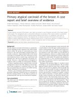
the dorsal aspect of the left great toe. An excisional biopsy of the lesion showed subcutaneous tissue containing multilobular and ill-defined tumor. The pathologic
diagnosis was consistent with acrospiroma; margins were
involved (Fig. 1a and b). Wide surgical excision was subsequently performed to clear the margins and the patient
was followed by clinical surveillance. Six months later,
the patient developed local recurrence and the pathological findings were similar to the original biopsy. At
this time, he was treated with radiation therapy (received
40 Gray) followed by clinical surveillance again.
Three years later, he presented with left groin mass. A
positron emission tomography-computed tomography
(PET-CT) scan showed uptake in the left inguinal lymph
© The Author(s). 2017 Open Access This article is distributed under the terms of the Creative Commons Attribution 4.0
International License ( which permits unrestricted use, distribution, and
reproduction in any medium, provided you give appropriate credit to the original author(s) and the source, provide a link to
the Creative Commons license, and indicate if changes were made. The Creative Commons Public Domain Dedication waiver
( applies to the data made available in this article, unless otherwise stated.
Diab et al. BMC Cancer (2017) 17:221
Page 2 of 4
Fig. 1 a Malignant cells are arranged in cords and sheets separated by a markedly desmoplastic stroma and extending deep into the dermis (H&E at
40X magnification). b Neoplastic cells showing squamous, sebaceous and mucinous differentiation. (H&E at 100X magnificent). c Liver tissue (upper)
with metastatic malignant acrospiroma (lower). The neoplastic cells have uniformly hyperchromatic nuclei (H&E at 400X magnification)
nodes and multiple hepatic lesions. A biopsy from the
hepatic lesion was consistent with metastatic acrospiroma (Fig. 1c). His laboratory studies were within normal
limits, except for an elevated carcinoembryonic antigen
(CEA) of 206.1 ng/mL (normal is less than 5.1 ng/mL).
Patient was subsequently started on systemic chemotherapy. He was initially treated with capecitabine. After
2 months of therapy, however, disease progressed and
treatment was discontinued. The second line of chemotherapy utilized carboplatin. He completed a total of
6 cycles on this combination until disease progression.
On progression, he was referred to our institution for
consideration of experimental therapy. Tissue from hepatic metastasis was sent for genomic profiling using next
generation sequencing Knight diagnostic gene panel.
Unfortunately, no actionable genomic alterations were
identified. Genes tested included EGFR, ALK, BRAF,
JAK2, NOTCH1, NRAS, or PTEN (Table 1), and the
sequencing coverage ranged from 92.4 to 100%. Our
patient was then placed on a phase I trial utilizing a PI3
kinase AkT mTOR inhibitor and ultimately progressed
and succumbed to his disease.
Pathology
Sections from the initial diagnostic biopsy revealed
tumor that composed of epithelial cells with foci of clear
cell morphology and cystic changes, suggesting trichilemmal differentiation (Fig. 1a and b). Some foci exhibited a sclerotic stroma with cystic changes and others
showed mucinous stroma and mucinous changes within
the epithelial cells (mucinous metaplasia). There was no
ductal differentiation. The tumor showed pushing
boarders, focal infiltrative pattern and small areas of increased pleomorphic features and atypia with mitotic activity and focal necrosis. The surrounding connective
tissue margins were involved. No immunohistochemical
studies were performed and the pathological diagnosis
was that of acrospiroma with atypia suggesting foci of
malignant transformation. Apart from metastasis to liver,
all subsequent metastasis sites, focal and distant, showed
findings similar to those seen in the original biopsy. In a
specimen that showed liver metastasis (Fig. 1c), the
tumor exhibited more cells with clear cytoplasm and
hyperchromatic nuclei. Interspersed with the clear cells
is a small population of goblet cells which appear to
contain mucinous materials. Because of the clear cell
morphology in a small population, CD10 immunohistochemical stain was also performed to exclude clear cell
type renal cell carcinoma, and it was negative.
Table 1 Mutation screening by next generation sequencing on
tissue from hepatic metastasis∞
Gene
Percentage of expected
sequencing coverage*
AKT1, BRAF, CDKN2A, DDR2, EGFR, ERBB2,
HRAS, JAK2, KDR, KRAS, MAP2K1, NRAS,
NTRK1, NTRK2, PIK3R1, PTPRD, TP53
100%
PIK3CA
96.9%
NOTCH1, NTRK3
96%
ALK
95%
PTEN
92.4%
PIK3R2
78.8%
∞ No mutations were identified
*Percent of gene covered by a minimum of 100 sequence reads, as compared
with expected coverage based on data from 10 normal DNA samples
Diab et al. BMC Cancer (2017) 17:221
Discussion and conclusion
Malignant acrospiroma is a rare tumor of the eccrine
sweat glands that usually displays an aggressive behavior.
Disease can arise de novo or transform from a previous
benign lesion. Our patient did not have a history of prior
malignancy. One of the challenges in establishing the
disease is that it is often mistaken for benign lesions,
and patients are monitored for some time before tumors
cause symptoms that prompt seeking medical attention.
Wenzel described a case of malignant acrospiroma that
was initially mistaken for osteomyelitis. The patient was
initially started on antibiotics before pathology on the
surgically excised digit confirmed malignancy [14]. In
other instances, disease masquerades as more common
malignant lesions, like malignant melanoma [16]. The
other challenge lies in complete sampling of the lesion.
Disease might be focal and/or may reside in the periphery of the biopsied sample.
Wide surgical excision is the mainstay of treatment
[12]. Wildemore compared outcomes with Mohs micrographic surgery to conventional surgery; all 19 patients
who underwent Mohs micrographic surgery had no local
recurrence at a 29-month follow up [16]. Our patient
was initially treated with surgical excision after the initial
biopsy showed positive margins. Even wide surgical excision, recurrence rates of as high as 50% have been documented [17]. Due to the aggressive nature and the
tendency for lymphatic invasion, some authors advocate
for prophylactic lymph node dissection [18]. Evidence is
lacking on the efficacy of adjuvant chemotherapy [19,
20]. Furthermore, whether the reported results are due
to a real response to chemotherapy or due to a slow
growth of the tumor is questionable. In a case of metastatic disease, complete response was achieved after
3 months of capecitabine (1500 mg/m2 in a split daily
dose, on a 3-weeks-on/1-week-off schedule) [21]. Treatment was complicated with grade 2 fatigue. At
24-month follow up, Lerner’s patient was disease free.
The use of second-line sunitinib was associated with an
8-month progression-free survival in one patient [22].
The role of radiotherapy appears to be more established
[4, 12, 13]. In one report of 3 patients who received adjuvant radiotherapy, 2 patients remained disease free at
27 and 35 months, respectively, after the completion of
treatment; the third died of rapidly progressive systemic
disease [23]. The dose of radiation was 70 Gray for the
surgical bed and 50 Gray for the regional lymphatic
chains.
With the advent of targeted therapy, the use of
genomics in the management of tumors has gained
popularity. Several mutations have been previously
identified in different cutaneous neoplasms, including
t(11;19) in clear cell hidradenomas, hidradenocarcinomas, and mucoepidermoid carcinomas [2, 24, 25],
Page 3 of 4
resulting in a TORC1-MAML2 gene fusion; TP53 mutations and amplification of the Her2/neu gene in hidradenocarcinomas [25]; and BRAF-V600E in aggressive
digital papillary adenocarcinoma [26]. Despite reports of
using next generation sequencing in other cutaneous
neoplasms, there have been no reports on its application
in malignant acrospiroma. Unfortunately, although next
generation sequencing was conducted in our case, it
failed to show mutations that would mandate targeted
therapy. Furthermore, none of the aberrations described
in other cutaneous tumors were identified.
Our patient was treated with chemotherapy when his
disease metastasized. He then entered a phase I trial utilizing a PI3 kinase AkT mTOR inhibitor. The patient was
treated on a phase I PI3K/AKt clinical trial that didn’t
require PI3K/AKt dysregulation. This was a dose finding
study and allowed patients with any tumor type regardless of their PI3K/AKt mutation status to be included.
The choice of PI3K/Akt was arbitrary as this is a commonly dysregulated pathway in cancer, and at the
current time there is no single test available to predict
PI3K/AKt dysregulation or response to Pi3K/Akt inhbitors. However, he ultimately progressed and succumbed
to his disease. The lack of identified mutations might
explain the aggressive nature of the disease.
Malignant acrospiroma is a rare tumor that originates
from the eccrine sweat glands. Aggressive in its nature,
its diagnosis is challenging, and specific tumor markers
and gene mutations are not defined. Wide surgical excision, with or without prophylactic lymph node dissection, is the treatment of choice. Evidence is lacking on
the efficacy and/or advised regimen of chemoradiotherapy. Prognosis is poor with systemic disease. More
studies are needed for the management of this disease.
Abbreviations
CEA: Carcinoembryonic antigen; Mg/m2: Milligrams per squared meters; Ng/
mL: Nanograms per milliliter; PET-CT: Positron emission tomography-computed
tomography
Acknowledgements
The authors have no acknowledgements to disclose.
Funding
This article received no funding sources for its preparation.
Availability of data and materials
This manuscript does not have any datasets to be provided.
Authors’ contributions
MD prepared the manuscript. AG provided the Pathology input. AG and MK
proofread the manuscript. All authors have read and approved the final
version of the manuscript.
Consent for publication
Consent to publish the case was obtained from the deceased patient’s wife.
Competing interests
None of the authors have competing interests to disclose.
Diab et al. BMC Cancer (2017) 17:221
Ethics approval and consent to participate
Ethics approval was not required.
Endnotes
The manuscript text does not include endnotes. The endnotes belonging to
the tables are provided in the table legends.
Page 4 of 4
25. Kazakov DV, et al. Cutaneous hidradenocarcinoma: a clinicopathological,
immunohistochemical, and molecular biologic study of 14 cases, including
Her2/neu gene expression/amplification, TP53 gene mutation analysis, and
t(11;19) translocation. Am J Dermatopathol. 2009;31(3):236–47.
26. Bell D, et al. Next-generation sequencing reveals rare genomic alterations in
aggressive digital papillary adenocarcinoma. Ann Diagn Pathol.
2015;19(6):381–4.
Author details
1
Department of Internal Medicine, Wayne State University, School of
Medicine, Detroit, MI, USA. 2Department of Pathology, Wayne State
University, School of Medicine, Detroit, MI, USA. 3Cardinal Bernardin Cancer
Center, Loyola University Chicago Stritch School of Medicine, Maywood, IL,
USA.
Received: 21 February 2016 Accepted: 22 March 2017
References
1. Mehregan AH, Hashimoto K, Rahbari H. Eccrine adenocarcinoma. A
clinicopathologic study of 35 cases. Arch Dermatol. 1983;119(2):104–14.
2. Tingaud C, et al. Lymph node location of a clear cell hidradenoma: report
of a patient and review of literature. J Cutan Pathol. 2016;43(8):702–6.
3. Crowson AN, Magro CM, Mihm MC. Malignant adnexal neoplasms. Mod
Pathol. 2006;19(Suppl 2):S93–S126.
4. Long WP, Dupin C, Levine EA. Recurrent malignant acrospiroma. Treatment
by chest wall excision. Dermatol Surg. 1998;24(8):908–12. discussion 911-2
5. Gortler I, et al. Metastatic malignant acrospiroma of the hand. Eur J Surg
Oncol. 2001;27(4):431–5.
6. Kauderer C, Clarke HD, Fatone CT. Malignant eccrine acrospiroma. A case
study. J Am Podiatr Med Assoc. 1995;85(2):116–7.
7. Cruz DJ. Sweat gland carcinomas: a comprehensive review. Semin Diagn
Pathol. 1987;4(1):38–74.
8. Deckelbaum S, et al. Eccrine poromatosis: case report and review of the
literature. Int J Dermatol. 2014;53(5):543–8.
9. Ogilvie JW. Malignant eccrine acrospiroma. A case report. J Bone Joint Surg
Am. 1982;64(5):780–2.
10. Cyrlak D, Barr RJ, Wile AG. Malignant eccrine acrospiroma of the breast. Int J
Dermatol. 1995;34(4):271–3.
11. Wilson KM, Jubert AV, Joseph JI. Sweat gland carcinoma of the hand
(malignant acrospiroma). J Hand Surg [Am]. 1989;14(3):531–5.
12. Andreoli MT, Itani KM. Malignant eccrine spiradenoma: a meta-analysis of
reported cases. Am J Surg. 2011;201(5):695–9.
13. Bandyopadhyay A, et al. Malignant acrospiroma of chest and abdominal
wall treated with chemotherapy. Indian J Dermatol. 2013;58(3):241.
14. Wenzel E, et al. Malignant eccrine acrospiroma: a case report. J Am Podiatr
Med Assoc. 2012;102(3):247–51.
15. Piris A, et al. Epidermal growth factor receptor gene status by fluorescence
in situ hybridization in malignant, atypical, and benign hidradenomas. Am J
Dermatopathol. 2010;32(6):586–92.
16. Wildemore JK, Lee JB, Humphreys TR. Mohs surgery for malignant eccrine
neoplasms. Dermatol Surg. 2004;30(12 Pt 2):1574–9.
17. Keasbey LE, Hadley GG. Clearcell hidradenoma; report of three cases with
widespread metastases. Cancer. 1954;7(5):934–52.
18. El-Domeiri AA, et al. Sweat gland carcinoma: a clinico-pathologic study of 83
patients. Ann Surg. 1971;173(2):270–4.
19. Kersting DW. Clear cell hidradenoma and hidradenocarcinoma. Arch
Dermatol. 1963;87:323–33.
20. Lopez-Burbano LF, et al. Malignant clear-cell hidradenoma. Plast Reconstr
Surg. 1987;80(2):300–3.
21. Lerner A, et al. Complete response of metastatic malignant
hidradenocarcinoma to capecitabine treatment. Arch Dermatol.
2011;147(8):998–9.
22. Battistella M, et al. Sunitinib efficacy in the treatment of metastatic skin
adnexal carcinomas: report of two patients with hidradenocarcinoma and
trichoblastic carcinoma. J Eur Acad Dermatol Venereol. 2010;24(2):199–203.
23. Harari PM, et al. The role of radiotherapy in the treatment of malignant
sweat gland neoplasms. Cancer. 1990;65(8):1737–40.
24. Behboudi A, et al. Clear cell hidradenoma of the skin-a third tumor type
with a t(11;19)–associated TORC1-MAML2 gene fusion. Genes Chromosom
Cancer. 2005;43(2):202–5.
Submit your next manuscript to BioMed Central
and we will help you at every step:
• We accept pre-submission inquiries
• Our selector tool helps you to find the most relevant journal
• We provide round the clock customer support
• Convenient online submission
• Thorough peer review
• Inclusion in PubMed and all major indexing services
• Maximum visibility for your research
Submit your manuscript at
www.biomedcentral.com/submit