The Yin/Yan of CCL2: A minor role in neutrophil anti-tumor activity in vitro but a major role on the outgrowth of metastatic breast cancer lesions in the lung in vivo
Bạn đang xem bản rút gọn của tài liệu. Xem và tải ngay bản đầy đủ của tài liệu tại đây (1.4 MB, 15 trang )
Lavender et al. BMC Cancer (2017) 17:88
DOI 10.1186/s12885-017-3074-2
RESEARCH ARTICLE
Open Access
The Yin/Yan of CCL2: a minor role in
neutrophil anti-tumor activity in vitro but a
major role on the outgrowth of metastatic
breast cancer lesions in the lung in vivo
Nicole Lavender1,2†, Jinming Yang1,2†, Sheau-Chiann Chen2,4, Jiqing Sai1,2, C. Andrew Johnson1,2, Philip Owens1,2,
Gregory D. Ayers3,4 and Ann Richmond1,2*
Abstract
Background: The role of the chemokine CCL2 in breast cancer is controversial. While CCL2 recruits and activates
pro-tumor macrophages, it is also reported to enhance neutrophil-mediated anti-tumor activity. Moreover, loss of
CCL2 in early development enhances breast cancer progression.
Methods: To clarify these conflicting findings, we examined the ability of CCL2 to alter naïve and tumor entrained
neutrophil production of ROS, release of granzyme-B, and killing of tumor cells in multiple mouse models of breast
cancer. CCL2 was delivered intranasally in mice to elevate CCL2 levels in the lung and effects on seeding and
growth of breast tumor cells were evaluated. The TCGA data base was queried for relationship between CCL2
expression and relapse free survival of breast cancer patients and compared to subsets of breast cancer patients.
Results: Even though each of the tumor cell lines studied produced approximately equal amounts of CCL2,
exogenous delivery of CCL2 to co-cultures of breast tumor cells and neutrophils enhanced the ability of tumor-entrained
neutrophils (TEN) to kill the less aggressive 67NR variant of 4T1 breast cancer cells. However, exogenous CCL2 did not
enhance naïve or TEN neutrophil killing of more aggressive 4T1 or PyMT breast tumor cells. Moreover, this anti-tumor
activity was not observed in vivo. Intranasal delivery of CCL2 to BALB/c mice markedly enhanced seeding and
outgrowth of 67NR cells in the lung and increased the recruitment of CD4+ T cells and CD8+ central memory T
cells into lungs of tumor bearing mice. There was no significant increase in the recruitment of CD19+ B cells, or
F4/80+, Ly6G+ and CD11c + myeloid cells. CCL2 had an equal effect on CD206+ and MHCII+ populations of
macrophages, thus balancing the pro- and anti-tumor macrophage cell population. Analysis of the relationship
between CCL2 levels and relapse free survival in humans revealed that overall survival is not significantly different
between high CCL2 expressing and low CCL2 expressing breast cancer patients grouped together. However,
examination of the relationship between high CCL2 expressing basal-like, HER2+ and luminal B breast cancer
patients revealed that higher CCL2 expressing tumors in these subgroups have a significantly higher probability
of surviving longer than those expressing low CCL2.
(Continued on next page)
* Correspondence:
†
Equal contributors
1
Department of Veterans Affairs, Tennessee Valley Healthcare System,
Nashville, TN, USA
2
Department of Cancer Biology, Vanderbilt University Medical Center, 432
Preston Research Building, 2220 Pierce Avenue, Nashville, TN 37232, USA
Full list of author information is available at the end of the article
© The Author(s). 2017 Open Access This article is distributed under the terms of the Creative Commons Attribution 4.0
International License ( which permits unrestricted use, distribution, and
reproduction in any medium, provided you give appropriate credit to the original author(s) and the source, provide a link to
the Creative Commons license, and indicate if changes were made. The Creative Commons Public Domain Dedication waiver
( applies to the data made available in this article, unless otherwise stated.
Lavender et al. BMC Cancer (2017) 17:88
Page 2 of 15
(Continued from previous page)
Conclusions: While our in vitro data support a potential anti-tumor role for CCL2 in TEN neutrophil- mediated
tumor killing in poorly aggressive tumors, intranasal delivery of CCL2 increased CD4+ T cell recruitment to the
pre-metastatic niche of the lung and this correlated with enhanced seeding and growth of tumor cells. These
data indicate that effects of CCL2/CCR2 antagonists on the intratumoral leukocyte content should be monitored
in ongoing clinical trials using these agents.
Keywords: CCL2, Breast cancer, Neutrophil killing, Metastasis
Background
C-C chemokine ligand 2 (CCL2), also known as MCP-1,
was first described as a gene induced in response to
platelet-derived growth factor that encodes monocyte
chemoattractant protein-1 [1, 2]. This chemokine mediates its actions by binding to C-C chemokine receptor 2
(CCR2), a seven-transmembrane G-protein coupled receptor [3]. Though CCL2 affects multiple cell types, its
affects mediated through neutrophils or macrophages
can be quite different in the presence or absence of activation of TGFβ signaling [4]. CCL2 is both positively
and negatively associated with the growth of several
tumor types, including breast cancer [5, 6].
The effect of CCL2 on tumor growth and metastasis
has been linked to its role in the recruitment of pro-tumor
or anti-tumor leukocytes into the tumor microenvironment. CCL2 has been reported to recruit myeloid-derived
suppressor cells and pro-tumorigenic macrophages into the
tumor microenvironment [6, 7], to promote the invasive
and metastatic properties of solid tumors. CCL2 secreted
by endothelial cells has been found to stimulate angiogenesis, and ultimately support tumor progression [8]. A recent
report by Kitamura et al. also found that CCL2 stimulates
breast cancer metastasis through the recruitment of macrophages via CCR2 signaling, followed by a CCL3 mediated
enhancement of invasion [9]. Estrogen receptor (ER) negative breast cancers exhibit increased expression of inflammatory chemokines CCL2, CCL4, and CXCL8 compared to
ER+ breast cancers and this correlates with the phenotype
of the inflammatory infiltrate in the tumor [10]. In an
immunohistochemical analysis of CCL2 expression in
205 breast cancer patients, CCL2 was lower in those tumors with ER and progesterone receptor (PR) positivity
and higher in basal like breast cancer [11].
While some reports imply that CCL2 can slow tumor
progression and metastasis, data from multiple laboratories indicate that inhibiting CCL2 will alter the tumor
microenvironment and antagonize tumor growth. The
capacity of CCL2 to attract tumor-promoting and immunosuppressive cells or their precursors provides a
strong rationale for attempting to therapeutically reduce
CCL2 levels in the setting of established neoplasms [12].
Indeed, CCL2 and CCR2 antagonists are currently in
clinical trials for treatment of solid tumors in combination
with standard chemotherapy (NCT01204996) and for
metastatic cancers (NCT01015560, NCT02723006) [13].
Depending on whether CCL2 recruits pro-tumor or
anti-tumor neutrophils and monocytes to the tumor will
positively or negatively effect tumor growth [14, 15].
CCL2 may attract anti-tumor immune cells that are required for efficient immunosurveillance, such that inhibition of CCL2 may promote neo-carcinogenesis as well
as the development of metastases. MMTV-PyMT mice
with a genetic deletion of either CCL2 or CCR2 exhibited earlier onset of tumor growth and increased metastasis, though the rate of primary tumor growth was
enhanced, implying an anti-tumor role for CCL2 in early
stages of tumor progression and in metastasis [16].
Moreover, CCL2 was been reported to increase the cytotoxicity of neutrophils against murine and human breast
cancer models, an activity referred to as ‘entrainment’
[17]. When CCL2 was added to co-cultures of naive
neutrophils isolated from non-tumor bearing BALB/c
mice and 4 T1 cells, tumor cell killing by neutrophils
was increased. This same effect was observed when neutrophils were isolated from healthy volunteers and cultured with MDA-MB-231 cells and CCL2 [17]. The
same report also demonstrated that neutrophils isolated
from tumor bearing mice and patients possess higher
levels of CCL2, which contributed to their killing ability.
Tumor “entrained” neutrophils (TEN) were reported to
kill tumor cells through direct contact in an NADPH
Oxidase-H2O2-dependent mechanism [17]. Thus it is
possible that CCL2 can enhance neutrophil-mediated
killing of tumor cells.
Based on these conflicting data, we wanted to further
evaluate whether CCL2 can “entrain” naïve neutrophils
to enhance tumor cell killing using three different tumor
models (i.e., 4T1, 67NR, and PyMT). These models were
chosen for their varied aggressiveness, comparing the
metastatic 4T1 and PyMT cell line with the non-metastatic
67NR cell line. We observed in vitro that CCL2 did increase killing by TEN but not naïve neutrophils in less aggressive 67NR models. However, CCL2 did not enhance
killing of 4T1 or PyMT tumor cells by naïve or TEN. Although naïve neutrophils isolated from one mouse genetic
background did kill tumor cells derived from another genetic background, exogenous addition of CCL2 did not
Lavender et al. BMC Cancer (2017) 17:88
affect this cytotoxicity. Importantly, intranasal delivery of
CCL2 increased the recruitment of leukocytes into the
BAL fluid and increased subsets of T cells in the lung, but
enhanced the outgrowth of the 67NR breast cancer cells in
the lung. Taken together, our findings suggest that CCL2
may have a more pro-tumor effect on tumor growth than
an anti-tumor effect.
Methods
Cell lines and animals
4T1 (ATTCC-CRL-2539) were obtained from ATCC and
the 67NR cells were obtained through an materials
transfer agreement from the Karmanos Cancer Institute
and cultured according to manufacturer’s specifications. MMTV-PyMT cells were derived from FVB or
C57BL/6 mouse strains and passaged in DMEM supplemented with 5% FBS. The more metastatic TGFβR2KO
PyMT cells (TbR2KO), isolated from both FVB and
C57BL/6 mice were developed in the laboratory of Hal
Moses (Vanderbilt University) [18, 19]. The less aggressive
TGFβR2WT PyMT and the more aggressive TGFβR2KO
PyMT cells were evaluated on mouse backgrounds that
are permissive (FVB) and less permissive (C57BL/6) to
tumor growth [20]. To selectively determine tumor cell
killing, tumor cells were transfected with a GFP2-Firefly
luciferase vector. BALB/c, FVB, and C57BL/6 mice were
purchased from Charles River Laboratories (Charleston,
SC). All animal experiments were approved by the ethics committee of the Vanderbilt Institutional Animal
Care and Use Committee review board and were conducted under protocol M/13/052 in compliance with
guidelines set forth by the US Department of Health
and Human Services Guide for the Care and use of
Laboratory Animals.
Neutrophil isolation
Neutrophils (naïve or TEN) were isolated from the
peritoneal wash of BALB/c, FVB, or C57BL/6 mice
aged 6–8 weeks using Histopaque-1077 and −1119
(Sigma-Aldrich, Saint Louis, MO). The peritoneal wash
was layered on top of Histopaque mediums and spun at
700 g for 30 min without brake. The PMN layer was
collected at the interface of Histopaque-1077 and −1119,
washed with PBS and re-suspended in Opti-MEM with
0.5% FBS. The isolated cells were >95% neutrophils. Cultures of tumor cells alone, naïve or TEN neutrophils alone,
and tumor cells + naïve or TEN neutrophils were seeded
into 12-well plates and incubated overnight at 37 °C. A dose
response curve was performed to determine the optimal
ratio of neutrophils to tumor cells for killing. The maximal
ratio for detection of tumor cell killing occurred with a
ratio of 30 neutrophils to 1 luciferase expressing tumor cell
(30:1). Co-cultures of neutrophils and tumor cells were incubated for 18 h in the presence and absence of 50 ng/mL
Page 3 of 15
CCL2 (R&D Systems, Minneapolis, MN) or 50 ng/mL
CCL2-neutralizing antibody (BD Biosciences, #554440
San Jose, CA).
FACS analysis of neutrophil content and CCR2 expression
To prepare single cell suspensions tumors were diced,
processed using gentle MACS dissociator (Miltenyi
Biotec) and subjected to enzymatic digestion with 1500
CDU Collagenase I, 1 mg/mL Dispase II, and 0.01 MU
DNase I per sample for 1 h. Cell suspensions were
strained through 70 μm nylon mesh. Samples were
washed with PEB buffer (0.5% BSA in PBS) and 1x10⁶
cells from each sample were stained with antibody
cocktail (CD45-APC/Cy7 (Biolegend, # 103116, San
Diego, CA), CD11b-FITC (BD Pharmingen, #553310,
San Jose, CA), Ly6G-PE (BD Pharmingen, #551461, San
Jose, CA). The amount of each antibody to use was determined based on prior titration experiments. Purified antimouse CD16/CD32 antibody (BD Pharmingen, #553142
San Jose, CA) was added to prevent non-specific antibody
binding. After 30 min incubation with antibodies, cells were
washed twice with PEB buffer, fixed in 0.5% buffered PFA
and analyzed on a custom 5-laser LSRII (BD Biosciences,
San Jose, CA).
ELISA assays
After incubation of neutrophils alone or tumor cells alone
for 18 h, media were collected from cell cultures and
stored at 4 °C until subjected to ELISA assay for murine
CCL2. All ELISAs were preformed according to the manufacturer’s instructions (R&D Systems, Minneapolis, MN).
Luciferase reporter killing assays
For reporter assays, luciferase expressing tumor cells
were washed with 1X PBS buffer after removing media,
then lysed using Promega Reporter Lysis Buffer (Luciferase
Assay System, Promega, Madison, WI). Cell lysates were
transferred from plates to microcentrifuge tubes, and spun
to remove remaining cellular debris. Subsequently, 20 μl of
cell lysate supernates were pipetted into opaque 96-well
plates, mixed with Luciferase Substrate (20 μl of Luciferin),
and luminescence was read immediately for 10 s with a
Luminescence reader (Promega, Madison, WI).
Determination of Reactive Oxygen Species (ROS) and
Granzyme-B Release
ROS was measured by L-012 (Wako Chemicals USA,
Inc, Richmond, VA) or Luminol (Fisher Scientific, Sewanee,
GA). For L-012 assays, media from single and co-cultured
samples was collected after the 18 h incubation period.
Samples were seeded into an opaque 96-well plate with L012 in the absence or the presence of Catalase. Luminol
experiments were performed with isolated neutrophils
(naïve or TEN) that were immediately seeded into opaque
Lavender et al. BMC Cancer (2017) 17:88
plates and incubated with Luminol at room temperature
for 15 min. Stimulants were then added and luminescence
was measured over a 10 min period. For both assays, samples were protected from light and read on luminometer.
Granzyme-B release was measured by ELISA (R&D
Systems, Minneapolis, MN) using conditioned media
collected after overnight incubation at 37 °C.
Intranasal Delivery of CCL2
Mice were anesthetized using an isoflurane vaporizer and
then 100 ng of CCL2 in 10 μl of PBS was delivered by the
intranasal route. The solution of CCL2 was gently placed
on the nares of the mice where it is readily taken in.
Analysis of outgrowth of 67NR cells in the lung after
intranasal delivery of CCL2
1 × 10⁶ 67NR cells were intravenously injected into mice.
These mice received intranasal delivery of 100 ng of
murine CCL2 daily. After two weeks of CCL2 treatment,
mice were sacrificed and lungs were removed, photographed, and weighed. The lung tumor weights were
normalized to the weight of tumor-free lungs.
Page 4 of 15
overall difference among groups for Luciferase Reporter
Assays and ELISAs (Figs. 1, 2, 3, and 4a, b, and 5).
Dunn’s post-test was used for pair-wise multiple comparison among groups if the KW test was statistically
significant (p < 0.05). Analysis of variance with a Bonferroni
correction for multiple comparisons was used in Fig. 4c
due to a decrease in sample size. The Wilcoxon rank sums
test was used to test for statistically significant differences
in tumor weight between PBS and CCL2 treated tumorbearing mice (Fig. 6). Analysis of variance with blocking
(two experiments) was performed to test for an overall
difference in number of lung metastasis among MFP-PBS,
MFP + TbR2KO tail vein injected (t.v.), and TbR2KO
groups, t.v. injected alone groups. Tukey’s honestly significant difference (HSD) was used for pair-wise multiple
comparisons. The log rank test was performed to test for
differences in the distributions of relapse-free-survival
(RFS) and CCL2 expression (i.e., high versus low) among
all breast cancers as well as within several the subtypes of breast cancer, respectively. Hereafter, * = p < 0.05,
** = p < 0.01, and *** = p < 0.001, respectively.
Results
Analysis of BAL Fluid Leukocytes after Intranasal Delivery
of CCL2
Effects of CCL2 on In vitro killing of tumor cells by naïve
neutrophils
Murine leukocytes were isolated and subsets analyzed by
FACS as we have previously described [21, 22] (see reference 18 Supplemental Data for a complete listing of
antibody sources). CCR2 expression in BALB/c and
FVB neutrophils was determined by FACS analysis
using PE-conjugated anti-CCR2 from R&D Systems,
Minneapolis, MN.
To evaluate the capacity of CCL2 to entrain neutrophils
to enhance tumor cell killing, we utilized a combination
of in vitro experiments with exogenous delivery of CCL2
to co-cultures of neutrophils and either aggressive 4T1
breast cancer cells compared to a less aggressive 4T1
variant, 67NR, or co-cultures of neutrophils with either
C576Bl/6 or FVB-PyMT breast tumor cells. This experimental design allowed us to examine the ability of exogenous CCL2 to enhance the ability of naïve neutrophils or
TEN to kill luciferase expressing aggressive and less aggressive breast tumor cells. Naïve neutrophils were isolated
from non-tumor bearing BALB/c mice (for luciferase expressing 4T1 and 67NR cultures), FVB, or C57BL/6 mice
(for PyMT cultures). Both FVB and C57BL/6 mice were
used for the PyMT model since the FVB strain is known to
be more permissive for tumor growth and C57BL/6 is
much less permissive [20, 23–25]. We first determined that
the optional ratio of neutrophils to tumor cells was 30:1.
When naïve neutrophils from BALB/c mice were cocultured at a ratio of 30 to 1 with 4 T1 cells, the neutrophils were indeed able to kill the tumor cells based upon a
reduction in intracellular luminescence (RLU) comparing
tumor cells alone to tumor cells plus neutrophils as illustrated in Fig. 1a (p = 0.002). Moreover, addition of CCL2
(50 ng/ml) to co-cultures of naïve neutrophils and 4 T1
cells did not increase the tumor cell killing over that produced by naïve neutrophils without CCL2 addition (Dunn’s
test, p = 0.12) (Fig. 1a). That is, there was no statistically
significant change in luminescent signal between the tumor
Analysis of the ability of less aggressive PyMT breast
tumors in the mammary Fat Pad to reduce the lung
colonization of more aggressive TGFβR2 knock Out PyMT
tumors after tail vein injection
Female FVB mice (10 weeks old) were injected into the
4th mammary fat pad (MFP) with either PBS alone or
PBS containing 15,000 PyMT breast cancer cells. Two
weeks later when the tumor was palpable, either PBS
alone (MFP-PBS) or 1 × 10⁶ TGFβR2 knockout PyMT
breast cancer cells in 200 μl of PBS (MFP + TbR2KO)
were delivered to the tumor-bearing mice by tail vein injection. A third group of mice (non-tumor bearing) received 1 × 10⁶ TGFβR2KO PyMT cells via tail vein (t.v.)
injection (t.v. TbR2KO). Three weeks later, mice were
sacrificed and lungs were removed, weighed, fixed in
paraformaldehyde, embedded in paraffin, subjected to
H&E staining, then the number of metastases counted.
Statistical analyses
The Kruskal-Wallis (KW) test, a nonparametric analog
of analysis of variance, was performed to test for an
A
p=0.039
1.2x1008
p=0.002
p=0.12
8.0x1007
_
4.0x1007
_
_
0
C
p<0.001
1.5x1008
p=0.05
8
p=0.058
_
1.0x1008
5.0x1007
_
_
0
RLU per 1.0x10⁶ tumor cells
Page 5 of 15
RLU per 1.0x10⁶ tumor cells
RLU per 1.0x10⁶ tumor cells
RLU per 1.0x10⁶ tumor cells
Lavender et al. BMC Cancer (2017) 17:88
B
2.0x1006
p=0.058
p<0.001
1.5x1006
P=0.058
_
1.0x1006
_
_
0.5x1006
0
D
4x1007
p=0.004
P=0.278
p=0.02
3x1007
_
_
2x1007
_
1x1007
0
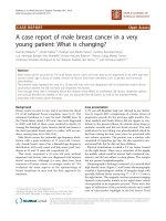
Fig. 1 CCL2 enhances killing of 67NR cells but not 4 T1 cells by neutrophils. Tumor cells were seeded with and without neutrophils at a ratio of
30 neutrophils to 1 tumor cell in the absence and presence of CCL2. After 18-h incubation at 37 °C, cells were lysed and luciferase was measured
to determine tumor cell killing. Luminescence was analyzed using the Kruskal-Wallis (KW) test with Dunn’s post-test if the KW test was statistically
significant (p < 0.05). a & b Naïve as well as tumor entrained neutrophils were able to kill 4 T1 tumor cells (p = 0.002 and p < 0.001, respectively).
c Naïve neutrophil killing of 67NR cells resulted in a p value of 0.058, but the addition of CCL2 resulted in a statistically significant killing of 67NR
cells (p = 0.001) d. TEN were not capable of killing tumor cells, but addition CCL2 to TEN enhanced this effect in 67NR models (p-0.02 for 67NR +
TEN vs. 67NR + TEN + CCL2, p = 0.004 for 67NR vs. 67NR + TEN + CCL2) Kruskal-Wallis test with Dunn’s test for multiple comparisons. Values are
graphed as mean ± SD
cells plus naïve neutrophils samples and tumor cells plus
naïve neutrophils plus CCL2 samples. Naïve neutrophils
from BALB/c mice did not significantly reduce the viability
of the 67NR cells based upon RLU measurements (adj. p =
0.058) (Fig. 1c), and addition of CCL2 did not significantly
change the viability of 67NR cells co-incubated with naïve
neutrophils alone (p = 0.058) (Fig. 1c). However, cocultures of 67NR cells and naïve neutrophils treated with
CCL2 (50 ng/ml) did significantly reduce the viability of
67NR cells (p = 0.001) (Fig. 1c). While naïve neutrophils
from FVB mice did not significantly reduce the viability of
PyMT tumor cells (p = 0.101) (Fig. 2a), addition of exogenous CCL2 did lead to a decrease in viability of the PyMT
cells in this co-culture compared to PyMT cells without
neutrophils (p = 0.005) (Fig. 2a). In the C57BL/6 PyMT
model, there was no reduction in viability of the PyMT
cells upon incubation with naïve neutrophils from C57BL/
6 (adj. p = 0.058) and we observed enhanced tumor cell viability in co-cultures of naïve neutrophils treated with CCL2
(adj. p = 0.001) (Fig. 2c).
Effects of CCL2 on In vitro tumor cell killing by tumor
entrained neutrophils
We next performed these experiments using neutrophils
isolated from mice bearing 4T1, 67NR, or PyMT tumors,
which we refer to as tumor entrained neutrophils (TEN).
TENs from BALB/c or FVB mice were able to kill 4T1
tumor cells and PyMT tumor cells, respectively in vitro
(p < 0.001 and p = 0.009, respectively) (Figs. 1b and 2b).
When we cultured 4T1 tumor cells with TEN, we observed a >56% reduction in luminescent signal, indicating that as with naïve neutrophils from BALB/c mice,
TENs were also able to kill 4T1 cells (adj. p = 0.058)
(Fig. 1b). As we saw with naïve neutrophils, exogenous
CCL2 did not enhance tumor cell killing by BALB/c
TEN (adj. p = 0.058) (Fig. 1b). In the case of 67NR cells,
TENs were not effective at killing tumor cells (p = 0.278)
(Fig. 1d). However, the addition of CCL2 did increase
tumor cell killing by the TENs in these co-cultures over
that by the TENs alone (p = 0.005) (Fig. 1d). In PyMT
mouse models, TENs from FVB mice were able to kill
3x1006
p=0.00
5
p=0.101
p=0.101
2x1006
_
_
_
1x1006
0
p=0.058
1.0x1007
p<0.001
5.0x1006
_
p=0.058
7.5x1006
_
_
2.5x1006
0
RLU per 1.0x10⁶ tumor cells
C
Page 6 of 15
RLU per 1.0x10⁶ tumor cells
RLU per 1.0x10⁶ tumor cells
A
RLU per 1.0x10⁶ tumor cells
Lavender et al. BMC Cancer (2017) 17:88
B
p=0.009
4x1005
p=0.00
9
3x1005
p=0.5
_
_
_
2x1005
1x1005
0
D
p=0.005
3x1006
p=0.163
p=0.058
_
2x1006
_
_
1x1006
0
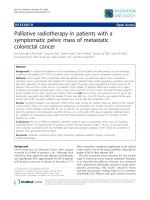
Fig. 2 Naïve or tumor entrained neutrophils (TENs) are able to kill PyMT tumor cells in vitro, but CCL2 does not increase this effect. PyMT cultures
were seeded the same as 4T1 and 67NR with the exception that neutrophils were isolated from either FVB or C57BL/6 mice, depending on cell
line background. a Naïve neutrophils from FVB mice were not capable of killing tumor cells, but CCL2 addition to these naïve neutrophils significantly
killed tumor cells (p = 0.005). b TENs were capable of killing tumor cells (p = 0.009). However, CCL2 did not significantly increase killing (p = 0.5). c Naïve
neutrophils from C57BL/6 mice did not kill C57BL/6 PyMT cells (p = 0.058), and CCL2 addition to this co-culture enhanced the number of viable PyMT
tumor cells (p < 0.001). d C57BL/6 TEN had little effect on the viability of autologous PyMT tumor cells, but CCL2 addition to these TENs resulted in a
modest reduction in viable PyMT tumor cells (p = 0.005). Kruskal-Wallis test with Dunn’s test for multiple comparisons. Values are graphed as mean ± SD
PyMT tumor cells (adj. p = 0.028), but exogenous CCL2
did not increase that killing (p = 0.50) (Fig. 2b). In
contrast, with the C57BL/6 PyMT model, TEN did not
significantly reduce PyMT tumor cell viability, but
addition of CCL2 to these co-cultures resulted in a very
small but significant change in tumor viability based
upon cell luminescence compared to the PyMT cells not
cultured with TENs (p =0.005) (Fig. 2d). However, CCL2
did not enhance killing of C57BL/6 TEN neutrophils cocultured with PyMT tumor cells compared to PyMT
plus TEN alone (Fig. 2d).
We did not observe any biologically significant increase
in tumor cell killing in response to CCL2 with 4T1 tumor
cells, likely because the naïve neutrophils and TEN alone
killed most of the 4T1 tumor cells, leaving little room for
enhanced killing. Moreover, the increases in TEN and
naïve neutrophil killing in response to CCL2 for PyMT
cells in FVB or C57BL/6 models were minimal. One possibility considered to explain these differences in tumor cell
killing ability was that naïve neutrophils isolated from
BALB/c mice are more effective than FVB or C57BL/6
neutrophils in vitro, particularly in less aggressive models.
To determine whether the naïve neutrophils from BALB/c
are more aggressive in killing than those of C57BL/6 mice,
we tested the ability of naïve BALB/c neutrophils to kill
PyMT tumor cells from the FVB mouse background
(Additional file 1: Figure S1). We found that naïve neutrophils isolated from BALB/c mice are indeed able to kill
PyMT tumor cells in vitro (p = 0.005), but exogenous
CCL2 dids not enhance killing (p = 0.347). This implies
that there may be something different about naïve BALB/
c neutrophils as compared to FVB or C57BL/6 naïve neutrophils with regard to their ability to kill tumor cells.
However, the ability of CCL2 to increase TEN killing appears to be limited to less aggressive 67NR cells.
Assays to evaluate factors in conditioned media that
affect neutrophil anti-tumor activity
Since the effects of CCL2 on TEN appeared to be limited
to less aggressive tumor cells, we examined whether
Lavender et al. BMC Cancer (2017) 17:88
Page 7 of 15
ELISA, but there was an increase in the amount of CCL2
in the cell lysate (data not shown). This disparity is likely
because the CCL2 produced by the tumor cells was taken
up by the neutrophils. Moreover, there may have been an
increased production of CCL2 by the cells in co-culture
that was not secreted. Thus, differences in ability to respond to exogenous CCL2 did not result from differences
in the levels of CCL2 produced by tumor cells or neutrophils (naïve or TEN) isolated from BALB/c or FVB mice.
However, addition of anti-CCL2, but not isotype matched
control IgG, was able to reverse the naïve neutrophil killing
of 67NR cells, indicating that CCL2 was needed for
the neutrophil killing of tumor cells (Additional file 1:
Figure S4). We also examined the level of CCR2 expression
A
CCL2 expression (ng/ml per
1x10⁶ cells
differences in CCL2 secretion may influence the response
to exogenous CCL2 to enhance tumor cell killing. Neutrophils isolated from naïve or tumor bearing BALB/c or FVB,
as well as tumor cells, were seeded into 6-well plates and
incubated for 18 h. Conditioned media were collected and
the CCL2 level was measured by ELISA. In these experiments, naïve and TEN neutrophils produced very low levels
of murine CCL2 (with the exception of 2/5 isolates of 4 T1
TEN), while tumor cells tended to secrete much higher
levels of CCL2. However, there were no statistical differences in secretion of CCL2 between 4T1, 67NR or PyMT
cells or between BALB/C and FVB neutrophils (Fig. 3).
Interestingly, co-culture of naïve or TEN with tumor cells
resulted in a decrease in CCL2 in the media as detected by
p=0.311
p=0.014
15
p=0.494
p=0.142
p=0.152
P=0.246
10
_
_
_
5
_
_
0
_
_
_
-5
B
BALB/c neutrophils
FVB neutrophils
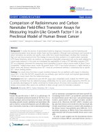
Fig. 3 Tumor cells secrete significantly higher levels of CCL2 compared to naïve and tumor entrained neutrophils. a Conditioned media was
collected from cultured cells after 18-h incubation at 37 °C, and then CCL2 levels were analyzed by ELISA. Tumor cells tended to secrete higher
levels of CCL2 than naïve neutrophils, but there were no statistical differences among the groups. Kruskal-Wallis test with Dunn’s test for multiple
comparisons; mean ± SD are graphed. b CCR2 expression on neutrophils isolated from BALB/c versus FVB mice. Membrane expression of CCR2
was evaluated on neutrophils isolated from BALB/c and FVB mice using protocols described in Methods using PE-conjugated anti-murine CCR2.
While CCR2 was expressed by only 6.03% of the neutrophils from BALB/c mice, 33.1% of the neutrophils from FVB mice expressed cell surface CCR2
Lavender et al. BMC Cancer (2017) 17:88
Page 8 of 15
on naïve neutrophils isolated from BALB/c mice as compared to naïve neutrophils from FVB mice. We observed
CCR2 receptor levels were higher in the FVB neutrophils,
indicating that the failure of the FVB neutrophils to respond
to exogenous CCL2 with enhanced tumor cell killing was
not due to a lack of CCR2 receptor expression (Fig. 3b).
Evaluation of CCL2 effects on neutrophil ROS and
granzyme-B release
Neutrophils are able to kill tumor cells and invading
pathogens by several means. This includes production of
reactive oxygen species (ROS) and release of lytic enzymes
from granules [26]. To examine the killing mechanisms in
our cell cultures, we tested for ROS production using L012 and/or Luminol as well as granzyme-B secretion via
ELISA. We found that 67NR TENs but not 4T1 TENs
produce more ROS than naïve BALB/c neutrophils, as
shown in Fig. 4a (p = 0.008 and p = −.081, respectively).
The addition of catalase to these conditioned media samples caused a decrease in ROS, illustrating naïve neutrophils and TENs are producing hydrogen peroxide. Despite
the higher levels of ROS produced, this did not correlate
with increased tumor cell killing in luciferase reporter assays (Fig. 1). That is, 4T1 TENs and 67NR TENs were less
B
p=0.008
1.0x1008
p=0.159
p=0.081
7.5x1007
_
5.0x1007
_
2.5x1007
_
_
_
_
0
ROS RLU per 1.0x10⁶ tumor cells
ROS RLU per 1.0x10⁶ tumor cells
A
p=0.008
p=0.055
6x1007
p=0.029
p=0.212
p=0.018
p=0.003
p<0.001
_
_
4x1007
2x1007
_
_
_
_
0
Log Granzyme pg/mL per 1.0x10⁶ cells
C
13
*
***
11
_
_
_
_
9
7
_
_
5
Fig. 4 Tumor entrained neutrophils produce greater amounts of ROS than naïve neutrophils. a Conditioned media was collected after overnight
incubation and tested for ROS using L-012 luminescent probe. Since this reagent measures all ROS, the addition of catalase determined the presence of
hydrogen peroxide. 67NR TEN produce more ROS, including hydrogen peroxide, than naïve neutrophils (p = 0.008). b ROS Levels do not correlate with
tumor cell killing. Intra- and extracellular ROS were measured in the Luminol assay. Single cell suspensions of tumor cells or freshly isolated
naïve neutrophils were incubated with Luminol for 15 min at room temperature. CCL2 or tumor cells were then added to naïve neutrophils
and luminescence was immediately measured. CCL2 did not significantly increase the ROS signal (p = 0.212); moreover, a decrease in ROS
signal was observed when tumor cells and neutrophils were co-cultured (p = 0.003 for 4 T1 vs. Neutrophils + 4 T1 and p < 0.001 for 67NR vs.
Neutrophils + 67NR). c Granzyme-B release when neutrophils are co-cultured with tumor cells and CCL2. Granzyme-B levels in conditioned
media from cultured cells were determined by ELISA. 4T1 cells co-cultured with naïve neutrophils exhibited higher granzyme-B release than
neutrophils alone (adj. p = 0.025). Also 67NR cells co-cultured with naïve neutrophils resulted in a significant increase in granzyme-B release
over that of neutrophils alone (adj. p < 0.001). For Fig. 4a and b, Kruskal-Wallis test with Dunn’s test for multiple comparisons. For Fig. 4c, a log
transformation was used to meet the normality assumption. ANOVA for an overall comparison and t-test for multiple comparisons with Bonferroni
p-value adjustment was used. Values are graphed as mean ± SD
Lavender et al. BMC Cancer (2017) 17:88
efficient at killing tumor cells than naïve neutrophils
(Fig. 1). 4T1 TENs were able to reduce tumor cell viability
by roughly 50% (p < 0.001, Fig. 1b), while naïve BALB/c
neutrophils were able to kill nearly 100% of the tumor
cells (p = 0.002, Fig. 1a). We then examined intracellular
and extracellular ROS in naïve neutrophils (Fig. 4a and b).
This analysis revealed that 67NR TENs possess significantly higher levels of ROS than naïve neutrophils (p =
0.008 for 67NR TEN vs. BALB/c neutrophils) (Fig. 4a).
Moreover, tumor cells produced more ROS than naïve
neutrophils (p = 0029 for 4T1 vs. neutrophil, p = 0.018 for
67NR vs. neutrophil) (Fig. 4b), and addition of naïve neutrophils to 4T1 cells or 67NR cells actually decreased ROS
(p = 0.003 and p < 0.001, respectively) (Fig. 4b), likely due
to loss of tumor cell ROS due to tumor cell killing. Also,
addition of naive neutrophils to 67NR cells resulted in
ROS levels that were much lower than those produced
when CCL2 was added to naïve neutrophils (p = 0.008)
(Fig. 4b). Hence, ROS detection as measured here correlates with tumor cell killing only in the sense that when
naïve neutrophils kill 4T1 or 67NR cells, there is a concordant reduction of ROS, since the ROS is mainly derived from the tumor cells. We postulated the killing
mechanism utilized by naïve and TEN likely involves
mechanisms other than induction of ROS. Consequently,
we examined granzyme-B release in conditioned media
collected from cell cultures (Fig. 4c). When 4T1 tumor
cells were added to naïve neutrophils we observed increased granzyme-B release compared to neutrophils
alone (adj. p = 0.025, Fig. 4c). Also 67NR cells co-cultured
with naïve neutrophils resulted in a significant increase in
granzyme-B release over that of naïve neutrophils alone
(adj. p < 0.001) (Fig. 4c). These data indicate that neutrophil killing of 4T1 and 67NR cells was associated with
granzyme-B activity. However, addition of exogenous
CCL2 did not increase that granzyme-B activity.
Effect on less aggressive breast cancer implants on the
colonization of more aggressive breast cancer cells
Granot et al. argued that CCL2 produced by tumor cells
could both enhance the growth of the primary tumor and at
the same time entrain neutrophils in the lung to kill tumor
cells and inhibit lung metastasis [17]. Since we observed that
the PyMT tumor cells make a substantial amount of CCL2
(Fig. 3), as Granot argued, it could be postulated that CCL2
released into the blood stream by a primary PyMT tumor
might impair the outgrowth of tumor cells in the lung after
intravenous injection. TGFβ is known to suppress CCL2 expression, thus it is expected that TGFβR2KO PyMT cells
will express more CCL2 and thus provide a good model for
exploring the role of CCL2 production by tumor cells in
metastasis. Fridlender et al. showed that TGFβ has the ability to inhibit the anti-tumor activity of TENs [4], thus we
reasoned that loss of response to TGFβ by PYMT cells
Page 9 of 15
should allow for increased CCL2 production and enhanced
anti-tumor activity of TEN at the pre-metastatic site. We
tested this hypothesis by implanting 15,000 PYMT tumor
cells into the 4th mammary fat pad (MFP) and when palpable tumors developed in the MFP, the tumor bearing mice
received intravenous injection of 1 × 10⁶ of the more aggressive TGFβR2KO PyMT breast tumor cells [27], or vehicle
control. A second group of mice did not have tumors implanted into the MFP, but received only the intravenous injection of the TGFβR2KO PyMT tumor cells at the same
time as the MFP tumor bearing mice. After allowing two
additional weeks for the outgrowth of the intravenously
injected tumor cells, all mice were euthanized and the lungs
were examined for metastasis based upon visual examination, weight, and histology. Mice that only received an
orthotopic implantation of PYMT tumor cells (expressing
TGFβR2) in the MFP did not develop tumors in the lung
during this period of time. Interestingly, there was a trend
toward fewer tumors in the lungs of mice with PyMT
tumors growing in the MFP that also received tail vein
injections of the TGFβR2KO PyMT tumor cells, as compared to mice that only received the tail vein injection of
TGFβR2KO PyMT breast tumor cells (adj. p = 0.091)
(Additional file 1: Figure S2). These data imply that signals
emanating from the orthotopic tumor might indeed have
an adverse impact on the colonization of circulating
tumor cells. This concept is compatible with the idea that
less aggressive tumors may be able to “entrain” the microenvironment in the lung to inhibit the growth of more
aggressive tumors. Moreover, we know these tumor cells
produce significant amounts of CCL2 (Fig. 3), even though
they continue to express TGFβR2. We did not measure the
CCL2 levels in the serum or lung after implantation of the
TβR2WT PyMT into the MFP as compared to normal
lung or lung after tail vein injection of PyMT-TβR2KO
alone, so we cannot definitely equate the suppression of
tumor outgrowth in the lungs to elevations in CCL2. In
fact, other investigators have shown CCL2 elevation in the
tumor microenvironment and premetastatic niche enhances tumor growth and metastasis [6, 22].
In vivo experiments to evaluate How delivery of CCL2 to
the lung affects colonization of the lung by breast cancer
cells
To evaluate the idea that higher tissue levels of CCL2 might
make changes in the microenvironment that can inhibit
tumor growth, we delivered increasing amounts of CCL2
intranasally to mice and monitored the concentration of
CCL2 in the lung (Fig. 5a). However, the delivery of 100 ng
vs 500 or 500 ng vs 1000 ng did not reveal statistically significant differences in the concentration of CCL2 in the
lung (p = 0.133 and p = 0.482, respectively) (Fig. 5a). This
may be due to the uptake of exogenous CCL2 by leukocytes and other stromal cells in the lung since endothelial
Concentration (pg/mL per ug of protein)
Lavender et al. BMC Cancer (2017) 17:88
Page 10 of 15
p=0.015
p=0.016
p=0.482
p=0.133
4x1004
p=0.088
p=0.397
_
_
_
2x1004
0
_
_
Fig. 5 Intranasal delivery of CCL2 increases the concentration of
CCL2 in the lung. Daily intranasal delivery of increasing concentrations of
CCL2 (0, 50, 100, 500 and 1000 ng) resulted in increasing concentrations
of CCL2 in the lung with saturation achieved by the 500 ng delivery or
1000 ng delivery compared to 50 ng delivery (p = 0.016 and p = 0.015,
respectively). Kruskal-Wallis test with Dunn’s test for multiple
comparisons. Data are graphed as mean ± SD
cells express high levels of CCR2 [28]. In contrast, increasing intranasal delivery of CCL2 enhanced recruitment of
CD8+ T lymphocytes into the BAL fluid and the number
of CD45+ cells in the BAL that were CD8+ tended to show
a dose dependent increase. Also there was a tendency to increase Ly6G+/F4/80+ cell recruitment into the BAL fluid
in response to increasing delivery of CCL2 (Additional file
1: Figure S3A and B). In the absence of intranasal delivery
of CCL2, BAL from PBS controls did not exhibit a measureable lymphocyte population and macrophages constituted <10% of live cells.
To examine the pro-tumor or anti-tumor function of
CCL2 in vivo, syngeneic 1 × 10⁶ 67NR cells were intravenously injected into BALB/c mice. Subsequently, 100 ng of
CCL2 was delivered daily by the intranasal route for two
weeks, then mice were sacrificed and lung tumor burden
was examined. After two weeks of exogenous CCL2 delivery, we observed that the net tumor contribution to the
weight of the lung in the CCL2 treated group was significantly increased [314 ± 83 mg, n = 5] in comparison with
the PBS control group [184 ± 45 mg, n = 7] (Wilcoxon rank
sum test, p = 0.006) (Fig. 6a and b, comparing 6Bb to Ba).
Thus, exogenous CCL2 favors 67NR tumor colonization in
vivo, even though it can enhance 67NR tumor cell killing by
TEN in vitro (Fig. 1d). These data point to the significance
of the tumor microenvironment with regard to chemokine
responses.
When we performed analysis of the leukocyte infiltrate
in the lungs of mice given intranasal injection of PBS
versus CCL2 (100 ng/ 5 days/week for 2 weeks) prior to
receiving tail vein injection of 1 × 10⁶ 67NR cells, we
did not observe a significant increase in the CD45 cells
recruited to the lung following intranasal delivery of
CCL2, though a substantial percentage of the cells in the
lung were CD45+ (40-45%) (Fig. 6c, p = 0.15). Of the
CD45 cells in the lung, ~27% were F4/80+, <5% were
Ly6G+, 5-8% were CD11c+, 17-18% were CD19+, 1718% were CD4+, and ~5% were CD8+ (Fig. 6d). While
CCL2 did not significantly affect the total F4/80, Ly6G,
CD11c, CD19, and CD8 cell content in the lung as a
percentage of the total CD45+ cells, there was a significant increase in the percentage of CD4+ T cells (p <
0.01, n = 5, Student’s t-test) (Fig. 6d). We also observed
that CCL2 increased the population of central memory
CD8+ T cells (p < 0.05, Student’s t-test), but did not alter
the percentage of CD4+ T cell central memory cells, the
effector memory CD4 + T cells, or CD8+ T cells (Fig. 6e).
Though CCL2 treatment did not increase the population
of F4/80 or Ly6G cells in the tumor, it did increase the
percentage of F4/80 cells that expressed CD206, a
marker for M2 macrophages. In contrast, CCL2 intranasal delivery increased the F4/80 population expressing
MHCII, a marker for M1 macrophages (Student’s t test,
p < 0.01 and p < 0.05, respectively) (Fig. 6Fa). There were
no significant changes in the population of F4/80 cells
producing IFNγ or IL-4, though there was a trend toward increased IL-4 in the CCL2 group (Fig. 6Fb).
Correlation between CCL2 mRNA expression in sub-classes
of human breast cancer and prognosis
Another way to examine the impact of CCL2 expression
by tumor cells is to determine whether CCL2 expression
correlates positively or negatively with relapse free survival. When we examined the TCGA and kmplot.com
data base to query expression of CCL2 (i.e., high vs. low
expression) in human breast cancer with respect to
relapse-free survival (RFS), the association of high CCL2
expression with RFS did not reach statistical significance
(p = 0.071) (Fig. 7b). However, with some subtypes of
breast cancer, patients expressing high levels of CCL2
exhibited improved RFS. For example high CCL2 expression suggested improvement in RFS for basal (p = 0.047),
HER2+ (p < 0.001) and luminal B (p = 0.047) breast cancers
(Fig. 7c, d and f). However, among patients with luminal A
breast cancer, the most abundant of the sub-group, RFS
differences among patients with high and low CCL2 expression was equivocal (p = 0.1) (Fig. 7e).
Discussion
CCL2 has been described as both supporting breast cancer
growth and progression and inhibiting breast cancer
Lavender et al. BMC Cancer (2017) 17:88
Page 11 of 15
P=0.006
Fig. 6 CCL2 promotes lung tumor growth. A BALB/c mice were intravenously implanted with 1 × 10⁶ 67NR cells and 100 ng CCL2 cytokine or
PBS vehicle were delivered daily by the intranasal route for 5 days/week for 2 weeks. Two weeks after treatment, mice were sacrificed and the
lung weight was determined. Wilcoxon rank sum test, p = 0.006. Mean ± SD. B Photographs of Lungs from CCL2 or PBS treated mice prior to i.v.
delivery of 1 × 10⁶ 67NR cells. Lungs from mice in Fig. 6a were removed from euthanized mice and representative ones were photographed.
PBS-treated mouse lung (a), CCL2-treated mouse lung (b), or tumor-free un-treated lung (c). C Lungs from CCL2-treated mice do not exhibit
significant increase infiltrate of CD45+ cells. Mice treated as described in 6A were euthanized; lungs were harvested then prepared for FACS
analysis of infiltrating CD45+ leukocytes. Data are reported as % of CD45+ cells total lung cells analyzed. Student’s t-test, p = 0.15. D CCL2
enhanced immune cells infiltrating into lung tumor. BALB/c mice were treated as described in 6a. Two weeks after treatment, the lung tumor
microenvironment was analyzed for the infiltration of immune cells by multicolor FACS. Data were analyzed by Student’s t-test. **p < 0.01, n = 5.
E CCL2 increases central memory CD8+ T cells but not effector memory T cells. Ea BALB/c mice were treated as described in 6A. Two weeks after
treatment, the lung tumor microenvironment was analyzed for memory T cells by multicolor FACS. Data were analyzed by the Student’s
t-test. *p < 0.05 vs. PBS controls, n = 5. Eb a representative graph indicating CD44 and CD62L expression on CD8+ T cells from PBS-treated mouse
lung or from CCL2-treated murine lung. F CCL2 effects on the polarization of lung macrophages. BALB/c mice were treated as described in 6A.
Two weeks after treatment, the lung tumor microenvironment was analyzed by FACS for a) the infiltration of F4/80+ macrophages expressing CD206
(**p < 0.01, n = 5.) and MHC II markers on the cell surface (* p < 0.05, n = 5) (or b) for the intracellular cytokines IFNγ and IL-4 (ns = not significantly different)). Data were analyzed by the Student’s t-test, n = 5
progression. When 4T1E/M3 cells with enhanced capacity
to metastasize to bone were altered to increase CCL2 production, there was an inhibition of metastasis of these cells
to bone and lung as compared to 4T1E/M3 cells expressing normal amounts of CCL2 [29]. CCL2 may play a protective role by mediating early immune surveillance in the
tumor progression process [16] through a process that involves recruitment of γ,δ tumor infiltrating lymphocytes
(TILS) to the tumor microenvironment [30]. CCR2, the
receptor for CCL2, is expressed on human effector memory CD4+ T cells that are useful for rapid recall responses
[31], on Th17 cells recruited to the lung during allergic reactions [32], on γ,δ T-cells infiltrating tumors [30], and on
CD4+ T cells where they can have negative influence in
Crohn’s disease [33].
Other reports show that CCL2 production by tumor
cells impairs T cell-mediated anti-tumor activity [34].
For example, Fujimoto described a role for stromal
CCL2 (MCP-1) in the recruitment of tumor promoting
macrophages into early breast cancer lesions, a process
which promotes tumor progression. MCP-1 mRNA
expression was enhanced when stromal cells were cocultured with breast tumor cells, and when immune deficient mice received blocking antibodies to MCP-1 (CCL2),
there was a reduction in angiogenesis, macrophage infiltration into the tumors and also the growth of the tumors
Lavender et al. BMC Cancer (2017) 17:88
Page 12 of 15
A
PAM50 Subtype
6%
CCL2
Genetic Alteration
Amplification
PAM50 Subtype
NA
Deep Deletion
Basal-like
mRNA Upregulation
HER2-enriched
B
Luminal A
mRNA Downregulation
Luminal B
C
D
E
F
Normal-like
Fig. 7 CCL2 Expression in Sub-Groups of Human Breast Cancer Correlates with Relapse-Free Survival. a CCL2 expression in the TCGA (The Cancer
Genome Atlas) as queried by the cBIO.org portal selected for the Breast Cancer Nature 2012 study containing PAM50 subtyping of breast tumors.
b Probability of RFS from kmplot.com in comparison to CCL2 expression using median and auto-cutoffs is shown. c Basal; d HER2+; e Luminal A;
f Luminal B
slowed. Hembruff et al. showed that TGFβR2 KO in fibroblasts increases CCL2 production and when these fibroblasts are co-implanted with 4T1 cells, there is enhanced
primary tumor growth and metastases as compared to
4 T1 cells co-implanted with TGFβR2 expressing fibroblasts
[35]. Moreover, the Pollard laboratory has shown that inhibition of CCL2 /CCR2 signaling blocks the recruitment
of inflammatory macrophages and reduces metastasis to
the lung [6]. Additionally, expression of this chemokine was
associated with a poor prognosis in breast cancer [36].
Similar findings were observed with 230 samples of
human breast cancer primary lesions where CCL2 expression in tumor cells and accumulating tumor associated macrophages (TAMs), increased angiogenesis, and
vessel invasion of tumor cells [37]. In still another study
of 427 invasive ductal carcinoma breast cancer cases, the
expression of CCL2 in the stroma of basal-like breast
cancer correlated with significant reduction in recurrencefree survival [38]. While other studies have demonstrated
CCL2 as a prognostic factor by evaluating selected cell
populations or distinct location of metastases, we have
chosen to look at a large dataset of more than 3000 primary breast cancers to evaluate the overall expression of
CCL2 mRNA. While we find that CCL2 cannot be used as
a prognostic factor of all breast cancer, but that it can be
prognostic for distinct subtypes of breast cancer. These
findings support the concept that in order to understand
CCL2 expression as tumor promoting or suppressive factor, one must use additional molecular or cellular features
to identify either the cells, or intrinsic subtype of the
breast cancer expressing CCL2 to determine whether
CCL2 is playing a significant role in RFS.
While we expected that the delivery of exogenous CCL2
might reduce the outgrowth of the tumor cells based upon
Granot’s work, we observed the opposite, potentially because there was no significant increase in neutrophil recruitment the lung. When we delivered 100 ng exogenous
CCL2 daily by the intranasal route to mice, we did not observe a significant increase in the level of CCL2 at the end
point of the experiment when the lungs were removed
Lavender et al. BMC Cancer (2017) 17:88
2 weeks later. This is likely due to rapid clearance of the
chemokine through the lung and to the uptake of CCL2
by the leukocytes, endothelial cells, and other cells in lung
[28]. Though only subtle changes were observed in the infiltrating leukocyte populations in the lung in response to
intranasal delivery of CCL2 (increased CD4+ T cells, increased central memory CD8+ T cells, increased CD206+
macrophages, and increased MHCII expressing macrophages), the end result was enhanced outgrowth of the
67NR cells in the lung. Our data are in accordance with
the work from the Pollard laboratory and other groups
showing that CCL2 can promote metastasis [6, 9, 39–43].
There are reports that the tumor promoting effect of
CCL2 applies to both ER+ and triple negative breast cancers [39, 44]. Taken together, our data suggest that while
less aggressive tumors may indeed alter the microenvironment of the pre-metastatic niche of the lung to inhibit
outgrowth of metastatic tumor cells, our attempts to
mimic this with delivery of exogenous CCL2 resulted in
enhanced seeding and outgrowth of breast cancer cells in
the lung. Moreover, increased CCL2 produced by stromal
cells in the tumor microenvironment has been reported to
promote metastasis of 4T1 TN breast cancer cells and ER
+ breast cancer to the lung [35, 44] and to support a cancer stem cell phenotype [45]. In addition, CCL2-mediated
activation of SMAD3 and the MAPK pathway are involved in the increased survival of tumor cells and an enhanced metastatic phenotype of these cells [46].
Conclusions
In conclusion, the link between CCL2 in breast cancer
metastasis remains obtuse. While there are some potential
anti-tumor effects of CCL2 in vitro and in vivo during
early tumor formation, there are strong data from many
groups, including the data reported here, indicating that
intra-tumoral CCL2 can promote breast tumor growth
and/or metastases. Clinical trials are currently ongoing for
solid tumor patients using CCL2/CCR2 inhibitors in combination with other therapies [13]. However, there are
some concerns about this since in mouse lung cancer
models, stopping anti-CCL2 therapy results in a rapid regrowth of tumor with enhanced metastasis [41]. The data
reported here may be important in evaluating response to
CCL2 or CCR2 inhibitors in these ongoing trials. While
our data support a potential anti-tumor role for CCL2 in
TEN-mediated tumor killing in the poorly aggressive
67NR BALB/c mouse tumor model, when CCL2 was delivered via the intranasal route, an increase in CCL2 associated CD4+ T cell and CD206+ macrophage recruitment
was associated with enhanced seeding and growth of tumor
cells in the lung. Thus evaluation of effects of CCL2/CCR2
inhibitor treatment in clinical trials on the specific subsets
of intra-tumoral leukocytes may be informative for evaluating response to therapy.
Page 13 of 15
Additional file
Additional file 1: Figure S1. Naïve BALB/c neutrophils can kill PyMT
(FVB) tumor cells, but CCL2 does not increase killing. PyMT cells from
FVB mice seeded with and without naïve BALB/c neutrophils (30
neutrophils: 1 tumor cell), in the absence and presence of CCL2. After
18-h at 37 °C, cells were lysed and luciferase was measured to determine tumor cell killing. Although from a different mouse strain, naïve
BALB/c neutrophils were able to kill FVB PyMT tumor cells (p = 0.005).
However, CCL2 did not enhance this effect (p = 0.347), Kruskal-Wallis
test with Dunn’s test for multiple comparisons. Values are graphed as
mean ± SD. Figure S2. Entrainment properties of less aggressive PyMT
tumor cells on the metastatic outgrowth of more aggressive TGFβR2
knock out PyMT tumors. Female FVB mice (10 weeks old) were injected
with either 15,000 PyMT breast cancer cells (MFP) or PBS (Non-tumor
bearing) in the 4th mammary fat pad. Two weeks later either 1 × 10⁶
TGFβR2 knockout PyMT (TbR2KO) breast cancer cells or PBS alone (in
200 μl) were delivered by tail vein injection to mice bearing PyMT tumors or
into non-tumor bearing mice (t.v. TbR2KO). Three weeks later, mice were sacrificed and lungs were removed, fixed, H&E stained and the number of metastases counted. Analysis of variance with blocking (two experiments) was
performed for an overall comparison (p < 0.001). Tukey’s honestly significant difference (HSD) for multiple comparisons among groups (adj. p =
0.009 for MFP-PBS vs. MFP + TbR2KO, adj. p < 0.001 for MFP-PBS vs. t.v.
TbR2KO). NS = not significant, p < 0.1, *p < 0.05, **p < 0.01, ***p < 0.001. Values
are graphed as mean ± SD. Figure S3. Intranasal delivery of CCL2 facilitates
the recruitment of leukocytes into BAL fluid. 3A. BAL fluid isolated from mice
receiving intranasal delivery of CCL2 showed an increase in CD8+ T cells as
CCL2 delivery increased from 100 ng to 1000 ng. Data are shown as % CD45+
cells and as % total cells. 3B. BAL fluid from mice receiving intranasal delivery
of CCL2 exhibited a trend toward increased numbers of neutrophils and NK
cells with increasing concentrations. Data are shown as % live cells in BAL fluid.
Figure S4. Blocking antibody to CCL2 reverses the neutrophil killing of 67NR
cells in vitro. 67NR cells(T) expressing luciferase were seeded with and without
neutrophils (E for effector cells) at a ratio of 30 neutrophils: 1 tumor cell in the
presence of control IgG or blocking antibody to CCL2. After an 18-hs at 37 °C,
cells were lysed and luciferase was measured to determine tumor cell killing.
Anti-CCL2 (50 ng/ml BD Biosciences) reversed the 67NR tumor cell killing of
BALB/c TEN, p < 0.01, Student’s t test, n = 5 per group. (PPTX 124 kb)
Abbreviations
CCL2: CC chemokine 2; CCL4: CC chemokine 4; CCR2: CC Chemokine
Receptor 2; CXCL8: CXC chemokine 8; ELISA: Enzyme-linked immunosorbent
assay; ER: Estrogen receptor; FBS: Fetal bovine serum; GFP2: Green fluorescent
protein 2; H&E: Hematoxylin and Eosin; HSD: Honestly significant difference; IL4: Interleukin 4; KO: Knock out; MCP-1: Macrophage chemotactic protein-1;
MFP: Mammary fat pad; MHC: Major histocompatibility complex; MMTV: Mouse
mammary tumor virus; NADPH: Nicotinamide adenine dinucleotide phosphate
(reduced form); Opti-MEM: Optimized minimal essential medium;
PyMT: Polyoma Middle T antigen; RFS: Relapse-free survival; ROS: Reactive
oxygen species; TbR2KO: TGF-β receptor 2 knock-out; TEN: Tumor entrained
neutrophil; TGF-β: Transforming growth factor-beta; TGFβR2: Transforming
growth factor-beta receptor 2
Acknowledgements
We are indebted to the following persons for their contributions to this
project:
Linda W. Horton for laboratory management; Logan Northcutt for technical
support; Anna E Vilgelm for consultation and advice; the Flow Cytometry
Core Resource for help with FACS analysis.
Funding
This work was supported by grants from the Department of Veterans Affairs and
the NCI: Senior Research Career Scientist Award (AR); Experimental work was
funded by R01-CA34590 (AR); Training grant support for NL, T32CA171895: and
Core Facility Support from the VICC Cancer Center Support Grant P30-CA068485.
Availability of data and materials
All data generated or analyses during this study are included in this article
and its Additional file 1.
Lavender et al. BMC Cancer (2017) 17:88
Authors’ contributions
Conception and design: AR; Development of methodology: NL, JY, JS, PO;
Acquisition of data: (provided cells, animals, facilities, etc.): NL, JY, JS, CAJ, PO;
Analysis and interpretation of data (statistical analysis, biostatistics, and
computational analysis): NL, JY, JS, S-CC, GDA; Writing, review and/or revision
of the manuscript: NL, JY, JS, AR, S-CC, GDA: Administrative, technical, or
material support: CAJ. Study supervision: AR. All the authors have read and
approved this manuscript.
Competing interests
The authors declare that they have no competing interests.
Consent for publication
Not applicable.
Ethics approval and consent to participate
This study does not involve human data other than that retrieved from the
TCGA data set. All mouse studies were performed utilizing protocols approved
by the Vanderbilt University IACUC (protocol ID number: M1600058).
Author details
1
Department of Veterans Affairs, Tennessee Valley Healthcare System,
Nashville, TN, USA. 2Department of Cancer Biology, Vanderbilt University
Medical Center, 432 Preston Research Building, 2220 Pierce Avenue,
Nashville, TN 37232, USA. 3Department of Biostatistics, Vanderbilt University,
Nashville, TN, USA. 4Division of Cancer Biostatistics, Department of
Biostatistics, Center for Quantitative Sciences, Nashville, TN, USA.
Received: 10 September 2016 Accepted: 18 January 2017
References
1. Rollins BJ, Morrison ED, Stiles CD. Cloning and expression of JE, a gene
inducible by platelet-derived growth factor and whose product has
cytokine-like properties. Proc Natl Acad Sci U S A. 1988;85(11):3738–42.
2. Yoshimura T, Yuhki N, Moore SK, Appella E, Lerman MI, Leonard EJ. Human
monocyte chemoattractant protein-1 (MCP-1). Full-length cDNA cloning,
expression in mitogen-stimulated blood mononuclear leukocytes, and sequence
similarity to mouse competence gene JE. FEBS Lett. 1989;244(2):487–93.
3. Charo IF, Myers SJ, Herman A, Franci C, Connolly AJ, Coughlin SR. Molecular
cloning and functional expression of two monocyte chemoattractant
protein 1 receptors reveals alternative splicing of the carboxyl-terminal tails.
Proc Natl Acad Sci U S A. 1994;91(7):2752–6.
4. Fridlender ZG, Sun J, Kim S, Kapoor V, Cheng G, Ling L, Worthen GS, Albelda
SM. Polarization of tumor-associated neutrophil phenotype by TGF-beta:
“N1” versus “N2” TAN. Cancer Cell. 2009;16(3):183–94.
5. Rollins BJ, Sunday ME. Suppression of tumor formation in vivo by expression of
the JE gene in malignant cells. Mol Cell Biol. 1991;11(6):3125–31.
6. Qian BZ, Li J, Zhang H, Kitamura T, Zhang J, Campion LR, Kaiser EA, Snyder
LA, Pollard JW. CCL2 recruits inflammatory monocytes to facilitate breasttumour metastasis. Nature. 2011;475(7355):222–5.
7. Huang B, Lei Z, Zhao J, Gong W, Liu J, Chen Z, Liu Y, Li D, Yuan Y, Zhang
GM, et al. CCL2/CCR2 pathway mediates recruitment of myeloid suppressor
cells to cancers. Cancer Lett. 2007;252(1):86–92.
8. Salcedo R, Ponce ML, Young HA, Wasserman K, Ward JM, Kleinman HK,
Oppenheim JJ, Murphy WJ. Human endothelial cells express CCR2 and
respond to MCP-1: direct role of MCP-1 in angiogenesis and tumor
progression. Blood. 2000;96(1):34–40.
9. Kitamura T, Qian BZ, Soong D, Cassetta L, Noy R, Sugano G, Kato Y, Li J,
Pollard JW. CCL2-induced chemokine cascade promotes breast cancer
metastasis by enhancing retention of metastasis-associated macrophages.
J Exp Med. 2015;212(7):1043–59.
10. Chavey C, Bibeau F, Gourgou-Bourgade S, Burlinchon S, Boissiere F, Laune
D, Roques S, Lazennec G. Oestrogen receptor negative breast cancers
exhibit high cytokine content. Breast Cancer Res. 2007;9(1):R15.
11. Wang J, Zhuang ZG, Xu SF, He Q, Shao YG, Ji M, Yang L, Bao W. Expression
of CCL2 is significantly different in five breast cancer genotypes and
predicts patient outcome. Intl J Clin Exp Med. 2015;8(9):15684–91.
12. Fridlender ZG, Buchlis G, Kapoor V, Cheng G, Sun J, Singhal S, Crisanti MC,
Wang LC, Heitjan D, Snyder LA, et al. CCL2 blockade augments cancer
immunotherapy. Cancer Res. 2010;70(1):109–18.
Page 14 of 15
13. Brana I, Calles A, LoRusso PM, Yee LK, Puchalski TA, Seetharam S, Zhong B,
de Boer CJ, Tabernero J, Calvo E. Carlumab, an anti-C-C chemokine ligand 2
monoclonal antibody, in combination with four chemotherapy regimens for
the treatment of patients with solid tumors: an open-label, multicenter
phase 1b study. Targeted Oncol. 2015;10(1):111–23.
14. Mitchem JB, DeNardo DG. Battle over CCL2 for control of the metastatic
niche: neutrophils versus monocytes. Breast Cancer Res. 2012;14(4):315.
15. Mitchem JB, Brennan DJ, Knolhoff BL, Belt BA, Zhu Y, Sanford DE,
Belaygorod L, Carpenter D, Collins L, Piwnica-Worms D, et al. Targeting
tumor-infiltrating macrophages decreases tumor-initiating cells, relieves
immunosuppression, and improves chemotherapeutic responses. Cancer
Res. 2013;73(3):1128–41.
16. Li M, Knight DA, A Snyder L, Smyth MJ, Stewart TJ. A role for CCL2 in both
tumor progression and immunosurveillance. Oncoimmunol. 2013;2(7), e25474.
17. Granot Z, Henke E, Comen EA, King TA, Norton L, Benezra R. Tumor
entrained neutrophils inhibit seeding in the premetastatic lung. Cancer Cell.
2011;20(3):300–14.
18. Forrester E, Chytil A, Bierie B, Aakre M, Gorska AE, Sharif-Afshar AR, Muller
WJ, Moses HL. Effect of conditional knockout of the type II TGF-beta
receptor gene in mammary epithelia on mammary gland development and
polyomavirus middle T antigen induced tumor formation and metastasis.
Cancer Res. 2005;65(6):2296–302.
19. Bierie B, Stover DG, Abel TW, Chytil A, Gorska AE, Aakre M, Forrester E, Yang
L, Wagner KU, Moses HL. Transforming growth factor-beta regulates
mammary carcinoma cell survival and interaction with the adjacent
microenvironment. Cancer Res. 2008;68(6):1809–19.
20. Davie SA, Maglione JE, Manner CK, Young D, Cardiff RD, MacLeod CL, Ellies
LG. Effects of FVB/NJ and C57BL/6 J strain backgrounds on mammary tumor
phenotype in inducible nitric oxide synthase deficient mice. Transgenic Res.
2007;16(2):193–201.
21. Vilgelm AE, Pawlikowski JS, Liu Y, Hawkins OE, Davis TA, Smith J, Weller KP,
Horton LW, McClain CM, Ayers GD, et al. Mdm2 and aurora kinase a
inhibitors synergize to block melanoma growth by driving apoptosis and
immune clearance of tumor cells. Cancer Res. 2015;75(1):181–93.
22. Vilgelm AE, Johnson CA, Prasad N, Yang J, Chen SC, Ayers GD, Pawlikowski
JS, Raman D, Sosman JA, Kelley M et al.: Connecting the Dots: TherapyInduced Senescence and a Tumor-Suppressive Immune Microenvironment.
J Natl Cancer Inst 2016, 108(6).
23. Coussens LM, Hanahan D, Arbeit JM. Genetic predisposition and parameters
of malignant progression in K14-HPV16 transgenic mice. Am J Pathol. 1996;
149(6):1899–917.
24. Rose-Hellekant TA, Gilchrist K, Sandgren EP. Strain background alters
mammary gland lesion phenotype in transforming growth factor-alpha
transgenic mice. Am J Pathol. 2002;161(4):1439–47.
25. Woodworth CD, Michael E, Smith L, Vijayachandra K, Glick A, Hennings H,
Yuspa SH. Strain-dependent differences in malignant conversion of mouse
skin tumors is an inherent property of the epidermal keratinocyte.
Carcinogenesis. 2004;25(9):1771–8.
26. Mantovani A, Cassatella MA, Costantini C, Jaillon S. Neutrophils in the
activation and regulation of innate and adaptive immunity. Nature Rev
Immunol. 2011;11(8):519–31.
27. Pickup MW, Laklai H, Acerbi I, Owens P, Gorska AE, Chytil A, Aakre M,
Weaver VM, Moses HL. Stromally derived lysyl oxidase promotes metastasis
of transforming growth factor-beta-deficient mouse mammary carcinomas.
Cancer Res. 2013;73(17):5336–46.
28. Mordelet EDH, Hillyer P, Romero IA, Male D. Chemokine transport across
human vascular endothelial cells. Endothelium. 2007;14(1):7–15.
29. Takahashi M, Miyazaki H, Furihata M, Sakai H, Konakahara T, Watanabe M,
Okada T. Chemokine CCL2/MCP-1 negatively regulates metastasis in a
highly bone marrow-metastatic mouse breast cancer model. Clin
ExpMetastasis. 2009;26(7):817–28.
30. Lanca T, Costa MF, Goncalves-Sousa N, Rei M, Grosso AR, Penido C,
Silva-Santos B. Protective role of the inflammatory CCR2/CCL2 chemokine
pathway through recruitment of type 1 cytotoxic gammadelta T
lymphocytes to tumor beds. J Immunol. 2013;190(12):6673–80.
31. Zhang HH, Song K, Rabin RL, Hill BJ, Perfetto SP, Roederer M, Douek DC,
Siegel RM, Farber JM. CCR2 identifies a stable population of human effector
memory CD4+ T cells equipped for rapid recall response. J Immunol. 2010;
185(11):6646–63.
32. Wang A, Wang Z, Cao Y, Cheng S, Chen H, Bunjhoo H, Xie J, Wang C, Xu Y,
Xiong W. CCL2/CCR2-dependent recruitment of Th17 cells but not Tc17
Lavender et al. BMC Cancer (2017) 17:88
33.
34.
35.
36.
37.
38.
39.
40.
41.
42.
43.
44.
45.
46.
Page 15 of 15
cells to the lung in a murine asthma model. Intl Arch Allergy Immunol.
2015;166(1):52–62.
Connor SJ, Paraskevopoulos N, Newman R, Cuan N, Hampartzoumian T, Lloyd
AR, Grimm MC. CCR2 expressing CD4+ T lymphocytes are preferentially
recruited to the ileum in Crohn’s disease. Gut. 2004;53(9):1287–94.
Vitiello PF, Shainheit MG, Allison EM, Adler EP, Kurt RA. Impact of tumorderived CCL2 on T cell effector function. Immunol Lett. 2004;91(2–3):239–45.
Hembruff SL, Jokar I, Yang L, Cheng N. Loss of transforming growth
factor-beta signaling in mammary fibroblasts enhances CCL2 secretion to
promote mammary tumor progression through macrophage-dependent
and -independent mechanisms. Neoplasia. 2010;12(5):425–33.
Fujimoto H, Sangai T, Ishii G, Ikehara A, Nagashima T, Miyazaki M, Ochiai A.
Stromal MCP-1 in mammary tumors induces tumor-associated macrophage
infiltration and contributes to tumor progression. Intl J Cancer. 2009;125(6):
1276–84.
Saji H, Koike M, Yamori T, Saji S, Seiki M, Matsushima K, Toi M. Significant
correlation of monocyte chemoattractant protein-1 expression with
neovascularization and progression of breast carcinoma. Cancer. 2001;
92(5):1085–91.
Yao M, Yu E, Staggs V, Fan F, Cheng N. Elevated expression of chemokine
C-C ligand 2 in stroma is associated with recurrent basal-like breast cancers.
Modern Pathol. 2016;29(8):810–23.
Svensson S, Abrahamsson A, Rodriguez GV, Olsson AK, Jensen L, Cao Y,
Dabrosin C. CCL2 and CCL5 are novel therapeutic targets for estrogendependent breast cancer. Clin Cancer Res. 2015;21(16):3794–805.
Fang WB, Yao M, Jokar I, Alhakamy N, Berkland C, Chen J, Brantley-Sieders
D, Cheng N. The CCL2 chemokine is a negative regulator of autophagy and
necrosis in luminal B breast cancer cells. Breast Cancer Res Treat. 2015;
150(2):309–20.
Bonapace L, Coissieux MM, Wyckoff J, Mertz KD, Varga Z, Junt T, Bentires-Alj
M. Cessation of CCL2 inhibition accelerates breast cancer metastasis by
promoting angiogenesis. Nature. 2014;515(7525):130–3.
Ksiazkiewicz M, Gottfried E, Kreutz M, Mack M, Hofstaedter F, KunzSchughart LA. Importance of CCL2-CCR2A/2B signaling for monocyte
migration into spheroids of breast cancer-derived fibroblasts. Immunobiol.
2010;215(9–10):737–47.
Soria G, Ben-Baruch A. The inflammatory chemokines CCL2 and CCL5 in
breast cancer. Cancer Lett. 2008;267(2):271–85.
Yoshimura T, Howard OM, Ito T, Kuwabara M, Matsukawa A, Chen K, Liu Y,
Liu M, Oppenheim JJ, Wang JM. Monocyte chemoattractant protein-1/CCL2
produced by stromal cells promotes lung metastasis of 4 T1 murine breast
cancer cells. PLoS One. 2013;8(3), e58791.
Tsuyada A, Chow A, Wu J, Somlo G, Chu P, Loera S, Luu T, Li AX, Wu X, Ye W,
et al. CCL2 mediates cross-talk between cancer cells and stromal fibroblasts
that regulates breast cancer stem cells. Cancer Res. 2012;72(11):2768–79.
Fang WB, Jokar I, Zou A, Lambert D, Dendukuri P, Cheng N. CCL2/CCR2
chemokine signaling coordinates survival and motility of breast cancer cells
through Smad3 protein- and p42/44 mitogen-activated protein kinase
(MAPK)-dependent mechanisms. J Biol Chem. 2012;287(43):36593–608.
Submit your next manuscript to BioMed Central
and we will help you at every step:
• We accept pre-submission inquiries
• Our selector tool helps you to find the most relevant journal
• We provide round the clock customer support
• Convenient online submission
• Thorough peer review
• Inclusion in PubMed and all major indexing services
• Maximum visibility for your research
Submit your manuscript at
www.biomedcentral.com/submit