Acetazolamide potentiates the anti-tumor potential of HDACi, MS-275, in neuroblastoma
Bạn đang xem bản rút gọn của tài liệu. Xem và tải ngay bản đầy đủ của tài liệu tại đây (13.58 MB, 23 trang )
Bayat Mokhtari et al. BMC Cancer (2017) 17:156
DOI 10.1186/s12885-017-3126-7
RESEARCH ARTICLE
Open Access
Acetazolamide potentiates the anti-tumor
potential of HDACi, MS-275, in
neuroblastoma
Reza Bayat Mokhtari1,2,3,6*, Narges Baluch5, Micky Ka Hon Tsui1, Sushil Kumar1, Tina S. Homayouni1, Karen Aitken1,
Bikul Das6, Sylvain Baruchel4 and Herman Yeger1,2,3*
Abstract
Background: Neuroblastoma (NB), a tumor of the primitive neural crest, despite aggressive treatment portends a
poor long-term survival for patients with advanced high stage NB. New treatment strategies are required.
Methods: We investigated coordinated targeting of essential homeostatic regulatory factors involved in cancer
progression, histone deacetylases (HDACs) and carbonic anhydrases (CAs).
Results: We evaluated the antitumor potential of the HDAC inhibitor (HDACi), pyridylmethyl-N-{4-[(2-aminophenyl)carbamoyl]-benzyl}-carbamate (MS-275) in combination with a pan CA inhibitor, acetazolamide (AZ) on NB SH-SY5Y,
SK-N-SH and SK-N-BE(2) cells. The key observation was that the combination AZ + MS-275 significantly inhibited
growth, induced cell cycle arrest and apoptosis, and reduced migration capacity of NB cell line SH-SY5Y. In addition,
this combination significantly inhibited tumor growth in vivo, in a pre-clinical xenograft model. Evidence was
obtained for a marked reduction in tumorigenicity and in the expression of mitotic, proliferative, HIF-1α and CAIX.
NB xenografts of SH-SY5Y showed a significant increase in apoptosis.
Conclusion: MS-275 alone at nanomolar concentrations significantly reduced the putative cancer stem cell (CSC)
fraction of NB cell lines, SH-SY5Y and SK-N-BE(2), in reference to NT2/D1, a teratocarcinoma cell line, exhibiting a
strong stem cell like phenotype in vitro. Whereas stemness genes (OCT4, SOX2 and Nanog) were found to be
significantly downregulated after MS-275 treatment, this was further enhanced by AZ co-treatment. The significant
reduction in initial tumorigenicity and subsequent abrogation upon serial xenografting suggests potential elimination
of the NB CSC fraction. The significant potentiation of MS-275 by AZ is a promising therapeutic approach and one
amenable for administration to patients given their current clinical utility.
Keywords: Neuroblastoma, Histone deacetylases, Carbonic anhydrases, HDAC inhibitor, Acetazolamide, MS-275
Background
Neuroblastoma (NB) is a tumor derived from the primitive neural crest that forms the peripheral sympathetic
nervous system. Despite aggressive treatment long-term
survival for high-risk NB is less than 40%, due mainly to
metastasis and relapse [1]. Intensive multimodal therapy
has failed to improve long-term survival significantly [1].
Although NB constitutes only 7% of pediatric malignancies, it accounts for more than 10% of mortality from
* Correspondence: ;
1
Developmental and Stem Cell Biology, The Hospital for Sick Children,
Toronto, ON, Canada
Full list of author information is available at the end of the article
childhood cancer [1]. Therefore, newer treatment
strategies are needed to address the therapeutic challenges of this highly aggressive pediatric cancer. As expression of both carbonic anhydrases (CA) and histone
deacetylases (HDACs) are reported to be elevated in
NB, they represent potential novel therapeutic targets
for NB [1–3]. The benzamide class I specific HDAC inhibitor (HDACi), pyridylmethyl-N{4-[(2-aminophenyl)carbamoyl]-benzyl}-carbamate (MS-275) alone or in
combination with other compounds (ex. azacytidine,
an inhibitor of DNA methylation), has been in clinical
trials for leukemia and other solid tumors [4, 5]. HDACi
has been proven to be effective in NB preclinical
© The Author(s). 2017 Open Access This article is distributed under the terms of the Creative Commons Attribution 4.0
International License ( which permits unrestricted use, distribution, and
reproduction in any medium, provided you give appropriate credit to the original author(s) and the source, provide a link to
the Creative Commons license, and indicate if changes were made. The Creative Commons Public Domain Dedication waiver
( applies to the data made available in this article, unless otherwise stated.
Bayat Mokhtari et al. BMC Cancer (2017) 17:156
studies [6]. MS-275 is noted for its potent anti-cancer
abilities, long serum half life, and selective HDACi
properties [7]. In particular, Jaboin et al. reported that
MS-275 induced apoptosis of NB KNCR in vitro after
48 h, and significantly reduced growth of adrenal
orthotopic xenografts [8]. MS-275 decreased cell
viability and induced differentiation of NB cell lines
(BE(2)-C and Kelly) [9, 10]. Other studies have shown
synergistic effects of HDACi with some of the conventional chemotherapeutic agents [11].
Maintaining pH homeostasis, as governed by carbonic
anhydrases (CAs) [12] is essential for tumor cell survival
and progression. One of the 15 CA isoforms, CAIX, is
associated with malignant progression and metastasis
[12]. CAIX in particular correlates with metastasis and
tumor progression, in many cancers including NB
[12, 13]. Further, upregulation of HIF1-α in the hypoxic
tumor microenvironment upregulates CAIX, its downstream target [12, 14]. This occurs in NB cell lines exposed to chronic hypoxia [13]. In NB patients higher
expression of membrane CAIX in NB biopsies is inversely associated with overall survival and event free
survival [13]. In addition, higher levels of membrane
CAIX are correlated with the less well-differentiated
phenotype, MYCN amplification and unfavorable pathology [14]. The critical role of CAs in tumor survival has
encouraged research into the efficacy of CA inhibitors
against several types of cancer [15].
The pan-CA inhibitor, acetazolamide (AZ), is routinely
administered for the treatment of high altitude sickness
and glaucoma [16]. We previously reported that AZ reduces cell viability colony formation, and inhibited
tumor growth in lung carcinoid and bladder cancer cell
lines in a concentration-dependent manner [17]. In these
studies AZ potentiated the anti-tumor effect of sulforaphane, an isothiocyanate with HDACi activity. In
human renal carcinoma and cervical cancer cells, AZ
and AZ-based derivatives, as single agent or in combination therapy with synthesized aromatic sulfonamides with
high affinity for CAIX demonstrated antitumor activity
including inhibition of cell proliferation, induction of apoptosis and suppression of tumor cell invasiveness [18, 19].
More recent evidence suggests that combining a carbonic anhydrase inhibitor with a HDACi might indeed
be more effective than either agent alone since they
target different steps in the response of tumor cells to
hypoxia prevalent in almost all cancers [17, 20]. In fact,
the hypoxic microenvironment positively enhances expansion of cancer stem cells (CSCs) where upregulation
of HIF1-α drives expression of CAIX associated with
CSC expansion [21, 22]. Further, MS-275 can increase
senescence in mesenchymal stem cells, and decreases
expression of stemness genes (e.g. Sall-4 and BMI-1)
[23]. Therefore, we postulated that combining AZ with
Page 2 of 23
MS-275, a potent selective HDACi, would be more
effective than either single agent alone against NB. MS275 at low μM concentrations has previously been
shown to negatively affect NB cell viability in vitro [8].
We confirmed this observation and provide evidence of
the ability of AZ to significantly potentiate the MS-275
HDACi effect on NB in vitro and in vivo.
Methods
Materials
Acetazolamide (AZ), cisplatin (CDDP), dimethyl sulfoxide
(DMSO) and MS-275 were obtained from Sigma-Aldrich
(Oakville, ON, Canada). Culture media, AMEM, DMEM/
F12 and DMEM, and supplements, fetal bovine serum
(FBS) and penicillin-streptomycin, were purchased from
Gibco (Burlington, ON, Canada). Bovine serum albumin
(BSA) was obtained from Invitrogen (Grand Island, NY,
USA). Matrigel was purchased from BD Biosciences company (La Jolla, CA, USA). Methylcellulose was obtained
from StemCell Technologies (Vancouver, BC, Canada)
and phosphate buffered saline (PBS) from Multicell
(St. Bruno, QC, Canada).
Cell lines
Cells were purchased from the American Type Culture
Collection (ATCC) as follow: N-type, MYCN nonamplified SH-SY5Y (CRL-2266) and SK-N-SH (HTB-11)
and MYCN amplified, SK-N-BE(2) (CRL-2271), and
teratocarcinoma NT2/D1 (CRL-1973). Cells were cultured
in AMEM (SH-SY5Y and SK-N-BE(2)) and DMEM/F12
(NT2/D1) supplemented with 10% fetal bovine serum and
1% penicillin/streptomycin (Multicell, St. Bruno, Quebec)
at 37 °C in a humidified atmosphere of 5% CO2. Reference
normal neuronal stem cell strains (NSC6539 and
NSC6562) were kindly provided by Dr. Peter Dirks
(SickKids) and maintained in Neurocult and Neuronal
stem cell Expansion media-Human from Stemcell Technologies (Stemcell Technologies,Vancouver, BC, Canada)
supplemented with 2 mM L-Glutamine, 75 μg/ml BSA,
10 ng/ml (B27, EGF and FGF), 2 μM/ml heparin and 1%
penicillin/streptomycin (Multicell, St. Bruno, Quebec,
Canada). The cells were grown on poly-L-ornithine and
laminin (Sigma-Aldrich, Oakville, ON, Canada) coated
plates at 37 °C and 5% CO2. The cells were fed every 3–4
days.
Trypan blue exclusion assay
Standard procedures were performed as described [17].
Briefly, cell viability was assessed by trypan blue exclusion assay by observing the number of trypan blue positive cells versus total cells counter per microscopic field.
Bayat Mokhtari et al. BMC Cancer (2017) 17:156
AlamarBlue cytotoxicity assay
Standard protocol was performed as described [17].
Percent survival vs. control (DMSO- 0.2x10⁻⁴μM) of
cells when treated with AZ, MS-275 and AZ + MS-275
were observed using AlamarBlue agent (AbD Serotec,
MorphoSys, Raleigh, NC, USA) agent (10% of total
volume) was added to each well for 4 h before fluorometric detection. Fluorescence was measured using the
SPECTRAmax Gemini Spectrophotometer (excitation
540 nm; emission 590 nm).
In-cell western assay
105 cells were seeded into 96 well plates and treated for
48 h with 1.5 μM MS-275. Following treatment, cells
were washed twice with PBS and fixed with 4% paraformaldehyde for 20 min. Cells were then permeabilized
with 0.1% Triton-X 100 for 5 min and washed twice with
PBS. Cells were blocked in 1x Odyssey blocking buffer
(LI-COR, Guelph, ON, Canada) for 2 h at room
temperature. Primary antibodies against p16 (1/50; Santa
Cruz, Santa Cruz, CA, USA), p21 (1/20; Santa Cruz,
Santa Cruz, CA, USA), p27 (1/20; Santa Cruz, Santa
Cruz, CA, USA), BCL-2 (1/100; Cell Signaling Technology,
Toronto, ON, Canada), cyclin D1 (1/10; Neomarkers/
Lab Vision,ThermoScientific, Fremont, CA, USA), and
CDK4 (1/100; Neomarkers/Lab Vision,ThermoScientific,
Fremont, CA, USA) were diluted in Odyssey blocking buffer at indicated ratios and added to cells overnight at 4 °C.
Pan-actin antibody (1/100; CEDARLANE, Burlington,
Ontario, Canada) was also added in conjunction with the
other antibodies to serve as a control of cell content. Cells
were then washed with a 0.1% Tween 20 (Fisher Scientific
Co, Markham, ON, Canada) solution for 5 min and repeated five times. Fluorescently labeled Li-COR secondary
antibody (Goat-anti-Rabbit IRDye 680; LI-COR, Guelph,
ON, Canada), (Goat-anti-Mouse IRDye 800; LI-COR,
Guelph, ON, Canada) were then added at a dilution of
1/500 and cells treated for 1 h at RT. In wells where
actin was not used as a cell content control, DRAQ5/
Sapphire700 were added at 1 mM and 1/1000 dilution
respectively. Cells were then again washed with 0.1%
Tween 20 solution five times for 5 min. Cells were imaged on the Odyssey Infrared Imaging System at excitation of 700 nm and 800 nm. Total fluorescence was
quantified and adjusted to cell content control of either
actin or DRAQ5/Sapphire700.
Propidium Iodide cell cycle assay
Briefly, 2 × 10⁶ cells treated with AZ and/or MS-275
were lifted by citrate saline and fixed in 80% ice-cold ethanol for 48 h. Cells were then pelleted and re-suspended in
2 mg/mL RNase A (Sigma-Aldrich, Oakville, ON, Canada)
for 5 min. A 0.1 mg/mL propidium iodide solution
(Sigma-Aldrich, Oakville, ON, Canada) was added,
Page 3 of 23
incubated for 30 min at RT, and cells filtered through a
cell-strainer into a 5 mL polystyrene tube. Labeled cells
were analyzed on a BD FACSCAN flow cytometer. Data
was fitted by the Watson-Pragmatic model on FlowJo
Software (Tree Star, Ashland, OR, USA).
Methylcellulose clonogenic assay
Standard protocol was performed as follows [17], cultures were trypsinized and single cells were suspended
in methycellulose medium (Methocult; StemCell Technologies, Vancouver, BC, Canada). In this process,
1.2x10⁴ SH-SY5Y, 7.5 x 10³ SK-N-BE(2) and 2 × 10³
NT2/D1 cells/mL were placed into a 40% methycellulose
solution supplemented with 10% FBS, 1% antibiotics and
49% culture medium. MS-275 concentrations ranging
from 10nM to 3 μM were added to the methycellulose.
Cells in methylcellulose were gently vortexed and distributed into non-adherent 35 mm tissue culture dishes
with a blunt end 16 gauge needle. Samples were placed
in a 37 °C incubator in 5% CO2. After 2 weeks colonies
were photographed and counted on a phase contrast
microscope using a grading dish. Clonogenicity was determined as the average of number of colonies per dish
for each group of cells.
Side Population (SP) assay
Briefly, 106 cells/ml were lifted with citrate saline
(0.05 M) and incubated for 1.5 h with 5 μg/mL Hoechst
33342 (bisbenzimide trihydrochloride); (Sigma-Aldrich,
Oakville, ON, Canada) in a 37 °C water bath. Negative
controls were prepared by prior addition of 50 μM
Verapamil HCl (Sigma-Aldrich, Oakville, ON, Canada),
calcium channel blocker. Cells were washed, counterstained with 1 μg/mL propidium iodide (Sigma-Aldrich,
Oakville, ON, Canada) and analyzed on a BD LSRII
flow cytometric analyzer.
Flow cytometry for cell surface ABCG2
For flow cytometry, 105 cells/ml were lifted with trypsin
and were blocked in cold 5% BSA/PBS solution at 4 °C
for 15 min. Next, cells were treated with anti-ABCG2
conjugated to phycoerythrin (R&D Systems, Mineapolis,
MN, USA) for 45 min. After being washed three times
with cold PBS, cells were resuspended in PBS solution
containing 7-AAD (BD Pharmingen, San Jose, CA, USA)
and then analyzed on a BD LSRII flow cytometric
analyzer. Cells negative for 7-AAD were gated to exclude
non-viable cells. Gating was determined from the negative trypsin controls.
Flow cytometry for OCT4, SOX2, Nanog
Adherent cells were lifted by trypsin, washed and fixed
with 4% paraformaldehyde (Canemco, St. Laurent, Quebec,
Canada) in PBS. 3 × 10⁶ cells were permeabilized with 0.1%
Bayat Mokhtari et al. BMC Cancer (2017) 17:156
Triton-X in PBS, washed twice with PBS and blocked with
5% BSA/PBS solution for 1 h at RT. Cells were incubated
overnight at 4 °C in primary antibody against OCT4
(1/200; Cell Signaling, Danvers, MA, USA), SOX2 (1/
200; R&D Systems, Mineapolis, MN, USA) or Nanog
(1/200; Cell Signaling Technology, Toronto, ON, Canada)
in 5% BSA/PBS. Cells were subsequently washed three
times with PBS and incubated with a chicken-anti-rabbit
Alexa Fluor-488 (1/3500; Invitrogen, Carlsbad, CA, USA)
or goat-anti-mouse R-Phycoerythrin (1/500; Caltag,
Burlingame, CA, USA) secondary antibody in 5% BSA/
PBS, for 1 h in room temperature. Cells were washed and
analyzed on a BD FACSCAN flow cytometer.
Immunofluorescence labeling
Cells were grown on glass coverslips until 75% confluent
and then treated with MS-275. Immunofluorescence
was performed [24] with primary antibodies to OCT4
(1/300; Cell Signaling Technology, Toronto, ON, Canada),
SOX2 (1/250; R&D Systems, Minneapolis, MN, USA) and
Nanog (1/200; Cell Signaling Technology, Toronto, ON,
Canada) followed by incubation in AlexaFluor secondary
antibodies (Invitrogen, Grand Island, NY, USA), and
mounting in PBS/glycerol.
Western blot analysis
Cells were lysed with RIPA extraction buffer (MBiotech,
Seoul, Korea) supplemented with a CompleteMini protease inhibitor tablet (Roche, Indianopolis, IN, USA).
100ug of protein was loaded for SH-SY5Y lysates, and
20 μg for NT2/D1 lysates. OCT4, SOX2 (Cell Signaling
Technology, Toronto, ON, Canada) and Nanog (Cell
Signaling Technology, Toronto, ON, Canada) antibodies
were used at 1/1000 dilution. Secondary horseradish peroxidase conjugated antibodies (Jackson Immunoresearch,
West Grove, PA, USA) were used at a dilution of 1/6000
and signal was detected with the Supersignal chemiluminescence detection system (Pierce Biotechnology, Rockford,
Il, USA).
Page 4 of 23
Crystal violet in 20% methanol. Phase contrast light
microscopic images (10x original magnification) were
taken at time points of 0, 48 and 72 h of treatment.
Migrated cells were counted manually to quantify numbers of cells migrated to wound area using NIH Image J
program. Each experiment was conducted three times in
triplicate and one representative assay is shown.
Xenograft studies for determining the in vivo efficacy of
AZ, MS-275, and AZ + MS-275 combination
For the in vivo xenograft study, 4–6 weeks-old female
NOD/SCID mice were obtained from the animal facility
at The Hospital for Sick Children. The animal use protocols were approved by the Animal Care Committee,
Sickkids Research Institute. Animals were treated per
guidelines of Canadian Council on Animal Care
(CCAC). Subcutaneous xenograft tumors were developed
by injecting SH-SY5Y cells (2 × 10⁶) into the inguinal fat
pad of NOD/SCID mice. When tumor diameter
reached 0.5 cm, the mice were randomized into four
groups (5 mice per group). The control and treatment
groups received intraperitoneal injections of vehicle
(PBS) or AZ (40 mg/kg), MS-275 (20 mg/kg) or the
combination, respectively, every day for 2 weeks. Experiments were terminated when tumor sizes exceeded
2 cm3 in volume or animals showed signs of morbidity.
Tumor diameters were measured on a daily basis until
termination. The long (D) and short diameters (d)
were measured with calipers. Tumor volume (cm3) was
calculated as V = 0.5 × D × d2. After euthanizing the
mice, tumors were resected, weighed and fixed in 10%
neutral-buffered formalin at room temperature and
processed for histopathology. For the in vivo serial
heterotransplantation analysis, 2x10⁶ untreated and
pretreated AZ + MS-275 cells, manually and enzymatically dissociated from treated tumors, were injected
subcutaneously to NOD/SCID mice. Growth rates
were measured 2–3 times per week. On the 38th day,
the animals were sacrificed, after which tumors were
removed and weighed.
Wound healing assay
SH-SY5Y cells were seeded in a 48-well plate on glass
cover slips and allowed to adhere overnight at a density
of 105 cells/well in 500 μl culture medium in triplicate.
Wells were marked with a straight black line on the
bottom for orientation. At the time of 90% confluence,
cell monolayers were scratched with a 200 μl pipette tip
using the marker guide. Loosened non-adherent cells
were washed off with medium. Fresh medium was added
to the cultures with additions of AZ (10 μM, 20 μM,
40 μM) and MS-275 (0.75 μM, 1.5 μM and 3 μM) and
cultured for 48 h. After the 48 h period cells were
washed with PBS and fixed in 4% paraformaldehyde.
After three washes in PBS, cells were stained with 1%
Electron microscopic analysis
Tumor fragments were fixed in 4% formaldehyde and
1% glutaraldehyde in phosphate buffer, pH 7.4, and post
fixed in 1% osmium tetroxide. Tumor tissues were then
dehydrated in a graded series of acetone from 50 to
100% and subsequently infiltrated and embedded in
Epon-Araldite epoxy resin. The processing steps from
post fixation to polymerization of resin blocks were carried out in a microwave oven, Pelco BioWave 34770
(Pelco International, Clovis, CA, USA). Ultrathin sections were cut with a diamond knife on the Reichert
Ultracut E (Leica Inc., Vienna, Austria). Uranyl acetate
and lead citrate were used to stain the sections before
Bayat Mokhtari et al. BMC Cancer (2017) 17:156
being examined in the JEM-1011 (JEOL USA Inc.,
Peabody, MA, USA). Digital electron micrographs were
acquired directly with a 1024 × 1024 pixels CCD camera
system (AMT Corp., Danvers, MA, USA) attached to
the ETM (1200 EX electron microscope).
Immunohistochemistry
Standard protocol was performed as described [17].
Immunohistochemistry (IHC) was performed on paraffin sections where slides underwent a series of deparaffinization and rehydration washes which were further
processed for antigen retrieval and blockage of endogenous peroxidase activity. Sections were then incubated with secondary antibody broad-spectrum poly
horseradish peroxidase, and incubation with DAB. The
percentage of positive cells was calculated by using the
formula [X (6 low power fields of positive staining)/
Y(total count per 6 fields) × 100]. The level of IHC of
the positive cells was also examined by ImageJ64
software.
Terminal deoxynucleotidyl transferase dUTP nick end
labelling (TUNEL) analysis
The TUNEL assay was performed on 5 μm sections prepared from formalin-fixed, paraffin-embedded xenografts, using the In Situ Cell Death Detection Kit
Page 5 of 23
(Roche, Indianopolis, IN, USA) and the protocol suggested by the manufacturer, except that the positive control was treated with 500 units/ml DNaseI (Roche,
Indianopolis, IN, USA) before adding the TUNEL reaction buffer. The peroxidase reaction was carried out with
stable DAB solution (Invitrogen, Grand Island, NY,
USA). Finally slides were counterstained with haematoxylin and examined under light microscopy.
Statistical analyses
The data are presented as mean +/− SD. Statistical
analysis of variance was run based on triplicate experiments and performed with Graphpad Prism 5.0 software (Hearne Scientific Software, Chicago, IL, USA)
using 2-tailed Student’s t-test or 2-tailed paired t-test.
Asterisks denote significance (*) p ≤ 0.05; (**) p ≤ 0.01;
(***) p ≤ 0.001. Coefficient of Drug Interaction (CDI)
was used for the assessment of drug interaction
(antagonistic, synergistic and additive). CDI was calculated by formula AB/AxB; where AB, A and B are the
cytotoxicity ratio of the combination, AZ single agent
and MS-275 single agent, respectively. Cytotoxicity
ratio at a certain drug concentration is the ratio of
%viability of cells at that concentration to % viability
of untreated cells. CDI equal to 1, < 1 and > 1 is
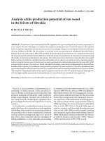
Fig. 1 AZ and/or MS-275 treatments produced a dose-dependent reduction in NB cells. a-c present graphic and tabulated evidence for the dose
response. Doses were AZ(0–160 μM), MS-275(0–3 μM) and AZ + MS-275(0–160 μM + 0.75 μM) for 48 h treatment of NSC6539, NSC6562, SH-SY5Y,
SK-N-SH and SK-N-BE(2) cells compared to the untreated group. The greatest decrease in IC50 was observed for AZ + MS-275 combination as
follow: SH-SY5Y = 17.5 μM, SK-N-SH = 16.5 μM and SK-N-BE(2) = 19.2 μM
Bayat Mokhtari et al. BMC Cancer (2017) 17:156
Page 6 of 23
Table 1 Percentage of cell viability values
Cell line
Treatment modality
Concentration (μM)
Reduction in cell viability (%)
p value
NSC6539
AZ
10
6 ± 0.67
-
MS-275
AZ + MS-275
NSC6562
AZ
MS-275
AZ + MS-275
SH-SY5Y
AZ
MS-275
AZ + MS-275
SK-N-SH
AZ
MS-275
AZ + MS-275
SK-N-BE(2)
AZ
MS-275
AZ + MS-275
20
26 ± 0.87
p < 0.05
40
47 ± 0.07
p < 0.01
0.75
12 ± 0.85
-
1.5
32 ± 0.97
p < 0.05
3
51 ± 0.23
p < 0.01
10 + 0.75
28 ± 0.95
-
20 + 0.75
46 ± 0.56
p < 0.05
40 + 0.75
55 ± 0.50
p < 0.01
10
2 ± 0.07
-
20
22 ± 0.47
p < 0.05
40
45 ± 0.67
p < 0.01
0.75
10 ± 0.34
-
1.5
24 ± 0.08
p < 0.05
3
46 ± 0.39
p < 0.01
10 + 0.75
23 ± 0.86
-
20 + 0.75
38 ± 0.50
p < 0.05
40 + 0.75
50 ± 0.61
p < 0.01
10
16 ± 0.20
p < 0.05
20
39 ± 0.64
p < 0.01
40
35 ± 0.58
p < 0.001
0.75
32 ± 0.55
p < 0.001
1.5
60 ± 0.69
p < 0.001
3
86 ± 0.78
p < 0.001
10 + 0.75
35 ± 0.85
p < 0.01
20 + 0.75
55 ± 0.86
p < 0.001
40 + 0.75
64 ± 0.78
p < 0.001
10
22 ± 0.59
p < 0.01
20
34 ± 0.42
p < 0.01
40
61 ± 0.03
p < 0.001
0.75
37 ± 0.99
p < 0.001
1.5
66 ± 0.03
p < 0.001
3
87 ± 0.19
p < 0.001
10 + 0.75
37 ± 0.85
p < 0.01
20 + 0.75
58 ± 0.86
p < 0.001
40 + 0.75
69 ± 0.95
p < 0.001
10
15 ± 0.01
p < 0.01
20
37 ± 0.61
p < 0.01
40
61 ± 0.58
p < 0.001
0.75
26 ± 0.69
p < 0.001
1.5
55 ± 0.38
p < 0.001
p < 0.001
3
82 ± 0.21
10 + 0.75
34 ± 0.88
p < 0.01
20 + 0.75
52 ± 0.88
p < 0.001
40 + 0.75
62 ± 0.86
p < 0.001
Table 1 shows percentage of cell viability values of NSC6539, NSC6562, SH-SY5Y, SK-N-SH and SK-N-BE(2) by AZ(0–160 μM), MS-275(0–3 μM), and AZ + MS-275
(0–160 μM + 0.75 μM) treatments (48 h)
Bayat Mokhtari et al. BMC Cancer (2017) 17:156
Page 7 of 23
considered as additive, synergistic and antagonistic,
respectively.
Results
AZ, MS-275 and AZ + MS-275 treatments inhibit growth of
NB SH-SY5Y cells
To determine the effect of AZ, MS-275 and AZ + MS275 treatments on the growth of NB SH-SY5Y, SK-N-SH
and SK-N-BE(2) cells, we used the AlamarBlue and trypan blue assays. As reference normal controls, we included the neuronal stem cell strains, NSC6539 and
NSC6562, which represent neural lineage derived stem
cells albeit from the central nervous system [25]. We
chose clinically acceptable concentration ranges for AZ
(0-160 μM) [17, 26] and MS-275 (0–3 μM) [7–9, 27]. It
should be noted that AlamarBlue also indicates effects
on oxidative phosphorylation as it is a substrate for the
last step in oxidative phosphorylation. Thus it can also
reflect metabolic mitochondrial effects.
Figure 1a-c shows that both AZ and MS-275 had a
more moderate concentration-dependent inhibitory effect on neuronal stem cells while the effects of MS-275
and the AZ + MS-275 combination were significantly enhanced on all three tumor lines. The reduction in cell
viability and IC50 values of NSC6539, NSC6562, SHSY5Y, SK-N-SH and SK-N-BE(2) by AZ, MS-275, and
AZ + MS-275 (48 h) shows the highest percent reduction
with AZ + MS-275 in achievable plasma concentration
(Tables 1 and 2). A significant difference in IC50 values
for SH-SY5Y, SK-N-SH and SK-N-BE(2), indicates the
potentiation of MS-275 effect by AZ. Interestingly, CDI
analysis for the combination of AZ and MS-275 on
different cell lines reveal that the combination is antagonistic at all concentrations on NSC6539 cells (CDI =
1.14–1.42) and additive on NSC6562 cells at concentrations above 20 μM AZ (CDI = 1). On NB cell lines (SHSY5Y, SK-N-SH and SK-N-BE(2)) the combination was
found to be additive at 40 μM and 80 μM of AZ (CDI = 1)
and synergistic at 160 μM of AZ (CDI < 1). Since SHSY5Y showed moderate resistance to AZ and/or MS-275,
Table 2 Percentage of IC50 values
Cell
IC50 (μM)
AZ
MS-275
AZ + MS-275
NSC6539
53
2.8
23.5
NSC6562
56
3
35
SH-SY5Y
45
1.23
17.5
SK-N-SH
42
1
16.5
SK-N-BE(2)
49
1.48
19.2
Table 2 shows percentage of NSC6539, NSC6562, SH-SY5Y, SK-N-SH and SK-NBE(2) by AZ (0–160 μM), MS-275 (0–3 μM), and AZ + MS-275 (0–160 μM +
0.75 μM) treatments (48 h)
average concentration-response and IC50 compared to
the other two NB cell lines, we chose to focus on SHSY5Y cell line for the rest of the study. The AlamarBlue
assay results also raised the question of effects on apoptosis and cell cycle as a measure of possible growth arrest
and/or toxicity.
We characterized the effect on apoptosis and cell cycle
using a propidium iodide (PI) based FACS analysis.
Using a dose less than the IC50 dose, AZ (40 μM vs.
45 μM), MS-275(1.5 μM vs. 2.36 μM) and AZ + MS275(40 μM + 0.75 μM) it was found that AZ, MS-275
and AZ + MS-275 treatments increase entry of SH-SY5Y
cells into SubG0-phase (0.6%, %57 and %61) with decrease into S-phase (13%, 9% and 4%) and G2/M-phase
(6%, 2% and 3%), significantly following a 48 h dosage of
AZ(40 μM), MS-275(1.5 μM) and AZ + MS-275(40 μM
+0.75 μM), respectively (Fig. 2a-b). In addition, western
blot analysis of cell cycle inhibitor, p21, shows that MS275 and AZ + MS-275 treatments (48 h) cause of 4.5
and 5.5 induction of p21 (p < 0.01) of SH-SY5Y cells
compare to control, respectively (Fig. 2c-d). Results
would suggest that AZ, MS-275 and AZ + MS-275 treatments are inducing apoptosis and preventing entry into
S and G2/M-phases. This was further characterized by
in-cell western and western blot analysis of cell cycle
checkpoints, demonstrating a 2.5 fold induction of p16
(85% ± 0.19%; p < 0.001), a 1.97 fold induction of p21
(50% ± 0.64%; p < 0.001), a 1.48 fold induction of p27
(34% ± 0.31%; p < 0.05), cyclin dependent kinase inhibitors (Cdki), and downregulation of CDK4 (33% ± 0.15%;
p < 0.01) and cyclin D1 (31% ± 0.35%; p < 0.001) following
a 48 h dosage of 1.5 μM MS-275, respectively (Fig. 2e-f).
These results were confirmed by western blot analysis
(Fig. 2 g-h). Furthermore, western blot analysis of the expression of the Ki67 proliferative marker showed a down
regulation of Ki67 (Fig. 2 g-h). Ki67 is expressed at all
points during the cell cycle, except in G0. It should be
noted that inductions of Cdki p21 occurred at the
lower concentration of MS-275 but decreased somewhat at the higher concentrations suggesting a more
complex epigenetic regulatory mechanism. Overall
Cdki inductions varied except for p16. We also conducted a PI cell cycle analysis to determine the percentage of sub-G1 cell.
Since cell death due to mechanical damage is also
accounted for in sub-G1 cell analysis, we also assessed
Annexin/7-AAD and cleaved-caspase 3 expressions by
FACs. Annexin stains cells in the early stage of apoptosis, while positive stain for 7-AAD indicates loss of
membrane integrity. Cells at end stage of apoptosis are
characterized by double staining for Annexin and 7AAD. The results of FACs analysis demonstrated that
MS-275 induces both early (10.7%) and late stage apoptosis (7.66%). The parallel assay with a clinically used
Bayat Mokhtari et al. BMC Cancer (2017) 17:156
Page 8 of 23
Fig. 2 AZ and/or MS-275 treatments increased cell cycle arrest and apoptosis of NB cells. a-b show propidium iodide analysis of cell cycle at 48 h
after treatment with AZ(40 μM), MS-275(1.5 μM) and AZ + MS-275 (40 μM + 0.75 μM) in NB SH-SY5Y, indicating increase entry of SH-SY5Y cells into
SubG0-phase (0.6%, %57 and %61; p ≤ 0.001) with decrease into S-phase (13%, 9% and 4%; p ≤ 0.05) and G2/M-phase (6%, 2% and 3%; p ≤ 0.05).
c-d show western blot analysis of cell cycle inhibitor, p21, indicates 48 h treatment of MS-275 and AZ + MS-275 treatments cause of 4.5 and 5.5
induction of p21 (p < 0.01) of SH-SY5Y cells compare to control, respectively. e-f show in-cell western (0.75 μM and 1.5 μM MS-275 treatment;
48 h) analyses of proteins associated with cell cycle arrest and apoptosis in NB SH-SY5Y cells. Results indicate that levels of cyclin D1 (0.666 ± 0.010%;
p = 0.0006), CDK4 (0.690 ± 0.033%; p = 0.0002) and BCL2 decreased significantly (0.376 ± 0.014%; p = 0.0035) while p16 CDK inhibitor significantly
increased (2.528 ± 0.101%; p = 0.0002). g-h show western blot data using lysates from cells treated 48 h with 0.75 μM and 1.5 μM MS-275. p21 and
p27 showed constant levels following treatment whereas cyclin D1 and CDK4 were reduced in expression. Expression of Ki67 proliferative marker
decreased as well
chemotherapeutic 1 μM Etoposide (the IC50 dose)
showed 2.51% and 8.58% induction of early and late
stage apoptosis, respectively (Fig. 3a and Table 3). In
addition, using the apoptotic indicator, cleaved caspase
3, 0.75 μM and 1.5 μM MS-275 after 48 h treatment
yielded significantly 1% and 3% expression of cleavedcaspase 3, respectively (p = 0.0003 and p < 0.0001 respectively as compared to control and p = 0.0001 when
comparing doses) (Fig. 3b-c). The pro-apoptotic effect of
MS-275 was further investigated by western blot analysis
of BCL2 and BAX expression. The results demonstrated
that BCL2/BAX ratio is reduced (19.6% of control) by
1.5 μM MS-275 treatment (48 h);(Fig. 3d), indicating a
potent apoptotic effect. MS-275 increases the expression of apoptotic protein BAX, hence decreasing the
BCL2/BAX ratio, and coordinate with decrease in survival. Western blot analysis also demonstrated that
survivin, a potent inhibitor of apoptosis, is reduced in
expression following MS-275 treatment (Fig. 3e). Here
NT2/D1 is a neuronal subclone of the teratomas NT2
cell line used in comparison, and as it is more stem
cell like.
AZ, MS-275 and AZ + MS-275 treatments reduced migration
capacity in NB SH-SY5Y cells
Previous studies had shown that both AZ and MS-275
reduce migration capacity in bladder and liver cancer
cells [27, 28]. In the current study, we show the reduction of migration capacity in AZ (40 μM), MS-275
(1.5 μM) and AZ + MS-275 (40 μM + 0.75 μM) treated
NB SH-SY5Y cells in Table 4. In Fig. 4a it is evident that
AZ alone has an obvious inhibitory effect, and greatly
enhanced by MS-275 at 48 h. The difference between
MS-275 alone and the combo treatment was statistically significant (p < 0.001) at both 48 h and 72 h. AZ
therefore enhances the inhibitory effect of MS-275 on
SH-SY5Y cell migration capacity (Fig. 4a-b). In this assay
the cytotoxicity of the agents is demonstrated overtly
Bayat Mokhtari et al. BMC Cancer (2017) 17:156
Page 9 of 23
Fig. 3 a-b show the results of FACs analysis for 7-AAD and Annexin staining. Results demonstrate that compared to etoposide, MS-275 induces
entry into early and late stage of apoptosis in 10.7% versus 2.51% and 7.66% versus 8.58% of cells, respectively. c show cleaved caspase 3 expression
after 48 h treatment with 0.75 μM and 1.5 μM MS-275 that yielded 1% and 3% expression, respectively (p = 0.0003 and p < 0.0001, respectively as
compared to control and p = 0.0001 when comparing doses). d-e show increased expression of apoptotic protein BAX and decreased BCL2/BAX ratio
with coordinate decrease in survival following 0.75 μM and 1.5 μM MS-275 treatment to 15.59% and 14.86% of control. In addition, survivin expression
reduced following 0.75 μM and 1.5 μM MS-275 treatment to 7.8% and 11%, respectively
because of the initial cell confluence and may predict subsequent in vivo effects.
AZ significantly potentiates the inhibitory effect of
MS-275 on tumorigenesis in NB SH-SY5Y xenografts
A concentration of 24.5 mg/kg MS-275 reduced growth
of NB KCNR cell line as orthotopic xenografts [8]. A 14day treatment protocol with 40 mg/kg AZ and/or
20 mg/kg MS-275 was devised based on the observed
IC50 results and previously used safe doses in other preclinical studies [8, 29]. The inhibition of tumor volume
is presented in Table 5. Figure 5a grossly shows a dramatic reduction in tumor growth and volume after MS275 (20 mg/kg) and significantly greater when AZ
(40 mg/kg) was added to the treatment. Figure 5b shows
the changes in histology suggesting phenotypic alterations
Table 3 Percentage of apoptosis values
Unstained (Lower left)
Annexin (Lower right)
7-AAD (Upper left)
Annexin 7-AAD (Upper right)
Control
90
0.6
6.80
3.9
Etoposide (1 μM)
85
2.57
5.90
8.68
MS-275 (1.5 μM)
72.18
10.11
8.60
7.06
Table 3 shows the parallel assay with a clinically used chemotherapeutic Etoposide (1 μM);(2.51% and 8.58%) and MS-275 (1.5 μM);(10.7% and 7.66%) showed
induction of early and late stage apoptosis, respectively
Bayat Mokhtari et al. BMC Cancer (2017) 17:156
Page 10 of 23
Table 4 Percentage of cell migration capacity values
Time
Control
Treatment
% Cell migration capacity
Untreated
94
AZ
MS-275
AZ + MS-275
48 h
72 h
95.33
96.98
93.66
90.3
85.87
-
-
p < 0.01
94
42.33
38.56
p < 0.001
p < 0.001
37
26
p < 0.001
p < 0.001
95
Table 4 shows percentage of cell migration capacity values of SH-SY5Y by AZ
(40 μM), MS-275 (0.75 μM) and AZ + MS-275 (40 μM + 0.75 μM) treatments
(48 h and 72 h)
while Fig. 5c and d graphically reflect large reductions of
volumes and weights, over the 14 days treatment
period. The extirpated tumors also revealed grossly that
both MS-275 and AZ + MS-275 markedly reduced the
hematogeneous appearance of the tumors suggesting
significant loss of vascularization (Fig. 5a-b). The significant reductions in tumor growth and weight were
greatest with AZ + MS-275 (Table 5). The significant
anti-tumor growth potentiation effect of AZ on MS275 suggested an additive effect for this combination.
IHC results revealed a possible explanation for the dramatic reduction in tumor volumes in that there was a
strong inhibition of angiogenesis as revealed with staining
for the angiogenesis marker (CD31) (Fig. 5e). Compared
to the untreated group, expression of the CD31 was most
significantly reduced with the combination AZ + SFN
(Table 5; Fig. 5f).
AZ, MS-275 and the AZ + MS-275 treatments induce
apoptosis in NB SH-SY5Y xenograft cells
The initial histopathological assessment of the residual
tumors (Fig. 5a-e) revealed reductions in cell density and
size, presence of pyknotic nuclei and reduced nuclear
size, most prominently in the case of AZ + MS-275. This
suggested that apoptosis could account for cell loss. To
further confirm the histological changes, we performed
electron microscopy on the tumor xenografts. Ultrastructural analysis revealed cells with degradative cytoplasmic changes [8, 30, 31] and nuclear fragmentation
(pyknotic cells) indicative of apoptosis in SH-SY5Y xenografts. AZ + MS-275 treated cells had the highest
number of pyknotic cells (Table 6; Fig. 6a-b), further
supported the histological finding that AZ potentiated
the apoptotic effect of MS-275. Next, we assessed apoptosis with the TUNEL assay on the xenografts (Table 6).
We found that AZ + MS-275 treated cells had the
highest percent TUNEL positive cells (Fig. 6c-d), and
significantly greater than MS-275 alone. To further
confirm the apoptotic process, we performed IHC to
study the effect of AZ, MS-275 and AZ + MS-275 treatments on expression of the apoptotic marker (cleaved
PARP) in the SH-SY5Y xenografts. Compared to the
untreated group, expression of the apoptotic marker
(cleaved PARP) was significantly induced, versus untreated controls (Table 6; Fig. 6e-f ). Thus, AZ showed
an overt potentiation of the anti-tumor effect of MS275 in the SH-SY5Y xenografts.
AZ, MS-275 and AZ + MS-275 reduce expression of mitotic
and proliferative markers in NB SH-SY5Y xenografts
The results from the apoptosis assessment of treated xenografts suggested strong effects on tumor cell proliferation. We therefore performed IHC to study the effect of
AZ, MS-275 and AZ + MS-275 treatments on expression
of mitotic (phosphohistone-H3; pHH3) and proliferation
(Ki-67) markers in the SH-SY5Y xenografts. Compared
to the untreated group, expression of the mitotic marker
(pHH3) was moderately reduced after AZ treatment
alone, significantly after MS-275 treatment and further
enhanced with the combination AZ + MS-275 (Table 7;
Fig. 7a-b). In a similar manner expression of the proliferative marker, Ki67 was significantly reduced by all
treatments and the most by AZ + MS-275 (Table 7;
Fig. 7c-d). Thus, the large reductions in tumor growth
produced by MS-275 and the AZ + MS-275 combination
are additionally reflected in significant reductions in mitosis and proliferation paralleled by increased apoptosis.
It is noteworthy that the potent inhibitory effect of AZ
alone was revealed using these markers, and potentiation
of MS-275 was further confirmed.
AZ and/or MS-275 treatment reduced expression of
HIF1-α and CAIX in NB SH-SY5Y xenograft
Given the remarkable reductions in vascularization it
might be surmised that the tumors would experience
enhanced hypoxia under the treatments and thereby increased hypoxia induced gene expression. However, we
performed IHC on the xenografts and did observe that
control untreated tumors significantly expressed HIF1-α
and its downstream target CAIX (Table 7; Fig. 8a-d). We
further found that the number of HIF1-α positive cells
decreased significantly after all treatments and markedly
after AZ + MS-275 (Table 8; Fig. 8a and b). Additionally,
a markedly enhanced reduction in CAIX expression
(membrane localization) after AZ + MS-275 (Table 8;
Fig. 8c and d) paralleled that of HIF1-α. The major reduction in CAIX staining after AZ + MS-275 may reflect
much more than a loss of viable cells supporting the
concept of AZ potentiation of the epigenetic alterations
in expression.
Bayat Mokhtari et al. BMC Cancer (2017) 17:156
Page 11 of 23
Fig. 4 AZ and/or MS-275 treatments decreased migration capacity of NB cells. a-b represent the wound healing assay for AZ (40 μM), MS-275
(1.5 μM) and AZ + MS-275 (40 μM + 0.75 μM) treatment compared to untreated group in SH-SY5Y cells. AZ caused a 10 ± 0.35% (p = 0.025, 48 h)
and 12 ± 0.85% (p < 0.01,72 h) inhibition while MS-275 caused a 42 ± 0.13% (p < 0.001, 48 h) and 65 ± 0.30% (p < 0.001, 72 h) inhibition in migration.
The AZ + MS-275 combination significantly inhibited migration with a 63 ± 0.37% (p < 0.001, 48 h) and 74 ± 0.25% (p < 0.001, 72 h) inhibition in
migration ability
Table 5 Percentage of tumor volume, weight and expression of CD31 values
Treatment modality
Concentration (mg/kg)
% Inhibition of tumor volume
(p value)
% Reduction in tumor weight
(p value)
% Expression of CD31 marker
(p value)
AZ
40
13 ± 0.29 (p = 0.009)
29 ± 0.2 (p = 0.193)
87 ± 0.98 (p = 0.006)
MS-275
20
43 ± 0.50 (p = 0.001)
83 ± 0.74 (p < 0.001)
48 ± 0.94 (p = 0.008)
AZ + MS-275
40 + 20
60 ± 0.48 (p = 0.001)
89.5 ± 0.94 (p < 0.001)
27 ± 0.92 (p < 0.001)
Table 5 shows percentage of tumor volume and weight reduction and expression of CD31 values of SH-SY5Y tumors by AZ (40 mg/kg), MS-275 (20 mg/kg) and
AZ + MS-275 (40 + 20 mg/kg) treatments (14D)
Bayat Mokhtari et al. BMC Cancer (2017) 17:156
Page 12 of 23
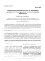
Fig. 5 AZ and/or MS-275 inhibited tumor growth of NB xenografts. a presents the gross morphology, b H&E histology, c volume, d weight,
e CD31 staining and f the percentage of CD31 positive cells after 14 days treatment with AZ and/or MS-275 compared to the untreated group in
SH-SY5Y xenografts. AZ caused 13% ± 0.29%, MS-275 43% ± 0.50% and AZ + MS-275 60% ± 0.48% (p = 0.0009) inhibition in tumor growth. Tumor
weights were reduced by 29 ± 0.2% (p = 0.193) for AZ, 83% ± 0.74% (p < 0.001) for MS-275 and 89.5 ± 0.49% (p < 0.001) for AZ + MS275. AZ caused
a 13% ± 0.98%(p = 0.006), MS-275 a 52% ± 0.0.94% (p = 0.0008) and AZ + MS-275 a 73% ± 0.92% (p < 0.0001) decrease in the number of CD31
positive cells compared to the control
AZ + MS-275 treatment significantly reduces the in vivo
tumorigenic potential of NB SH-SY5Y xenograft cells
The ultimate test for loss of tumorigenic potential is
whether treatments abrogate formation of tumors, accomplished by conducting serial heterotransplantation.
To test this, after the initial round of treatments with
the most effective AZ + MS-275 combination we serially
heterotransplanted a second set of 3x10⁴ cells into
NOD/SCID mice and in comparison to a similar number
of untreated cells. We found that the combination of
AZ + MS-275 caused a 69% ± 0.84% (p = 0.0002) inhibition
in resulting tumor volumes and weights (81% ± 0.78%;
p = 0.0001) respectively after 38 days (Fig. 9a-d). Note
however that only 3/5 injections yielded any tumor
Table 6 Percentage of pyknotic, TUNEL and Cleaved PARP positive cells values
Treatment modality
% Pyknotic cells (p value)
% TUNEL positive cells (p value)
% Cleaved PARP positive cells (p value)
AZ
8 ± 0.6 (p < 0.001)
7 ± 0.66 (p = 0.0015)
12 ± 0.7 (p < 0.01)
MS-275
36 ± 0.45
56 ± 0.33 (p < 0.001)
50 ± 0.23 (p < 0.001)
AZ + MS-275
51 ± 0.83 (p < 0.001)
82 ± 0.76 (p < 0.001)
69 ± 0.35 (p < 0.001)
Table 6 shows the percentage of pyknotic, TUNEL and Cleaved PARP positive cells values of SH-SY5Y tumors by AZ (40 mg/kg), MS-275 (20 mg/kg) and AZ +
MS-275 (40 + 20 mg/kg) treatments (14D)
Bayat Mokhtari et al. BMC Cancer (2017) 17:156
Page 13 of 23
Fig. 6 AZ and/or MS-275 treatments increase the apoptotic TUNEL index in NB xenografts. a-b present ultrastructural details [x5000, cytoplasmic
(arrow)] including the percentage of pyknotic cells. c-d present results from the TUNEL assay (x20 and x40) for detection of apoptotic cells (arrow)
and the number of apoptotic positive cells after 14 days treatment with AZ, MS-275 and AZ + MS-275 compared to untreated group in SH-SY5Y
xenografts. The number of TUNEL positive cells increased modestly after treatment with AZ (7 ± 0.66%; p = 0.0015), moderately after MS-275
(56 ± 0.33%; p < 0.001) and significantly after AZ + MS-275 (82 ± 76%; p < 0.001) compared to the untreated group. e-f present the expression
of the apoptotic marker (cleaved PARP) that was significantly induced by AZ (12% ± 0.16%; p = 0.001), MS-275 (63% ± 0.33%; p = 0.0001) and
further by AZ + MS-275 (78% ± 0.09%; p = 0.0001)
mass. A marked phenotypic alteration in morphological appearance (Fig. 9b) resembled the treated
tumors in Fig. 5a-e. These results suggest that the
AZ + MS-275 combination might permanently alter
cell phenotype and affect the presumptive CSCs in
the tumor.
AZ, MS-275 and AZ + MS-275 treatment affects stem cell
marker expression in NB SH-SY5Y xenograft cells
Our serial heterotransplantation results suggested that the
CSC fraction could be targeted by a potentiated effect of
AZ + MS-275. In parallel studies, we had examined the effect of MS-275 on another MYCN amplified NB cell line,
Table 7 Percentage of pHH3 and Ki67 positive cells values
Treatment modality
% Expression of pHH3 positive cells
(p value)
% Expression of Ki67 positive cells
(p value)
AZ
58 ± 0.66 (p < 0.05)
61 ± 0.77 (p = 0.01)
MS-275
22 ± 0.11 (p < 0.01)
29 ± 0.42 (p < 0.001)
AZ + MS-275
6 ± 0.73 (p < 0.001)
16 ± 0.85 (p < 0.001)
Table 7 shows percentage of pHH3 and Ki67 positive cells values of SH-SY5Y tumors by AZ (40 mg/kg), MS-275 (20 mg/kg) and AZ + MS-275 (40 + 20 mg/kg)
treatments (14D)
Bayat Mokhtari et al. BMC Cancer (2017) 17:156
Page 14 of 23
Fig. 7 AZ and/or MS-275 treatments affected the mitosis and proliferation of NB xenografts. a-b present the IHC study (x20 and x40) of the
mitotic index (pHH3, arrow) and the number of pHH3 positive cells. c-d present the immunodetection and number of Ki67 positive cells (arrow)
after 14 days treatment with AZ, MS-275 and AZ + MS-275 compared to untreated group in SH-SY5Y xenografts. Data shows that pHH3 expression was
significantly reduced by AZ (42 ± 0.43%; p = 0.02), MS-275 (78 ± 0.16%; p = 0.002) and further by AZ + MS-275 (94 ± 0.05%; p = 0.001). Ki67 expression
was moderately reduced by AZ (38 ± 0.70%; (p = 0.007), strongly reduced by MS-275 (70 ± 0.25%; p = 0.0002) and further reduced by AZ + MS-275
(84 ± 0.73%; p = 0.0001)
Fig. 8 AZ and/or MS-275 treatments affected hypoxia response and CAIX in NB xenograft cells. a-b present the IHC study (x20 and x40) on
HIF1-α expression, (arrow) and the number of HIF-1α positive cells. c-d present CAIX expression (arrow) and the number of CAIX positive cells
after 14 days treatment with AZ and/or MS-275 compared to untreated group in SH-SY5Y xenografts. HIF-1α expression was significantly reduced
by AZ (29 ± 0.7%; p < 0.001), MS-275 (66 ± 0.23%; p < 0.001) and further enhanced in AZ + MS-275 (83 ± 0.67%; p < 0.001). CAIX expression was
significantly reduced by AZ (18 ± 0.11%; p = 0.0138), MS-275 (73 ± 0.33%; p < 0.001) and markedly by AZ + MS275 (90 ± 0.16%; p < 0.001)
Bayat Mokhtari et al. BMC Cancer (2017) 17:156
Page 15 of 23
Table 8 Percentage of HIF-1α and CAIX positive cells values of SH-SY5Y tumors
Treatment modality
% Expression of HIF-1α positive cells
(p value)
% Expression of CAIX positive cells
(p value)
AZ
71 ± 0.70 (p < 0.001)
82 ± 0.11 (p = 0.0138)
MS-275
34 ± 0.23 (p < 0.001)
27 ± 0.33 (p < 0.001)
AZ + MS-275
17 ± 0.73 (p < 0.001)
10 ± 0.16 (p < 0.001)
Table 8 shows the percentage of SH-SY5Y tumors by AZ (40 mg/kg), MS-275 (20 mg/kg) and AZ + MS-275 (40 + 20 mg/kg) treatments (14D)
SK-N-BE(2), and in comparison to the teratocarcinoma
cell line, NT2 with a prevalent stem cell phenotype. MS275 significantly inhibited clonogenic potential of SHSY5Y and SK-N-BE(2) cells (Fig. 10a-c). Here we first
established an effective concentration range for abrogating
clonogenicity in methycellulose, the reduction in the presumptive tumor initiating SP cell fraction (Fig. 11a-b),
compared MS-275 with TSA and found MS-275 to be
more potent (Fig. 11c-d) while cisplatin (CDDP) was ineffective and reduction in expression of stem cell markers
OCT4 and SOX2 by FACs and western blot analysis
(Fig. 12a-h). These studies established the ability of MS275 to reduce the tumor initiating potential of NB MYCN
NB amplified cell lines. However, to expand upon these
studies using immunophenotyping to determine the role
of specific stemness markers (OCT4, SOX2, Nanog), we
applied IHC to our SH-SY5Y xenografts after AZ, MS-275
and AZ + MS-275 treatments. We found that the AZ +
MS-275 treatment produced the highest reduction in
OCT4 expression, number of SOX2 positive cells, and
Nanog immunopositive cells (Table 9; Fig. 13a-f). It is interesting to note that OCT4 and SOX2 expressions were
most affected by AZ treatment, and relative to Nanog,
proportionally greatest after AZ + MS-275 treatment,
again supporting the idea of AZ potentiation of MS-275.
Discussion
HDACis are currently being evaluated in cancer clinical trials including NB with still promising results [32]. Whether
these like SAHA and MS-275 could become routinely
Fig. 9 AZ + MS-275 decreased the in vivo tumorigenic potential of pretreated NB SH-SY5Y cells. a presents the in vivo serial heterotransplantation
study (x20 and x40) with pretreated NB xenograft cells showing the morphology, b H&E histology, c volume and d weight after 37 days treatment with
AZ + MS-275 compared to untreated group in SH-SY5Y xenografts. Notably, AZ + MS-275 caused a significant reduction in tumor volumes (69 ± 0.84%;
p = 0.0002) and weights (81 ± 0.78%; p = 0.0001) after 37 days compared to xenografts generated from untreated cells
Bayat Mokhtari et al. BMC Cancer (2017) 17:156
Page 16 of 23
Fig. 10 MS-275 treatment reduced clonogenic potential of NB and teratocarcinoma cells. a clonogenic potential was negatively affected with
increasing doses of MS-275. b dose response curves of MS-275 treatment of NT2/D1, SH-SY5Y and SK-N-BE(2) over a 2 week growth period in
methycellulose compared to control. Similarly, clonogenic capacity was negatively affected in all three cell lines despite different initial clonogenic
efficiencies. IC50 values were calculated to be 0.13 μM, 0.20 μM and 0.18 μM for NT2/D1, SH-SY5Y and SK-N-BE(2), respectively. c representative
images for control, 0.2 μM and 0.75 μM MS-275 treatments in methycellulose clonogenic assay run on NT2/D1 and SK-N-BE(2)
administered is currently undecided. However, little has
been done to determine if these could be potentiated with
other approved drugs and in particular drugs like AZ
which can be repurposed based on sound reasoning given
knowledge about pH regulation in tumor cells. We took
this latter approach and now report that AZ, MS-275 and
especially the AZ + MS-275 combination inhibited migration, in vitro growth, induced cell cycle arrest and apoptosis of NB SH-SY5Y. In addition, the combination
markedly inhibited tumor growth in vivo, reduced tumorigenicity and expression of mitosis, proliferative, HIF1-α
and CAIX markers in NB SH-SY5Y xenografts. Importantly, we provide additional evidence that MS-275, at
nanomolar concentrations, significantly reduced the tumor
initiating cell fraction in NB SH-SY5Y and SK-N-BE. The
significant reduction in initial tumorigenicity and subsequent serial heterotransplantation suggests either potential elimination or reprogramming of NB tumor
initiating cells. Moreover, stemness genes (OCT4,
SOX2 and Nanog) were found to be significantly
down-regulated after MS-275 and the effect was enhanced by AZ + MS-275 treatment.
MS-275 has been previously shown to induce a potent
G1 cell cycle arrest in NB studies [33, 34]. We confirmed
this key G1 cell cycle arrest and provided evidence that
dysregulation of the G1 entry checkpoint in NB is likely
due to Cyclin D1 overexpression [34]. Cell cycle inhibitors that modulate cyclinD/CDK4 complex are important in G1 cell cycle arrest [8, 34]. Cyclin D1 and CDK4
knockdown results in proliferation inhibition, G1 cell
cycle arrest and neuronal differentiation [35]. In this
study we show that MS-275 treatment significantly reduced the expression of cyclin D1 and CDK4 relative to
controls. It is not clear whether this reduction results
from a direct effect of MS-275 or involves a more
downstream mechanism. It has been shown that
HDACi can induce the p21 cell cycle inhibitor [36].
Similarly, we found that p21 and p27 were upregulated
Bayat Mokhtari et al. BMC Cancer (2017) 17:156
Page 17 of 23
Fig. 11 MS-275 treatment decreased the SP fraction of NB and teratocarcinoma cells. a using Hoechst 33342 dye exclusion, the SP fraction in
NT2/D1, SH-SY5Y and SK-N-BE(2) was determined to be 3.32 ± 1.26%, 0.74 ± 0.35% and 0.32 ± 0.11%, respectively. b representative FACs SP profiles
for SH-SY5Y control and cells treated with 1.5 μM MS-275, 100nM TSA and 10 μM CDDP. Gating was determined by verapamil negative control.
c normalizing for control, 0.75 μM MS-275 reduced the SP fraction significantly (p < 0.0001, p = 0.0098 and p = 0.0237). Similar treatments with
1.5 μM MS-275 also reduced the SP population in NT2/D1, SH-SY5Y and SK-N-BE(2) significantly (p < 0.0001, p = 0.0177 and p < 0.0001). d low nM
doses, 0.1 μM HDACi TSA and 0.75 μM MS-275, significantly reduced the SP fraction of both NB lines but greater in SK-N-BE(2), a MYCN amplified
cell line (p = 0.0062, p = 0.045 respectively); in contrast highly toxic levels of CDDP did significantly alter the SP fraction (p > 0.05)
with MS-275 treatment. Interestingly, we observed a
dramatic increase in the expression of p16 CDKi. Deregulation of p16 is a common finding in a variety of
neoplasms [37], and HDACi have been found to induce
p16 in certain types of cancer such as colon carcinoma
[38]. Induction of multiple cell cycle inhibitors would
be predicted to strongly block cell cycle progression.
MS-275 induces apoptosis through different mechanisms including induction of oxidative stress, the intrinsic and extrinsic pathways of apoptosis [39]. It has been
shown by Muhlethaler-Mottet [37] that inducing the intrinsic pathway of apoptosis is the most common mechanism by which HDACi such as TSA, SAHA and NaB
induce apoptosis [40]. Helminthosporium carbonum
(HC)-toxin (a natural HDACi) has been shown to decrease the expression of anti-apoptotic BCL2 in NB [41].
We found that the BCL-2/BAX ratio was significantly
decreased by MS-275 treatment, indicating induction of
the intrinsic pathway of apoptosis. BCL-2/BAX ratio also
serves as a predictor of drug efficacy and cancer invasiveness [42].
We surmised that targeted inhibition of CAs by AZ
could interfere with the hypoxia induced HIF1-α mediated regulation of tumor cell pH homeostasis with likely
consequences to other HIF1-α regulated processes required for tumor cell growth, progression, and survival
[43]. Since HDACi, such as MS-275 and SAHA, also target HIF1-α activity (e.g. translation [42]) the combination might produce a synergistic effect by blocking the
ability of tumor cells to overcome hypoxic stress induced apoptosis [8, 44, 45]. Since the hypoxic niche favors localization of CSCs and their growth and survival
[21], our results suggest that CSCs are targeted by the
AZ + HDACi combination.
Previous studies have shown that HDAC inhibitors
affect migration capacity of tumor cells [46]. The
Bayat Mokhtari et al. BMC Cancer (2017) 17:156
Page 18 of 23
Fig. 12 MS-275 treatment decreased expression of stem cell markers in NB and teratocarcinoma cells. a in the high OCT4, SOX2 and Nanog expressing
NT2/D1 teratocarcinoma cell line, MS-275 was able to reduce nuclear expression of these stem cell markers as shown by immunofluorescence labeling.
b Western blot analysis demonstrated a MS-275 reduction in expression of OCT4 and Nanog in NT2/D1, and OCT4 in SY5Y. c representative
FACs profiles for OCT4 in SH-SY5Y and NT2/D1 cells. d calculated from FACs data in c. SH-SY5Y contains 0.38 ± 0.06% OCT4 positive and
0.33 ± 0.04% SOX2 positive cells. e MS-275 treatment significantly reduced expression of OCT4, SOX2 and Nanog in NT2/D1 at 0.75 μM
(p = 0.0024, p < 0.0001 and p = 0.0031, respectively) and at 1.5 μM (p = 0.0096, p < 0.0001 and p = 0.0023, respectively). f when normalized to
percentage of control, MS-275 treatment significantly reduced expression of OCT4 and SOX2 in SH-SY5Y at 0.75 μM (p = 0.0480 and p = 0.0391,
respectively) and 1.5 μM (p = 0.0003 and p = 0.0066, respectively). g representative FACS profile for ABCG2 staining in SH-SY5Y and SK-N-BE(2) NB cell
lines. h ABCG2 expression significantly decreases following MS-275 treatment in SH-SY5Y, SK-N-BE(2) and NT2/D1 cells at 0.75 μM (p = 0.0111, p = 0.0131
and p = 0.0086, respectively) and at 1.5 μM (p = 0.027, p = 0.0022 and p = 0.0084, respectively). Trypsin cleavage of cell surface ABCG2 was used
as a negative control
combination of trichostatin A (HDACi) and decitabine
effectively decreased migration capacity of ovarian cancer cell line SKOV3 [47]. MS-275 treatment reduced migration capacity of leukemia cells [48]. Epigenetic
modifications by HDACi play a key role in regulating
the expression of proteins that promote or suppress
tumor cell migration [49]. Tumor cell migration capacity
is also enhanced by activation of the HIF1-α pathway
[50], which in turn regulates CA activity. Therefore, CA
inhibitors could decrease migration capacity of tumor
cells. Invasion and migration are key components of the
metastatic process. Here we show that the AZ + MS-275
Bayat Mokhtari et al. BMC Cancer (2017) 17:156
Table 9 Percentage of OCT4, SOX2 and Nanog positive cells
values of SH-SY5Y tumors
Stem cell marker
Treatment modality
% Expression of positive
cells (p value)
OCT4
AZ
63 ± 0.35 (p < 0.05)
MS-275
37 ± 0.85 (p < 0.001)
AZ + MS-275
18 ± 0.45 (p < 0.05)
AZ
68 ± 0.60 (p < 0.01)
MS-275
39 ± 0.50 (p < 0.009)
AZ + MS-275
18 ± 0.46 (p < 0.002)
AZ
89 ± 0.60 (p < 0.01)
MS-275
46 ± 0.45 (p < 0.01)
AZ + MS-275
30 ± 0.76 (p < 0.01)
SOX2
Nanog
Table 9 shows the percentage of SH-SY5Y tumors by AZ (40 mg/kg), MS-275
(20 mg/kg) and AZ + MS-275 (40 + 20 mg/kg) treatments (14D)
combination significantly affected tumor cell migration
using a wound healing assay concomitant with effects on
growth and tumor cell survival. Thus the wound healing
assay has limitations but effects on confluent cultures
may predict subsequent in vivo results.
Our observations of a potentiated anti-tumor effect
by AZ + MS-275 using the different assays questioned
whether the effects were synergistic. In fact simple interpretations of the data might deduce synergism with
certain parameters and additively with others. We
therefore undertook to analyze the effects by CDI
analysis and found that in monolayer cultures the combination was antagonistic at all concentrations on
NSC6539 cells and additive on NSC6562 cells at concentrations above 20 μM AZ. On NB tumor cell line
SH-SY5Y the combination was additive at 40 μM and
80 μM of AZ and synergistic at 160 μM of AZ. Since
the combination was additive or antagonistic at all concentrations on neural stem cells, while additive or synergistic above IC50 on SH-SY5Y cells, CDI values
indicate that the combination of acetazolamide and
MS-275 was specifically cytotoxic on tumor cells. In
fact, further in vivo results support the notion that the
AZ + MS-275 combination was indeed potently cytotoxic for NB tumor cells.
Metastasis is a major problem in advanced stage NB.
As indicated we found that the AZ + MS-275 combination significantly decreased migration of the SH-SY5Y
tumor cells. We asked whether a key molecular contributor to the metastatic phenotype, CAIX, which has
been well documented to play a role in tumor development and metastasis, was affected [51]. CAIX expression was found to be dramatically decreased by AZ +
MS-275, suggesting that this combination treatment
might indeed block metastatic behavior. CAIX is induced by HIF1-α, and HIF1-α knockdown significantly
Page 19 of 23
decreased proliferation, migration and invasiveness of
NB cell lines [50, 52]. HIF1-α expression is regulated by
PI3K/AKT signaling, which is blocked by HDAC inhibition [53–55]. Similar inhibitory effect on the expression of the hypoxia mediated axis in breast cancer cells
has been reported for MS-275 [56]. Here, we showed
that the AZ + MS-275 combination was most effective
in coordinately reducing the number of HIF1-α and
CAIX positive cells, correlating with a significant reduction in tumorigenic and likely metastatic potential.
Taken together our findings indicate the AZ + MS-275
treatment regimen could interfere with NB metastasis
at multiple levels.
MS-275 has recently been determined to be an inhibitor of the mTOR pathway by upstream modulation of
AKT [55]. The mTOR pathway has also been associated
with the regulation and modulation of HIF1-α, which
can modulate and regulate CSCs and the stem cell
phenotype [57]. Indeed, we previously showed that hypoxia induced HIF1-α signaling enhances the CSC
phenotype in NB side populations (SP) [58]. CSCs are
enriched in the SP fraction, a subpopulation defined by
the ability to exclude the DNA-binding Hoechst 33342
dye [59, 60]. These SP cells in NB express high levels of
stem cell markers, show increased tumorigenicity, and
expand under hypoxia [56]. HDACi have been shown
to modulate the HIF1-α mediated pathway by targeting
HIF1-α towards proteosomal degradation and by
repressing transactivation [61, 62]. While the modulation of AKT, mTOR and the HIF mediated pathways by
HDACi is not yet fully characterized, it offers mechanistic insight showing the multiple targets for how MS275 could be targeting CSCs (schematically shown in
Fig. 14). Commonalities in signaling and gene expression has been found between normal stem cells and
CSC; as such, drugs targeting normal stem cells should
be investigated for their potential ability to deplete the
CSC compartment.
Cancers with high percentages of cells expressing the
stemness markers, OCT4 and Nanog, have been associated with prognostically poor phenotypes [63]. In this
study, we showed that the combination of AZ + MS-275
significantly decreased the number of OCT4, Nanog and
SOX2 positive cells in NB xenografts. In addition, we
found that treatment with MS-275 significantly reduced
the percentage of cells expressing OCT4, SOX2 and
Nanog in NB and in a high stemness phenotype teratocarcinoma cell line. Decrease in expression of these
stem cell markers in teratocarcinoma is associated
with differentiation and loss of the stem cell phenotype. We also found MS-275 treatment reduced the
expression of these stemness related genes in the SP
fraction, taken as another means of identifying a fraction containing CSCs. NB SP cells expel Hoechst
Bayat Mokhtari et al. BMC Cancer (2017) 17:156
Page 20 of 23
Fig. 13 AZ and/or MS-275 treatments reduced the expression of stem cell markers in NB xenografts. a-b present IHC staining (x20 and x40) for
OCT4 cell localization and number of OCT4 positive cells, c-d SOX2 cell localization and number of SOX2 positive cells, and e-f Nanog cell localization
and number of Nanog positive cells after 14 days treatment with AZ, MS-275 and AZ + MS-275 compared to untreated group in SH-SY5Y xenografts.
The number of OCT4 positive cells was reduced after treatment with AZ by 37 ± 0.35% (p < 0.05), MS-275 by 63 ± 0.85% (p < 0.001) and AZ + MS-275
by 82 ± 0.45% (p < 0.001). The number of SOX2 positive cells was reduced in AZ by 32 ± 0.60% (p = 0.01), MS-275 by 61 ± 0.5% (p = 0.0009) and
AZ + MS-275 by 82 ± 0.46% (p = 0.0002). The number of Nanog positive cells was reduced in AZ by 11 ± 0.60% (p > 0.05), MS-275 by 54 ± 0.45%
(p = 0.0005) and AZ + MS-275 by 70 ± 0.76% (p = 0.0002)
33342 primarily by means of the ATP-dependent drug
pump ABCG2 [58–60]. ABCG2 expression is regulated by AKT/PI3K signaling pathway, which HDACi
is known to inhibit [64].
Finally, it is significant that MYCN non-amplified
SH-SY5Y and MYCN amplified SK-N-BE(2) cell line SP
fractions were equally targeted by MS-275 treatment.
This critical result indicated that MS-275 could target
MYCN amplified NB tumors, a group associated with
the most aggressive NB phenotype and poor prognosis
[1]. Cortes et al. has recently shown that actinomycin D
(a member of the actinomycine family) could synergistically potentiate the efficacy of the histone deacetylase inhibitor, SAHA in NB clinical trials. In addition, the
combination of actinomycin D with SAHA could inhibit
tumor growth in SK-N-JD NB-xenografts [65]. While MS275 or other HDACi such as SAHA are currently in
clinical trials as promising anticancer therapeutics,
however they still have limitations as single agents [66].
Therefore, a combination of a HDACi with a CA inhibitor affecting pH homeostasis, may constitute a
more effective alternative therapeutic option, as we
have reported in other types of cancer [17, 20] and as
shown here for NB. We surmise that concomitant compromise of pH homeostasis drives tumor cells into a
homeostatic crisis difficult to surmount. Since AZ has
been in clinical use over a long period and the pharmacokinetics and side effects are well known and managed
[67], it could be readily combined with HDACi already
being evaluated in clinical trials for NB.
Bayat Mokhtari et al. BMC Cancer (2017) 17:156
Page 21 of 23
Fig. 14 Schema depicting a potential mechanism of AZ and/or MS-275 treatments in NB cells. As illustrated, AZ, MS-275 and AZ + MS-275 treatments
target survival pathways in NB cells. Alterations in cell cycle, and the HIF1-α/CAIX axis by AZ, MS-275 and AZ + MS-275 could affect the NB cell transit
amplifying cell population, and its proliferation and survival. Importantly, these treatments could additionally target the CSC in NB by perturbation of
self-renewal potential, stem cell state and the SP phenotype. The pan-inhibition of these pathways and critical components would ultimately decrease
the tumorigenic potential of NB cells and lead to elimination of the tumor cells
Conclusions
We present convincing evidence suggesting that the
AZ + MS-275 combination is likely targeting a substantial portion of NB tumor cells in vivo and in particular
the CSC population in NB. This finding increases its
value in terms of possible management of NB tumor
progression and metastasis using two drugs with known
parameters. Since CAIX expression was markedly reduced or abrogated, we propose that AZ + MS-275
should be considered for treatment of the most aggressive NB cases and could find utility for other stages and
cases where metastatic progression is predictable from
molecular genetic and expression analyses.
Abbreviations
CAs: Carbonic anhydrases; HDAC: Histone deacetylases; HDACi: Histone
deacetylases inhibitor; MS-275: Pyridylmethyl-N-{4-[(2-aminophenyl)carbamoyl]-benzyl}-carbamate; NB: Neuroblastoma
Acknowledgments
We thank Dr. Johann Hitzler, M.D. (The Hospital for Sick Children, Toronto)
for critical review of the manuscript.
Funding
This study was partially supported by James Birrell Neuroblastoma Fund, The
Hospital for Sick Children, and the Cancer Research Society, Canada, and
the Rally Foundation. Funding agencies had no role in the design of the
study and collection, analysis, and interpretation of data and in writing the
manuscript.
Availability of data and materials
All relevant materials are included in the manuscript.
Authors' contributions
RBM initiated the study, designed the experimental approach, conducted the
experiments, analyzed the data and wrote the original manuscript. NB assisted
the experiments, made the statistical analysis and drafted the manuscript. MKHT,
SK and TSH participated in the experiments, assisted the statistical analysis,
samples collection and drafting the manuscript. KA, BD and SB assisted in design
of the study, coordinated and helped to draft and revised the manuscript. HY
conceived the study idea, initiated and supervised the study and revised the
manuscript. All authors read and approved the final manuscript.
Competing interests
The authors declare that they have no competing interests.
Consent for publication
Not applicable.
Ethics approval and consent to participate
The animal use protocols were approved by the Animal Care Committee,
Sickkids Research Institute. Animals were treated per guidelines of Canadian
Council on Animal Care (CCAC).
Author details
1
Developmental and Stem Cell Biology, The Hospital for Sick Children,
Toronto, ON, Canada. 2Department of Paediatric Laboratory Medicine, The
Hospital for Sick Children, Toronto, ON, Canada. 3Institute of Medical Science,
University of Toronto, Toronto, ON, Canada. 4Department of Paediatrics,
Division of Hematology/Oncology, The Hospital for Sick Children, Toronto,
ON, Canada. 5Department of Pathology and Molecular Medicine, Queen’s
University, Kingston, ON, Canada. 6Department of Immunology and
Infectious Diseases, The Forsyth Institute, Cambridge, MA, USA.
Bayat Mokhtari et al. BMC Cancer (2017) 17:156
Received: 15 October 2016 Accepted: 8 February 2017
References
1. Irwin MS, Park JR. Neuroblastoma: paradigm for precision medicine. Pediatr
Clin North Am. 2015;62(1):225–56.
2. Dungwa JV, Hunt LP, Ramani P. Carbonic anhydrase IX up-regulation is
associated with adverse clinicopathologic and biologic factors in
neuroblastomas. Hum Pathol. 2012;43(10):1651–60.
3. Bolden JE, Peart MJ, Johnstone RW. Anticancer activities of histone
deacetylaseinhibitors. Nat Rev Drug Discov. 2006;5(9):769-84.
4. Ngamphaiboon N, Dy GK, Ma WW, Zhao Y, Reungwetwattana T, DePaolo D,
Ding Y, Brady W, Fetterly G, Adjei AA. A phase I study of the histone
deacetylase (HDAC) inhibitor entinostat, in combination with sorafenib in
patients with advanced solid tumors. Invest New Drugs. 2015;33(1):225–32.
5. Gore S: MS-275 and Azacitidine in Treating Patients With Myelodysplastic
Syndromes, Chronic Myelomonocytic Leukemia, or Acute Myeloid
Leukemia. National Cancer Institute (NCI).2016; NCT00101179.
6. Rettig I, Koeneke E, Trippel F, Mueller WC, Burhenne J, Kopp-Schneider A,
Fabian J, Schober A, Fernekorn U, von Deimling A, et al. Selective inhibition
of HDAC8 decreases neuroblastoma growth in vitro and in vivo and enhances
retinoic acid-mediated differentiation. Cell Death Dis. 2015;6, e1657.
7. Bieliauskas AV, Pflum MK. Isoform-selective histone deacetylase inhibitors.
Chem Soc Rev. 2008;37(7):1402–13.
8. Jaboin J, Wild J, Hamidi H, Khanna C, Kim CJ, Robey R, Bates SE, Thiele CJ.
MS-27-275, an inhibitor of histone deacetylase, has marked in vitro and in
vivo antitumor activity against pediatric solid tumors. Cancer Res. 2002;
62(21):6108–15.
9. Frumm SM, Fan ZP, Ross KN, Duvall JR, Gupta S, VerPlank L, Suh BC, Holson E,
Wagner FF, Smith WB, et al. Selective HDAC1/HDAC2 inhibitors induce
neuroblastoma differentiation. Chem Biol. 2013;20(5):713–25.
10. Thurn KT, Thomas S, Moore A, Munster PN. Rational therapeutic
combinations with histone deacetylase inhibitors for the treatment of
cancer. Future Oncol. 2011;7(2):263–83.
11. Groh T, Hrabeta J, Khalil MA, Doktorova H, Eckschlager T, Stiborova M. The
synergistic effects of DNA-damaging drugs cisplatin and etoposide with a
histone deacetylase inhibitor valproate in high-risk neuroblastoma cells. Int J
Oncol. 2015;47(1):343–52.
12. Ilardi G, Zambrano N, Merolla F, Siano M, Varricchio S, Vecchione M, De Rosa
G, Mascolo M, Staibano S. Histopathological determinants of tumor resistance.
a special look to the immunohistochemical expression of carbonic anhydrase
IX in human cancers. Curr Med Chem. 2014;21(14):1569–82.
13. Ameis HM, Drenckhan A, Freytag M, Izbicki JR, Supuran CT, Reinshagen K,
Holland-Cunz S, Gros SJ. Carbonic anhydrase IX correlates with survival and
is a potential therapeutic target for neuroblastoma. J Enzyme Inhib Med
Chem. 2016;31(3):404–9.
14. Kaluz S, Kaluzova M, Stanbridge EJ. Proteasomal inhibition attenuates
transcriptional activity of hypoxia-inducible factor 1 (HIF-1) via specific effect
on the HIF-1alpha C-terminal activation domain. Mol Cell Biol. 2006;26(15):
5895–907.
15. Monti SM, Supuran CT, De Simone G. Anticancer carbonic anhydrase
inhibitors: a patent review (2008 - 2013). Expert Opin Ther Pat. 2013;23(6):
737–49.
16. Scozzafava A, Supuran CT. Glaucoma and the applications of carbonic
anhydrase inhibitors. Subcell Biochem. 2014;75:349–59.
17. Mokhtari RB, Kumar S, Islam SS, Yazdanpanah M, Adeli K, Cutz E, Yeger H.
Combination of carbonic anhydrase inhibitor, acetazolamide, and
sulforaphane, reduces the viability and growth of bronchial carcinoid cell
lines. BMC Cancer. 2013;13:378.
18. Carlin S, Khan N, Ku T, Longo VA, Larson SM, Smith-Jones PM. Molecular
targeting of carbonic anhydrase IX in mice with hypoxic HT29 colorectal
tumor xenografts. PLoS One. 2010; 5(5):e10857.
19. Cianchi F, Vinci MC, Supuran CT, Peruzzi B, De Giuli P, Fasolis G, Perigli G,
Pastorekova S, Papucci L, Pini A et al. Selective inhibition of carbonic
anhydrase IX decreases cell proliferation and induces ceramide-mediated
apoptosis in human cancer cells. J Pharmacol Exp Ther. 2010;334(3):710–9.
20. Islam SS, Mokhtari RB, Akbari P, Hatina J, Yeger H, Farhat WA. Simultaneous
Targeting of Bladder Tumor Growth, Survival, and Epithelial to-Mesenchymal
Transition with a Novel Therapeutic Combination of Acetazolamide (AZ)
and Sulforaphane (SFN). Target Oncol. 2016;11(2):209–27.
Page 22 of 23
21. Lock FE, McDonald PC, Lou Y, Serrano I, Chafe SC, Ostlund C, Aparicio
S, Winum JY, Supuran CT, Dedhar S. Targeting carbonic anhydrase IX
depletes breast cancer stem cells within the hypoxic niche. Oncogene.
2013;32(44):5210–9.
22. Ledaki I, McIntyre A, Wigfield S, Buffa F, McGowan S, Baban D, Li JL, Harris
AL. Carbonic anhydrase IX induction defines a heterogeneous cancer cell
response to hypoxia and mediates stem cell-like properties and sensitivity
to HDAC inhibition. Oncotarget. 2015;6(23):19413–27.
23. Di Bernardo G, Squillaro T, Dell'Aversana C, Miceli M, Cipollaro M,
Cascino A, Altucci L, Galderisi U. Histone deacetylase inhibitors promote
apoptosis and senescence in human mesenchymal stem cells. Stem
Cells Dev. 2009;18(4):573–81.
24. Ramachandran SIJ, Göttgens E, Krieg E, and Hammond E. Epigenetic
therapy for solid tumors: highlighting the impact of tumor hypoxia. Genes.
2015;6:935–56.
25. Gallo M, Ho J, Coutinho FJ, Vanner R, Lee L, Head R, Ling EK, Clarke ID, Dirks
PB. A tumorigenic MLL-homeobox network in human glioblastoma stem
cells. Cancer Res. 2013;73(1):417–27.
26. Xue-Jun Li, YX BM, and Xiao-Qiang Qi. Effects of Acetazolamide Combined
with or without NaHCO3 on Suppressing Neoplasm Growth, Metastasis and
Aquaporin-1(AQP1) Protein Expression. Int J Mol Sci. 2007;8(3):229–40.
27. Wen Hui Lin JLM DJM, Michelle M. Jack, and Robert C. Baxter. Involvment of
insulin-like growth factor binding protein-3 in the effects of the histone
deacetylase inhibitor MS-275 in hepatoma cells. J Biol Chem. 2011;1-25.
28. Bin K, Shi-Peng Z. Acetazolamide inhibits aquaporin-1 expression and colon
cancer xenograft tumor growth. Hepatogastroenterology. 2011;58(110-111):
1502–6.
29. Richalet JP, Rivera M, Bouchet P, Chirinos E, Onnen I, Petitjean O, Bienvenu A,
Lasne F, Moutereau S, Leon-Velarde F. Acetazolamide: a treatment for chronic
mountain sickness. Am J Respir Crit Care Med. 2005;172(11):1427–33.
30. Juergens RA, Wrangle J, Vendetti FP, Murphy SC, Zhao M, Coleman B,
Sebree R, Rodgers K, Hooker CM, Franco N, et al. Combination epigenetic
therapy has efficacy in patients with refractory advanced non-small cell
lung cancer. Cancer Discov. 2011;1(7):598–607.
31. Henry-Mowatt J, Dive C, Martinou JC, James D. Role of mitochondrial membrane
permeabilization in apoptosis and cancer. Oncogene. 2004;23(16):2850–60.
32. Mottamal M, Zheng S, Huang TL, Wang G. Histone deacetylase inhibitors in
clinical studies as templates for new anticancer agents. Molecules. 2015;
20(3):3898–941.
33. Magnaghi-Jaulin L, Ait-Si-Ali S, Harel-Bellan A: Histone acetylation and the
control of the cell cycle. Prog Cell Cycle Res. 2000;4:41–7.
34. Molenaar JJ, Ebus ME, Koster J, van Sluis P, van Noesel CJ, Versteeg R, Caron
HN. Cyclin D1 and CDK4 activity contribute to the undifferentiated
phenotype in neuroblastoma. Cancer Res. 2008;68(8):2599–609.
35. Hardwick LJ, Ali FR, Azzarelli R, Philpott A. Cell cycle regulation of
proliferation versus differentiation in the central nervous system. Cell Tissue
Res. 2015;359(1):187–200.
36. Takai N, Ueda T, Nishida M, Nasu K, Narahara H. Histone deacetylase
inhibitors induce growth inhibition, cell cycle arrest and apoptosis in
human choriocarcinoma cells. Int J Mol Med. 2008;21(1):109–15.
37. Romagosa C, Simonetti S, Lopez-Vicente L, Mazo A, Lleonart ME, Castellvi J,
Ramon y Cajal S. p16(Ink4a) overexpression in cancer: a tumor suppressor
gene associated with senescence and high-grade tumors. Oncogene. 2011;
30(18):2087–97.
38. Wang L, Chen H, Yu J, Lin X, Qi J, Cui C, Xie L, Huang S. CPP2-p16MIS
treatment-induced colon carcinoma cell death in vitro and prolonged
lifespan of tumor-bearing mice. BMC Cancer. 2016;16:571.
39. Gao S, Mobley A, Miller C, Boklan J, Chandra J. Potentiation of reactive
oxygen species is a marker for synergistic cytotoxicity of MS-275 and 5azacytidine in leukemic cells. Leuk Res. 2008;32(5):771–80.
40. Muhlethaler-Mottet A, Flahaut M, Bourloud KB, Auderset K, Meier R, Joseph
JM, Gross N. Histone deacetylase inhibitors strongly sensitise neuroblastoma
cells to TRAIL-induced apoptosis by a caspases-dependent increase of the
pro- to antiapoptotic proteins ratio. BMC Cancer. 2006;6:214.
41. Deubzer HE, Ehemann V, Westermann F, Heinrich R, Mechtersheimer G,
Kulozik AE, Schwab M, Witt O. Histone deacetylase inhibitor
Helminthosporium carbonum(HC)-toxin suppresses the malignant
phenotype of neuroblastoma cells. Int J Cancer. 2008;122(8):1891–900.
42. Matsumoto H, Wada T, Fukunaga K, Yoshihiro S, Matsuyama H, Naito K.
Bax to Bcl-2 ratio and Ki-67 index are useful predictors of neoadjuvant
chemoradiation therapy in bladder cancer. Jpn J Clin Oncol. 2004;34(3):124–30.
Bayat Mokhtari et al. BMC Cancer (2017) 17:156
43. Parks SK, Chiche J, Pouyssegur J. pH control mechanisms of tumor survival
and growth. J Cell Physiol. 2011;226(2):299–308.
44. Deubzer HE, et al. Anti-neuroblastoma activity of Helminthosporium
carbonum(HC)-toxin is superior to that of other differentiating compounds
in vitro. Cancer Lett. 2008;264(1):21–8.
45. Hutt DM, Roth DM, Vignaud H, Cullin C, Bouchecareilh M. The histone
deacetylase inhibitor, Vorinostat, represses hypoxia inducible factor 1 alpha
expression through translational inhibition. PLoS One. 2014;9(8):e106224.
46. Dokmanovic M, Clarke C, Marks PA. Histone deacetylase inhibitors: overview
and perspectives. Mol Cancer Res. 2007;5(10):981–9.
47. Meng F, Sun G, Zhong M, Yu Y, Brewer MA. Anticancer efficacy of cisplatin
and trichostatin A or 5-aza-2'-deoxycytidine on ovarian cancer. Br J Cancer.
2013;108(3):579–86.
48. Vire B, de Walque S, Restouin A, Olive D, Van Lint C, Collette Y. Antileukemia activity of MS-275 histone deacetylase inhibitor implicates 4–1BBL/
4-1BB immunomodulatory functions. PLoS One. 2009;4(9):e7085.
49. Nagelkerke A, Bussink J, Mujcic H, Wouters BG, Lehmann S, Sweep FC, Span
PN. Hypoxia stimulates migration of breast cancer cells via the PERK/ATF4/
LAMP3-arm of the unfolded protein response. Breast Cancer Res. 2013;15(1):R2.
50. Chen SZM, Xing L, Wang Y, Xiao Y, Wu Y. HIF-1alpha contributes to
proliferation and invasiveness of neuroblastoma cells via SHH signaling. PloS
One. 2015;10(3):e0121115.
51. Robertson N, Potter C, Harris AL. Role of carbonic anhydrase IX in human
tumor cell growth, survival, and invasion. Cancer Res. 2004;64(17):6160–5.
52. KL PG. Hypoxia inducible factor-1alpha as a cancer drug target. Mol Cancer
Therapeutics. 2004;3(5):647–54.
53. Jiang BH, Liu LZ. PI3K/PTEN signaling in tumorigenesis and angiogenesis.
Biochim Biophys Acta. 2008;1784(1):150–8.
54. Jiang BH, Liu LZ. PI3K/PTEN signaling in angiogenesis and tumorigenesis.
Adv Cancer Res. 2009;102:19–65.
55. Zhang P, Guo Z, Wu Y, Hu R, Du J, He X, Jiao X, Zhu X: Histone Deacetylase
Inhibitors Inhibit the Proliferation of Gallbladder Carcinoma Cells by
Suppressing AKT/mTOR Signaling. PLoS One. 2015;10(8):e0136193.
56. Wilson-Edell KA, Yevtushenko MA, Rothschild DE, Rogers AN, Benz CC.
mTORC1/C2 and pan-HDAC inhibitors synergistically impair breast cancer
growth by convergent AKT and polysome inhibiting mechanisms. Breast
Cancer ResTreat. 2014;144(2):287–98.
57. Marhold M, Tomasich E, El-Gazzar A, Heller G, Spittler A, Horvat R, Krainer M,
Horak P. HIF1alpha Regulates mTOR Signaling and Viability of Prostate
Cancer Stem Cells. Mol Cancer Res. 2015;13(3):556–64.
58. Das B, Tsuchida R, Malkin D, Koren G, Baruchel S, Yeger H. Hypoxia
enhances tumor stemness by increasing the invasive and tumorigenic side
population fraction. Stem Cells. 2008;26(7):1818–30.
59. Patrawala LCT, Schneider-Broussard R, Zhou J, Claypool K, Tang DG. Side
population is enriched in tumorigenic, stem-like cancer cells, whereas
ABCG2+ and ABCG2- cancer cells are similarly tumorigenic. Cancer Res.
2005;65(14):6207–19.
60. Bleau AM, Hambardzumyan D, Ozawa T, Fomchenko EI, Huse JT, Brennan CW,
Holland EC. PTEN/PI3K/Akt pathway regulates the side population phenotype and
ABCG2 activity in glioma tumor stem-like cells. Cell Stem Cell. 2009;4(3):226–35.
61. Masoud GN, Li W. HIF-1alpha pathway: role, regulation and intervention for
cancer therapy. Acta Pharm Sin B. 2015;5(5):378–89.
62. Liang D, Kong X, Sang N. Effects of histone deacetylase inhibitors on HIF-1.
Cell Cycle. 2006;5(21):2430–5.
63. Chiou SH, Yu CC, Huang CY, Lin SC, Liu CJ, Tsai TH, Chou SH, Chien CS, Ku HH,
Lo JF. Positive correlations of Oct-4 and Nanog in oral cancer stem-like cells and
high-grade oral squamous cell carcinoma. Clin Cancer Res. 2008;14(13):4085–95.
64. Dragu DL, Necula LG, Bleotu C, Diaconu CC, Chivu-Economescu M.
Therapies targeting cancer stem cells. Current trends and future challenges.
World J Stem Cells. 2015;7(9):1185–201.
65. Cortes CL, Veiga SR, Almacellas E, Hernandez-Losa J, Ferreres JC, Kozma SC,
Ambrosio S, Thomas G, Tauler A: Effect of low doses of actinomycin D on
neuroblastoma cell lines. Mol Cancer. 2016;15:1.
66. Pharmaceuticals S. A Phase 2 Multi-Center Study of Entinostat (SNDX-275) in
Patients With Relapsed or Refractory Hodgkin's Lymphoma. Beverly Hills:
Tower Cancer Research Foundaton; Denver: University of Colorado;
Baltimore: Johns Hopkins; Omaha: University of Nebraska Medical Center;
Buffalo: Roswell Park Cancer Institute; Houston: MD Anderson Cancer
Center; 2016; NCT00866333.
67. Seda G, Tsai S, Lee-Chiong T. Medication effects on sleep and breathing.
Clin Chest Med. 2014;35(3):557–69.
Page 23 of 23
Submit your next manuscript to BioMed Central
and we will help you at every step:
• We accept pre-submission inquiries
• Our selector tool helps you to find the most relevant journal
• We provide round the clock customer support
• Convenient online submission
• Thorough peer review
• Inclusion in PubMed and all major indexing services
• Maximum visibility for your research
Submit your manuscript at
www.biomedcentral.com/submit