Validation of an NGS mutation detection panel for melanoma
Bạn đang xem bản rút gọn của tài liệu. Xem và tải ngay bản đầy đủ của tài liệu tại đây (655.17 KB, 7 trang )
Reiman et al. BMC Cancer (2017) 17:150
DOI 10.1186/s12885-017-3149-0
RESEARCH ARTICLE
Open Access
Validation of an NGS mutation detection
panel for melanoma
Anne Reiman1,2, Hugh Kikuchi1,2, Daniela Scocchia1, Peter Smith1, Yee Wah Tsang1, David Snead1
and Ian A Cree1,3,4*
Abstract
Background: Knowledge of the genotype of melanoma is important to guide patient management. Identification
of mutations in BRAF and c-KIT lead directly to targeted treatment, but it is also helpful to know if there are driver
oncogene mutations in NRAS, GNAQ or GNA11 as these patients may benefit from alternative strategies such as
immunotherapy.
Methods: While polymerase chain reaction (PCR) methods are often used to detect BRAF mutations, next generation
sequencing (NGS) is able to determine all of the necessary information on several genes at once, with potential
advantages in turnaround time. We describe here an Ampliseq hotspot panel for melanoma for use with the
IonTorrent Personal Genome Machine (PGM) which covers the mutations currently of most clinical interest.
Results: We have validated this in 151 cases of skin and uveal melanoma from our files, and correlated the data with
PCR based assessment of BRAF status. There was excellent agreement, with few discrepancies, though NGS does have
greater coverage and picks up some mutations that would be missed by PCR. However, these are often rare and of
unknown significance for treatment.
Conclusions: PCR methods are rapid, less time-consuming and less expensive than NGS, and could be used as triage
for patients requiring more extensive diagnostic workup. The NGS panel described here is suitable for clinical use with
formalin-fixed paraffin-embedded (FFPE) samples.
Keywords: Melanoma, NGS, PCR, Mutation, BRAF, NRAS
Background
Driver genetic abnormalities within growth pathways can
be identified in most tumour types and define response to
targeted anti-cancer drugs. In melanoma, validated drug
targets with companion diagnostic utility now include
BRAF and c-KIT, as these are driver oncogenes in which
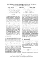
activating mutations occur (Fig. 1) and can be targeted by
tyrosine kinase inhibitors (TKIs). Demonstration of other
mutations in other driver oncogenes, particularly NRAS,
GNAQ and GNA11, can be useful in determining treatment strategy, such as an early decision to use immunotherapy with CTLA4 or PDL1 inhibitors. The number of
* Correspondence:
1
Department of Pathology – Coventry and Warwickshire Pathology Services
(CWPS), University Hospitals Coventry and Warwickshire, Coventry CV2 2DX,
UK
3
Institute of Ophthalmology, University College London, Bath Street, London
EC1V 9EL, UK
Full list of author information is available at the end of the article
drugs and the complexity of their associated companion
diagnostic pathways is increasing rapidly. Additional information from gene and protein expression can also assist
comprehensive analysis of melanoma biology [1, 2].
Pathology laboratories need to provide a rapid, reliable
and comprehensive diagnostic molecular pathology service to oncologists [3]. This is challenging, particularly
when diagnostics budgets are under pressure. However,
advances in the automation of DNA and RNA retrieval
from FFPE samples, as well as improvements in immunohistochemistry mean that tumours can be characterised rapidly for mutational status [4].
There are a large number of commercial methods
based on polymerase chain reaction (PCR) available for
BRAF determination in melanoma. Those most commonly
used include cobas® (Roche Diagnostics, Burgess Hill, UK)
and Therascreen® (Qiagen, Manchester, UK). Many laboratories still use older sequencing technologies including
© The Author(s). 2017 Open Access This article is distributed under the terms of the Creative Commons Attribution 4.0
International License ( which permits unrestricted use, distribution, and
reproduction in any medium, provided you give appropriate credit to the original author(s) and the source, provide a link to
the Creative Commons license, and indicate if changes were made. The Creative Commons Public Domain Dedication waiver
( applies to the data made available in this article, unless otherwise stated.
Reiman et al. BMC Cancer (2017) 17:150
Page 2 of 7
Fig. 1 Growth pathway in melanoma, showing the genes included in the IonTorrent panel which can be targeted by tyrosine kinase inhibitors
Sanger and Pyrosequencing, and the use of NGS methods
is becoming more common. Different methods tolerate different degrees of DNA quality, require different levels of
operator skill, and need different levels of expertise for
their interpretation [5]. Despite this, comparative studies
are relatively uncommon. A recent study compared Sanger
sequencing with cobas® for BRAF V600E mutation [6]. The
detection rate did not differ significantly, but six cases were
missed by cobas which does not cover all of the mutations
potentially present. Conversely, while cobas PCR produced
results in every case tested, Sanger sequencing failed in ten
cases [6]. Sanger sequencing usually has lower sensitivity
than targeted PCR, but the assays also have different requirements for input DNA. Sensitivity is not the only issue:
the amount of input DNA required varies between technologies and can have a major effect on the feasibility of
testing small samples. The BRAF cobas test requires 125
ng input DNA, while the Sanger sequencing method used
in the study cited above [6] needed just 50 ng DNA [7].
TaqMan castPCR technology (ThermoFisher, Paisley, UK)
has been shown comparable to pyrosequencing [8], and requires 50 ng of DNA per reaction in 96 well plates (200 ng
in total), but only 100 ng DNA per 48 wells in Taqman
array format [9], while Therascreen requires 80 ng DNA
per reaction (320 ng in total) [10]. The simplicity of the
PCR method chosen is also important to busy routine
pathology laboratories, where the availability of expert staff
is a major factor [5].
We recently participated in the validation of a 22 gene
Ampliseq panel for lung and colorectal cancer on the
IonTorrent Personal Genome Machine (PGM) (ThermoFisher) [11], and have now designed a melanoma panel
covering all actionable melanoma mutations, though it
would also be feasible to use validated comprehensive
gene panel if this is preferred. Despite recent improvements, NGS is challenging to use in a routine molecular
pathology environment and many laboratories still prefer
a PCR solution. Taqman Array™ technology (ThermoFisher) can be used with castPCR mutation detection assays to provide an alternative and less expensive panel
testing facility to NGS methods [9]. Our current panel
incorporates KRAS, NRAS, EGFR, and BRAF for combined screening of common mutations in colorectal cancer,
lung cancer and melanoma, known as the REB array [9].
While multiple gene PCR is helpful, there is also a
need for validated NGS solutions suitable for clinical use
to assist the molecular characterisation of melanoma,
particularly when these do not identify driver mutations.
We therefore compared our Ampliseq panel for use on
the IonTorrent PGM NGS platform with routine PCR
methods and the recently validated REB array.
Methods
The study was designed as a direct comparison between
multi-gene PCR (TaqMan™ array) and (IonTorrent) NGS
based against routine Therascreen® or cobas® testing for
BRAF alone.
Study population
A total of 151 samples were sourced from 2014 and
2015 from 132 melanoma patients attending University
Hospital Coventry and Warwickshire (UHCW) for diagnosis and treatment, including 18 uveal melanomas blocks
obtained from the Institute of Ophthalmology London for
comparison. All advanced melanomas are routinely tested
for BRAF, either in house, or at a referral centre using
Therascreen® (Qiagen, Manchester UK) or cobas® (Roche
Diagnostics, Burgess Hill, UK). Clinical data supplied with
Reiman et al. BMC Cancer (2017) 17:150
the specimens were collected from the Pathology Laboratory Information Management System (ULTRA, Cirdan,
Lisburn, Northern Ireland, UK). To ensure that all melanoma types can be handled, we have included 2 mucosal
and 4 acral lentiginous melanomas, as well as 18 uveal
melanomas.
DNA extraction
Areas of high cellularity melanoma (>50% neoplastic cells)
were marked on a slide by a histopathologist. These were
matched with the corresponding block. Samples from the
areas marked were then punched out using a 1 mm diameter skin punch with plunger (Integra Miltex, Plainsboro,
NJ, USA). DNA extraction was performed using the Promega Maxwell™ instrument, according to the manufacturer’s instructions. DNA content and quality was checked
by Qubit 2.0 fluorimeter (ThermoFisher) according to the
manufacturer’s instructions using their HS dsDNA kit
before mutation detection. We used a volume of 1 μl
(less than 50 ng DNA) to conserve as much of the sample
as possible, and worked with a range of concentrations,
depending on the extraction concentration achieved.
REB Taqman array
CastPCR™ provides a platform for sensitive mutation detection and in combination with TaqMan Array provides
a robust solution with minimal pipetting steps. Extracted
DNA (100 ng) is diluted with Taq polymerase and added
to a port of a manufactured microfluidic array [9]. TaqMan
arrays can be run on AB7900HT, ViiA7 and Quantstudio
PCR machines (ThermoFisher), although for this study, we
used the ViiA7 instrument. The REB array method requires little operator time or experience: it covers all BRAF
Page 3 of 7
mutations of relevance and >95% of reported NRAS
mutations.
IonTorrent NGS
The panel design was based on the Catalogue of Somatic
Mutations in Cancer (COSMIC) database and literature
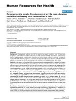
searches, and included seven genes important in melanoma
tumorigenesis (Fig. 2, Table 1 and Additional file 1: Table
S1). A total 95 hotspot mutations in these genes were submitted to the Ampliseq designer tool ( The resultant primer pool
consisted of 21 primer pairs with 98% of predicted
coverage of the target region (hotspots COSM1139 and
COSM1142 were not included in the final design). The
final target region was 2.47kb with the amplicon range
125-175 bp in length. The codons listed for each gene
included in the Ampliseq panel are shown in Table 1.
Sequencing was performed as previously described [11],
with the following changes to accommodate IonChef loading of the IonTorrent 314 chips. 10 ng of gDNA from
each of the samples chosen for NGS analysis was combined with the Ampliseq™ reagents and primer pool for
the melanoma gene panel and amplified initially for 23 cycles, which was later optimised to 28 cycles. After initial amplification, the amplified products were partially
digested before IonExpress Adapters and Barcode sequences were ligated to the library fragments. Following
barcoding the libraries were cleaned up using a magnetic
bead method (IonTorrent, ThermoFisher) and then quantified by the Ampliseq Q-PCR method (ThermoFisher).
Once the libraries were successfully quantified they were
combined and diluted to 50 pM. For IonTorrent 314
chips, 7 libraries on average were combined per chip.
These library pools were then loaded into the IonChef
Fig. 2 Diagram showing the codons for each gene within the Ion Torrent melanoma panel: the majority of mutations tested were within BRAF,
NRAS and c-KIT
Reiman et al. BMC Cancer (2017) 17:150
Page 4 of 7
Table 1 Summary of custom Ampliseq gene panel for melanoma, including common driver mutations in BRAF, KRAS, NRAS, MEK,
GNAQ, and GNA11
Gene
Reference
Codons included
Number of Mutations
BRAF
NM_004333.4
466, 469, 583, 584, 586, 592, 594, 595, 597, 600, 601, 605, 614, 618
29
GNAQ
NM_002072.3
209, 359
6
GNA11
NM_002067.2
183, 209, 223
3
NRAS
NM_002524.4
12, 13, 18, 50, 59, 61, 68
7
c-KIT
NM_000222.2
553, 557, 559, 560, 566, 569, 576, 642, 655, 816, 820, 822, 823, 829, 853
18
KRAS
NM_033360
12, 61
5
MEK1/MAP2K1
NM_002755.2
111, 124, 203, 264
5
instrument for further library preparation and chip loading. The loaded 314 chips were then run on the IonTorrent PGM instrument according to manufacturer’s
instructions. The Variant Caller plugin (included in the
provided Ion Torrent Suite software version 4.2) was used
to analyse the aligned sequence data for the identification
of hotspot mutations and novel variants. The variant caller
files were subjected for further analysis with the Ion Reporter online tool. Coverage was regarded as acceptable if
mean was at least 300×, and the Variant caller default setting interrogating somatic variants with high stringency
was used for hotspot calling, to exclude those < 2% allele
frequency. Only known mutations with COSMIC references were included. For the purposes of this analysis,
SNPs and variants of unknown significance were ignored.
Data and economic analysis
A sample size of 150 biopsies is expected to be sufficient
to meet the need for comparison of the two methods,
based on an expected Kappa of 0.9, with a 2-tailed Null
value of 0.6 and 80% power. Data from both PCR (all
methods) and NGS were compared using kappa statistics,
and discrepant results tested independently by another laboratory. Costing data were obtained from timesheets and
invoice information held within the pathology laboratory
for use with the CMD Impact tool available on-line
from the Royal College of Pathologists (www.rcpath.org/
cmd-impact.html).
Ethics
The development of service improvements does not require specific research ethics committee approval as stated
in the EU Clinical Trials Directive (2001/20/EC). However,
in this instance, we obtained most samples from the Arden Tissue Bank, (ATB15-001) which has NRES ethics approval (12/SC/0526) to conduct studies on anonymised
samples that are not required for diagnosis. In practice,
written consent for use of tissue was obtained from all patients as part of the surgical consent process.
Results
A total of 151 blocks from 148 resections were submitted
for NGS testing, representing 132 patients with repeat
samples available from metastases and primary tumours
in 11 patients. There were 76 males and 37 females with
cutaneous melanoma. The average age of the patients was
66 years (range 22 - 95 years, median 70 years). Duplicate
blocks were tested from the same sample for three samples repeated as controls, producing the same results in
each case.
Patients
A total of 105 patients had cutaneous melanoma (including 4 acral lentiginous melanoma and 2 malignant blue
naevus), 2 had mucosal melanoma, 19 uveal melanomas
(18 primary, 1 metastatic), and 6 unknown primary site.
Mutations
The mutations identified in each group are summarized
in Table 2. Driver mutations were identified in 70% of
the patients studied, and in 74% of the cutaneous melanomas (all types). BRAF was the commonest mutation
found, present in 44 of the 105 cutaneous patients
(42%). NRAS was present in 28 of the 105 cutaneous
melanomas (27%), and small numbers of KIT and MEK1
mutations were identified in cutaneous melanomas (3 and
4 cases respectively). Both mucosal melanomas tested
were wild-type, but 4 of the 6 melanomas of unknown primary origin had mutations, including one GNAQ mutation suggesting that this might have been of uveal origin.
Mutations were found in 10 of the 19 uveal melanomas,
with equal numbers of GNA11 and GNAQ mutations,
but there were also two NRAS mutations.
BRAF correlation
No molecular pathology was done routinely by the pathologist in 33 cases, mainly due to the presence of intercurrent disease, and the 19 historical uveal melanoma
cases. In the remaining 80 cases, molecular investigation
for BRAF mutations was done by cobas PCR. For BRAF,
Reiman et al. BMC Cancer (2017) 17:150
Page 5 of 7
Table 2 Summary of mutations identified by melanoma type
Melanoma Type
BRAF
NRAS
KRAS
KIT
MEK
GNA11
GNAQ
WT
Total
Cutaneous SSM
15
11
0
1
1
0
0
6
34
Cutaneous Nodular
15
11
0
0
1
0
0
11
38
Cutaneous NOS
12
3
0
1
1
0
0
5
22
LMM
1
2
0
0
0
0
0
2
5
Acral Lentiginous
0
1
0
0
1
0
0
2
4
Malignant blue naevus
1
0
0
0
0
0
0
1
2
Mucosal
0
0
0
0
0
0
0
2
2
Unknown Primary
0
3
0
1
0
0
1
1
6
Uveal
0
2
0
0
0
4
4
9
19
Total
44
33
0
3
4
4
5
39
132
patient-based correlation with cobas detection in 80/132
cases was 92.5%, with six discrepancies, as shown in
Table 3. For the 33 cases without cobas, the REB array
was run with residual DNA from the NGS sample. The
assay failed in 2 cases, but resulted in a further 15 cases
with complete correlation BRAF mutation between
PCR and NGS, including 2 cases with V600K mutations:
the remainder were confirmed as wild-type (100%
correlation).
Other mutations
NRAS is present on the REB array (though not on
cobas) and was completely concordant in 5 samples
found to have the Q61R mutation founds by NGS. Three
KIT mutations were identified (Table 2), and correlated
with known KIT mutations by pyrosequencing performed
at University Hospital Birmingham in 2 cases, which was
from a skin primary and melanoma from an unknown primary site. MEK mutation was seen in 1 case, which was
under treatment with a BRAF inhibitor (vemurafenib),
and a KIT mutation was identified in another case on
treatment (Table 3).
The costs of NGS are compared with the PCR Taqman
Array previously published in Table 4, showing that PCR
is less expensive at £135 per case than NGS at £257 per
case. There are differences in the cost of reagents, but
the major difference in costs is due to the staff costs for
NGS (£ per case) in comparison with PCR (£ per case).
Discussion
Our results validate the use of a custom design targeted
Ampliseq panel for melanoma mutation detection. Our
panel includes the genes commonly mutated in ocular
(uveal) and mucosal melanoma as well as the more common cutaneous tumours. The panel described here has
been used in one patient with metastatic melanoma who
had a history of both skin and uveal primary lesions.
The clinical question being which one had given rise to
his metastatic disease. The sample contained a GNA11
mutation, indicative of metastasis from the uveal tumour.
We provide further evidence for the utility of the REB
array in melanoma, and this could be used to triage samples for the more expensive NGS method, which has a
longer turnaround time (90 min compared with 2 days,
from DNA to result). However, it should be noted that
rapid turnaround is of limited clinical relevance to melanoma patients, unless they present with widespread
metastases.
The ability of NGS to look for MEK mutations associated with resistance to vemurafenib may be helpful in
guiding patient treatment, and there is a case to be made
for re-testing of samples taken on progression. In this
series, only two patients had documented resistance, one
of whom developed a MEK mutation, while the other had
a KIT mutation, both of which are potentially treatable.
Ampliseq™ provides targeted NGS for hotspots. The
NRAS and BRAF primers used have been previously
Table 3 Discrepancies between cobas PCR for BRAF and NGS. As cobas does not distinguish mutations at BRAF codon 600, these
are designated V600X in the table, WT is wild-type
Case (Sample)
cobas
NGS
Comment
True discrepancy
30
WT
BRAF
Mutation not present on cobas
Yes
33
WT
BRAF
Mutation not present on cobas
Yes
35
WT
BRAF
Mutation not present on cobas
Yes
48
V600X
MEK1
Resistance gene due to vemurafenib treatment
No
85
V600X
NRAS
Resistance gene due to vemurafenib treatment
No
100
V600X
KIT
Resistance gene due to vemurafenib treatment
No
Reiman et al. BMC Cancer (2017) 17:150
Page 6 of 7
Table 4 Costs of the Taqman PCR array per sample for mutation
analysis [9], versus IonTorrent PGM analysis using the Ampliseq
method for our laboratory. The major differences are due to the
high cost of consumables for NGS
Cost (£/sample)
PCR Array
(per sample)
NGS panel
(per sample)
Cost of consumables per test
40
134
External quality assurance
6
5
Equipment rental and maintenance
35
45
Staff costs
54
73
Total per sample
135
257
validated with the 22 gene Onconetwork IonTorrent
PGM panel which includes both [11]. For known mutations, results from variant caller can be obtained directly
or uploaded to IonReporter for more detailed analysis.
We chose the former approach, due to security of data
concerns which prevent us from routinely uploading
data to IonReporter over non-hospital systems. However,
the default IonTorrent analysis settings worked well in
this study and variant caller does provide COSMIC references for known variants, allowing rapid checking of
mutations for individual samples.
Comparison of different methods is important, but can
be complicated. Each method designed to identify actionable mutations or hotspots, rather than to sequence
entire genes, has slightly differing coverage, and differing
sensitivity for each mutation included. Discrepancies between methods are therefore inevitable and expected. Investigation usually reveals that such discrepancies often
involve rare variants or variants of unknown significance.
We have identified a number of comparative studies in
the literature, but perhaps less than one might expect
given the number of methods commercially available,
let alone those used as in-house laboratory-developed
(‘homebrew’) tests. The majority of these compare inhouse tests with Sanger sequencing and commercially
available PCR assays, with some variation in sensitivity,
but few discrepancies overall [4, 6, 12–16]. There are
the expected issues with coverage: for instance, some
PCR methods do not detect the relatively common
V600K mutation [16]. If there is a clinical need to identify other mutations than those tested, the use of cascade testing with other methods should be considered.
We now use the REB array [9] as a triage for NGS:
those patients who show a BRAF or NRAS mutation
can be excluded from further testing, while those in
whom it is clinically relevant to identify other driver
mutations (e.g. c-KIT) can be pursued further.
The costs of the two methods differ substantially
(Table 4). As expected, the REB array proved much less
expensive than NGS, but also has less coverage. In reality, the REB array can be used to exclude tumours with
common BRAF or NRAS mutations from further testing,
allowing resources to be concentrated on sequencing
cases where the driver mutation is not known, and knowledge of the KIT status of the tumour may be helpful. This
is not a large panel, and we rarely require NGS on more
than 8 patients at a time, and we therefore selected the
314 chip as optimal. The number of cases could be scaled
up for the 316 and 318 chips, but this is unlikely to alter
the cost per patient. The degree of automation now available using the IonChef™ for chip loading releases operator
time, but library preparation is not yet automated and
NGS does need considerable molecular pathology expertise. This is the major source of the increased cost of NGS
in comparison with PCR methods, and should decline as
automation is introduced.
Conclusion
The methods compared here all have the ability to find
mutations within melanoma, particularly those in BRAF.
We have found it helpful to know of NRAS mutations to
manage resources, and increasingly to direct patients towards immunotherapy at an earlier stage of their management. Those without an identified driver mutation may
well benefit from the NGS approach used here, or a more
comprehensive panel, depending upon workflow within
the laboratory.
Additional file
Additional file 1: Table S1. Complete mutation hotspot list included in
Custom Ampliseq gene panel for melanoma, including common driver
mutations in BRAF, NRAS, KRAS, MEK, GNAQ, and GNA11. (DOCX 125 kb)
Abbreviations
BRAF: B-Raf proto-oncogene, serine/threonine kinase; COSMIC: Cataloque of
somatic mutations in cancer database; CTLA4: cytotoxic T-lymphocyte-associated
protein 4 (CD152); DNA: Dexoxyribose nucleic acid; dsDNA: double stranded
DNA; EGFR: Epidermal growth factor receptor; FFPE: Formalin-fixed and
paraffin-embedded; GNA11: Guanine nucleotide-binding protein subunit
alpha-11; GNAQ: Guanine nucleotide-binding protein G(q) subunit alpha;
KIT: Mast/stem cell growth factor receptor (SCFR, CD117); MEK: Dual specificity
mitogen-activated protein kinase kinase 1; NGS: Next generation sequencing;
PCR: Polymerase chain reaction; PDL1: Programmed death ligand 1 (B7-H1,
CD274); REB: RAS, EGFR and BRAF (array)
Acknowledgements
We are grateful to the Departments of Pathology at Queen Alexandra
Hospital, Portsmouth and Birmingham University Hospital for their assistance
with access to uveal melanoma tissue and KIT pyrosequencing results. This
study benefitted from funding and advice from Novartis, to whom we are
most grateful. We are particularly grateful to Drs Fiona Read, Rory Buchan,
Mike Lau, and John Millholland for their support.
Funding
Novartis provided funding for this investigator sponsored study.
Availability of data and materials
The complete list of mutation hotspots is listed in Additional file 1: Table S1.
The custom design including files required will be loaded onto www.ampliseq.com
following publication.
Reiman et al. BMC Cancer (2017) 17:150
Authors’ contributions
IC and AR conceived the idea. The sequencing was performed by AR, HK,
and DSc. Patient follow-up and histopathology support involved PS, DSn, YT,
and IC. IC wrote the paper with assistance from all authors, who contributed
amendments and approved the final version.
Authors’ information
AR is a senior lecturer at Coventry University, HK is a Warwick University PhD
student, DSc is an Erasmus student Daniela Scocchia1, PS is an advanced
practitioner in pathology, DSn and YT are histopathologists. IC is a molecular
pathologist, and holds honorary chairs at UCL and Coventry University.
Competing interests
IC has shares in CanTech Ltd, and has acted as a consultant to the
pharmaceutical and diagnostics industry on personalised medicine,
including Astra-Zeneca, Amgen, Pfizer, Merck, MSD, Biocartis, Novartis,
Roche, and ThermoFisher.
Consent for publication
Not applicable.
Ethics approval and consent to participate
The development of service improvements does not require specific research
ethics committee approval as stated in the EU Clinical Trials Directive (2001/20/
EC). However, in this instance, we obtained most samples from the Arden Tissue
Bank, (ATB15-001) which has NRES ethics approval (12/SC/0526) to conduct
studies on anonymised samples that are not required for diagnosis. In practice,
written consent is obtained from patients as part of the surgical consent process.
Page 7 of 7
10. Garcia-Dios DA, Lambrechts D, Coenegrachts L, Vandenput I, Capoen A,
Webb PM, et al. High-throughput interrogation of PIK3CA, PTEN, KRAS,
FBXW7 and TP53 mutations in primary endometrial carcinoma. Gynecol
Oncol. 2013;128(2):327–34.
11. Tops B, Normanno N, Kurth H, Amato E, Mafficini A, Rieber N, et al.
Development of a semi-conductor sequencing-based panel for genotyping
of colon and lung cancer by the Onconetwork consortium. BMC Cancer.
2015;15(1):26.
12. Ihle MA, Fassunke J, Konig K, Grunewald I, Schlaak M, Kreuzberg N, et al.
Comparison of high resolution melting analysis, pyrosequencing, next
generation sequencing and immunohistochemistry to conventional Sanger
sequencing for the detection of p.V600E and non-p.V600E BRAF mutations.
BMC Cancer. 2014;14:13.
13. Machnicki MM, Glodkowska-Mrowka E, Lewandowski T, Ploski R, Wlodarski P,
Stoklosa T. ARMS-PCR for detection of BRAF V600E hotspot mutation in
comparison with Real-Time PCR-based techniques. Acta Biochim Pol.
2013;60(1):57–64.
14. Huang T, Zhuge J, Zhang WW. Sensitive detection of BRAF V600E mutation
by Amplification Refractory Mutation System (ARMS)-PCR. Biomark Res.
2013;1(1):3.
15. Colomba E, Helias-Rodzewicz Z, Von Deimling A, Marin C, Terrones N,
Pechaud D, et al. Detection of BRAF p.V600E mutations in melanomas:
comparison of four methods argues for sequential use of immunohistochemistry
and pyrosequencing. J Mol Diagn. 2013;15(1):94–100.
16. Ahn S, Lee J, Sung JY, Kang SY, Ha SY, Jang KT, et al. Comparison of three
BRAF mutation tests in formalin-fixed paraffin embedded clinical samples.
Korean J Pathol. 2013;47(4):348–54.
Author details
Department of Pathology – Coventry and Warwickshire Pathology Services
(CWPS), University Hospitals Coventry and Warwickshire, Coventry CV2 2DX,
UK. 2Centre for Research in Applied Biological and Exercise Sciences,
Coventry University, Coventry CV1 5FB, UK. 3Institute of Ophthalmology,
University College London, Bath Street, London EC1V 9EL, UK. 4Centre for
Technology Enabled Health Research (CTEHR), Faculty of Health & Life
Sciences, Coventry University, Coventry CV1 5FB, UK.
1
Received: 1 October 2016 Accepted: 18 February 2017
References
1. Jewell R, Conway C, Mitra A, Randerson-Moor J, Lobo S, Nsengimana J, et al.
Patterns of expression of DNA repair genes and relapse from melanoma.
Clin Cancer Res. 2010;16(21):5211–21.
2. Parker KA, Glaysher S, Polak M, Gabriel FG, Johnson P, Knight LA, et al. The
molecular basis of the chemosensitivity of metastatic cutaneous melanoma
to chemotherapy. J Clin Pathol. 2010;63(11):1012–20.
3. van Krieken JH, Normanno N, Blackhall F, Boone E, Botti G, Carneiro F, et al.
Guideline on the requirements of external quality assessment programs in
molecular pathology. Virchows Arch. 2013;462(1):27–37.
4. Ehsani L, Cohen C, Fisher KE, Siddiqui MT. BRAF mutations in metastatic
malignant melanoma: comparison of molecular analysis and immunohistochemical
expression. Appl Immunohistochem Mol Morphol. 2014;22(9):648–51.
5. Cree IA, Deans Z, Ligtenberg MJ, Normanno N, Edsjo A, Rouleau E, et al.
Guidance for laboratories performing molecular pathology for cancer patients.
J Clin Pathol. 2014;67:923–931.
6. Jurkowska M, Gos A, Ptaszynski K, Michej W, Tysarowski A, Zub R, et al.
Comparison between two widely used laboratory methods in BRAF V600
mutation detection in a large cohort of clinical samples of cutaneous melanoma
metastases to the lymph nodes. Int J Clin Exp Pathol. 2015;8(7):8487–93.
7. Rutkowski P, Gos A, Jurkowska M, Switaj T, Dziewirski W, Zdzienicki M, et al.
Molecular alterations in clinical stage III cutaneous melanoma: correlation with
clinicopathological features and patient outcome. Oncol Lett. 2014;8(1):47–54.
8. Pisareva E, Gutkina N, Kovalenko S, Kuehnapfel S, Hartmann A, Heinzerling L,
et al. Sensitive allele-specific real-time PCR test for mutations in BRAF codon
V600 in skin melanoma. Melanoma Res. 2014;24(4):322–31.
9. Kikuchi H. RA, Nyoni, J, Lloyd K., Savage R, Wotherspoon T, Berry L, Snead D,
Cree IA. Development and validation of a Taqman array for cancer mutation
analysis. Pathogenesis. 2016: In press.
Submit your next manuscript to BioMed Central
and we will help you at every step:
• We accept pre-submission inquiries
• Our selector tool helps you to find the most relevant journal
• We provide round the clock customer support
• Convenient online submission
• Thorough peer review
• Inclusion in PubMed and all major indexing services
• Maximum visibility for your research
Submit your manuscript at
www.biomedcentral.com/submit