Increased sensitivity of African American triple negative breast cancer cells to nitric oxide-induced mitochondria-mediated apoptosis
Bạn đang xem bản rút gọn của tài liệu. Xem và tải ngay bản đầy đủ của tài liệu tại đây (2.01 MB, 16 trang )
Martinez et al. BMC Cancer (2016) 16:559
DOI 10.1186/s12885-016-2547-z
RESEARCH ARTICLE
Open Access
Increased sensitivity of African American
triple negative breast cancer cells to nitric
oxide-induced mitochondria-mediated
apoptosis
Luis Martinez1, Easter Thames2, Jinna Kim3, Gautam Chaudhuri4,5, Rajan Singh3,4,5 and Shehla Pervin3,4,5,6*
Abstract
Background: Breast cancer is a complex heterogeneous disease where many distinct subtypes are found. Younger
African American (AA) women often present themselves with aggressive form of breast cancer with unique biology
which is very difficult to treat. Better understanding the biology of AA breast tumors could lead to development of
effective treatment strategies. Our previous studies indicate that AA but not Caucasian (CA) triple negative (TN)
breast cancer cells were sensitive to nitrosative stress-induced cell death. In this study, we elucidate possible
mechanisms that contribute to nitric oxide (NO)-induced apoptosis in AA TN breast cancer cells.
Methods: Breast cancer cells were treated with various concentrations of long-acting NO donor, DETA-NONOate
and cell viability was determined by trypan blue exclusion assay. Apoptosis was determined by TUNEL and caspase
3 activity as well as changes in mitochondrial membrane potential. Caspase 3 and Bax cleavage, levels of Cu/Zn
superoxide dismutase (SOD) and Mn SOD was assessed by immunoblot analysis. Inhibition of Bax cleavage by
Calpain inhibitor, and levels of reactive oxygen species (ROS) as well as SOD activity was measured in NO-induced
apoptosis. In vitro and in vivo effect of NO treatment on mammary cancer stem cells (MCSCs) was assessed.
Results and discussion: NO induced mitocondria-mediated apoptosis in all AA but not in CA TN breast cancer
cells. We found significant TUNEL-positive cells, cleavage of Bax and caspase-3 activation as well as depolarization
mitochondrial membrane potential only in AA TN breast cancer cells exposed to NO. Inhibition of Bax cleavage and
quenching of ROS partially inhibited NO-induced apoptosis in AA TN cells. Increase in ROS coincided with
reduction in SOD activity in AA TN breast cancer cells. Furthermore, NO treatment of AA TN breast cancer cells
dramatically reduced aldehyde dehydrogenase1 (ALDH1) expressing MCSCs and xenograft formation but not in
breast cancer cells from CA origin.
Conclusions: Ethnic differences in breast tumors dictate a need for tailoring treatment options more suited to the
unique biology of the disease.
Keywords: Breast cancer, Health disparity, African American, Unique biology
* Correspondence: ;
3
Charles R. Drew University of Medicine and Science, Los Angeles, CA 90059,
USA
4
Department of Obstetrics and Gynecology, David Geffen School of Medicine
at UCLA, Los Angeles, CA 90095, USA
Full list of author information is available at the end of the article
© 2016 The Author(s). Open Access This article is distributed under the terms of the Creative Commons Attribution 4.0
International License ( which permits unrestricted use, distribution, and
reproduction in any medium, provided you give appropriate credit to the original author(s) and the source, provide a link to
the Creative Commons license, and indicate if changes were made. The Creative Commons Public Domain Dedication waiver
( applies to the data made available in this article, unless otherwise stated.
Martinez et al. BMC Cancer (2016) 16:559
Background
Breast cancer is a complex disease where heterogeneous
cell types contribute to its initiation and progression [1, 2].
It has been broadly classified into estrogen receptor positive
(ER+) and estrogen receptor negative (ER-) sub types, each
of which are dependent on specific environmental cues and
signaling pathways for their development [3]. Frequent
diagnosis of aggressive triple negative (TN) (ER-, progesterone receptor negative (PR-) and Her2 Neu-) form of breast
cancer in young African American (AA) women suggest
disparity in development of this deadly disease [4, 5].
Limited treatment options for these aggressive TN breast
tumors causes high mortality rates in AA women [6]. In
sharp contrast, Caucasian (CA) women are usually postmenopausal when they develop ER+ or TN breast tumors,
which usually have better prognosis and lower mortality
rates when compared to AA patients [7]. Even after adjustments for socio-economic factors, AA breast tumors appear to exhibit specific aggressive characteristics suggesting
existence of unique biology contributed both by tumor cells
and host microenvironment [8].
Using laser capture microdissection and genome-wide
mRNA expression analysis, it has been reported that
stroma of AA breast tumors had higher inflammation and
angiogenesis when compared to similar tumors from the
Caucasian populations [9]. In addition, several cell cycle
regulators like p16, CCNA2, CCNB1 and CCNE2 as well
as several biological processes including endoplasmic
reticulum (ER)-associated degradation was found much
higher in the tumor epithelium of AA than in the CA
populations [9]. Ethnic differences were found in the increased expression of AMFR, a candidate oncogene that
promotes metastasis, in AA when compared to CA breast
tumors [9]. Furthermore, a tumor suppressor CDH13, was
found hyper-methylated in AA when compared to CA
breast tumors [10].
Our previous studies have indicated that AA and CA
breast cancer cells respond differently to nitrosative
stress, which is induced by nitric oxide (NO), a pleiotropic molecule that is produced by nitric oxide synthase
(NOS) [11–13]. Oxidative/nitrosative stresses, which are
produced by reactive oxygen species (ROS) /reactive nitrogen species (RNS) respectively influence all subtypes
of breast cancer [14, 15]. Both endothelial nitric oxide
synthase (eNOS) as well as inducible nitric oxide synthase (iNOS) have been detected in a large number of
human breast tumors, where their expression patterns
correlate with tumor grades [16]. However, ethnic differences in expression of NOS and response of AA and CA
breast cancer cells to oxidative/nitrosative stress remains
understudied. We have previously reported that AA TN
breast cancer cell line, MDA-MB-468, was highly sensitive to NO-induced apoptosis [17]. NO was able to up
regulate MAP kinase phosphatase (MKP-1) expression
Page 2 of 16
in MDA-MB-468 cells that promoted inactivation of
ERK1/2, Bax integration into mitochondrial membrane
leading to caspase-9 and -3 activation [17, 18]. However,
NO was unable to increase MKP-1 expression or induce
apoptosis in MDA-MB-231, a CA TN breast cancer cell
line [17]. On the other hand, low (nM) concentrations of
NO significantly up regulated proliferation of MDA-MB231 cells by increasing translation of cyclin D1 and ornithine decarboxylase [19]. In this study, we have further
examined responses of three additional TN breast cancer
cell lines, from each of the ethnic populations, to nitrosative stress. Consistent with our previous studies, we
found striking differences between AA and CA TN
breast cancer cell lines towards nitrosative stress. NO
specifically inactivated superoxide dismutase (SOD) to
increase ROS that partly contributed to apoptosis in AA
TN breast cancer cell lines. More importantly, NO treatment of AA breast cancer cell lines reduced mammary
cancer stem cell (MCSC) content in vitro and attenuated
xenograft formation in vivo. Our studies therefore, provide further evidence that there are ethnic differences in
the biology of TN breast tumors and specific players in
each population should be targeted for effective therapeutic interventions.
Methods
Materials
DETA-NONOate was purchased from Cayman Biochemicals (Ann Arbor, MI), Calpain Inhibitor III was from
Calbiochem (Darmstadt, Germany), and N-acetyl-l-cysteine
(NAC) was purchased from Sigma Aldrich (St. Louis, MO).
Ac-DEVD-AMC was purchased from Pharmingen (San
Diego, CA, USA). ApoAlert DNA Fragmentation Assay kit
was obtained from Clontech (Mountain View, CA). SOD
activity was measured by using an assay kit obtained from
Cayman Chemical Company (Ann Arbor, MI). Aldetect
Lipid Peroxidation Assay Kit was from Enzo Life Sciences
(Ann Arbor, MI, USA). MitoTracker Red CMX-Ros dye
was obtained from Life Technologies (Grand Island, NY).
Human cell lines
All human breast cancer cell lines were obtained from
American Type Culture Collection (ATCC) (Manassas,
VA) in 2013. ATCC uses Promega PowerPlex 1.2 system
and the Applied Biosystems Genotyper 2.0 software for
analysis of amplicon. We have not done any further testing in our lab. MDA-MB-231, MDA-MB-157 and MDAMB-436 breast cancer cell lines were propagated in
Leibovitz’s L-15 medium containing 10 % FBS. HCC1806, HCC-70, MDA-MB-468 and HCC-1395 were
propagated in RPMI 1640 containing 10 % FBS. BT-549
was propagated in DMEM F-12 containing 10 % FBS.
Martinez et al. BMC Cancer (2016) 16:559
Cell viability
Cells seeded in six-well plate (7.5 × 10⁵ per well) were
allowed to grow overnight. The cells treated with various
concentrations of DETA-NONOate for 24 h were collected, and viability was determined by trypan blue
exclusion method. The number of viable cells at each
concentration and time point was determined in triplicate with a hemacytometer [20].
TUNEL assay
The TUNEL assay was performed using ApoAlert DNA
Fragmentation Assay kit from Clontech as described previously [21]. Briefly, cells (2×10⁵) were plated in 6 well
plates, fixed in 1 % formaldehyde-PBS at 4 °C for 20 min.
The cells were washed with PBS, and stored overnight in
70 % ethanol. The cells were treated with nucleotide mixture containing terminal deoxynucleotidyltransferase
(Tdt) enzyme and incubated at 37 °C for 1 h. Cells were
washed and analyzed under fluorescent microscope.
Caspase-3 assay
Cells were lysed in insect cell lysis buffer {50 mm
HEPES, 100 mM NaCl, 2 mM EDTA, 0.1 % 3-[(3-cholamidopropyl)dimethylammonio]-1-propanesulfonic acid
(CHAPS), 10 % sucrose, 5 mM DTT, and 1× protease inhibitor} for 30 min at 4 °C. The lysates were used for
caspase-3 (3 μg) assay using Ac-DEVD-AMC substrate,
which after specific cleavage releases fluorescent AMC
that was quantified using a fluorometer (Versa Fluro;
Bio-Rad) with excitation at 380 nm and emission at 440
nm as described previously [22].
Page 3 of 16
Piscataway, NJ) for 1 h. Immunoreactive bands were visualized by enhanced chemiluminescence (ECL) detection system (Amersham) as described previously [20, 22].
Measurement of MMP by flow cytometry
Cells (1X10⁶) were harvested after various treatments,
washed twice with cold 1xPBS and incubated with 100
nM MitoTracker Red CMX-Ros dye at 37 °C for 15 min
in the dark, washed twice with cold PBS, and analyzed immediately by flow cytometry, as described previously [20].
Measurements of malondialdehyde and 4-hydroxy-alkenals
Levels of malondialdehyde and 4-hydroxy-alkenals
(4-HAE) was measured using Aldetect Lipid Peroxidation
Assay Kit (cat # BML-AK170-0001, Enzo Life Sciences,
Ann Arbor, MI, USA) as per manufacturer’s instructions,
described previously [23].
Mitochondria and cytosolic cell fractionation
The cell fractionation was performed using mitochondria and cytoplasmic extraction reagents from Thermo
Scientific (Rockford, IL, USA). The fractionation was
done as described previously [17].
Superoxide dismutase activity assay
Cells (1×10⁶) were seeded in six well plates to confluence and collected without use of proteolytic enzyme.
SOD activity was measured by using an assay kit obtained from Cayman Chemical Company (Ann Arbor,
MI). The activity assay was performed according to the
manufacturer’s protocol.
Western analysis
Aldefluor assay and flow cytometry
For analysis of cytosolic proteins, cells were lysed in cell
lysis buffer [50 mm HEPES (pH 7.5); 1 mm DTT, 150
mM NaCl, 1 mM EDTA, 0.1 % Tween 20, 10 % glycerol,
10 mm β-glycerophosphate, 1 mM NaF, 0.1 mm orthovanadate, 10 μg/ml leupeptin, 10 μg/ml aprotinin, and
0.1 mM PMSF] and were incubated at 4 °C for 30 min.
Protein concentration was measured using Bio-Rad protein assay dye concentrate. Lysates (30 μg) were resolved
electrophoretically on 10 % SDS-polyacrylamide gel and
electrotransferred to a polyvinylidine difluoride membrane
(Bio-Rad) using a tank blot procedure (Bio-Rad Mini Protean II). The membranes were incubated with the following
primary antibodies: Heme oxygenase-1 (HO-1) (Santa Cruz,
Cat # sc-10789), cleaved caspase-3 (Cell Signaling, Cat #
9661), Bax (Santa Cruz Biotechnologies, Cat # 20067), Bcl2
(BD-Transduction Laboratories, Cat # 551052), β-actin (Cell
Signaling Technologies, Cat # 4967), NOX4 (Abcam, Cat #
ab60940), Mn-SOD (Abcam, Cat # 13533), Cu/Zn SOD
(Abcam, Cat #13498), COX IV (Abcam, ab14744) and
1:1000 dilutions of respective horseradish peroxidase-linked
F(ab) fragment secondary antibody (Amersham Corp.,
Aldefluor assay was carried out as described previously
[24, 25] according to manufacturer’s (cat # 01700, Stem
cell Technologies, Vancouver, Canada) guidelines.
Briefly, breast cancer cells were suspended in Aldefluor
assay buffer containing an ALDH substrate, bodipyaminoacetaldehyde (BAAA) at 1.5 μM, and incubated
for 40 min at 37 °C. To distinguish between ALDH+ and
ALDH− cells, a fraction of cells was incubated under
identical condition in the presence of a 10-fold molar
excess of the ALDH inhibitor, diethyl amino benzaldehyde (DEAB). This results in a significant decrease in
the fluorescent intensity of ALDH+ cells and was used to
compensate the flow cytometer. To determine CD44 expression, MDA-MB-231 and HCC1806 cells suspended
in PBS were exposed to PE conjugated anti-human
CD44 antibody (cat # 555479, BD Pharmingen™, CA,
USA) and subjected to flow cytometry analysis.
Xenograft formation
Six to eight week old nude mice (Harlan Laboratories Inc.
Indianapolis, IN) were used for xenograft engraftment.
Martinez et al. BMC Cancer (2016) 16:559
Control or DETA-NONOate treated (24h) MDA-MB-468,
HCC-70, HCC-1806 and MDA-MB-231 cells (2x10⁶ cells/
100μl) were mixed with matrigel (1:1) and implanted
subcutaneously (posterior dorsolateral) in the nude mice.
Tumors were monitored over a period of 15 weeks and
tumor volume was calculated as described previously
[24, 26]. This study was carried out in strict accordance
with the recommendations in the Guide for the Care and
Use of Laboratory Animals of the National Institutes of
Health. The protocol was approved by the Institutional
Animal Care and Use Committee on the Ethics of Animal
Experiments of the Charles R. Drew University of Medicine and Science (permit number: I-1103-261).
Statistical analysis
Data are presented as mean ± S.D. and between-group
differences were analyzed using ANOVA. If the overall
ANOVA revealed significant differences, then pairwise
comparisons between groups were performed by
Newman–Keuls multiple comparison test. All comparisons were two-tailed, and p-values <0.05 were considered statistically significant. The experiments were
repeated at least three times, and data from representative experiments are shown.
Results
Nitric Oxide preferentially induced cell death in AA TN
breast cancer cells
Effect of NO on viability and proliferation of breast cancer
cell was examined by treatment with various concentrations of DETA NONOate, a long acting NO-donor. We
analyzed four different breast cancer cell lines obtained
from each of the ethnic populations. AA (HCC-1806,
HCC-70, MDA-MB-157 and MDA-MB-468) and CA
(BT-549, MDA-MB-436, HCC-1395 and MDA-MB-231)
breast cancer cell lines were treated with various concentrations of DETA-NONOate for different time points. We
and others have previously demonstrated that lower
concentrations (1-100μM) of DETA-NONOate released
physiological range (nM) while at higher concentrations
(0.2-1mM) released high pathophysiological range (μM) of
NO respectively [27, 28]. In this study, we observed that
DETA-NONOate at 50μM induced (HCC-1806: 19.37 ±
2.58 %; MDA-MB-468: 43.94 ± 1.26 %); 100μM (HCC1806: 17.21 ± 0.18 %; MDA-MB-468: 47.01 ± 0.36 %);
200μM (HCC-1806: 41.11 ± 0.89 %; MDA-MB-468: 76.46
± 1.25 %); and 300μM (HCC-1806: 23.54 ± 0.27 %; MDAMB-468: 84.78 ± 0.58 %) cell death at 48 h (Fig. 1a). In
addition, high NO at 48 h. induced significantly higher cell
death at 500μM (HCC-70: 52.10 ± 5.76 %; HCC-1806:
82.84 ± 3.36 %; MDA-MB-468: 95.35 ± 1.82 %; and MDAMB-157: 94.71 ± 1.71 %) and at 1mM (HCC-70: 84.72 ±
1.83 %; HCC-1806: 92.80 ± 2.98 %; MDA-MB-468: 96.66
± 1.57 %; and MDA-MB-157: 99.23 ± 0.769 %) (Fig. 1b). In
Page 4 of 16
all the four CA cell lines, no apparent cell death was observed at low concentrations of NO, while at high concentrations only 20-28 % cell death was observed at 48h in
both MDA-MB-231 (27.07 ± 4.56 %) and MDA-MB-436
(27.19 ± 5.57 %) cancer cells (Fig. 1b). Even at 24h, 1mM
DETA-NONOate was able to induce significant cell death
selectively in AA TN breast cancer cells (HCC-70: 40.00 ±
3.99 %; HCC1806: 46.23 ± 3.42 %; and MDA-MB-468:
98.14 ± 0.66 %), while no significant cell death was observed in CA cells (Fig. 1c). We further performed terminal deoxynucleotidyl transferase dUTP nick end
labeling (TUNEL) assay to assess whether DNA fragmentation occurred in TN AA breast cancer cells after 24 and
48 h of DETA-NONOate treatments. There was a significant increase in TUNEL-positive cells in AA but not in
CA breast cancer cell line following DETA-NONOate
(1mM) treatment (Fig. 1d-e). Quantitative analysis of
TUNEL-positive cells in MDA-MB-468 cells showed significant increase at both 24h (28.71 ± 4.49 %) and 48h
(43.27 ± 7.78 %) (Fig. 1e). No significant increase of
TUNEL-positive cells were found in MDA-MB-231 cells
at both time points following similar treatments (Fig. 1e).
As DNA fragmentation is a hallmark of cells undergoing
apoptosis where caspase-3 is the main executioner enzyme [29], we simultaneously examined caspase-3 activity
in TUNEL-positive AA TN cells treated with NO. We
found increased caspase-3 activity in AA TN cells (HCC1806 and MDA-MB-468) with 1mM DETA-NONOate
treatment as early as 24h, while no increase in activity was
found in CA TN cells (Fig. 1f). Since the breast cancer
cells from CA did not undergo apoptosis with NO treatment, we further examined the induction of HO-1, which
is an early stress response marker [30]. We were able to
detect early (8h after treatment) up-regulation of HO-1
protein levels in all the AA cell lines examined, while no
significant change was found in CA TN breast cancer cells
(Fig. 1g). These data indicate that NO increased cell death
due to apoptosis in AA, while no detectable response was
observed in CA TN breast cancer cells.
NO induced mitochondria-mediated apoptosis in AA TN
breast cancer cells
We further examined whether mitochondria was involved
in NO-induced apoptosis in AA TN breast cancer cells. Integrity of mitochondrial membrane, which is maintained by
the ratio of pro- apoptotic and anti-apoptotic proteins,
releases cytochrome C when compromised to activate
caspase-3 [31]. Cleaved Bax, a pro-apoptotic molecule, has
been found to integrate into the mitochondrial membrane
to release cytochrome C [17]. Cleaved caspase-3 as well as
cleaved Bax, which is an active form of Bax, was detected
in all AA but not in CA TN breast cancer cells with 48h of
DETA-NONOate (1mM) treatment (Fig. 2a-b). Interestingly, basal levels of Bcl2, an anti-apoptotic molecule was
Martinez et al. BMC Cancer (2016) 16:559
*** ***
**
0
0.5
1
***
***
***
50
***
**
***
***
AA
CA
Hrs
E
24
48
TUNEL-positive cells / field
0
0
24
48
60
***
40
**
20
ns
0
MDA-MB-231
AA
F
G
Hrs
0
24
48
***
1000
***
500
***
***
ns
ns
Fig. 1 (See legend on next page.)
B
T54
M
9
D
A
-M
B
-2
31
H
C
C
-1
80
M
6
D
A
-M
B
-4
68
0
AA
M
M
D
D
A
-M
B
-4
68
MDA-MB-468
1500
CA
***
*
AA
CA
D
Caspase 3 Activity
***
***
H
C
C
HC -70
C
M
DA 180
6
-M
B46
8
M
CA
M BT
D
A - 54
M -MB 9
DA - M 23 1
B
H -4
C C 36
-1
39
5
H
C
H CC
M C 70
D -1
A
8
M -MB 06
D
A- -46
M
8
B
-1
57
1
-2
3
D
A
B
-M
B
T54
9
8
-4
6
-M
B
-1
80
6
D
A
M
AA
*
0
0
C
***
*
*
H
C
***
50
* *
0
0
8
16
24
** ***
*
100
A
-M
B
-2
31
20
***
***
***
40
***
*** ***
*** ***
BT
-5
D
49
AM
B
-2
31
HC
C13
95
60
100
Hrs
150
M
***
***
C
DN (mM)
% Cell Death
% Cell Death
100
80
B
DN (mM)
0
0.01
0.05
0.1
0.2
0.3
% Cell Death
A
Page 5 of 16
CA
Martinez et al. BMC Cancer (2016) 16:559
Page 6 of 16
(See figure on previous page.)
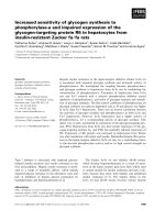
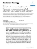
Fig. 1 Nitric oxide preferentially induced cell death in AA breast cancer cells. AA and CA breast cancer cell lines were propagated in their
respective media and treated with various concentrations of a) DETA-NONOate (DN)(0.01-0.3mM) for 48h. b) DETA-NONOate (0.5-1mM) for 48h c)
DETA-NONOate (1mM) for 8-24h. Following these various treatments, the cells were harvested and their viability was determined by trypan blue
exclusion method using hemocytometer for cell counting. d) Cells propagated on 8 well chamber slides were treated with DETA NONOate
(1mM) for 24 and 48h. After these treatments the cells were fixed with 4% paraformaldehyde for 30 mins and DNA fragmentation determined by
performing TUNEL assay. A representative picture of control and TUNEL positive MDA-MB-468 and MDA-MB-231 cells are shown. e) Quantitation
of TUNEL positive cells was performed on images taken at 100 × magnification. Number of TUNEL positive cells/field (average of 3 fields) for each
time points/cell line is shown. f) Cells treated with DETA-NONOate (1mM) for 48h were lysed in insect cell lysis buffer and 3μg of total cell lysates
were subjected to caspase-3 activity assay using florescent substrates. Results from 3 different experiments were represented. g) Immunoblot
analysis for heme oxygenase-1 (HO-1) was performed using 100μg of total cell lysates obtained from cells treated with DETA-NONOate (1mM) for
various time points (0-24h). We used β-actin as housekeeping control. Two way ANOVA statistics was performed for A, B, C, E and F. Data are
represented as mean ± SD with significant values presented as *p < 0.01, **p < 0.001, ***p < 0.0001
undetectable in HCC-1806, while its levels dramatically declined in MDA-MB-468 and MDA-MB-157 AA TN
breast cancer cells with NO treatment (Fig. 2a-b). In
sharp contrast, significant Bcl2 levels were detected in
all CA TN breast cancer cells and remained relatively
stable or even slightly increased upon NO exposure
(Fig. 2a-b). Since Bax cleavage was a common occurrence in all AA TN cells undergoing apoptosis, we
inhibited Bax cleavage to examine its contribution to
NO-induced apoptosis. Calpain, which gets activated
by oxidative stress, cleaves Bax at the N-terminal to
generate a potent pro-apoptotic 18-kDa fragment that
promotes Bcl-2-independent cytochrome C release
and apoptotic cell death [32]. We found that pretreatment with Calpain inhibitor III caused 52.74 ±
4.56 % reduction in cell death with NO treatment in
HCC-1806 AA TN breast cancer cells as assessed by
trypan blue exclusion assay (Fig. 2c). No change in
cell viability was observed in NO treated CA breast
cancer cells with or without Calpain inhibitor III. Immunoblot analysis shows that in addition to reduction
in cell death, there was reduced Bax and caspase-3
cleavage with Calpain inhibitor III treatment in AA
but not in CA breast cancer cells (Fig. 2d-e).
Apoptotic stimuli initiates a series of changes in the
mitochondria that are crucial to the death program [33,
34]. One of the changes is opening of large pores in the
mitochondrial membrane leading to mitochondrial permeability transition (PT) and disruption in the mitochondrial membrane potential (MMP), which are early
obligatory step in the death program [35]. We, further
examined changes in MMP in AA and CA TN breast
cancer cells with NO treatment. We found AA TN
breast cancer cells upon NO exposure underwent significant depolarization of MMP as early as 24h with further
increase at 48h (Fig. 2f ). On the contrary, with NO treatment, there was hyperpolarization of mitochondrial
membranes in CA breast cancer cell lines (Fig. 2f ).
These data indicate that NO induced mitochondriamediated apoptosis specifically in AA but not CA TN
breast cancer cells.
NO increased oxidative stress to induce mitochondriamediated apoptosis in AA TN breast cancer cells
NO has been found to react with ROS, more specifically
with superoxide (O-2) , to generate peroxynitrite and oxidative/nitrosative stress that could contribute to depolarization
of MMP and mitochondria-mediated apoptosis [28]. We
measured malondialdehyde and 4-hydroxyalkenals, which
have been used as an indicator of lipid peroxidation and oxidative stress, in NO treated cells. Concentrated lysates of
control and NO treated cells were subjected to colorimetric
assay to detect malondialdehyde and 4-hydroxyalkenals
using kit and manufacturer’s instructions as mentioned in
Materials and Method. There was increase in oxidative
stress with DETA-NONOate (1mM) treatment as
early as 6h in HCC-70 (273.79 ± 27.27 %), HCC-1806
(151.04 ± 9.38 %), MDA-MB-468 (204.20 ± 25.26 %),
and MDA-MB-157 (218.87 ± 40.07 %) AA TN breast cancer
cells (Fig. 3a). No significant increase in oxidative stress was
detected in any of the CA TN breast cancer cells examined
(Fig. 3a). We further pre-treated AA breast cancer cells with
N-acetyl cysteine (NAC), a ROS quencher, to determine
whether increase in ROS with NO treatment contributed to
apoptosis. Pre-treatment of cells with NAC significantly
reduced NO-mediated cell death in HCC-1806 (67.13 ±
1.57 %) (Fig. 3b). Immunoblot analysis showed that NOinduced high Bax cleavage in HCC-1806 cells was attenuated by pretreatment with NAC (Fig. 3c). On the contrary in
CA TN cell lines, NAC treatment led to slight increase in
cell death in a concentration-dependent manner (Fig. 3d).
No significant cleavage of Bax was observed in CA breast
cancer cells with any of the treatments (Fig. 3e). These
data indicate that increased levels of ROS in NO treated
AA TN breast cancer may contribute to the observed cell
death.
NO inactivated SOD to increase ROS only in AA TN breast
cancer cells
Since NO exposure was able to increase ROS levels in AA
TN breast cancer cells, we further examined the levels of
mitochondrial manganese superoxide dismutase (Mn-SOD)
and cytosolic copper/zinc SOD (Cu/Zn-SOD). These
Martinez et al. BMC Cancer (2016) 16:559
Fig. 2 (See legend on next page.)
Page 7 of 16
Martinez et al. BMC Cancer (2016) 16:559
Page 8 of 16
(See figure on previous page.)
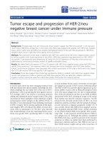
Fig. 2 Nitric oxide induced mitochondria mediated apoptosis in AA breast cancer cells. a-b) Cells were seeded to confluence and treated with
DETA NONOate (0.5-1mM) for 48h. Immunoblot analysis was performed for cleaved caspase-3 (19 and 17kDa band), total Bax (Intact Bax 20kD
and cleaved Bax 18kD band) and Bcl2. Expression of β-actin was used as housekeeping control. Neither caspase-3 nor Bax cleavage was found in
CA cell lines. c) Cells were exposed with DETA-NONOate (1mM) with or without pretreatment (1h) with various concentrations (10-30μM) of
Calpain III (Ci III) inhibitor. Data is presented as mean ± SD with significant values presented as ***p < 0.001. d, e) Cells treated with DETA-NONOate and
Calpain inhibitor III were subjected to Immunoblot analysis for cleaved caspase-3 and Bax. f) Cells treated with DETA NONOate for 24h (blue curve) or
48h (red curve) were harvested and incubated with Mito Tracker dye to measure mitochondrial membrane depolarization using flow cytometer.
Uncoupling agent carbonyl cyanide-4-(trifloromethoxy) phenyl hydrazine (FCCP) was used as positive control (green curve) while untreated cells served
as control (black curve)
metalloenzymes are most effective intracellular enzymatic
antioxidants, known to hydrolyze ROS to H2O2, which further dissociates to O2 and H2O [36]. Levels of both SODs
were examined after treatment with DETA-NONOate (0.51mM) for 24 and 48h. No significant decline in the levels of
Mn-SOD or Cu/Zn SOD was observed in either AA or CA
breast cancer cells at 24h of DETA-NONOate treatment
(data not shown). However, at 48h, DETA-NONOate (0.51mM) caused a dramatic reduction in the levels of both
Mn-SOD and Cu/Zn-SOD in AA but not in CA breast cancer cells (Fig. 4a-c). On the contrary, in some CA cell lines
there was an increase in the levels of Mn-SOD (BT-549
and HCC-1395), while in another cell line (MDA-MB-436)
there was no significant change from control with NO
treatment (Fig. 4a-c). With cell fractionation, we found considerable amount of Mn-SOD in the mitochondria of CA
(BT-549) but not in AA (HCC-70 and HCC-1806) breast
cancer cells (Fig. 4d). We did not find any difference in
levels of NOX4 protein, which is known to produce ROS,
in AA and CA breast cancer cells (Fig. 4a-d). The purity of
mitochondria was assessed by examining COX IV protein
levels in the mitochondrial fraction (Fig. 4d). Since reduction in the protein levels of SOD at later time points of NO
treatment did not coincide with early increase in ROS, we
further examined the SOD activity in these cells. We found
exposure to NO (DETA NONOate 1mM, 48h) reduced
SOD activity (HCC-70: 19.15 ± 7.81 %; HCC-1806: 17.22 ±
6.51 %) in AA breast cancer cells (Fig. 4e). In sharp contrast
to decline in AA breast cancer cells, there was some increase in SOD activity in all the CA TN breast cancer cells
with NO treatment (BT-549: 3.80 ± 2.87 %; MDA-MB-231:
8.63 ± 2.06) (Fig. 4e). We further examined SOD activity
after shorter exposures to various concentrations of NO in
an AA TN breast cancer cell line (HCC-1806). With lower
levels of NO (DETA-NONOate 500μM) we found a decline
in SOD activity as early as 3h (25.01 ± 9.94 %), which continued to reduce till 8h (72.83 ± 7.73 %) after which it
returned to near control by 16-24h (Fig. 4f). With higher
NO (DETA-NONOate 1mM), the SOD activity continued
to decline till 8h (76.91 ± 5.2) after which it remained lower
than the control (Fig. 4g). Since lower concentrations of
NO reduced proliferation of AA breast cancer cell lines, we
further examined its effect on Mn SOD and Cu/Zn SOD as
well as on various proliferation (cyclin D1 and PCNA),
apoptosis (Bax and Bcl2), and oxidative stress (NOX4)
markers. We did not find changes in the levels of Mn SOD
or Cu/Zn SOD in any of the cell lines examined, while the
levels of cyclin D1 declined in MDA-MB-468 cells treated
with lower concentrations of NO (Fig. 5a, b). In addition,
while no significant changes in Bax and Bcl2 was found,
there was some decline in NOX4 protein levels in both the
cell lines examined. We further examined the stability of
these proteins after treating the cells with non-specific protein synthesis inhibitor, cycloheximide, for different time
points. No significant changes in the levels of Cu/Zn SOD
was observed in MDA-MB-468, MDA-MB-231 and BT549 breast cancer cells, while in HCC-1806 cells, there was
an initial decline followed by an increase at later time
points of cycloheximide treatment (Fig. 5c-f). In addition,
no significant differences in the levels of Mn-SOD with cycloheximide treatment was detected. However the levels of
Bcl2 rapidly declined and remained lower in AA when
compared to CA breast cancer cells treated with cycloheximide (Fig. 5c-f). Levels of Bax however remained unchanged in these cells while its cleavage increased at later
time points of cycloheximide treatment (Fig. 5c-f). Levels
of proliferating cell nuclear antigen (PCNA) remained constant in all the cell lines with cycloheximide treatment
(Fig. 5c-f). The above data shows that NO inactivates SOD
to increase oxidative stress specifically in AA but not CA
TN breast cancer cells. In addition, there was no significant
difference in the stability of Mn-SOD or CU/Zn-SOD in
the AA and CA TN breast cancer cells.
NO treatment reduced Mammary Cancer Stem Cell
content in AA breast cancer cells
Mammary cancer stem cells (MCSCs) are a small subset
of undifferentiated cells in breast tumors that have been
highly implicated in its initiation and progression [37].
Most therapeutic and chemotherapeutic drugs has been
found to kill more differentiated cells while MCSCs
evade these treatments [38]. Heterogeneity exists within
MCSCs populations, which have high aldehyde dehydrogenase 1 (ALDH1) activity and increased expression of
CD44, a cell surface marker [39]. There are reports of
high expression of ALDH1 in breast tumors in women
from African origin [40, 41]. We found high ALDH1 expression in all the 3 AA TN breast cancer cells examined
Martinez et al. BMC Cancer (2016) 16:559
Page 9 of 16
Fig. 3 Nitric oxide–induced apoptosis in AA breast cancer cells was dependent on increase in ROS. a) Cells seeded in 96 well plates were treated
with DETA NONOate (1mM) for different time points (6-24h). 4-hydroxy-alkenal levels was measured using Aldetect Lipid Peroxidation Assay kit as
per manufacturer’s instruction. Cells were seeded to confluence and treated with DETA NONOate (1mM) for 24h with or without pretreatment
(1h) with N acetyl-cysteine (NAC) at various concentrations (10-30mM). b, d) Cell viability was measured by trypan blue exclusion method using
hemocytometer for cell counting. c, e) Immunoblot analysis of total cell lysates was performed for Bax (lower panels). Data are represented in
Mean ± SD with significant values presented as *p < 0.01 and **p < 0.001
Martinez et al. BMC Cancer (2016) 16:559
Fig. 4 (See legend on next page.)
Page 10 of 16
Martinez et al. BMC Cancer (2016) 16:559
Page 11 of 16
(See figure on previous page.)
Fig. 4 Nitric oxide inactivates SOD to increase ROS in AA TN breast cancer cells. a-c) Cells treated with DETA-NONOate (0.5 mM and 1mM) for
48h was subjected to immunoblot blot analysis for NOX4, Mn-SOD and Cu/Zn SOD where β-actin was used as control. d) Control and DETA NONOate
(1mM) treated cells were subjected to cell fractionation using kit to separate cytoplasmic and mitochondrial fraction as per manufacturer’s instructions.
These fractions were further subjected to immunoblot blot analysis for NOX4, Mn-SOD, and Cu/Zn SOD. COX4 was used as positive control for purity
of mitochondrial fraction. e, f, g) cells treated with DETA-NONOate (0.5 mM and 1mM) for various time points (3-36h) were subjected to SOD activity
assay using kit as per manufacturer’s instructions. Data are represented in Mean ± SD with significant values presented as *p < 0.01 and **p < 0.001
and NO treatment significantly reduced the expression
of ALDH1 in these cells (Fig. 6a). The higher expression
of ALDH1 in AA TN breast cancer cells sharply contrasts with its much lower basal expression in CA TN
breast cancer cells (Fig. 6a). We however, found much
higher CD44 expression in the CA breast cancer cells
that further increased with NO treatment (Fig. 6a). We
further confirmed by Western blot that the increase in
caspase cleavage in AA TN breast cancer cell lines with
NO treatment occurred simultaneously with decrease in
MCSC population (data not shown).
We further measured ALDH1 activity in HCC-1806 and
HCC-70 after treatment with DETA-NONOate (1mM,
24-36h). We found high basal activity of ALDH1 in HCC1806 (11.8 %) and HCC-70 (11.3 %) cells (Fig. 6b-c). Upon
NO exposure there was considerable decline in ALDH1
activity in both the cell lines, HCC-1806 (6.8 %) and
HCC-70 (5.6 %) (Fig. 6b-c). A large amount of dead cells
were found in both the cell lines after NO treatment. We
were unable to detect any ALDH1 activity in MDA-MB231 cells (Fig. 6d). On the other hand, a large number of
MDA-MB-231 cells (17.4 %) expressed CD44, which was
undetected in HCC-1806 cells (Fig. 6e-f).
We further examined whether NO treatment of AA and
CA breast cancer cells influenced tumor formation in vivo.
For this purpose, we implanted control and NO-treated
(for 24h) AA and CA TN breast cancer cells (2x10⁶) subcutaneously (at a ventro-lateral site) in nude mice and monitored tumor volume for 15 weeks. When compared to the
untreated control cells, NO treatment of AA TN breast
cancer cells dramatically reduced tumor volume in nude
mice (Fig. 6b, c). These results were in sharp contrast to
that observed in CA TN breast cancer cells where NO
treatment led to formation of larger xenografts when compared to similar treatment groups from AA TN breast cancer cells (Fig. 6d). Our data, therefore, indicate that NO
treatment of AA TN breast cancer cells significantly reduce
MCSC content and xenograft formation.
Discussion
Breast cancer in AA women usually have aggressive characteristics and unique biology. In this study, we report that
nitrosative stress induced by NO, promoted apoptosis
preferentially in all the AA but not in CA TN breast cancer cells. Specifically, NO treatment induced cleavage of
pro-apoptotic Bax and an increase in caspase-3 activity in
AA but not in CA TN breast cancer cells. Interestingly, a
decline in anti-apoptotic Bcl2 and depolarization of MMP
was observed in AA breast cancer cells. In sharp contrast,
Bcl2 levels remained steady or even increased along with
hyperpolarization of MMP in NO treated breast cancer
cells of CA origin. There was an early induction of HO-1,
a stress response gene, in AA breast cancer cells, which
were sensitive to nitrosative stress. Levels of HO-1 did not
change in any of the CA cells with NO exposure, further
indicating their insensitivity to nitrosative stress. In another study, NO treatment was found to induce higher increase migration and invasiveness of MDA-MB-231 (CA
TN origin) when compared to MDA-MB-468 (AA TN
origin) breast cancer cells [42]. It appears that NO elicits
tumor promoting responses in CA breast cancer cells that
sharply contrasts to the death-promoting signals in AA
cells. We have previously shown that low concentrations
of NO in addition to increasing proliferation of MDAMB-231 cells, activated mammalian target of rapamycin
(mTOR) to increase translation of proteins [18]. Studies
also show that ROS/RNS drive a continuous process of
DNA adducts that crosslinks as well as promoting posttranslational modification of lipids and proteins that increase survival, immunosuppression and inhibition of
apoptosis [43]. These could be some of the mechanisms
by which NO influenced tumor promoting behavior in
CA breast cancer cells.
In our study, the basal levels of ROS were comparable
in breast cancer cells from the two ethnic populations.
However, NO treatment specifically increased ROS only
in AA TN breast cancer cells. This increase in ROS coincided with reduction in the activity of total (mitochondrial and cytosolic) SOD in AA TN breast cancer cells.
Our study further shows that there was no significant
difference in the stability of SOD in AA and CA TN
breast cancer cells. There are reports of reduced SOD
activity associated with increased nitrosative stress in
AAs but not in CA umbilical vein endothelial cells
(HUVEC) [44]. Inverse relationship between NO and
SOD activity was also evident in plasma of AA patients
suffering from hypertension [45]. Older AA as well as
those that underwent spontaneous preterm birth had
lower SOD activity when compared to similar group of
CA patients [46]. No difference in Mn-SOD Ala-9Val
polymorphism in breast tumors from AA and CA patients has been detected [47]. However, a striking
Martinez et al. BMC Cancer (2016) 16:559
Page 12 of 16
Fig. 5 a-b) Cell lines (AA and CA TN cells) were treated with various concentrations (1-300μM) of DETA NONOate for 24h after which they were
harvested, lysed and subjected to immunoblot analysis for BAX, Bcl2, Mn-SOD, Cu/Zn SOD, NOX4, cyclin D, and PCNA. β-actin was used as control.
Experiments were repeated at least three and a representative data is shown. c-f) Cells treated with cycloheximide (CHX) for various concentrations
were subjected to immunoblot analysis for BAX, Bcl2, Mn-SOD, Cu/Zn SOD, NOX4, cyclin D, and PCNA. β-actin was used as control
Martinez et al. BMC Cancer (2016) 16:559
Page 13 of 16
A
B
C
D
E
G
F
H
I
1200
1000
Tumor volume (mm3)
Tumor volume (mm3)
Tumor volume (mm3)
1200
800
600
400
200
0
800
600
400
200
0
0
5
10
Weeks
MDA-MB-468
1000
HCC-1806
15
0
5
10
15
Weeks
MDA-MB-231
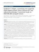
Fig. 6 a) Breast cancer cell lines treated with DETA NONOate (1mM) for 24 and 48h were examined for MCSC markers ALDH1 and CD44 by Immunoblot
analysis. b-c) Breast cancer cell lines, HCC1806 and HCC70 were treated with DETA NONOate (1mM) for 24-36h. Control and treated cells were suspended in
ALDH1 assay buffer and subjected to ALDH1 activity assay using florescence activated cytometric analysis. Both dead and live populations in control and treated
cells were gated to detect ALDH1 activity. d) MDA-MB-231 cell line was suspended in ALDH1 assay buffer and subjected to ALDH1 activity assay using
florescence activated cytometric analysis. e-f) Control MDA-MB-231 and HCC1806 cells were stained with PE conjugated CD44 and subjected to cytometric
analysis. g-i) Breast cancer cells (2x10⁶) with and without treatment with DETA NONOate (1mM) for 24h were implanted subcutaneously (ventro-lateral site) in
nude mice and tumor volume was monitored for 15 weeks (n = 5). Data are represented as mean ± SD (**, p ≤ 0.01, ***, p ≤ 0.001, ns: non-significant)
Martinez et al. BMC Cancer (2016) 16:559
difference in the frequency of 10T/9T intron 3 polymorphism in mitochondrial SOD in AAs has been reported [48]. Although sensitivity of AA population to
NO-mediated inactivation of SOD is well demonstrated,
molecular mechanisms and consequences of SOD inactivation remain poorly understood.
Prominent differences in the biology of AA and CA
breast tumors suggest differences in both the tumor cells
and the microenvironment in which these tumors develop.
Despite significant differences in biology there is not much
difference in the treatment strategies for these tumors.
Tumor infiltrating macrophages in poorly differentiated
breast carcinoma, express active iNOS that promote angiogenesis, increase tumor size and cause poor survival of patients [16]. More recently, targeting endogenous iNOS in
two CA breast cancer cell lines MDA-MB-231 and BT-549,
by selective iNOS and pan-NOS inhibitors, was found effective in reducing tumor volume in mouse model [49].
Our previous studies indicate AA TN breast cancer
cells utilize arginine preferentially to synthesize polyamines [21, 22]. Arginase and nitric oxide synthase (NOS)
compete for cellular arginine to produce polyamines and
NO respectively [21, 22]. Elevated arginase activity in cells
has been found to both decrease cellular availability of NO
by competing with NOS to increase L-ornithine. We have
shown that arginase expression is up-regulated in AA breast
cancer cells, which are dependent on polyamines for survival
and proliferation [21]. Therefore exploiting ethnic differences in arginine metabolism has the potential to provide
selective druggable targets that could effectively reduce aggressiveness of CA and AA TN breast tumors. Although
there is ample evidence of unique biology of AA TN breast
tumors, no potential druggable target has so far been identified or exploited to reduce the severity of the disease.
The potential of using NO to induce apoptosis in AA
breast tumors appears promising for therapeutic intervention. Depending on the cell type, NO has shown promise
as a therapeutic agent to reduce tumor volume [50, 51].
The therapeutic potential of NO-donors depends on its
capacity to release NO at optimum concentrations and in
a temporally regulated manner to kill tumor cells. A number of potential NO-donors include organic nitrites glyceryl trinitrite (GTN), metal-nitrosyl complexes sodium
nitroprusside (SNP), S-nitrosoglutathione (GSNO) and
diazeniumdiolates. Studies have shown that GTN induces
apoptosis in colon cancer, inhibit hypoxia-mediated metastatic potential of B16F10 murine melanoma cells and increase the chemosensitivity of human prostate tumor
xenografts [52–54]. Diethylene diazeniumdiolates (NONOates) has been found to be an effective chemo preventive
agent against bone metastatic breast cancer as well as
aggressive breast cancer cell lines [55]. Unfortunately,
many of these NO-donors lack target specification and
controlled release kinetics, therefore cannot be tested for
Page 14 of 16
their efficacy in vivo. Considering dual effects of NO and
lack of ideal NO-donors, we are not equipped technically
to use NO for therapy.
Conclusions
AA and CA TN breast cancer cells respond differently to
nitrosative stress suggesting differences in their biology. Increased sensitivity of AA TN breast tumors to nitrosative
stress-mediated apoptosis suggest exploring the potential of
NO as a therapeutic agent. Therapeutic strategies should
be tailored to the unique biology of the disease.
Abbreviations
AA, African American; CA, Caucasian; TN, Triple negative; NO, Nitric Oxide;
SOD, Superoxide dismutase; ROS, Reactive oxygen species; ALDH1, Aldehyde
dehydrogenase1; MCSCs, Mammary cancer stem cells; ER, Estrogen receptor; PR,
Progesterone receptor; NOS, Nitric oxide synthase; RNS, Reactive nitrosative
stress; MKP-1, MAP kinase phosphatase-1; TUNEL, Terminal deoxynucleotidyl
transferase dUTP nick end labeling; HO-1, Hemeoxygenase1; MMP, Mitochondrial
membrane potential; PT, Mitochondria permeability transition; NAC, N –acetyl
cysteine
Acknowledgements
This work was supported by National Institute of Health Grants SC1CA165865
(SP) and SC1AG049682 (RS), and in part by U54MD007598 (RS), and
S21MD000103 (RS, SP) grants.
Availability of data and materials
Not applicable.
Authors’ contributions
Study Concept and design: S.P, L.M, R.S. Acquisition of data: L.M, E.T, J.K.
Analysis and interpretation of data: S.P, L.M, G.C, R.S. Writing and review of
the manuscript: S.P, L.M, G.C, R.S. Study supervision: S.P, R.S. All authors have
read and approved the manuscript.
Competing interests
The authors declare that they have no competing interests.
Consent for publication
Not applicable.
Ethics approval and consent to participate
No human subjects were involved for this study. This study was carried out
in strict accordance with the recommendations in the Guide for the Care
and Use of Laboratory Animals of the National Institutes of Health. The
protocol was approved by the Institutional Animal Care and Use Committee
on the Ethics of Animal Experiments of the Charles R. Drew University of
Medicine and Science (permit number: I-1103-261). This information has
been added in the Methods section page 8 of this manuscript.
Author details
1
California State University, Dominguez Hills, Los Angeles, CA, USA.
2
Columbia University New York, New York, NY 10027, USA. 3Charles R. Drew
University of Medicine and Science, Los Angeles, CA 90059, USA.
4
Department of Obstetrics and Gynecology, David Geffen School of Medicine
at UCLA, Los Angeles, CA 90095, USA. 5Jonsson Comprehensive Cancer
Center at UCLA, Los Angeles, CA 90095, USA. 6Division of Endocrinology and
Metabolism, Charles R. Drew University of Medicine and Science, 1731 East
120th Street, Los Angeles, CA 90059, USA.
Received: 26 January 2016 Accepted: 11 July 2016
References
1. Polynak K. Heterogeneity in breast cancer. J Clin Invest. 2011;121:3786–88.
2. Koren S, Bentires-Alj M. Breast tumor heterogeneity: source of fitness, hurdle
for therapy. Mol Cell. 2015;60:537–46.
Martinez et al. BMC Cancer (2016) 16:559
3.
4.
5.
6.
7.
8.
9.
10.
11.
12.
13.
14.
15.
16.
17.
18.
19.
20.
21.
22.
Cadoo KA, Fornier MN, Morris PG. Biological subtypes of breast cancer:
current concepts and implications for recurrence patterns. Q J Nucl Med
Mol Imaging. 2013;57:312–21.
Carey LA, Perou CM, Livasy CA, Dressler LG, Cowan D, Conway K, Karaca G,
Troester MA, Tse CK, Edminston S, Deming SL, Geradts J, Cheang MC, Nielsen TO,
Moorman PG, Earp HS, Millikan RC. Race, breast cancer subtypes, and subtypes,
and survival in the Carolina Breast Cancer Study. JAMA. 2006;295:2492–502.
Kanaan YM, Sampey BP, Beyene D, Esnakula AK, Naab TJ, Ricks-Santi LJ, Dasi S,
Day A, Copeland RL Sr BKWFW, Gabrielson E, Dewitty Jr RL. Metabolic profile of
triple-negative breast cancer in African-American women reveals potential
biomarkers of aggressive disease. Cancer Genomics Proteomics. 2014;11:279–94.
Dietze EC, Sistrunk C, Miranda-Carboni G, O’regan R, Seewaldt VL. Triplenegative breast cancer in African-American women: disparities versus
biology. Nat Rev Cancer. 2015;15:248–54.
Balmanoukian A, Zhang Z, Jeter S, Slater S, Armstrong DK, Emens LA,
Fetting JH, Wolff AC, Davidson NE, Jacobs L, Lange J, Tsangaris TN, Zellars R,
Gabrielson E, Stearns V. African American women who receive primary
anthracycline- and taxane- based chemotherapy for triple-negative breast
cancer suffer worse outcomes compared with white women. J Clin Oncol.
2009;27:e35–7.
Anders CK, Carey LA. Biology, metastatic patterns, and treatment of patients
with triple-negative breast cancer. Clin Breast Cancer. 2009;9 Suppl 2:S73–81.
Martin DN, Boersma BJ, Yi M, Reimers M, Howe TM, Yfantis HG, Tsai YC,
Williams EH, Lee DH, Stephens RM, Weissman AM, Ambs S. Differences in
the tumor microenvironment between African-American and EuropeanAmerican Breast cancerpatients. PLoS One. 2009;4, e4531.
Wang S, Dorsey TH, Terunuma A, Kittles RA, AMbs S, Kwabi-Addo B.
Relationship between tumor DNA methylation status and patient
characteristics in African-American and European-American women with
breast cancer. PLoS One. 2012;7, e37928.
Espey MG, Miranda KM, Feelisch M, Fukuto J, Grisham MB, Vitek MP, Wink
DA. Mechanisms of cell death governed by the balance between nitrosative
and oxidative stress. Ann N Y Acad Sci. 2000;899:209–21.
Korde SD, Basak A, Chaudhary M, Goval M, Vagga A. Enhanced nitrosative and
oxidative stress with decreased total antioxidant capacity in patients with oral
precancer and oral squamous cell carcinoma. Oncology. 2011;80:382–89.
Seril DN, Liao J, Yang GY. Colorectal carcinoma development in inducible
nitric oxide synthase-deficient mice with dextran sulfate sodium-induced
ulcerative colitis. Mol Carcinog. 2007;46:341–53.
Switzer CH, Ridnour LA, Cheng R, Heinecke J, Burke A, Glynn S, Ambs S,
Wink DA. S-Nitrosation mediates multiple pathways that lead to tumor
progression in estrogen receptor-negative breast cancer. For Immunopathol
Dis Therap. 2012;3:117–24.
Das Gupta S, So JY, Wall B, Wahler J, Smolarek AK, Sae-Tan S, Soewono KY,
Yu H, Lee MJ, Thomas PE, Yang CS, Suh N. Tocopherols inhibit oxidative
and nitrosative stress in estrogen-induced early mammary hyperplasia in
ACI rats. Mol Carcinog. 2015;54:916–25.
Ambs S, Glynn SA. Candidate pathways linking inducible nitric oxide
synthase to a basal-like transcription pattern and tumor progression in
human breast cancer. Cell Cycle. 2011;10:619–24.
Pervin S, Singh R, Chaudhuri G. Nitric-oxide-induced Bax integration into the
mitochondrial membrane commits MDA-MB-468 cells to apoptosis:
essential role of Akt. Cancer Res. 2003;63:5470–9.
Pervin S, Singh R, Freije WA, Chaudhuri G. MKP-1-induced
dephosphorylation of extracellular signal-regulated kinase is essential for
triggering nitric oxide-induced apoptosis in human breast cancer cell lines:
implications in breast cancer. Cancer Res. 2003;63:8853–60.
Pervin S, Singh R, Hernandez E, Wu G, Chaudhuri G. Nitric oxide in
physiologic concentrations targets the translational machinery to increase
the proliferation of human breast cancer cells: involvement of mammalian
target of rapamycin/eIF4E Pathway. Cancer Res. 2007;67:289–99.
Pervin S, Singh R, Gau CL, Edamatsu H, Tamanoi F, Chaudhuri G.
Potentiation of nitric oxide-induced apoptosis of MDA-MB-468 cells by
farnesyltransferase inhibitor: implications in breast cancer. Cancer Res. 2001;
61:4701–6.
Singh R, Pervin S, Wu G, Chaudhuri G. Activation of caspase-3 activity and
apoptosis in MDA-MB-468 cells by N(omega)-hydroxy-L-arginine, an
inhibitor of arginase, is not solely dependent on reduction in intracellular
polyamines. Carcinogenesis. 2001;22:1863–9.
Singh R, Pervin S, Karimi A, Cederbaum S, Chaudhuri G. Arginase activity in
human breast cancer cell lines: N(omega)-hydroxy-L-arginine selectively
Page 15 of 16
23.
24.
25.
26.
27.
28.
29.
30.
31.
32.
33.
34.
35.
36.
37.
38.
39.
40.
41.
42.
inhibits cell proliferation and induces apoptosis in MDA-MB-468 cells.
Cancer Res. 2000;60:3305–12.
Pervin S, Tran L, Urman R, Braga M, Parveen M, Li SA, Chaudhuri G, Singh R.
Oxidative stress specifically downregulates survivin to promote breast
tumour formation. Br J Cancer. 2013;108:848–58.
Pervin S, Hewison M, Braga M, Tran L, Chun R, Karam A, Chaudhuri G, Norris
K, Singh R. Down-regulation of vitamin D receptor in mammospheres:
implications for vitamin D resistance in breast cancer and potential for
Combination therapy. PLoS. 2013;8, e53287.
Singh R, Parveen M, Basgen JM, Fazel S, Meshesha MF, Thames EC, Moore B,
Martinez L, Howard CB, Vergnes L, Reue K, Pervin S. Increased Expression of
Beige/Brown Adipose Markers from Host and Breast Cancer Cells Influence
Xenograft Formation in Mice. Mol Cancer Res. 2016;14(1):78–92.
Singh R, Avliyakulov KV, Braga M, Haykinson MJ, Martinez L, Singh V,
Parveen M, Chaudhuri G, Pervin S. Proteomic identification of mitochondrial
targets of arginase in human breast cancer. PLoS One. 2013;8, e79242.
Pervin S, Singh R, Chaudhuri G. Nitric oxide-induced cytostasis and cell cycle
arrest of a human breast cancer cell line (MDA-MB-231): potential role of
cyclin D1. Proc Natl Acad Sci USA. 2001;98:3583–8.
Thomas DD, Espey MG, Ridnour LA, Hofseth LJ, Mancardi D, Harris CC, Wink
DA. Hypoxic inducible factor 1alpha, extracellular signal-regulated kinase,
and p53 are regulated by distinct threshold concentrations of nitric oxide.
Proc Natl Acad Sci USA. 2004;101:8894–9.
Namura S, Zhu J, Fink K, Endres M, Srinivasan A, Tomaselli KJ, Yuan J,
Moskowitz MA. Activation and cleavage of caspase-3 in apoptosis induced
by experimental cerebral ischemia. J Neurosci. 1998;18:3659–68.
Beyrich C, Löffler J, Kobsar A, Speer CP, Kneitz S, Eignethaler M. Infection of
human coronary artery endothelial cells by group B streptococcus
contributes to dysregulation of apoptosis, hemostasis, and innate immune
responses. Mediators Inflamm. 2011;2011:971502.
Zhao B, He T. Chidamide, a histone deacetylase inhibitor, functions as a
tumor inhibitor by modulating the ratio of Bax/Bcl-2 and P21 in pancreatic
cancer. Oncol Rep. 2015;33:304–10.
Gao G, Dou QP. N-terminal cleavage of bax by calpain generates a potent
proapoptotic 18-kDa fragment that promotes bcl-2-independent cytochrome
C release and apoptotic cell death. J Cell Biochem. 2000;80:53–72.
SUen DF, Norris KL, Youle RJ. Mitochondrial dynamics and apoptosis. Genes
Dev. 2008;22:1577–90.
Wolff S, Erster S, Palacios G, Moll UM. p53's mitochondrial translocation and
MOMP action is independent of Puma and Bax and severely disrupts
mitochondrial membrane integrity. Cell Res. 2008;18:733–44.
Alavian KN, Beutner G, Lazrove E, Sacchetti S, Park HA, Licznerski P, Li H, Nabili P,
Hockensmith K, Graham M, Porter Jr GA, Jonas EA. An uncoupling channel within
the c-subunit ring of the F1FO ATP synthase is the mitochondrial permeability
transition pore. Proc Natl Acad Sci USA. 2014;111:10580–5.
Becuwe P, Ennen M, Klotz R, Barbieux C, Grandemange S. Manganese superoxide
dismutase in breast cancer: from molecular mechanisms of gene regulation to
biological and clinical significance. Free Radic Biol Med. 2014;77:139–51.
Dontu G, Abdallah WM, Foley JM, Jackson KW, Clarke MF, Kawamura MJ,
Wicha MS. In vitro propagation and transcriptional profiling of human
mammary stem/progenitor cells. Genes Dev. 2003;17:1253–70.
Nandy SB, Ganwani L, Nahleh Z, Subramani R, Arumugam A, de la Rosa JM,
Lakshmanaswamy R. Recurrence and metastasis of breast cancer is
influenced by ovarian hormone's effect on breast cancer stem cells. Future
Oncol. 2015;11:983–95.
DA Cruz PA, Marques O, Rosa AM, DE Fátima FM, Rêma A, Lopes C. Coexpression of stem cell markers ALDH1 and CD44 in non-malignant and
neoplastic lesions of the breast. Anticancer Res. 2014;34:1427–34.
Schwartz T, Stark A, Pang J, Awuah B, Kleer CG, Quayson S, Kingman S,
Aitpillah F, Abantanga F, Jiagge E, Oppong JK, Osei-Bonsu E, Martin I, Yan X,
Toy K, Adjei E, Wicha M, Newman LA. Expression of aldehyde
dehydrogenase 1 as a marker of mammary stem cells in benign and
malignant breast lesions of Ghanaian women. Cancer. 2013;119:488–94.
Proctor E, Kidwell KM, Jiagge E, Bensenhaver J, Awuah B, Gyan K, Toy K,
Oppong JK, Kyei I, Aitpillah F, Osei-Bonsu E, Adjei E, Ohene-Yeboah M,
Brewer RN, Fondjo LA, Owusu-Afriyie O, Wicha M, Merajver S, Kleer C,
Newman L. Characterizing Breast Cancer in a Population with Increased
Prevalence of Triple-Negative Breast Cancer: Androgen Receptor and ALDH1
Expression in Ghanaian Women. Ann Surg Oncol. 2015;22:3831–5.
Glynn SA, Boersma BJ, Dorsey TH, Yi M, Yfantis HG, Ridnour LA, Martin DN,
Switzer CH, Hudson RS, Wink DA, Lee DH, Stephens RM, Ambs S. Increased
Martinez et al. BMC Cancer (2016) 16:559
43.
44.
45.
46.
47.
48.
49.
50.
51.
52.
53.
54.
55.
Page 16 of 16
NOS2 predicts poor survival in estrogen receptor-negative breast cancer
patients. J Clin Invest. 2010;120:3843–54.
Tudek B, Winczura A, Janik J, Siomek A, Foksinski M, Olinski R. Involvement
of oxidatively damaged DNA and repair in cancer development and aging.
Am J Transl Res. 2010;15:254-84.
Feairheller DL, Park JY, Sturgeon KM, Williamson ST, Diaz KM,
Veerabhadrappa P, Brown MD. Racial Differences in oxidative stress and
inflammation: in vitro and in vivo. Clin Transl sci. 2011;4:32–7.
Zhou L, Xiang W, Potts J, Floyd M, Sharan C, Yang H, Ross J, Nyanda AM,
Guo Z. Reduction in extracellular superoxide dismutase activity in AfricanAmerican patients with hypertension. Free Radic Biol Med. 2006;41:1384–91.
Herway C, Kanninen T, Witkin SS, Saade G, Fortunato SJ, Menon R. Ethnic
disparity in amniotic fluid levels of hyaluronan, histone H2B and superoxide
dismutase in spontaneous preterm birth. J Perinat Med. 2013;41:277–82.
Millikan RC, Player J, de Cotret AR, Moorman P, Pittman G, Vannappagari V,
Tse CK, Keku T. Manganese superoxide dismutase Ala-9Val polymorphism
and risk of breast cancer in a population-based case-control study of
African Americans and whites. Breast Cancer Res. 2004;6:R264–74.
Shao J, Chen L, Marrs B, Lee L, Huang H, Manton KG, Martin GM, Oshima J.
SOD2 polymorphisms: unmasking the effect of polymorphism on splicing.
BMC Med Genet. 2007;8:7.
Grenados-Principal S, Liu Y, Guevara ML, Blanco E, Choi DS, Qian W, Patel T,
Rodriguez AA, Cusimano J, Weiss HL, Zhao H, Landis MD, Dave B, Gross SS,
Chang JC. Inhibition of iNOS as a novel effective targeted therapy against
triple-negative breast cancer. Breast Cancer Res. 2015;17:25.
Rapozzi V, Della Pietra E, Zorzet S, Zacchigna M, Bonavida B, Xodo LE. Nitric
oxide-mediated activity in anti-cancer photodynamic therapy. Nitric Oxide.
2013;30:26–35.
Kudo S, Nagasaki Y. A novel nitric oxide-based anticancer therapeutics by
macrophage-targeted poly (l-arginine)-based nanoparticles. J Control
Release. 2015;217:256–62.
Millet A, Bettaieb A, Renaud F, Prevotat L, Hammann A, Solary E, Mignotte B,
Jeannin JF. Influence of the nitric oxide donor glyceryl trinitrate on
apoptotic pathways in human colon cancer cells. Gastroenterology. 2002;
123:235–46.
Postovit LM, Adams MA, Lash GE, Heaton JP, Graham CH. Nitric oxidemediated regulation of hypoxia-induced B16F10 melanoma metastasis. Int J
Cancer. 2004;108:47–53.
Frederiksen LJ, Sullivan R, Maxwell LR, Macdonald-Goodfellow SK, Adams MA,
Bennett BM, Siemens DR, Graham CH. Chemosensitization of cancer in vitro
and in vivo by nitric oxide signaling. Clin Cancer Res. 2007;13:2199–206.
Simeone AM, Colella S, Krahe R, Johnson MM, Mora E, Tari AM. N-(4Hydroxyphenyl) retinamide and nitric oxide pro-drugs exhibit apoptotic and
anti-invasive effects against bone metastatic breast cancer cells.
Carcinogenesis. 2006;3:568–77.
Submit your next manuscript to BioMed Central
and we will help you at every step:
• We accept pre-submission inquiries
• Our selector tool helps you to find the most relevant journal
• We provide round the clock customer support
• Convenient online submission
• Thorough peer review
• Inclusion in PubMed and all major indexing services
• Maximum visibility for your research
Submit your manuscript at
www.biomedcentral.com/submit