ROR2 is epigenetically inactivated in the early stages of colorectal neoplasia and is associated with proliferation and migration
Bạn đang xem bản rút gọn của tài liệu. Xem và tải ngay bản đầy đủ của tài liệu tại đây (2.55 MB, 12 trang )
Ma et al. BMC Cancer (2016) 16:508
DOI 10.1186/s12885-016-2576-7
RESEARCH ARTICLE
Open Access
ROR2 is epigenetically inactivated in the
early stages of colorectal neoplasia and is
associated with proliferation and migration
Sean S. Q. Ma1, Sameer Srivastava2,3, Estelle Llamosas1, Nicholas J. Hawkins4, Luke B. Hesson2, Robyn L. Ward2
and Caroline E. Ford1*
Abstract
Background: Colorectal cancer (CRC) is closely linked to Wnt signalling, with 94 % of cases exhibiting a Wnt related
mutation. ROR2 is a receptor tyrosine kinase that is thought to repress β-catenin dependent Wnt signalling. Our
study aims to determine if ROR2 is epigenetically silenced in CRC and determine if in vitro silencing of ROR2
potentiates Wnt signalling, and alters the proliferative, migratory or invasive potential of cells.
Methods: ROR2 expression was examined in CRC cell lines and patient adenomas using qRT-PCR, while COBRA and
bisulphite sequencing was used to analyse ROR2 promoter methylation. 258 patient primary tumour samples from
publicly available databases were also examined for ROR2 expression and methylation. In addition, the functional
effects of ROR2 modulation were investigated in HCT116 cells following ROR2 siRNA knockdown and in RKO and
SW620 cells following ectopic ROR2 expression.
Results: Reduced ROR2 expression was found to correlate with ROR2 promoter hypermethylation in colorectal
cancer cell lines, carcinomas and adenomas. ROR2 expression was downregulated in 76.7 % (23/30) of CRC cell lines
with increasing ROR2 promoter hypermethylation correlating with progressively lower expression. Analysis of 239
primary tumour samples from a publicly available cohort also found a significant correlation between reduced ROR2
expression and increased promoter methylation. Methylation analysis of 88 adenomas and 47 normal mucosa
samples found greater percentage of adenoma samples to be methylated. Additional analysis also revealed that
adenoma samples with reduced ROR2 expression also possessed ROR2 promoter hypermethylation. ROR2
knockdown in the CRC cell line HCT116 significantly decreased expression of the β-catenin independent Wnt
targets genes JNK and NFATC1, increased cellular proliferation and migration but decreased invasion. When ROR2
was ectopically expressed in RKO and SW620 cells, there was no significant change to either cellular proliferation or
migration.
Conclusion: ROR2 is frequently epigenetically inactivated by promoter hypermethylation in the early stages of colorectal
neoplasia and this may contribute to colorectal cancer progression by increasing cellular proliferation and migration.
Keywords: Colorectal cancer, ROR2, Epigenetic silencing, Hypermethylation, Wnt
* Correspondence:
1
Metastasis Research Group, Adult Cancer Program, School of Women’s and
Children’s Health, Lowy Cancer Research Centre, UNSW Australia, Sydney,
NSW 2052, Australia
Full list of author information is available at the end of the article
© 2016 The Author(s). Open Access This article is distributed under the terms of the Creative Commons Attribution 4.0
International License ( which permits unrestricted use, distribution, and
reproduction in any medium, provided you give appropriate credit to the original author(s) and the source, provide a link to
the Creative Commons license, and indicate if changes were made. The Creative Commons Public Domain Dedication waiver
( applies to the data made available in this article, unless otherwise stated.
Ma et al. BMC Cancer (2016) 16:508
Background
Colorectal cancer (CRC) is the third most common cancer
worldwide with an estimated 1 million cases each year
contributing to over 608,000 deaths [1–3]. CRCs develop
from benign intraepithelial neoplasms known as adenomas, which progress to cancer after an accumulation of
mutations [4, 5]. The Wnt signalling pathway is frequently
altered in CRC with ~94 % of cases possessing a mutation
in a Wnt pathway gene [6]. One of the early precipitating
events for colorectal adenoma development is mutation of
the APC gene, an important tumour suppressor and regulator of β-catenin dependent Wnt signals [5, 7, 8]. APC
along with AXIN and GSK3β are responsible for degradation of cytosolic β-catenin and loss of APC leads to βcatenin accumulation, Wnt pathway hyperactivation and
increased cellular proliferation and migration [8–15].
In contrast, the β-catenin independent Wnt pathway affects planar cell polarity (PCP), cell adhesion and motility
and is not reliant on β-catenin levels [16–20]. The receptor
tyrosine kinase-like orphan receptor 2 (ROR2) is a receptor
tyrosine kinase which binds with WNT5A to activate the
β-catenin independent Wnt pathway [21–23]. In addition
to activating β-catenin independent Wnt/JNK signalling,
ROR2 and WNT5A interaction has been shown to antagonise downstream targets of β-catenin dependent Wnt;
specifically inhibition of AXIN2 expression and the TCF/
LEF transcription factors [16, 20, 23–26]. Consistent with
its reported antagonism of β-catenin dependent Wnt signals, a 2010 study found ROR2 to be silenced in colorectal
cancer, resulting in increased cellular proliferation [27].
However, other reports in colorectal cancer, melanoma and
osteosarcoma have found elevated ROR2 expression in tumours compared to normal tissue [28–32]. These conflicting reports have raised questions as to the role ROR2 plays
in cancer and presents the possibility that the downstream
effects of ROR2 are dependent on other Wnt genes and the
cellular context of the cancer itself [33–35].
In this study, we investigated whether ROR2 expression
is altered in colorectal cancers and adenomas. We also
assessed the effects of altered ROR2 expression on βcatenin dependent Wnt signalling, proliferation, migration
and invasion properties in colorectal cancer cells.
Results
ROR2 is epigenetically silenced by promoter
hypermethylation in colorectal cancer cell lines
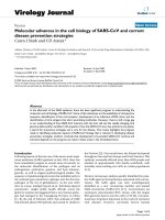
Quantitative reverse transcriptase polymerase chain reaction (qRT-PCR) showed 23 out of 30 CRC cell lines lacked
expression of ROR2 at the mRNA level (Fig. 1a). Methylation analysis using combined bisulphite restriction analysis
(COBRA) showed 25 out of the 30 cell lines had methylation in the ROR2 promoter (Additional file 1).
Bisulphite sequencing revealed that C170 and HCT116
cell lines, which had the highest levels of ROR2 expression,
Page 2 of 12
had little to no methylation across the examined promoter
molecules. SW480, SNUC2B and HCT15 cell lines which
have low levels of ROR2 expression were revealed to have
higher levels of methylation across their promoter molecules. The cell lines RKO and SW620 with no detectable
levels of ROR2 expression were found to have heavy promoter methylation (Fig. 1b).
Treatment of 2 methylated cell lines (SW620 and RKO)
with the DNA methyltransferase inhibitor 5-aza-2′-deoxycytidine (5-aza-dC) resulted in ROR2 promoter demethylation and restoration of ROR2 expression (Fig. 1c).
Epigenetic inactivation of ROR2 is an early event in
colorectal neoplasia
To determine if ROR2 expression was also reduced in
primary tumour samples, we examined publicly available
data from The Cancer Genome Atlas (TCGA). Data
from 12 paired CRC patient samples showed that on
average, 11 of the patient primary tumours had a twofold decrease in ROR2 expression compared to the normal mucosa samples (P < 0.01) (Fig. 2a).
ROR2 methylation was examined in a larger cohort of
239 CRCs and 19 normal mucosa samples and significantly
greater methylation was found in the CRCs (P < 0.001)
(Fig. 2b). Examination of the RNA-Seq data within the
cohort also found significantly lower ROR2 expression (P <
0.05) in the CRCs compared to the normal (Fig. 2c). A
direct comparison of methylation and expression in the
colorectal tumour samples of the cohort revealed that
samples with high methylation (beta values > 0.25) had significantly lower ROR2 expression (P < 0.0001) (Fig. 2e,
Additional file 2). This analysis of publicly available data reveals that loss of ROR2 expression is present in CRCs as well
as cell lines and that hypermethylation of the ROR2 CpG island (CGI) is the cause.
To determine whether hypermethylation of the ROR2
promoter was an early or late event in colorectal neoplasia,
we compared the number of methylated samples in 88
non-invasive adenomas to 47 normal mucosa specimens.
COBRA assays revealed methylation in 80.7 % of adenomas
while only 15.5 % of the normal mucosa showed signs of
methylation (Fig. 2d). ROR2 expression and methylation
were examined in 6 adenoma samples chosen for their absence of submucosal infiltration and non-serrated histological profile (Additional file 3). qRT-PCR revealed 5 of
the 6 adenomas had reduced ROR2 expression compared
to matching normal mucosa samples. Bisulphite sequencing
showed that 4 of those adenomas were hypermethylated
across the ROR2 CGI promoter (Fig. 2f).
In vitro silencing of ROR2 in colorectal cancer increases
proliferation and migration and decreases invasion
To explore the effects of loss of ROR2 expression on Wnt
signalling, we utilised siRNA knockdown of ROR2 mRNA
Ma et al. BMC Cancer (2016) 16:508
Page 3 of 12
Fig. 1 ROR2 expression loss in colorectal cancer cell lines caused by promoter hypermethylation. a qRT-PCR of 30 different colorectal cancer cell
lines showing ROR2 expression normalised against 3 housekeeping genes. Insert shows the relative position of ROR2 qRT-PCR primers relative to
ROR2 gene. b Bisulphite sequencing of 7 colorectal cancer cell lines (C170, HCT116, SW480, SNUC2B, HCT15, SW620, RKO) showing increased
methylation index (MI) of ROR2 promoter correlating with decreased levels of ROR2 mRNA expression. Black squares represent methylated CpG
dinucleotides. White squares represent unmethylated CpG dinucleotides. Grey squares represent CpG dinucleotide with an inconclusive finding. Gene map
of ROR2 indicates the region of the ROR2 CpG island analysed in bisulphite sequencing. c qRT-PCR of RKO and SW620 cells after 5-aza-2-deoxycytidine
(5-aza-dC) treatment compared with control cells (n = 3). ROR2 expression was normalised against 3 housekeeping genes. Corresponding bisulphite
sequencing reveals loss of ROR2 promoter methylation and decreased methylation index (MI) resulting from 5-aza-dC treatment
and assessed the expression of the β-catenin independent
Wnt genes JNK and NFATC1, the β-catenin dependent
genes AXIN2 and CCND1 and the epithelial-mesenchymal
transition markers VIM and CDH1. Silencing ROR2 in the
HCT116 cell line was associated with a 34 % reduction in
JNK (P < 0.01) and 29 % reduction in NFATC1 (P < 0.05)
and 31 % reduction in CCND1 (P < 0.05). ROR2 knockdown did not result in significant changes to VIM, AXIN2
and CDH1 expression levels (Fig. 3a). To assess the effects
of ROR2 loss on cell behaviour, we next assessed proliferation, migration and invasion kinetics.
ROR2 silencing significantly increased the proliferation at
HCT116 cells (P < 0.05) (Fig. 3b). Transwell migration assays suggested a marginal increase in cellular migration,
though this did not reach statistical significance (P = 0.056)
(Fig. 3c). However, the ability of cells to invade through an
extracellular matrix was decreased following ROR2 silencing (P < 0.01) (Fig. 3d). These data show ROR2 loss in
HCT116 cells results in changes in the expression of a specific subset of Wnt signalling genes, increases proliferation
and migration but decreases invasion.
Ectopic expression of ROR2 in RKO and SW620 cell lines
did not significantly alter cellular proliferation, migration
and invasion
ROR2 was ectopically expressed in RKO and SW620
cells using a ROR2 pFLAG plasmid (pROR2) with reexpression of the receptor confirmed using qRT-PCR
(Fig. 4a). Following ectopic ROR2 expression in RKO
and SW620 cells, there was no significant change to
Ma et al. BMC Cancer (2016) 16:508
Page 4 of 12
Fig. 2 ROR2 promoter hypermethylation and silencing in adenomas and patient tumour samples. a Matching normal and tumour samples from
TCGA database showing differences in ROR2 expression as assessed using Agilent microarray (n = 12) (P < 0.01). b ROR2 methylation comparison
in entire cohort of tumour and normal samples from TCGA database as assessed using Illumina Infinium (HumanMethylation450) arrays (n = 258) (P < 0.001).
Methylation values were obtained by averaging the beta values of the methylation probes that fell within the ROR2 CpG island. c Average normalised ROR2
expression in entire cohort of tumour and normal samples from TCGA database as assessed using Illumina RNA-Seq (n = 258) (P < 0.05). d Methylation
percentages in colorectal adenomas and normal samples as analysed using COBRA assays (n = 47 & n = 88 respectively). e Comparison of ROR2 expression to
methylation in colorectal tumour samples from TCGA database (n = 239) (P < 0.0001). Samples with average beta values <0.25 were categorised as low
methylation whilst samples with average beta values >0.25 were categorised as high methylation. The results shown here are based upon data generated by
the TCGA Research Network: f qRT-PCR of 6 patient adenoma samples with matching normal tissue showing differences in
ROR2 expression. Expression was normalised against 3 housekeeping genes. Bisulphite sequencing revealing a corresponding change in ROR2 promoter
methylation between samples of patient adenomas and adjacent normal tissue
Ma et al. BMC Cancer (2016) 16:508
Page 5 of 12
Fig. 3 Increased proliferative, metastatic and invasive potential following ROR2 knockdown in HCT116 cells. a qRT-PCR of Wnt & EMT associated genes in
HCT116 cell lines after ROR2 siRNA knockdown. All expression results normalised against 3 housekeeping genes (n = 3) (P < 0.05). b CCK-8 proliferation assay
of HCT116 cells with and without ROR2 siRNA knockdown (n = 3) (P < 0.01). c Images of transwell migration assay of HCT116 cells with and without ROR2
siRNA knockdown at 10× magnification. d Average cell count comparison between HCT116 cells with and without ROR2 siRNA knockdown. Average count
taken from 4 independent image fields at 20× magnification (n = 3). e Images of transwell invasion assay of HCT116 cells with and without ROR2 siRNA
knockdown at 10× magnification. f Average cell count comparison between HCT116 cells with and without ROR2 siRNA knockdown. Average count taken
from 4 independent image fields at 20× magnification (n = 3) (P < 0.01)
Ma et al. BMC Cancer (2016) 16:508
Page 6 of 12
Fig. 4 Functional consequences of ectopic ROR2 expression in RKO and SW620 cell lines. a ROR2 qRT-PCR of RKO and SW620 cell lines with ectopic ROR2
expression (pROR2 transfection) and control (pFLAG-CMV-4™ transfection) relative to expression in HCT116 cell lines. All expression results normalised
against 3 housekeeping genes (n = 1). b CCK-8 proliferation assay of RKO and SW620 cells with and without ectopic ROR2 expression (n = 3). c Wound
healing assay comparing percentage area of wound covered by RKO cells with and without ectopic ROR2 expression over a 4 day period (n = 1). d Images
of RKO cells in wound healing assay on day 0 and day 3 comparing cells with and without ectopic ROR2 expression. e Wound healing assay comparing
percentage area of wound covered by SW620 cells with and without ectopic ROR2 expression over a 12 day period (n = 1). f Images of SW620 cells in
wound healing assay on day 0 and day 9 comparing cells with and without ectopic ROR2 expression
cellular proliferation (Fig. 4b). When cellular migration
was examined in RKO and SW620 cells using wound
healing assays, there was no significant change detected
between the rate of wound closure between cells with
and without ectopic ROR2 expression (Fig. 4c-f), indicating that although ROR2 knockdown may resulted in
functional changes to CRC cell lines, the same may not
be true with ectopic ROR2 expression.
Ma et al. BMC Cancer (2016) 16:508
Discussion
Although ROR2 is not normally expressed in mature adult
cells, evidence from prior studies indicate that it is present
in the colon epithelium as well as in parathyroid, testicular
and uterine tissue [27, 36]. Previous publications examining ROR2 in CRC found both upregulation and downregulation of the receptor in CRC [27, 28]. Both publications
used qRT-PCR to document ROR2 expression in 20
matching tumour and normal samples yet report different
findings. The reasons for the conflicting results in these 2
publications remain unclear although differences in study
cohort and methodology may explain this discrepancy.
In our study, analysis using qRT-PCR found ROR2 expression loss in the majority of both CRC cell lines (n = 23)
and colorectal adenoma (n = 6) samples. In addition, analysis of 258 patient samples from the publicly available
TCGA database found a significant decrease of ROR2 expression in primary tumour samples compared to the normal mucosa, providing strong evidence that ROR2 is
downregulated in CRC.
Our study also uses COBRA and bisulphite sequencing
to show for the first time that not only is promoter hypermethylation present in the majority of CRC cell lines but it
is also present in early colorectal adenomas. Along with the
methylation, there was also a corresponding loss of ROR2
expression in the adenoma samples, leading us to hypothesise that the observed downregulation was caused by epigenetic silencing through promoter hypermethylation. This
was supported by our analysis of both CRC cell lines and
primary tumours samples from the publicly available TCGA
database as well as data from the previous publication from
Lara et al. [27]. Our cell line experimentation also supported this hypothesis as ROR2 expression was restored in
RKO and SW620 cells following demethylation using the
DNA methyltransferase inhibitor 5-aza-2′deoxycytidine.
These findings of ROR2 expression loss and promoter
hypermethylation are particularly important as they have
been conducted on not only cell lines but also on clinical
samples from both adenomas and primary tumours. Together, the clinical data along with the analysis and experimentation of cell lines provides strong evidence that
epigenetic silencing of ROR2 through promoter hypermethylation occurs early in colorectal carcinogenesis.
Although we have shown ROR2 to be epigenetically silenced in the majority of CRC cases, the exact molecular
outcomes of this loss in the colon epithelium remains
unclear. Knockdown experiments confirm that ROR2 expression loss results in a subsequent decrease of the
downstream β-catenin independent Wnt genes JNK and
NFATC1. Although previous studies have shown ROR2
loss resulted in increased expression of the β-catenin
dependent Wnt target AXIN2 [23], we did not observe
this in our in vitro cell line model. Our examination of
the β-catenin dependent Wnt target AXIN2 and CCND1
Page 7 of 12
not only revealed no apparent increase but CCND1 expression levels were instead found to be significantly decreased. A likely explanation for this difference in
findings may be the differences in the biological models
used, as the previous publication which reported increased AXIN2 expression following ROR2 silencing
used in vivo mouse models incorporating the tumour
microenvironment [24]. It is possible that in our experiments on an immortal cancer cell line, the cellular and
genetic context was significantly different and that the
loss of ROR2 resulted in the activation of different signalling pathways. This is supported by recent publications which show ROR2 and other Wnt associated genes
to be capable of activating both the β-catenin dependent
and β-catenin independent halves of the Wnt signalling
pathway [20, 24, 33, 34, 37]. ROR2 has been shown to
interact with different co-receptors [38, 39] and ligands
[40] as well being the target of phosphorylation by different intracellular proteins [25, 41]. As the exact signalling
consequences of these ROR2 interactions are as yet uncertain, it is possible that ROR2 downregulation resulted
in different signalling cascades in in vivo mice and in
immortal cancer cells.
Although there was no observed upregulation in the βcatenin dependent Wnt target genes following ROR2
knockdown as reported in the literature [23, 24], our in
vitro assays on HCT116 cells still revealed an increase in
proliferation and migration. There was a significant increase to cellular proliferation following ROR2 knockdown
while the observed increase to migration was close to significance with a P value of only 0.056. The effect of ROR2
knockdown on cellular invasion was also investigated in
HCT116 cells, with the results revealing a decrease in
cellular invasion. These results are consistent with our
findings that ROR2 was initially lost in precancerous adenomas which possess no invasive properties. Analysis of
gene expression also found no changes to the key EMTrelated genes CDH1 and VIM following ROR2 knockdown, suggesting that invasion capacity in CRC only
occurs later during disease progression. Our combined
functional analysis indicates that ROR2 downregulation
may cause increased proliferation and migration in early
non-invasive adenomas, resulting in a more metastatic
phenotype. The lack of observed increase in β-catenin
dependent Wnt target genes indicate that these changes
were not influenced by the inhibition of β-catenin
dependent Wnt signals. It is possible that ROR2 loss affected both arms of the Wnt signalling pathways as had
been previously reported in breast and renal cancer,
resulting in the observed phenotypic changes [20, 33–35].
Another possibility is that the interaction between Wnt
signalling and another signalling pathway resulted in unexpected circumstances [42–44]. It is evident that ROR2
plays a much more complex role in CRC and the Wnt
Ma et al. BMC Cancer (2016) 16:508
signalling pathway than previously thought. Further investigations examining the direct interactions ROR2 has with
Wnt and EMT associated genes through techniques such
as DNA microarrays or RNA-seq would help reveal the
exact mechanism in which ROR2 affects cellular proliferation and migration in the context of CRC progression.
As we had found loss of ROR2 function to increase cellular proliferation and migration, we hypothesised that reexpression of the receptor may have the opposite effect.
However, when ROR2 was ectopically expressed in RKO
and SW620 cells, there was no significant change observed in cellular proliferation and migration. This may
have been because the level of ROR2 expression generated
by plasmid transfection was significantly higher than that
of normal ROR2 expression levels. This could have adversely affected Wnt signalling as certain pathways are
sensitive to the ratios of receptors and ligands [45, 46].
It is also possible that the RKO cell line was not functionally affected by ectopic ROR2 expression because it
did not originate from a CRC caused by aberrant Wnt signalling. Although ~94 % of CRC cases possess a mutation
in a Wnt pathway gene with APC being the gene most
predominantly mutated [6], not all CRC cases arise from a
dysfunctional Wnt signalling pathway. A significant proportion of CRC cases result from other causes such as
mutations and methylation in mismatch repair (MMR)
genes [47, 48]. RKO cells do not have a mutant APC gene
but they do have methylated MMR genes as well as possessing the CpG island methylator phenotype [49]. This
suggests that RKO cells originally became carcinogenic
through methylation and loss of function in MMR genes
rather than through aberrant Wnt signalling.
In SW620 cells, the absence of any change in proliferation and migration following ectopic ROR2 expression
may have been because the cell line originated from a
secondary tumour site. SW620 and SW480 cells originated from the same patient with SW620 cells obtained
from a lymph node metastasis while SW480 were from
the primary tumour [50]. Having already metastasised to
a secondary site, SW620 cells would possess a markedly
different genetic composition than that of a primary
tumour and may be resistant to any functional effects
resulting from restoration of expression in an early gene
target such as ROR2.
This is a potential issue for all cell line models as they
are cancer cells that are different to the colorectal adenomas in which we believe ROR2 methylation and expression loss first occurs. To truly determine if early ROR2
loss is involved in CRC progression in adenomas, a biological model which more closely resembles colorectal adenomas would be needed. Future research could possibly
investigate functional effects of ROR2 loss in colorectal
adenomas grown in in vitro organoids [51]. Another possibility would be to use an inducible mouse knockout
Page 8 of 12
model that targeted ROR2 in the colon. Using a mouse
strain that had a high prevalence for adenomas such as
the APC heterozygous 57BL/6 J-ApcMin/J mouse line,
would allow for the determination of whether or not early
ROR2 loss potentiates adenoma growth and development.
Conclusion
Our study has found that ROR2 promoter hypermethylation and subsequent expression loss is an early event in
CRC progression that first occurs in non-invasive adenomas. ROR2 expression was found to be downregulated in
the majority of CRC cases, with subsequent in vitro experimentation indicating that the silencing of the receptor
may facilitate increased cellular proliferation and migration. Although it was hypothesised that hyperactivation of
the β-catenin dependent Wnt signals was the cause, decreases in both β-catenin dependent and independent
genes following ROR2 knockdown suggested that the effects of ROR2 modulation are context dependent and that
the observed effects on proliferation and migration may
be influence by interactions with pathways other than βcatenin dependent Wnt [35, 43, 52, 53]. Future research
investigating the interaction of ROR2 with various Wnt
and EMT associated proteins would help elucidate the
exact mechanism in which ROR2 affects cellular proliferation and migration. Examination of ROR2 loss in a more
adenoma like biological model instead of in cancer cell
lines would also aide in determining if the silencing of the
receptor promoted CRC progression.
Methods
Cell lines
All colorectal cancer cells were obtained from ATCC
(American Type Culture Collection, Manassas, VA,
USA). HCT116 cells were cultured in McCoy’s media
(Life Technologies, Rockville, MD) supplemented with
10 % foetal bovine serum, 1× glutamine (200 mM) and
penicillin/streptomycin (10 units/ml). RKO cells were
cultured in RPMI media (Life Technologies, Rockville,
MD) supplemented with 10 % foetal bovine serum, 1×
glutamine (200 mM) and penicillin/streptomycin (10
units/ml). SW620 cells were cultured in DMEM (Life
Technologies, Rockville, MD) supplemented with 10 %
foetal bovine serum, 1× glutamine (200 mM) and penicillin/streptomycin (10 units/ml). Cells were grown in
incubators with humidified atmosphere of 5 % CO2 at
37 °C. Cells were tested on a monthly basis to ensure
there was no mycoplasma contamination.
ROR2 pFLAG plasmid construction
A ROR2 pFLAG plasmid (pROR2) was constructed by isolating the ROR2 cDNA transcript from the Addgene ROR2
plasmid using Primer 1 (CTGATATCGATGGCCCGGG
GCTCGGCGCTCCCGC) and Primer 2 (TCCTCTAGAT
Ma et al. BMC Cancer (2016) 16:508
CAAGCTTCCAG CTGGACTTGG). The resulting PCR
fragment then underwent restriction enzyme digestion with
both EcoRV and XbaI. The DNA was then subcloned into
the pFLAG-CMV™-4 plasmid containing an N-terminal
epitope tag following a similar restriction enzyme digest.
ROR2 siRNA Knockdown
Cells were seeded at 1 × 10⁶ cells into 60 mm plates
(Nunc™, Thermo Fisher Scientific, Rockford, IL USA)
and allowed to adhere over a 6 h period. Cells were then
serum starved for 18 h before being transfected with either 60 pmoles of ROR2 siRNA or scrambled control
siRNA (Life Technologies, Rockville, MD). siRNA were
premixed in 250 μl of serum free McCoy’s media (Life
Technologies, Rockville, MD). siRNA mixture was then
combined with 6 μl of Lipofectamine® 2000 (Life Technologies, Rockville, MD) premixed in 250 μl of serum
free McCoy’s media before addition to cells. After transfection, cells were incubated at 5 % CO2 at 37 °C before
being used in subsequent experimentation.
Ectopic ROR2 expression
Cells were seeded at 1 × 10⁶ cells into 60 mm plates
(Nunc™, Thermo Fisher Scientific, Rockford, IL USA)
and allowed to adhere over a 6 h period. Cells were then
serum starved for 18 h before being transfected with either 1.4 μg of empty pFLAG-CMV™-4 plasmid or 1.4 g
of pmoles of ROR2 pFLAG plasmid. Plasmid solutions
were premixed in 250 μl of serum free RPMI media (Life
Technologies, Rockville, MD) for RKO cells and DMEM
(Life Technologies, Rockville, MD) for SW620 cells. The
plasmid solutions were then combined with 6 μl of Lipofectamine® 2000 (Life Technologies, Rockville, MD) premixed in 250 μl of the appropriate serum free media
before addition to cells. After transfection, cells were incubated at 5 % CO2 at 37 °C before being used in subsequent experimentation.
Quantitative real time PCR
Cell samples underwent cell lysis using 2-mercaptoethanol
and RNA extraction was carried out using the RNeasy Extraction Kit (Qiagen 74106). 1 μg of RNA was quantified
and treated with RNase-free DNase (Life Technologies
18068–015). The DNase treated RNA was used for cDNA
synthesis using Quantitect cDNA synthesis kit (Qiagen
205313) with appropriate negative controls. The primer sequence used for ROR2 qRT-PCR was designed to amplify a
region which included all known transcript variants of
ROR2 (Forward 5′-GTCCAACGCACAGCCCAAATC-3′
& Reverse 5′-CCGGTTGCCAATGAAGCGTG-3′). qRTPCR was performed using SYBR® Mastermix Reagent
(Qiagen 204056) and the M × 5000p Thermal Cycler. Each
sample was run in triplicate and the experiment was run
for 40 cycles. ROR2 results and those of Wnt & EMT
Page 9 of 12
related genes (AXIN2, CCND1, JNK, NFATC1, CDH1,
VIM) were normalised against 3 house-keeping genes
(SDHA, RPL13A, HSP90AB1). Primer sequences for additional genes can be found in Additional file 4. ROR2
knockdown qRT-PCR experiments were repeated in triplicate and statistical significance was evaluated using unpaired t-test.
Combined bisulphite restriction analysis (COBRA) Assay
DNA was extracted from samples before undergoing
bisulphite treatment using Ez DNA Methylation™ – Gold
Kit (Zymo Research, Australia). The ROR2 promoter region was amplified using ROR2 COBRA semi-nested
primers which covered a 436 bp region of the 1958 bp
ROR2 CpG island where MBD-Seq data indicated the
greatest level of coverage. (Forward 5′-GGGTTAYGTTTATTTTAGGATTTTGTTAGGT-3′ & Forward nested
5′-GTYGTGTGTTTTTGAAGGAGGAAGATT-3′ & Reverse 5′-CTCTCAATATCCCRAACTTCAAATAAAATCTAA-3′). The PCR product was digested with TaqI
restriction enzyme (Fermentas) before undergoing gel
electrophoresis in a 1.5 % agarose gel. Resulting bands
were visualised under UV light.
Bisulphite sequencing
DNA was extracted from samples before undergoing
bisulphite treatment using Ez DNA Methylation™ – Gold
Kit (Zymo Research, Australia). ROR2 COBRA seminested primers were used to amplify the ROR2 CpG island region. The resulting PCR product was then ligated
into pCR™2.1-TOPO® plasmid (Life Technologies, Rockville, MD) before being transformed into chemically
competent DH5α™ E. coli bacteria. The bacteria were
utilised to clone the PCR product before being plated
onto LB agar plates for blue white selection. Bacteria
which contained pCR™2.1-TOPO® plasmid with ROR2
PCR inserts were sequenced using BigDye® (Life Technologies, Rockville, MD) with ROR2 Reverse and Forward
nested primers before undergoing Sanger sequencing
(Ramaciotti Centre, UNSW Australia).
5-aza-2-deoxycytidine treatment
Cells were seeded at 1 × 10⁶ cells into 60 mm plates
(Nunc™, Thermo Fisher Scientific, Rockford, IL USA)
and allowed to adhere over a 24 h period. Cells were
subsequently treated to 2.5 μM concentrations of 5-aza2-deoxycytidine (Sigma A3656). Treatment was repeated
every 24 h over a 72 h period. Control cells were treated
with the vehicle control of acetic acid instead of 5-aza-2deoxycytidine.
Data analysis of TCGA cohort
Normalised ROR2 expression and methylation data of
tumour and matched normal tissue were obtained from
Ma et al. BMC Cancer (2016) 16:508
The Cancer Genome Atlas ( />and analysed by Agilent microarrays and Illumina HiSeq
2000 RNA Sequencing. Methylation values were analysed
using Illumina Infinium (HumanMethylation450) arrays
and the beta-value average was obtained from methylation
probes that fell within the 1958 bp ROR2 CpG island. Statistical significance of matched patient tumour and normal
samples were carried out using paired t-test. Statistical significance of expression and methylation comparison of the
entire cohort was evaluated using unpaired t-test. Statistical
significance of expression in low and high methylation samples was evaluated using unpaired t-test. The results shown
in these analyses are in whole or part based upon data generated by the TCGA Research Network; />Patient samples
Forty-seven normal and 88 adenoma samples were collected from patients at Westmead Hospital using endoscopic mucosal resection (Ethics committee approval
number 2008/6/4.6 and 11194, Sydney West Area
Health Service Human Research and Ethics Committee)
[54]. A further six fresh colorectal adenomas and paired
adjacent normal mucosa samples were taken from surgical resection specimens from 3 males and 3 females at
St Vincent’s Hospital, Sydney (Ethics committee approval number H00/022 and 00113) [55]. Informed consent was obtained from all patients participating in the
study. The adenomas obtained showed no evidence of
invasive malignancy (Additional file 3).
Proliferation assay
Twenty four h after ROR2 siRNA transfection, ROR2
knockdown and control HCT116 cells were lifted using
1× 0.5 % Trypsin EDTA and seeded into a clear 96-well
well plate (Nunc™, Thermo Fisher Scientific, Rockford,
IL USA) at 1 × 10⁴ cells/well. Cells were allowed to adhere for 2 h before 3 wells of ROR2 knockdown cells
and 3 wells of control cells were treated with 10 μl of
CCK-8 reagent (Dojindo Molecular Technologies, Inc.
Rockville, MD) before the plate was wrapped in foil.
10 μl of CCK-8 reagent was also added to 3 media only
wells to act as control blank readings. 2 h after addition
of CCK-8 reagent, the treated wells were read on Spectramax 190 plate reader at 450 nm absorbance using the
media only wells as blank readings. CCK-8 reagent was
applied to additional triplicate wells at 24 & 48 h after
the initial seeding and their 450 nm absorbance was read
to determine changes in cellular proliferation. All subsequent readings for each siRNA treatment was normalised against the initial reading 2 h after seeding. The
experiment was repeated in triplicate and statistical significance was evaluated using 2 way ANOVA.
Page 10 of 12
Migration assay
Seven hundred μl of media supplemented with 20 %
foetal bovine serum was added to the lower chamber of
transwell migration plates while 200 μl of media supplemented with 1 % foetal bovine serum was added to the
insert (Corning Incorporated – Life Sciences, One Becton Circle Durham, NC 27712 USA). 24 h after ROR2
siRNA transfection, knockdown and control HCT116
cells were lifted using 1× 0.5 % Trypsin EDTA and resuspended in 1 % FBS media to a concentration of 7 ×
105 cells/ml. 100 μl of cell solution was added to the inserts. The plates were incubated for 48 h at 37 °C before
the inserts were removed and washed twice in PBS.
Cells were then fixed with 100 % methanol for 20 min
before again being washed twice in PBS. Inserts were
then stained with 1 % crystal violet for 30 min before
being washed twice in PBS. Non-migrated cells on the
upper surface of inserts were removed using cotton
swabs. The transwell membrane was then excised and
mounted onto a glass slide with mounting medium
(Dako CS70330-2). 4 independent field counts at 20×
magnification using ImageQuant TL Software were used
to assess cell numbers. The experiment was repeated in
triplicate and statistical analysis was evaluated using unpaired t-test.
Invasion assay
Transwell invasion plates with pre-coated matrigel
were first rehydrated using warm serum free media for
2 h at 37 °C. Media was then removed and 750 μl of
media supplemented with 20 % foetal bovine serum
was added to the lower chamber while 100 μl of serum
free media was added to the insert (Corning Incorporated–Life Sciences, One Becton Circle Durham, NC
27712 USA). 24 h after ROR2 siRNA transfection,
knockdown and control HCT116 cells were lifted using
1× 0.5 % Trypsin EDTA and resuspended in 1 % FBS
media to a concentration of 7 × 10⁵ cells/ml. 200 μl of
cell solution was added to the inserts. The plates were
incubated for 48 h at 37 °C before the inserts were removed and washed twice in PBS. Cells were then fixed
with 100 % methanol for 20 min before again being
washed twice in PBS. Inserts were then stained with
1 % crystal violet for 30 min before being washed twice
in PBS. Non-migrated cells on the upper surface of inserts were removed using cotton swabs. The transwell
membrane was then excised and mounted onto a glass
slide with mounting medium (Dako CS70330-2). 4 independent field counts at 20× magnification using
ImageQuant TL Software were used to assess cell
numbers. The experiment was repeated in triplicate
and statistical analysis was evaluated using unpaired ttest.
Ma et al. BMC Cancer (2016) 16:508
Additional files
Additional file 1: COBRA of CRC cell lines showing methylation in majority
of samples. COBRA assays on 31 colorectal cancer cell lines reveals that 26 of
the cell lines possessed some level of ROR2 methylation. (EPS 3931 kb)
Additional file 2: ROR2 methylation and expression correlation. Analysis
of correlation between ROR2 methylation and expression in 239 primary
tumour samples from TCGA dataset. (EPS 568 kb)
Additional file 3: Table of qRT-PCR Primer Sequences. Histopathological
information on adenoma samples and matching normal mucosa. Age ranges
of patients 57–79. Samples listed in order from top to bottom in Fig 2F.
(TXT 552 bytes)
Additional file 4: Table of qRT-PCR Primer Sequences. List of qRT-PCR
primer sequences used in experimentation. (TXT 726 bytes)
Abbreviations
5-aza-dC, 5-aza-2′-deoxycytidine; ATCC, American Type Culture Collection; CGI,
CpG island; COBRA, combined bisulphite restriction analysis; CRC, colorectal
Cancer; MI, methylation index; MMR genes, Mismatch repair genes; PCP, planar
cell polarity; qRT-PCR, quantitative reverse transcriptase polymerase chain
reaction; ROR2, receptor tyrosine kinase-like orphan receptor 2; TCGA, The
Cancer Genome Atlas
Acknowledgements
Not applicable.
Funding
This work was supported by the NSW Cancer Council. Support and funding
was also provided by the Translational Cancer Research Network (TCRN).
Availability of data and materials
The data that support the findings of this study are available from The
Cancer Genome Atlas The results shown in
these analyses are in whole or part based upon data generated by the TCGA
Research Network ( />Authors’ contributions
SSQM participated in the expression and methylation analysis on cell lines
and adenomas, carried out expression and methylation analysis on publicly
available data, participated in the in vitro analysis and drafted the
manuscript. SS participated in performing the expression and methylation
analysis on cell lines and adenomas. EL participated in the in vitro analysis
and helped to draft the manuscript. NJH acquired adenoma data and
participated in the analysis of adenoma samples. LBH participated in
conceiving the study, and participated in its design and coordination and
helped to draft the manuscript. RLW participated in conceiving the study,
and participated in its design and coordination and helped to draft the
manuscript. CEF participated in conceiving the study, and participated in its
design and coordination and helped to draft the manuscript. All authors
have read and approved the manuscript.
Competing interests
The authors declare that they have no competing interests.
Consent to publish
Not applicable, no details, images or videos relating to individual participants
were documented in this study.
Ethics approval and consent to participate
Proper ethics approval was obtained for all research performed on both
normal mucosa and adenoma samples from patients. The ethics committee
approval numbers are 2008/6/4.6 and 11194, H00/022 and 00113 from the
Sydney West Area Health Service Human Research and Ethics Committee.
Informed consent was obtained from all patients participating in the study.
Author details
1
Metastasis Research Group, Adult Cancer Program, School of Women’s and
Children’s Health, Lowy Cancer Research Centre, UNSW Australia, Sydney,
NSW 2052, Australia. 2Colorectal Cancer Group, Adult Cancer Program, Lowy
Page 11 of 12
Cancer Research Centre, UNSW Australia, Sydney, NSW 2052, Australia.
3
Department of Biotechnology, Motilal Nehru National Institute of
Technology Allahabad, Uttar Pradesh 211004, India. 4Mayne Medical School,
University of Queensland, 288 Herston Road, Herston, Brisbane St Lucia, Qld
4072, Australia.
Received: 19 October 2015 Accepted: 18 July 2016
References
1. Ferlay J, et al. Estimates of worldwide burden of cancer in 2008: GLOBOCAN
2008. Int J Cancer. 2010;127(12):2893–917.
2. Jemal A, et al. Global cancer statistics. CA Cancer J Clin. 2011;61(2):69–90.
3. Siegel R, Desantis C, Jemal A. Colorectal cancer statistics. CA Cancer J Clin.
2014;64(2):104–17.
4. Fearon ER, Vogelstein B. A Genetic Model for Colorectal Tumorigenesis. Cell.
1990;61(5):759–67.
5. Markowitz SD, Bertagnolli MM. Molecular Basis of Colorectal Cancer REPLY.
N Engl J Med. 2010;362(13):1246–7.
6. Cancer Genome Atlas, N. Comprehensive molecular characterization of
human colon and rectal cancer. Nature. 2012;487(7407):330–7.
7. Fodde R, Smits R, Clevers H. APC, signal transduction and genetic instability
in colorectal cancer. Nat Rev Cancer. 2001;1(1):55–67.
8. Rowan AJ, et al. APC mutations in sporadic colorectal tumors: A mutational
“hotspot” and interdependence of the “two hits”. Proc Natl Acad Sci U S A.
2000;97(7):3352–7.
9. Ichii S, et al. Inactivation of both APC alleles in an early stage of colon
adenomas in a patient with familial adenomatous polyposis (FAP). Hum Mol
Genet. 1992;1(6):387–90.
10. Lamlum H, et al. APC mutations are sufficient for the growth of early
colorectal adenomas. Proc Natl Acad Sci U S A. 2000;97(5):2225–8.
11. Aguilera O, et al. Epigenetic alterations of the Wnt/beta-catenin pathway in
human disease. Endocr Metab Immune Disord Drug Targets. 2007;7(1):13–21.
12. Clevers H, Nusse R. Wnt/beta-catenin signaling and disease. Cell. 2012;
149(6):1192–205.
13. Voronkov A, Krauss S. Wnt/beta-catenin signaling and small molecule
inhibitors. Curr Pharm Des. 2013;19(4):634–64.
14. Rubinfeld B, et al. Association of the APC gene product with beta-catenin.
Science. 1993;262(5140):1731–4.
15. Fearon ER. Molecular genetics of colorectal cancer. Annu Rev Pathol. 2011;6:
479–507.
16. Oishi I, et al. The receptor tyrosine kinase Ror2 is involved in non-canonical
Wnt5a/JNK signalling pathway. Genes Cells. 2003;8(7):645–54.
17. Katoh M. WNT/PCP signaling pathway and human cancer (review). Oncol
Rep. 2005;14(6):1583–8.
18. Katoh M, Katoh M. WNT signaling pathway and stem cell signaling network.
Clin Cancer Res. 2007;13(14):4042–5.
19. Chien AJ, Conrad WH, Moon RT. A Wnt Survival Guide: From Flies to Human
Disease. J Investig Dermatol. 2009;129(7):1614–27.
20. Debebe Z, Rathmell WK. Ror2 as a Therapeutic Target in Cancer. Pharmacol
Ther. 2015.
21. Matsuda T, et al. Expression of the receptor tyrosine kinase genes, Ror1 and
Ror2, during mouse development. Mech Dev. 2001;105(1–2):153–6.
22. Afzal AR, Jeffery S. One gene, two phenotypes: ROR2 mutations in
autosomal recessive Robinow syndrome and autosomal dominant
brachydactyly type B. Hum Mutat. 2003;22(1):1–11.
23. Mikels A, Minami Y, Nusse R. Ror2 Receptor Requires Tyrosine Kinase Activity
to Mediate Wnt5A Signaling. J Biol Chem. 2009;284(44):30167–76.
24. Mikels AJ, Nusse R. Purified Wnt5a protein activates or inhibits beta-cateninTCF signaling depending on receptor context. Plos Biology. 2006;4(4):570–82.
25. Yamamoto H, et al. Wnt5a modulates glycogen synthase kinase 3 to induce
phosphorylation of receptor tyrosine kinase Ror2. Genes Cells. 2007;12(11):
1215–23.
26. Nomachi A, et al. Receptor tyrosine kinase Ror2 mediates Wnt5a-induced
polarized cell migration by activating c-Jun N-terminal kinase via actinbinding protein filamin A. J Biol Chem. 2008;283(41):27973–81.
27. Lara E, et al. Epigenetic repression of ROR2 has a Wnt-mediated, protumourigenic role in colon cancer. Mol Cancer. 2010;9:170.
28. Mei H, et al. High expression of ROR2 in cancer cell correlates with
unfavorable prognosis in colorectal cancer. Biochem Biophys Res Commun.
2014;453:703.
Ma et al. BMC Cancer (2016) 16:508
29. O’Connell MP, et al. The orphan tyrosine kinase receptor, ROR2, mediates
Wnt5A signaling in metastatic melanoma. Oncogene. 2010;29(1):34–44.
30. Morioka K, et al. Orphan receptor tyrosine kinase ROR2 as a potential
therapeutic target for osteosarcoma. Cancer Sci. 2009;100(7):1227–33.
31. Enomoto M, et al. Autonomous regulation of osteosarcoma cell
invasiveness by Wnt5a/Ror2 signaling. Oncogene. 2009;28(36):3197–208.
32. Lu BJ, et al. Expression of WNT-5a and ROR2 correlates with disease severity
in osteosarcoma. Mol Med Rep. 2012;5(4):1033–6.
33. Henry C, et al. Expression of the novel Wnt receptor ROR2 is increased in
breast cancer and may regulate both beta-catenin dependent and
independent Wnt signalling. J Cancer Res Clin Oncol. 2015;141(2):243–54.
34. Ford CE, et al. The dual role of the novel Wnt receptor tyrosine kinase,
ROR2, in human carcinogenesis. Int J Cancer. 2012;133(4):779–87.
35. Rasmussen NR, et al. Receptor tyrosine kinase-like orphan receptor 2 (Ror2)
expression creates a poised state of Wnt signaling in renal cancer. J Biol
Chem. 2013;288(36):26301–10.
36. Katoh M, Katoh M. Comparative genomics on ROR1 and ROR2 orthologs.
Oncol Rep. 2005;14(5):1381–4.
37. Ueno K, et al. Down-regulation of frizzled-7 expression decreases survival,
invasion and metastatic capabilities of colon cancer cells. Br J Cancer. 2009;
101(8):1374–81.
38. Nishita M, et al. Ror2/Frizzled complex mediates Wnt5a-induced AP-1
activation by regulating Dishevelled polymerization. Mol Cell Biol. 2010;
30(14):3610–9.
39. Asad M, et al. FZD7 drives in vitro aggressiveness in Stem-A subtype of
ovarian cancer via regulation of non-canonical Wnt/PCP pathway. Cell
Death Dis. 2014;5, e1346.
40. Liu Y, et al. Wnt5a induces homodimerization and activation of Ror2
receptor tyrosine kinase. J Cell Biochem. 2008;105(2):497–502.
41. Kani S, et al. The receptor tyrosine kinase Ror2 associates with and is
activated by casein kinase Iepsilon. J Biol Chem. 2004;279(48):50102–9.
42. Rios AC, et al. Neural crest regulates myogenesis through the transient
activation of NOTCH. Nature. 2011;473(7348):532–5.
43. Astudillo P, Larrain J. Wnt signaling and cell-matrix adhesion. Curr Mol Med.
2014;14(2):209–20.
44. Faux MC, et al. Restoration of full-length adenomatous polyposis coli (APC)
protein in a colon cancer cell line enhances cell adhesion. J Cell Sci. 2004;
117(Pt 3):427–39.
45. Shan J, et al. Synthesis of potent dishevelled PDZ domain inhibitors guided by
virtual screening and NMR studies. Chem Biol Drug Des. 2012;79(4):376–83.
46. Madeira M, et al. Estrogen receptor alpha/beta ratio and estrogen receptor
beta as predictors of endocrine therapy responsiveness-a randomized
neoadjuvant trial comparison between anastrozole and tamoxifen for the
treatment of postmenopausal breast cancer. BMC Cancer. 2013;13:425.
47. Mitchell RJ, et al. Mismatch repair genes hMLH1 and hMSH2 and colorectal
cancer: a HuGE review. Am J Epidemiol. 2002;156(10):885–902.
48. Li GM. Mechanisms and functions of DNA mismatch repair. Cell Res. 2008;
18(1):85–98.
49. Ahmed D, et al. Epigenetic and genetic features of 24 colon cancer cell
lines. Oncogenesis. 2013;2, e71.
50. Hewitt RE, et al. Validation of a model of colon cancer progression. J Pathol.
2000;192(4):446–54.
51. Sato T, et al. Long-term expansion of epithelial organoids from human
colon, adenoma, adenocarcinoma, and Barrett’s epithelium.
Gastroenterology. 2011;141(5):1762–72.
52. Ren D, Minami Y, Nishita M. Critical role of Wnt5a-Ror2 signaling in motility
and invasiveness of carcinoma cells following Snail-mediated epithelialmesenchymal transition. Genes Cells. 2011;16(3):304–15.
53. Li X, et al. Activation of Wnt5a-Ror2 signaling associated with epithelial-tomesenchymal transition of tubular epithelial cells during renal fibrosis.
Genes Cells. 2013;18(7):608–19.
54. Metz AJ, et al. A correlation of the endoscopic characteristics of colonic
laterally spreading tumours with genetic alterations. Eur J Gastroenterol
Hepatol. 2013;25(3):319–26.
55. Hesson LB, et al. Altered promoter nucleosome positioning is an early event
in gene silencing. Epigenetics. 2014;9(10):1422–30.
Page 12 of 12
Submit your next manuscript to BioMed Central
and we will help you at every step:
• We accept pre-submission inquiries
• Our selector tool helps you to find the most relevant journal
• We provide round the clock customer support
• Convenient online submission
• Thorough peer review
• Inclusion in PubMed and all major indexing services
• Maximum visibility for your research
Submit your manuscript at
www.biomedcentral.com/submit