Expression of circadian clock genes and proteins in urothelial cancer is related to cancer-associated genes
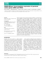
Bạn đang xem bản rút gọn của tài liệu. Xem và tải ngay bản đầy đủ của tài liệu tại đây (1.96 MB, 17 trang )
Litlekalsoy et al. BMC Cancer (2016) 16:549
DOI 10.1186/s12885-016-2580-y
RESEARCH ARTICLE
Open Access
Expression of circadian clock genes and
proteins in urothelial cancer is related to
cancer-associated genes
Jorunn Litlekalsoy1,2,7* , Kari Rostad3, Karl-Henning Kalland1,4, Jens G. Hostmark2,5 and Ole Didrik Laerum1,6
Abstract
Background: The purpose of this study was to evaluate invasive and metastatic potential of urothelial cancer by
investigating differential expression of various clock genes/proteins participating in the 24 h circadian rhythms and
to compare these gene expressions with transcription of other cancer-associated genes.
Methods: Twenty seven paired samples of tumour and benign tissue collected from patients who underwent
cystectomy were analysed and compared to 15 samples of normal bladder tissue taken from patients who
underwent cystoscopy for benign prostate hyperplasia (unrelated donors). Immunohistochemical analyses were
made for clock and clock-related proteins. In addition, the gene-expression levels of 22 genes (clock genes, casein
kinases, oncogenes, tumour suppressor genes and cytokeratins) were analysed by real-time quantitative PCR (qPCR).
Results: Considerable up- or down-regulation and altered cellular distribution of different clock proteins, a
reduction of casein kinase1A1 (CSNK1A1) and increase of casein kinase alpha 1 E (CSNK1E) were found. The pattern
was significantly correlated with simultaneous up-regulation of stimulatory tumour markers, and a down-regulation
of several suppressor genes. The pattern was mainly seen in aneuploid high-grade cancers. Considerable alterations
were also found in the neighbouring bladder mucosa.
Conclusions: The close correlation between altered expression of various clock genes and common tumour
markers in urothelial cancer indicates that disturbed function in the cellular clock work may be an important
additional mechanism contributing to cancer progression and malignant behaviour.
Keywords: Circadian clock genes, Casein kinases, Oncogenes, Tumour suppressor genes and cytokeratins
Background
Time is a fundamental part of all biological processes
in tissues and cells. Both in rodents and humans, the
circadian timing system affects many cellular and
physiological functions, including cell proliferation,
metabolic pathways, protein synthesis and energy metabolism [1]. Severe and prolonged disturbances of the
circadian timing system are believed to predispose to
cancer development in different organs, not only in the
mammary and prostate glands, but also in several other
types of cancer, including ovarian, kidney, brain, colorectal,
* Correspondence:
1
Department of Clinical Science, The Gade Laboratory of Pathology,
University of Bergen, Bergen, Norway
2
Department of Clinical Medicine, Section of Surgery, University of Bergen,
Bergen, Norway
Full list of author information is available at the end of the article
lung, head/neck, pancreatic cancer and hematological
malignancies [2–14].
The mammalian circadian clock system consists of
positive and negative regulators, with a complex autoregulatory transcriptional and translational feedback
program. By accumulating and binding to the promoter
region of the two transcriptions factors, BMAL1 and
CLOCK, PER and CRY proteins reduce the transcription
of many genes, including their own. This occurs during
ambient light exposure via the master clock in the brain,
the suprachiasmaticus nucleus (SCN). The corresponding proteins oscillate with a delayed phasing and with
maximum levels at dusk [15].
The transcription factors CLOCK and BMAL1 form a
heterodimer which in humans is acting stimulatory on gene
transcription during night time. CLOCK also contributes
© 2016 The Author(s). Open Access This article is distributed under the terms of the Creative Commons Attribution 4.0
International License ( which permits unrestricted use, distribution, and
reproduction in any medium, provided you give appropriate credit to the original author(s) and the source, provide a link to
the Creative Commons license, and indicate if changes were made. The Creative Commons Public Domain Dedication waiver
( applies to the data made available in this article, unless otherwise stated.
Litlekalsoy et al. BMC Cancer (2016) 16:549
to chromatin-remodelling and mediates acetylation of
BMAL1. The type of phasing can vary from organ to organ.
For instance, BMAL1 undergoes rhythmic acetylation in
the liver where the timing parallels the down-regulation of
circadian transcription in clock-controlled genes.
The 24 h clock generation is modified by posttranslational events such as phosphorylation and ubiquitination which contribute to precision, stability and nuclear
translocation of the core clock proteins. PER and BMAL1
have also been identified as tumour suppressors [15–20].
Casein kinase 1 epsilon and delta (CSNK1E and CSNK1D)
are critical in regulating the core circadian protein turnover
in mammals. Mutations in either of these kinases may thus
have dramatic effects on the circadian period [21].
Urothelial carcinoma of the bladder is a very complex
malignancy with multiple alterations in complementary
pathways. The advent of high-throughput methods of
molecular analysis, as microarray-based approaches, has
been used extensively to look for expression profiles in
effort to sub-classify bladder cancer (stage and pathways)
and to predict outcomes and response to systemic treatments. Several tissue and blood-based biomarkers have
been identified, but status as of today is that no biomarker
panel is yet validated for individual prognostic and daily
clinical practice. A problem is that most researchers combine biomarkers from a single pathway (cell-cycle, apoptosis or angiogenesis) while the focus rather should be in
investigating biomarker combinations that encompass a
variety of different pathways to increase the predictive
value and opportunity for targeted treatment. Standard
pathological features and imaging are insufficient to allow
accurate staging, prognostication and prediction of the patient’s outcome [22, 23]. This reveals an urgent need for
identifying novel biomarkers that can define the invasive
urothelial carcinomas with intrinsic property for recurrence and metastases.
The urinary system undergoes significant circadian
rhythms in humans. During day and night both urinary
excretion and extrusion of urine are actively regulated by
several internal factors, such as antidiuretic hormone [24].
Such circadian variations led us to postulate that similar to
other organs, perturbation of the clockwork may be a
contributory mechanism of dysregulation during the development of urothelial cancer. Since clock genes have a
modifying role in the gene regulation, they may interact
with the transcription of oncogenes and/or tumour
suppressor-genes. If so, they might be used as independent
or additional markers of malignant behaviour. Therefore,
ten key proteins of the clockwork were selected for a combined analysis of transcriptional activity and presence of
their proteins in the malignant cells. For comparison,
simultaneous analyses of gene-expression patterns were
performed for oncogenes and suppressor-genes that are
commonly altered in urothelial cancer.
Page 2 of 17
Methods
Patient material and tissue
Twenty-seven patients with invasive urothelial cancer
undergoing cystectomy from 2006 to 2009 were included.
General procedures for the cystectomy patients are that
the patients enter the operating room around 07:45 in the
morning. The anesthesia is completed around 08:20 and
within the next 5–10 min open surgery is performed. The
bladder is removed from the body around 10:00 whereupon the surgeon immediately collects tissue samples
from tumour and adjacent normal appearing mucosa into
separate tubes. Within twenty minutes, the harvested
bladder biopsies are cut into small pieces and snap frozen
at −80 °C. Patient details are given in Table 1. Normal
bladder biopsies were taken from 15 male patients who
had TUR-P (transurethral resection of the prostate) for
benign prostatic hyperplasia (BPH). The mucosal biopsies
consisted of the whole urothelial layer and some underlying connective tissue. A major part of the cell nuclei
were from urothelium as compared to sub-mucosal fibroblasts. Both the cystectomies and the unrelated normal
mucosa were harvested in the time period 9 to 12 AM.
Paraffin-embedded tissue slides were made for histological
diagnostics, and classified by the WHO and NM-system.
The study was approved by the Regional Ethical Committee (REK No. 12226/REK No. 2009/1527).
Immunohistochemistry
The paraffin blocks were cut in 5 μm sections and
stained with antibodies listed in Table 2. The sections
were de-paraffinised and pre-treated as listed in Table 2,
and stained as described earlier [25]. Sections of tissue
microarrays made of twelve different tissues, reported to
express one or more of our chosen proteins, served as
control.
Evaluation of staining results
The analyses were made separately for the tumour and
neighbouring benign tissue from cystectomies, and unrelated normal mucosa. Positive staining of epithelial cells
was estimated as weakly, moderately and strong, (separately for the nucleus (N) and the cytoplasm (C)). Counting was performed on cells from tumour, normal
appearing mucosa without atypia, and normal mucosa
from the 15 individuals (Table 3). For control, the same
staining procedure was performed on tissue microarrays
comprising other human tumours/normal tissues. All
cases were scored on coded specimens separately by
ODL and JGH.
Flow cytometry (FCM)
FCM was performed on single cell suspensions of tumour
tissue obtained by cutting the tissue into small pieces
which were shaken, filtered, spun down, re-suspended in
Litlekalsoy et al. BMC Cancer (2016) 16:549
Page 3 of 17
Table 1 Tumour grade, invasiveness, T-stage, ploidy and survival in the individual patients
No
G
1
Low
V.I.
2
High
3
High
4
Low
5
High
6
Low
7
High
8
Low
9
High
10
High
11
Low
12
High
13
Low
14
High
15
High
x
16
High
x
17
High
x
18
Low
19
20
pTa
pT1
pT2A
pT2B
pT3B
x
x
D
A
x
x
x
x
x
x
x
x
x
Survival A/D
A
7,25
1,9
-
17
D/1m
D/10m
7,7
A
x
8
A
x
3,5
A
x
x
x
A-S
x
x
x
D-S
4,22
x
12,89
23,04
D/6m
-
-
A
x
72
10,06
A
x
7,91
21
D/10m
x
8,13
26,2
D/14m
x
x
6,86
-
A
x
x
8,2
32
A
x
x
6,2
28
A
x
x
12
21
A
x
14
20
A
x
x
x
4,4
-
D/11m
High
x
x
x
64
15
A
High
x
x
x
24
18
D/17m
21
High
x
22
High
23
Low
24
High
25
Low
26
Low
27
High
x
x
x
x
x
x
x
x
x
x
18
18
A
84
20
D/3m
18
17
A
41
38
D/10m
41
15
x
x
x
x
x
A
x
x
x
x
7,2
x
3,3
x
13
A
D/24m
20
D/10m
No case number, G grade, V.I. vascular invasion, pTa-pT1-pT2A-pT2B-pT3B tumour stage, D Diploid, A Aneuploid, D-S Diploid S-phase, A-S Aneuploid S-phase,
Survival A/D survival after surgery (in months, m), A alive, D dead
PBS and fixed by addition of 96 % ethanol, stained with
propidium iodide as earlier described [26] and analysed on
a FACScan flow cytometer (Becton Dickinson, Palo Alto,
CA, USA). Normal human lymphocytes were used as
standard, and the ploidy index (PI) was calculated as a
ratio between the peak channel for the tumour cells and
the peak channel for the lymphocytes.
RNA extraction and real-time quantitative PCR (qPCR)
RNA purification and single-stranded cDNA synthesis
Biopsies were ground to powder under liquid N2. Total
RNA was extracted according to standard protocols
(Invitrogen Trizol LS protocol and Qiagen miRNeasy
protocol; Invitrogen, Carson City, CA). 30 μl of singlestranded cDNA for qPCR analysis was synthesised from
1 μg of total RNA according to Ambion (Ambion, TX,
USA) instructions.
Endogenous control and endogenous control cards
The different tissue types included in our study were
initially studied with respect to gene expression of 16
different housekeeping genes, to assess which one was
best suited as endogenous control for our purpose. Two
endogenous control cards accommodating 8 samples
each, in triplicate, were applied. β-actin (ACTB) proved
to be the most suitable endogenous control for our three
tissue types and therefore chosen when designing the
Taqman low density arrays (TLDA) cards. In addition
GAPDH was added in the TLDA cards as standard
(from the supplier), but was not used in our further
calculations.
Real-time quantitative PCR (qPCR) in low-density array
format
Taqman low density arrays (TLDA) are customizable,
384-well microfluidic cards for real-time qPCR (Applied
Litlekalsoy et al. BMC Cancer (2016) 16:549
Page 4 of 17
Table 2 Specifications of antigens and corresponding antibodies
Antigen
Specificities Purchaser
Dilution
Pre-treatment
PER1 (Per12-A)
Polyclonal
AH Diagnostics AS
Fjellgata 1, Oslo
1:50, overnight at 4 °C
Microwave treatment for 10 min at 750 W and
20 min at 500 W in 10 mmol/L citrate buffer pH6
PER2 (N-19, sc-7728)
Polyclonal
Santa Kruz Biotecnology 1:200, overnight at 4 °C
Inc. Europe
Microwave treatment for 10 min at 750 W and
20 min at 500 W in 10 mmol/L citrate buffer pH6
PER3 (Per32-A)
Polyclonal
AH Diagnostics AS
Fjellgata 1, Oslo
Microwave treatment for 10 min at 750 W and
20 min at 500 W in 10 mmol/L citrate buffer pH6
CRY1 (W-L5, sc-101006)
Monoclonal Santa Kruz Biotecnology 1:200, overnight at 4 °C
Inc. Europe
Microwave treatment for 10 min at 750 W and
20 min at 500 W in 10 mmol/L citrate buffer pH6
CRY2 (P-21, sc-130731)
Polyclonal
Santa Kruz Biotecnology 1:200, overnight at 4 °C
Inc. Europe
Microwave treatment for 10 min at 750 W and
20 min at 500 W in 10 mmol/L citrate buffer pH6
BMAL 1 (LS-B660/12275)
Polyclonal
Lifespan Biosciences
(Nordic biosite)
1:100, overnight at 4 °C
Microwave treatment for 10 min at 750 W and
20 min at 500 W in 10 mmol/L citrate buffer pH6
CLOCK (LS-B278/18928
Polyclonal
Lifespan Biosciences
(Nordic biosite)
1:500, overnight at 4 °C
Microwave treatment for 10 min at 750 W and
20 min at 500 W in 10 mmol/L citrate buffer pH6
Anti-CSNK1α1L
Polyclonal
Abcam.com England
1:150, overnight at 4 °C
Microwave treatment for 10 min at 750 W and
20 min at 500 W in 10 mmol/L citrate buffer pH6
Casein kinase 1Ɛ (Sc-25423)
Polyclonal
Santa Kruz Biotecnology 1:100, overnight at 4 °C
Inc. Europe
Microwave treatment for 10 min at 750 W and
20 min at 500 W in 10 mmol/L citrate buffer pH6
Casein kinase 1α (Sc-28886)
Polyclonal
Santa Kruz Biotecnology 1:100, overnight at 4 °C
Inc. Europe
Microwave treatment for 10 min at 750 W and
20 min at 500 W in 10 mmol/L citrate buffer pH6
1:50, overnight at 4 °C
Table 3 Mean scores of positivity in nucleus and cytoplasm for
the clock proteins
Protein
Cancer cells
Neighbouring mucosa
Nucleus Cytopl. Nucleus
PER 1
2.17
Normal mucosa
Cytopl.
Nucleus Cytopl.
0
1.71
0
2.00
0
+/− SEM 0.15
0
0.10
0
0
0
PER 3
0.43
0.67
0.73
0
1.15
0.22
+/− SEM 0.08
0.11
0.13
0.15
0
0.13
CRY 1
1.96
1.84
1.27
1.76
0.62
2.08
+/− SEM 0.16
0.11
0.14
0.20
0.13
0.15
CRY 2
0.83
2.31
2.75
2.00
2.00
0
+/− SEM 0
0.13
0.23
0.18
0.26
0.28
BMAL1
2.40
2.33
2.27
1.16
2.08
1.42
+/− SEM 0.20
0.12
0.19
0.20
0.21
0.20
CLOCK
2.04
2.23
2.57
2.52
2.75
2.91
+/− SEM 0.16
0.12
0.13
0.13
0.14
0.09
Casein kinase 1 alpha
1.93
2.70
2.78
3.00
2.90
3.00
+/− SEM 0.18
0.10
0.13
0.00
0.11
0.00
Casein kinase 1 alpha 1 L
+/−SEM
1.96
2.59
2.88
2.96
2.54
2.92
0.24
0.12
0.08
0.04
0.22
0.08
Casein kinase 1 epsilon
Biosystems (ABI)). Each TLDA card was configured
for 24 genes in duplicates, including β-actin and
GAPDH as endogenous controls, core clock-genes
and genes encoding several tumour markers (TaqMan
assays are listed in Table 4). Single-stranded cDNA
corresponding to 200 ng of total RNA was diluted in
Taqman Universal buffer (ABI) and added to each
loading well. The samples were distributed to the micro wells by centrifugation for 1 min at 343xg. The
cards were placed in an ABI PRISM 7900HT Sequence Detection System thermocycler for 40 cycles:
15 s at 95 °C and 60 s at 60 °C. The SDS2.3 and RQ
manager 1.2 software (ABI) were used for analysis
and data were exported to Excel for further visualization. Data Assist v.3.01 (ABI) was utilized for hierarchical cluster analysis and generation of correlation
plots. The gene expression data were analysed using
the comparative Ct-method (ΔΔCt). Gene expression
levels were normalized against ß-actin and calibrated
against a chosen calibrator to provide fold change
relative gene expression levels. Two separate gene expression analysis were performed in order to study
the relative differential gene expression (fold change
(Relative quantity (RQ)) in the respective tissues: tumour
and neighbouring benign tissue relative to unrelated normal mucosa, and relative gene expression levels in tumour
versus neighbouring mucosa.
2.93
2.07
2.75
1.95
3.00
2.00
Statistics
+/− SEM 0.05
0.05
0.10
0.09
0
0
Statistical Package for the Social Sciences (SPSS v.12)
(SPSS Inc. Chicago, Illinois) was utilized for statistical
+/− SEM: +/− standard error of the arithmetic means
Litlekalsoy et al. BMC Cancer (2016) 16:549
Page 5 of 17
Table 4 List of TaqMan gene expression assays and their corresponding proteins
Gene assay
Protein
Gene assay
Protein
Gene assay
Protein
Hs00978050_m1
H-RAS
Hs01034249_m1
p53
Hs00242988_m1
PER 1
Hs00364284_m1
K-RAS
Hs00923894_m1
p16
Hs00256143_m1
PER 2
Hs00180035_m1
N-RAS
Hs02621230_m1
pTEN
Hs00213466_m1
PER 3
Hs01076078_m1
EGFR
Hs00559840_m1
Cytokeratin 7
Hs01565974_m1
CRY 1
Hs00182181_m1
uPAR
Hs00196158_m1
Cytokeratin 1
Hs00323654_m1
CRY 2
Hs01126606_m1
PAI 1
Hs00361185_m1
Cytokeratin 5
Hs00154147_m1
BMAL 1
Hs00166289_m1
Cytokeratin10
Hs00231857_m1
CLOCK
Hs99999905_m1
GAPDH
Hs00265033_m1
Cytokeratin14
Hs01887794_m1
CK1A1L
Hs00793391_m1
CK1A1
Hs00266431_m1
CK1ε
analysis. The Spearman’s rank correlation (correlations coefficient, c) was used to determine significant correlation
between the various gene expressions. The MannWhitney non-parametric rank test was used to identify
correlation between the gene expressions in the tumours
compared to neighbouring mucosa. Data Assist v.3.01
(ABI) was applied on the gene expression data to calculate
Pearson’s product monument correlation coefficients (r)
for each sample represented in the various tissue types.
Pearson’s correlation was used for the hierarchical cluster
analysis and generation of heat maps of gene expression.
Data Assist v.3.01 (ABI) performed a two-sample, twotailed Student’s t-test for comparing the fold change values
(2(−deltaCt)) of the separate biological groups (normal bladder mucosa, neighbouring benign and tumour tissue), and
a p-value was calculated. The results were presented in
the mRNA fold change gene expression plots (log fold
change versus sample group).
Results
Immunohistochemistry
Stimulatory clock proteins/casein kinases
Cytoplasmic BMAL1 staining was slightly stronger in
the tumour and the neighbouring mucosal cells than in
the normal, unrelated mucosa. In the nuclei, BMAL1
was significantly increased in neighbouring tissue, and
also slightly increased in tumour tissue compared to
normal mucosal cells (Table 3). Six cases expressed neither BMAL1 nor CRY2 in the nucleus. When this was
compensated for, the remaining positive cases for
BMAL1 had a mean score in the nucleus of 1.84 +/−
SEM 0.15, which is significantly higher than in the normal mucosa. CLOCK was significantly reduced in the
tumour cells, but not in the nucleus or cytoplasm in the
neighbouring mucosa.
Casein kinase 1A and 1A1Like were both significantly
reduced in the tumour nuclei, but not in the cytoplasm.
Casein kinase 1E was equally expressed in both nucleus
and cytoplasm.
Inhibitory clock proteins
PER1 was positive in the nucleus and absent in cytoplasm of neoplastic, neighbouring and normal mucosa
(Table 3). PER2 did not give satisfactory staining and
was omitted. PER3 was absent in nucleus of normal mucosa, but expressed in cancer cells and their neighbouring mucosa. Opposite, it was lower in the cytoplasm of
cancer cells and neighbouring tissue compared to normal mucosa, and there seemed to be a significant shift
from cytoplasm to nucleus in malignancy. CRY1 was
significantly increased in tumour cytoplasm and neighbouring mucosal cells. The increased expression of
CRY1 in the cancer cells was three times higher than
in normal mucosa. CRY2 was absent in the nucleus in
cancer cells and low in the cytoplasm, while neighbouring and normal mucosal cells showed no major
differences.
Altogether, this indicates complex alterations, where
the main features were redistribution between nucleus
and cytoplasm, and an increase of both stimulatory
and inhibitory clock proteins, see in Additional file 1:
Figure S1.
Gene expression analysis
Raw data and general pattern
The over-all differences in gene expression pattern in
tumours compared to matched neighbouring mucosa
are shown in Table 5. The gene-expression signal correlation plot is visualized in Fig. 1. The mRNA fold
change in tumour and neighbouring mucosa from 27
patients relative to normal mucosa from 15 unrelated
donors are visualized in Fig. 2. Figures 3 and 4 display
relative quantity of mRNA in tumour compared to
neighbouring mucosa of 27 patients for the genes found
statistically significant. Figure 5 shows a hierarchical
cluster diagram (heat map) of differential expression of
22 genes in normal mucosa from 15 unrelated donors together with tumour/neighbouring mucosa from 27 patients (cystectomies).
A. Relative mRNA gene expression levels of clock genes and common tumour markers from cystectomies (Tumour/Benign-fold change)
GENES
Patient sample
BMAL CLOCK PER1 PER2 PER3 CRY1 CRY2 CSNK1A1L CSNK1A1 CSNK1E TP53 p16
PTEN EGFR HRAS KRAS NRAS Upar PAI-1 KRT7
KRT1 KRT5 KRT10 KRT14
1
1,3
0,8
0,1
0,1
0,3
0,5
0,3
34,8
0,6
0,5
0,7
8,1
0,5
0,8
0,9
0,6
0,9
0,1
0,1
0,5
1,1
0,0
0,1
9,5
2
2,1
1,0
0,9
1,2
0,8
1,5
1,3
0,0
1,3
3,5
2,0
6,3
2,4
0,8
1,9
1,0
2,3
1,6
0,6
83*
0,0
0,3
0,1
311*
3
4,9
3,2
0,4
0,7
5,9
3,9
0,8
3,2
3,2
11,3
0,9
131
1,9
8,7
8,9
2,2
10,1
0,5
0,4
8,8
2,6
477*
5,9
174
4
1,0
1,1
0,2
0,3
0,6
0,4
0,7
37,1
0,6
0,3
2,2
0,9
0,7
1,5
2,5
1,3
1,9
0,6
0,2
18,9
1,0
4,0
0,6
970*
5
1,3
1,2
0,5
0,3
0,8
0,7
0,5
0,1
0,4
0,5
0,8
0,6
0,7
0,5
0,5
1,3
0,9
0,2
0,3
0,0
1,3
0,1
0,0
0,3
6
3,6
1,6
1,4
0,5
0,7
2,3
1,2
177
1,3
3,0
2,9
88,0
0,8
1,0
2,2
1,5
7,9
6,8
3,1
8,1
0,1
17,4
4,5
110
7
1,4
2,3
0,9
1,0
0,9
2,4
1,0
0,0
1,3
7,4
1,9
1,1
0,7
1,1
1,3
1,2
2,5
1,2
3,8
13,8
0,7
2,8
8,4
0,8
8
2,8
2,1
0,0
0,6
1,6
2,8
3,1
6,6
1,9
8,5
1,8
12,9
39,0
2,2
0,2
3,0
2,8
0,4
0,2
129*
0,7
0,2
8,4
0,0
9
0,5
1,3
1,7
0,4
5,9
1,0
2,2
0,2
0,7
0,4
0,6
0,2
0,4
0,8
0,6
0,5
0,3
0,3
0,3
1,4
0,6
0,0
0,1
0,0
10
0,6
0,3
0,3
0,2
0,8
1,4
0,6
169
0,5
0,4
1,3
0,0
1,1
3,0
0,8
0,6
1,9
0,6
0,5
0,4
64,7
1,8
0,4
71,9
11
1,5
1,3
0,6
1,0
1,0
0,9
1,1
0,3
1,0
1,2
3,4
24,0
1,2
1,7
2,2
1,5
2,6
0,9
0,4
38,9
2,0
6,7
18,0
5,7
12
4,3
1,7
0,8
0,4
0,6
0,6
0,4
0,0
1,5
1,9
3,6
1,3
4,0
1,1
5,2
1,9
3,3
1,7
0,8
29,0
0,9
31,2
16,8
19.1
13
4,1
1,0
2,0
1,7
0,5
1,3
1,5
1,5
0,9
0,5
2,8
3,9
5,7
0,6
1,8
2,1
3,4
11,2
25,3
25,1
10,1
106*
628*
72,3
14
1,1
0,3
0,1
0,3
0,4
0,0
0,1
0,0
0,2
0,2
0,4
2,6
1,9
0,1
0,3
0,6
0,3
0,0
0,0
0,7
0,0
0,1
12,2
0,5
15
3,6
2,2
0,3
0,4
1,6
1,4
1,3
6,2
1,3
1,8
4,0
7,9
2,6
2,0
2,0
2,7
2,3
0,7
2,5
16,0
0,8
0,2
2,4
27,1
16
0,8
0,7
0,3
0,4
0,4
0,6
0,6
0,6
1,8
0,8
0,9
0,8
0,8
1,2
2,9
1,1
0,8
0,2
0,5
15,5
0,0
0,6
0,4
0,6
17
2,3
1,2
0,2
0,5
1,3
2,0
0,9
0,1
0,8
0,9
3,3
1,9
0,8
3,8
2,7
1,6
2,0
0,3
0,4
47,4
0,0
0,8
0,2
25,0
18
1,9
1,8
0,2
0,2
0,2
1,5
0,2
1,2
1,1
1,0
3,0
84,0
0,7
3,4
1,8
1,5
3,9
0,8
0,4
5,3
1,3
40*
0,1
4535*
19
0,8
0,4
0,3
0,3
0,3
0,7
0,6
0,6
0,7
0,5
1,2
10,0
1,1
4,2
3,2
0,7
1,2
0,9
2,8
1598*
0,4
7,9
1172*
633*
20
2,0
2,8
0,6
0,3
0,9
0,6
0,8
0,0
1,0
1,0
3,4
1,9
1,0
0,6
1,3
2,6
1,6
0,5
0,4
3,4
0,5
0,1
0,0
3,1
21
0,5
1,2
0,7
0,5
0,7
1,5
1,0
0,4
0,8
0,7
1,9
1,3
0,9
2,1
1,9
1,6
1,5
0,5
0,8
17,9
0,1
0,1
21,1
8,0
22
0,8
0,8
0,7
0,3
0,1
1,0
0,6
0,0
1,2
2,4
4,3
4,2
1,9
2,8
2,2
1,1
2,3
1,2
2,1
7,8
0,7
0,3
0,1
225
23
0,5
1,3
1,7
0,4
5,9
1,0
2,2
0,2
0,7
0,4
0,6
0,2
0,4
0,6
0,6
0,5
0,3
0,3
0,3
1,4
0,6
0,0
0,1
0,0
24
1,2
1,1
0,4
0,7
0,3
0,3
0,3
1,1
1,1
0,8
1,4
0,3
0,9
1,7
1,2
1,5
1,1
0,4
0,6
0,0
23,3
12,1
29,8
9,2
25
2,6
1,1
0,5
0,6
1,3
0,5
0,5
21,9
1,2
1,7
1,6
1,7
1,1
0,8
1,7
1,1
1,4
0,9
0,7
1,9
1,0
0,0
1,4
1,4
26
1,0
0,7
0,5
1,5
0,2
0,7
0,4
0,0
1,7
1,0
1,3
1,2
4,7
0,4
0,7
0,9
0,8
1,4
2,3
51,5
0,8
34,4
11,0
0,6
27
1,0
0,7
0,1
1,0
0,0
0,4
0,2
27,9
2,5
2,3
3,8
0,7
7,6
1,4
3,4
1,5
2,3
1,5
11,6
730*
0,5
135*
212*
65,1
Litlekalsoy et al. BMC Cancer (2016) 16:549
Table 5 Relative gene expression levels of clock genes and common tumour markers from cystectomies (Tumour/Benign-fold change)
Page 6 of 17
B. Average T/B fold change in mRNA gene expression of genes upregulated and downregulated in 27 cystectomy patients
Number of
patients
17
17
4
3
7
11
8
13
13
11
20
19
Average
2,47
up-regulation
1,67
1,70
1,50
3,34
1,99
1,72
37,63
1,57
4,08
2,56
st.dev
1,2
0,6
0,2
0,2
2,4
0,8
0,7
61,5
0,6
3,4
Number of
patients
7
8
23
21
19
13
17
14
12
Average
0,64
up-regulation
0,60
0,41
0,39
0,50
0,53
0,49
0,13
st.dev
0,2
0,3
0,2
0,3
0,2
0,2
0,2
0,2
14
16
19
19
20
8
8
22
8
13
15
19
20,69 5,44
2,60
2,65
1,69
2,91
3,32
6,69
129,69 13,30 67,51 143,45 382,79
1,0
37,2
9,9
1,9
1,8
0,6
2,2
3,7
8,2
361,9
22,1
129,9 328,1
1036,3
13
7
8
12
10
8
7
7
19
19
5
17
14
12
8
0,65
0,54
0,69
0,48
0,69
0,60
0,58
0,64
0,63
0,48
0,42
0,33
0,44
0,21
0,19
0,33
0,2
0,2
0,2
0,3
0,2
0,2
0,2
0,1
0,3
0,3
0,2
0,3
0,3
0,2
0,2
0,3
Litlekalsoy et al. BMC Cancer (2016) 16:549
Table 5 Relative gene expression levels of clock genes and common tumour markers from cystectomies (Tumour/Benign-fold change) (Continued)
B2. Average T/B fold change in mRNA gene expression of genes upregulated and downregulated in 27 cystectomy patients. Patient samples identified as outliers by SPSS for respective gene assys
have been excluded from the analysis (*)
Number of
patients
17
17
4
3
7
11
8
13
13
11
20
19
Average
2,47
up-regulation
1,67
1,70
1,50
3,34
1,99
1,72
37,63
1,57
4,08
2,56
st.dev
1,2
0,6
0,2
0,2
2,4
0,8
0,7
61,5
0,6
3,4
Number of
patients
7
8
23
21
19
13
17
14
12
Average
0,64
up-regulation
0,60
0,41
0,39
0,50
0,53
0,49
0,13
st.dev
0,2
0,3
0,2
0,3
0,2
0,2
0,2
0,2
14
16
19
19
20
8
8
22
8
13
15
19
20,69 5,44
2,60
2,65
1,69
2,91
3,32
6,69
129,69 13,30 67,51 143,45 382,79
1,0
37,2
9,9
1,9
1,8
0,6
2,2
3,7
8,2
361,9
22,1
129,9 328,1
1036,3
13
7
8
12
10
8
7
7
19
19
5
17
14
12
8
0,65
0,54
0,69
0,48
0,69
0,60
0,58
0,64
0,63
0,48
0,42
0,33
0,44
0,21
0,19
0,33
0,2
0,2
0,2
0,3
0,2
0,2
0,2
0,1
0,3
0,3
0,2
0,3
0,3
0,2
0,2
0,3
C. Average T/B fold change in mRNA gene expression in aneuploid and diploid patient tumour samples
Aneuploid (19 patients)
Average
1,9
1,3
0,6
0,6
1,2
1,2
0,8
12,3
1,2
2,1
2,2
13,7
2,0
2,1
2,4
1,4
2,3
1,3
2,9
137
5,7
43
111
325
st.dev
1,4
0,8
0,5
0,4
1,7
0,9
0,5
38,6
0,7
2,8
1,3
34,1
1,9
2,0
1,9
0,6
2,1
2,5
6,0
390
15,3
112
126
1032
Diploid (8 patients)
*
Average
1,6
1,3
0,6
0,6
1,4
1,2
1,2
32,1
1,0
1,9
1,7
17,0
6,0
1,1
1,2
1,3
2,3
1,3
0,9
31,1
0,9
7,9
5,4
137
st.dev
1,0
0,4
0,6
0,5
1,9
0,9
1,0
60,8
0,5
2,8
1,0
29,9
13,4
0,6
0,9
0,8
2,4
2,2
1,2
44,0
0,6
12,3
5,6
339
Gene expression levels identified as outliers by SPSS statistical analysis
Page 7 of 17
Litlekalsoy et al. BMC Cancer (2016) 16:549
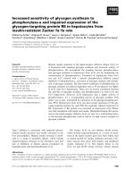
Fig. 1 (See legend on next page.)
Page 8 of 17
Litlekalsoy et al. BMC Cancer (2016) 16:549
Page 9 of 17
(See figure on previous page.)
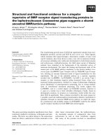
Fig. 1 Gene expression signal correlation plots. The plots display the correlations between mRNA normalized gene expression levels in the
normal control bladder tissue samples of 15 patients with BPH (a), benign tissue peripheral to the tumour (b) and tumour tissue (c) of 27
cystectomy patients, respectively. Pearson’s product moment correlation coefficients (r) for each pair of samples were calculated using DataAssist
from Applied Biosystems. Each cell represents a different scatter plot, coloured to indicate the strength of the correlations between the samples.
The higher the correlation between the gene expression levels in the two samples (the closer the correlation coefficient (r), is to 1), the colour
moves towards brighter red. The poorer the correlation between the gene expression levels in the two samples (the closer r is to 0), the colour
moves towards darker red and then green, indicating no correlation. All samples are correlated with each other for each of the selected genes
Gene expression correlation plots
The strength of the correlations of relative mRNAlevels in the different patient samples is visualized in
the gene expression signal correlation plots (Fig. 1). The
plots display the strength of the correlations between
normalised gene expression levels in 15 biopsies of normal
bladder mucosa (Fig. 1a), and 27 matched benign/tumour
biopsies taken from patients who underwent cystectomy
(Fig. 1b and c, respectively). An increasing dissimilarity in
gene expression levels and poorer correlations among
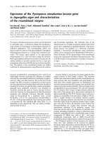
Fig. 2 mRNA fold change gene expression plots. Gene expression levels in benign neighbouring mucosa and tumour tissue relative to normal
bladder mucosa tissue from BPH patients. The relative quantity plots display the log2 fold change in mRNA levels in the benign (blue bars) and
tumour (red bars) tissue taken from cystectomies (27 patients) versus normal bladder tissue from BPH patients. The bars in a. display the log2 fold
change (log2 RQ) in mRNA levels of the clock genes, while the tumour marker genes are plotted in b. Genes with a negative value are downregulated, while genes with a positive value are up-regulated in the malignant bladder (tumour and benign tissue) versus the normal bladder
(whose log2 value is 0 for each gene). Statistical significance with a p-value ≤ 0.05 was found for KRT7, PER1, PER2, PTEN, uPAR and PAI-1 (Two-sample, two-tailed Student’s t-test)
Litlekalsoy et al. BMC Cancer (2016) 16:549
Page 10 of 17
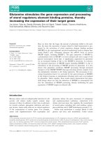
Fig. 3 Relative mRNA quantity of PER1, PER2, PER3 and CRY2. Real-time quantitative PCR expression levels normalized against the endogenous
control β-actin (ACTB). The figure gives the comparison between 27 tumour and matched benign bladder tissue samples. Columns, median; bars,
a: PER1, b: PER2, c: PER3 and d: CRY2. The relative gene expression of all four genes was significantly elevated in the benign versus malignant
bladder tissue. The changes were consistent for each pair of tumour - neighbouring mucosa, indicated by the p-value of the statistical test (nonparametric paired samples Mann-Whitney test)
patients were seen when moving from normal bladder
mucosa to neighbouring and tumour tissue.
mRNA gene expressions in tumour/neighbouring mucosa
from cystectomies compared with normal bladder mucosa
Gene expression patterns (Ct-values) of the normal unrelated mucosa (15 samples) were consistent regarding
the two housekeeping genes included in the study. The
gene expressions in the tumour and neighbouring tissue
collected from the cystectomies were, for all genes
included, compared relatively to the gene expression
pattern of these 15 samples.
BMAL1 was down-regulated in both neighbouring and
tumour tissue compared to normal mucosa, while CLOCK
Fig. 4 Relative mRNA quantity of KRT7, KRT14, NRAS, TP53 and UPAR. Real-time quantitative PCR expression levels normalized against the endogenous
control β-actin. The figure gives the comparison between 27 tumour and matched benign bladder tissue samples. Columns, median; bars, a: KRT7,
b: KRT14, c: NRAS, d: TP53 and e: UPAR. The gene expression levels of the cytokeratins, the NRAS and TP53 were significantly elevated in the tumour versus
benign bladder tissue, while the expression of UPAR was significantly elevated in the benign tissue compared to the tumour. The changes were consistent
for each pair of tumour - neighbouring mucosa, indicated by the p-value of the statistical test (non-parametric paired samples Mann-Whitney test)
Litlekalsoy et al. BMC Cancer (2016) 16:549
Page 11 of 17
Fig. 5 Unsupervised hierarchical cluster analysis of differentially expressed genes. Normal bladder tissue from 15 unrelated donors with BPH
together with tumour and matched benign tissue from 27 cystectomy patients were analysed. Real-time qPCR expression data were imported
into DataAssist (ABI) for unsupervised hierarchical cluster analysis. Distances between samples and assays were calculated based on the delta-Ct
values using Pearson’s correlation. Differentially expressed genes are represented in rows and the different samples are represented in columns.
Each cell in the heat map represents one samples relative expression of one gene. For each gene assay, the middle expression level was set as
the median of all of the delta-Ct-values of all samples for that gene assay. Gene expression colour codes in the heat map: Green colour represents
relative levels of mRNA lower than the middle value for that gene expression assay (decreased gene expression); Red colour represents levels of
mRNA higher than the middle expression level (increased gene expression); Dark colour reflects an mRNA expression level closer to the middle
expression level (no major increase or decrease in gene expression). Patient samples: Blue colour: The normal bladder tissue taken from BHP
patients is numbered 1–15N; Green colour: Normal benign tissue taken peripherally to the tumour is numbered 1–27N; Red colour: tumour tissue
is numbered 1–27T. Genes: Purple colour: Clock genes; Black colour: cancer associated genes. (Clustering method: complete linkage. Map type:
assay centric)
was slightly up-regulated in neighbouring tissue and
slightly down-regulated in tumour (Fig. 2).
PER1 and CRY1 were both up-regulated in neighbouring and tumour tissue compared to normal mucosa,
while PER2 and PER3 were up-regulated in neighbouring
mucosa and down-regulated in the tumour tissue. CRY2
was down-regulated in both tissue types compared with
normal mucosa. This corresponds well with the immunostaining results (Table 3).
The casein kinases CSNK1A1L and CSNK1E were downregulated in neighbouring mucosa and up-regulated in
tumour tissue, while CSNK1A1 was down-regulated in
both tissue types (Fig. 2a).
HRAS was down-regulated in neighbouring and tumour
tissue compared to the normal mucosa, while NRAS
seemed to be equally down-regulated in neighbouring and
up-regulated in tumour tissue. KRAS, EGFR and p16 were
all up-regulated in both tissue types compared to normal
mucosa (Fig. 2b). The tumour suppressors TP53 and
PTEN were moderately down-regulated in both tissue
types and uPAR and PAI-1 displayed similar patterns.
Cytokeratin 1 (KRT1) was down-regulated in neighbouring and up-regulated in tumour tissue, while the other
cytokeratins (KRT5-7-10-14) were all up-regulated in both
tissue types compared with normal mucosa. Only KRT7,
PER1, PER2, uPAR, PTEN and PAI-1 had p-values below
0.05. This might be explained by the heterogeneity of the
patient samples individual gene expression levels, but the
tendency described between the biological groups seemed
clear.
Differences in mRNA gene expression levels in tumour
versus benign neighbouring mucosa from cystectomies
Average down- or up-regulation with standard deviation
(SD) of each gene expression studied is given in Table 5B.
The clock and clock related genes (BMAL, CLOCK, PER1,
PER2, PER3, CRY1, CRY2, CSNK1A1, CSNK1E) tended to
be either up-regulated or down-regulated from 2-fold to
5-fold in tumour samples compared with matched benign
samples. CSNK1A1L, which is a homolog to CSNK1A1,
showed a much higher fold change, from approximately
thirty-fold to more than one hundred fold up-regulation
in 6 out of 27 patient samples, as well as being not detected and highly down-regulated in a subset of patients.
In the majority of the samples, the expression of BMAL
and CLOCK was down-regulated in the tumour tissue
compared to matched benign mucosa. PER1, PER2 and
PER3 were lower in the tumour when compared to neighbouring benign mucosa. For CRY1, the gene seemed to be
equally up- or down-regulated in the samples, and for
CRY2, the majority of the samples showed a downregulation in the tumour tissue. For the three clock related
Litlekalsoy et al. BMC Cancer (2016) 16:549
casein kinases, the samples were almost equally distributed between up- and down-regulated gene expression in
the tumour tissue, with a wide variation in gene expression levels and hence T/B-ratios.
The mRNA levels for the common tumour markers
showed that p16 was moderately to highly up-regulated in
19 of the 27 samples, while PTEN was mainly moderately
up-regulated or down-regulated in half the samples each.
TP53, EGFR, NRAS, HRAS, KRAS, UPAR and PAI-1 was
generally approximately 2-fold down-regulated or between
2- and 6-fold up-regulated, with some extreme exceptions.
The cytokeratins were different from the other genes studied, displaying extremely high T/B-fold changes (100- to
1000-fold up-regulated or highly down-regulated) in subsets of tumours. KRT1 was mainly down-regulated (17/27
of the samples), while KRT7 and KRT14 were mainly upregulated. KRT5 and KRT10 were up- and down-regulated
in approximately half of the samples, respectively.
Among the clock genes, the expression of PER1, PER2,
PER3 and CRY2 were significantly elevated in the benign
tissue compared to the tumour tissue (p = 0.001, 0.002,
0.037 and 0.001 respectively) (Fig. 3). The relative quantity
of mRNA was significantly elevated in the tumour tissue
compared to the benign tissue for KRT7, KRT14, NRAS
and TP53 (p = 0.004, 0.010, 0.008 and 0.004, respectively).
This also corresponds with Fig. 2b which reveals the same
pattern. The expression of TP53 is lower in the neighbouring mucosa compared to tumour tissue and even more
down-regulated in the normal unrelated mucosa. For
uPAR, the level of mRNA was statistically elevated in the
benign tissue compared to the tumour (p = 0.019) (Fig. 4),
this is also in accordance with the expressions pattern displayed in Fig. 2b.
Statistical correlations
Spearman’s rank correlation revealed correlations of the
estimated T/B ratios between the various clock-genes. The
ones found statistically significant, are listed in Table 6.
Statistical significance between the tumour associated
genes is listed in Table 7, and correlations between the
clock genes compared to other cancer-associated genes are
listed in Table 8. The relative quantity of mRNA in tumour
compared to neighbouring mucosa was found statistically
significant for the genes displayed in Figs. 3 and 4.
Hierarchical cluster analysis
An unsupervised hierarchical cluster analysis of the
relative mRNA-levels was performed and visualized in a
heat map (Fig. 5). There were substantial variations
between normal mucosal and tumour expression patterns. The neighbouring mucosa exhibited a series of
aberrations similar to the tumour and appeared considerably different from the unrelated donor mucosa.
Page 12 of 17
Table 6 Correlations between the different clock genes
Genes encoding
p-value
Correlation
coefficient, C
Stimulatory
BMAL1
CLOCK
- CLOCK
0.004
0.539
- CSNK1A1
0.003
0.544
- CSNK1E
0.002
0.566
- PER3
0.001
0.593
- CRY1
0.005
0.522
- CRY2
0.014
0.467
- CSNK1A1
0.029
0.421
- CSNK1E
0.013
0.471
- CRY2
0.007
0.509
- CSNK1A1L
0.049
−0.382
- CSNK1A1
0.001
0.620
- CSNK1E
0.009
0.495
- CRY1
0.012
0.475
- CRY2
0.000
0.687
- CRY2
0.000
0.643
- CSNK1E
0.014
0.469
- CSNK1E
0.000
0.900
Inhibitory
PER1
PER2
PER3
CRY1
Casein kinases
CSNK1A1
The correlation coefficients: C < 0.3: poor correlation, 0.3 < C < 0.5: fair correlation,
0.6 < C < 0.8: moderately strong correlation and 0.8 < C: Very strong correlation
The genes listed in the right column of the table are found to correlate to the
underlined genes in the corresponding left column
The genes uPAR and PAI-1 clustered and connected
to a cluster of p16 and KRT7. Five of the clock genes
were also clustered (PER1, PER2, PER 3, CRY1 and
CRY2). CLOCK clustered with H-K-N-RAS, EGFR and
TP53. They clustered with the two cytokeratins (KRT5
and KRT10), which in turn were connected to KRT14.
The casein kinases CSNK1A1 and CSNK1E clustered and
connected to the cluster of BMAL1 and PTEN, whereupon these clusters were connected to the cluster of
CSNK1A1L and KRT1.
Sorted by tissue type, all the normal bladder samples,
except for one (11N blue), clustered together. This outlier was placed among the neighbouring samples. There
was a similar expression pattern between 6N blue, the
outlier, and its adjacent tumour sample (23T red). They
seemed to have a lower level of mRNA expression for all
genes selected, and all samples in this cluster revealed a
low expression of uPAR and PAI-1 (which were strongly
correlated; p = 0.00, c = 0.781).
The neighbouring samples from the cystectomies were
mainly divided into two clusters. In the first, 12 of the
neighbouring samples clustered with four tumour samples (5, 7, 9 and 13T). This cluster revealed a lower expression or minor changes in the expression of BMAL1,
Litlekalsoy et al. BMC Cancer (2016) 16:549
Page 13 of 17
Table 7 Correlations between the selected tumour markers
Genes encoding
p-value
Correlation coeff, C
Genes encoding
p-value
Correlation coeff, C
TP53
- PTEN
0.042
0.395
HRAS
- NRAS
0.011
0.479
- HRAS
0.005
0.522
- UPAR
0.026
0.428
- KRAS
0.000
0.654
- PAI-1
0.017
0.455
- NRAS
0.000
0.660
- KRT7
0.004
0.536
- UPAR
0.000
0.627
- KRT5
0.002
0.569
- PAI-1
0.011
0.482
- KRT14
0.000
0.637
P16
PTEN
EGFR
- KRT7
0.017
0.455
KRAS
- NRAS
0.000
0.701
- KRT14
0.011
0.485
NRAS
- UPAR
0.000
0.627
- KRT7
0.041
0.396
- KRT5
0.001
0.605
- KRT14
0.002
0.561
- KRT7
0.035
0.407
0.603
- KRT5
0.006
0.518
0.424
- KRT14
0.011
0.479
- PAI-1
0.000
0.781
- KRT7
0.005
0.525
0.503
- KRT5
0.001
0.599
0.425
- KRT14
0.006
0.519
KRT7
- KRT5
0.017
0.456
- KRT10
0.017
0.457
KRT5
- KRT10
0.002
0.562
- KRT14
0.002
0.576
- KRAS
0.024
0.432
- NRAS
0.001
- KRT14
0.028
- KRAS
0.022
0.438
- NRAS
0.047
0.386
- UPAR
0.008
- PAI-1
0.027
- KRT7
0.002
0.570
- KRT5
0.025
0.430
- KRT10
0.006
0.511
- HRAS
0.005
0.526
- NRAS
0.013
0.471
- KRT14
0.006
0.511
PAI-1
UPAR
The correlation coefficients: C < 0.3: poor correlation, 0.3 < C < 0.5: fair correlation, 0.6 < C < 0.8: moderately strong correlation and 0.8 < C: Very strong correlation
CLOCK, tumour marker genes, cytokeratins and casein
kinases. The majority of these samples had a higher expression of PER1, PER2, PER3, CRY1 and CRY2. In the
second cluster (8 neighbouring samples, 23T and 11N
blue), CLOCK, HRAS, KRAS, NRAS, TP53, EGFR and
cytokeratin 14, revealed a lower level of expression/
minor changes in gene expression. Except for 23T and
11N blue, the neighbouring samples in this cluster also
revealed a higher expression of uPAR, PAI-1, p16, KRT7,
PER1, PER2, PER3, CRY1 and CRY2. Most of the tumour
samples accumulated into one cluster (17 samples). One
neighbouring sample (14N green) was included in this
sub-group. Lower expression of CLOCK, the stimulatory
clock genes and PTEN, together with increased expression of KRT7 and KRT14, characterized this cluster.
Some aneuploid tumours (15, 17, 19, 21, 22, and 27T)
grouped together in a sub-cluster, with increased expression of HRAS, KRAS, NRAS, TP53 and EGFR. A mixed
cluster of tumour and neighbouring mucosal samples
(normal green: 9, 10, 23, 24; tumour red: 3, 10, 12, 24)
revealed higher expression of tumour markers, cytokeratins and casein kinases.
Correlations between gene expressions and DNA ploidy
Histological stage and vascular invasion are listed in Table 1.
Diploid/aneuploid DNA stemline values are shown in
Tables 5C and 9. According to the ploidy of the cancer
cells, the average tumour/benign fold change in mRNA
levels were similarly expressed for the clock genes except
for BMAL1, CRY2 and CSNK1A1L. The average expression
of BMAL1 was slightly up-regulated in the aneuploid
cells while CRY2 was slightly down-regulated for the
aneuploid cells and up-regulated in the diploid cells.
The average for CSNK1A1L was up-regulated for both
categories, but more than the double for the diploid
cancer cells (Table 5C).
For the other cancer related genes, the total T/B averages for p16 and PTEN were found divergent in the two
categories; with four fold higher expression in the diploid compared to the aneuploid stem line. The opposite
Litlekalsoy et al. BMC Cancer (2016) 16:549
Page 14 of 17
Table 8 Correlations between the clock genes and common tumour markers
Genes encoding
BMAL
CLOCK
PER1
PER2
PER3
p-value
Correlation coeff, C
Genes encoding
- TP53
0.031
0.415
CSNK1A1
p-value
Correlation coeff, C
-TP53
0.005
- P16
0.001
0.527
0.601
- PTEN
0.001
0.582
- PTEN
- KRAS
0.029
0.419
- KRAS
0.004
0.536
0.000
0.670
- NRAS
0.000
0.635
- NRAS
0.000
0.694
-UPAR
0.001
0.611
- PAI-1
0.003
0.546
- KRAS
0.000
0.634
- KRT7
0.007
0.504
- NRAS
0.005
0.525
- KRT5
0.005
0.521
- UPAR
0.031
0.416
- P16
0.040
0.397
- PAI-1
0.047
0.386
- EGFR
0.035
0.407
- PTEN
0.017
0.455
- PAI-1
0.011
- KRT7
0.011
- KRT5
- KRT10
CRY-1
- NRAS
0.001
0.584
- TP53
0.008
0.499
0.480
- P16
0.013
0.471
0.480
- PTEN
0.021
0.442
0.044
0.390
- KRAS
0.008
0.497
0.034
0.409
- NRAS
0.000
0.659
-UPAR
0.010
0.487
- PAI-1
0.033
0.412
- KRT7
0.033
0.412
- KRT5
0.019
0.448
- KRT14
0.045
0.389
CSNK1E
The correlation coefficients: C < 0.3: poor correlation, 0.3 < C < 0.5: fair correlation, 0.6 < C < 0.8: moderately strong correlation and 0.8 < C: Very strong correlation
The genes listed in the right column of the table are found to correlate to the underlined genes in the corresponding left column
pattern was seen for EGFR and HRAS, with an average
of two fold higher expression in the aneuploid compared
to the diploid cells. The average of the PAI-1 was slightly
down-regulated in the diploid category and almost tree
fold up-regulated in the aneuploid cells. Due to individual samples with very high T/B ratios, it was difficult to
estimate the cytokeratins’ average in tumour/benign tissue. However, the trend among the five cytokeratins revealed an increased (several T/B-fold) level of gene
expression in the aneuploid as compared to the diploid
cancer cells.
Discussion
In the present tumour analyses there were fundamental
changes in the cellular clockwork, both as estimated by their
gene expression patterns and by immunohistochemistry.
The latter parameter not only showed quantitative changes
in the tumour cells, but also alterations in the distribution
Table 9 Survey of flow cytometric DNA ploidy in the tumours
WHO grade Diploid Aneuploid Dipl S-phase, Aneup S-phase, Survival
mean
mean
Low
7
3
10.7
16.0
8
High
1
16
25.93
19.55
8
between the nuclei and cytoplasm (Table 3). Several clock
genes showed a down-regulation when compared to their
own neighbouring mucosa, i.e. PER1, PER2 and PER3,
while CRY2 was down-regulated in both tumour and
neighbouring tissue when compared to normal mucosa
from unrelated donors (Fig. 2a). In contrast, PER1 and
CRY1 were up-regulated in tumour and neighbouring mucosa compared to the normal donor tissue. These findings
were consistent with the IHC data (Table 3). One of the
casein kinases (CSNK1A1), which is known to have a critical regulatory role in transmitting signals from the clock
genes, was reduced [27].
We also found a moderately strong correlation between the T/B ratios of PER2, CSNK1E and CSNK1A, respectively (Table 6). When using the neighbouring
mucosa as reference to tumour, the picture became
complex, since the mucosa may already have acquired
preneoplastic properties or different influences from malignant tissue. It was striking that clock gene aberrations
were found mainly in aneuploid tumours of high grade.
The same applied to increasing heterogeneity in tumour
as well as neighbouring mucosa.
When compared to normal unrelated mucosa, all the
cancer related oncogenes except HRAS, were strongly
up-regulated, while the two suppressor genes TP53 and
Litlekalsoy et al. BMC Cancer (2016) 16:549
PTEN were down-regulated (Fig. 2b). In line with other
studies [28], we have earlier reported that there is an accumulation of the p53 protein in these tumour cells, possibly
a non-functional suppressor protein, while PTEN seems
to be largely absent [25, 29]. The strong up-regulation of
high molecular weight cytokeratins found in the geneexpression analysis is also consistent with our earlier findings [25]. The accumulation of these proteins has been
related to a worse prognosis. The same relates to an upregulation of the plasminogen activator (uPAR) and the
inhibitor, PAI-1, which at high levels paradoxically stimulates invasive growth.
PAI-1 expression in different tissues is closely controlled by clock genes in vivo. Loss of clock genes may
result in an increased PAI-1 expression and constitutes a
contributing risk factor for cardiovascular disease. There
is also a possibility that CRY suppresses PAI-1 expression independent of its clock function. It has been suggested that clock genes and RAS may differentially affect
the circadian expression of PAI-1 in various tissues. Other
studies reveal that the basic helix-loop-helix (bHLH)/PAS
domain transcription factor plays a crucial role in controlling the biological clock that control the circadian
rhythms. In line with this, a novel bHLH/PAS protein
cycle-like factor (CLIF) regulates the circadian regulation
of PAI-1 gene expression in endothelial cells [30, 31].
A surprising finding was that oncogene overexpression was both correlated to the levels of stimulatory
and inhibitory clock genes (Table 8). We have earlier
reported a strong up-regulation of EGFR and p16, including H-, K- and N-RAS [25, 29], and in the
present mRNA analysis all of these tumour genes
were strongly correlated (Table 7 and 8). This extends
earlier findings that malignant behaviour in urothelial
cells may at least in part be due to a combined action
of oncogenes, altered suppressor genes and aberrant
clock gene expression [32–34]. However, the present
data do not give any information with regards to
which of these three gene classes is the primary cause
of this deviation. Alternatively, mutations and/or deletions
in either of them, leading to non-functional proteins could
be critical steps in development of biological malignancy.
The finding that such combined aberrations are almost
exclusively in high grade, aneuploid tumours, points in the
same direction. Thus, it has been known for several decades
that aneuploid urothelial cancers have a higher malignant
potential, accompanied by a higher frequency of aneuploidy
in the neighbouring normal appearing mucosa [26]. The expressions of uPAR and PAI-1, which mediate a cascade of
other cellular functions related to invasiveness and proteolysis, were also correlated to alterations of the clock gene
expression points in the same direction (see Table 8).
In rodents, it has been reported that mutation of the
CSNK1 priming site in PER2 (Ser662), leads to decreased
Page 15 of 17
phosphorylation of stabilizing sites in PER2 and accelerated circadian rhythms. PER1 and 2 have the highest amplitude oscillations of all the known core clock proteins,
with almost complete degradation near the end of the
subjective night in the SCN. PER2 also undergoes temporal changes in phosphorylation that reaches a zenith
just prior to its destruction. Both kinase/phosphatase activities are thought to regulate PER2 net phosphorylation
and stability [35–37]. PER2 has also been found to function as a tumour suppressor, with the absence of both its
copies causing an increased rate of radiation-induced cancers. It now seems evident that its anti-cancer action
arises from the ability to turn off Myc. In the absence of
PER2, Myc levels greatly rise, thereby explaining why
many types of tumours display higher levels of CSNK1E
than their normal cell equivalents [38].
The majority of all advanced human tumours have
mutations in the TP53 gene, and in rodents PER2
expression is also found directly regulated by p53 binding to a response element in the PER2 promoter. This
p53 response element is evolutionarily conserved and
overlaps with the E-Box element critical for BMAL1/
CLOCK binding and its transcriptional activation of PER
2 expression. In consequence, p53 may block BMAL1/
CLOCK binding to the PER2 promoter, where the cellular level of PER2 is inversely correlated with that of p53.
Studies also suggest that functional PER2 is important
for p53-mediated stress signals to reach the circadian
clock network and that p53 acts as a transcription factor
that regulates the circadian clock by direct control of
PER2 expression [39]. A common paradox is that that
there may be an accumulation of the protein in malignant cells in spite of their unrestricted growth. Our observation of down-regulation of the gene expression
combined with accumulation of p53 in urothelial cancer
(Fig. 2) is therefore a common finding in these tumours
[25, 40]. Surprisingly, the reduced transcription of the
tumour suppressor p53 was correlated to the expression
of clock genes and related casein kinases (Table 8).
Since we have only investigated cystectomies and unrelated normal mucosa harvested in the first part of the
light period, i.e. before noon, high transcriptional levels
of the inhibitory genes and low levels of the stimulatory
ones would be expected. As shown in our Results, this
was not the case, indicating a disturbance of the circadian timing in the malignant urothelial cells. However,
two open questions remain: Could the observed clock
gene alterations be due to a longstanding phase shift of
otherwise normal oscillations and not a disruption of the
clock work per se? Although our data do not warrant a
firm conclusion on these questions, they mainly suggest
a severe perturbation of the cellular clocks. One can
speculate whether the preparation for surgery and surgery/anesthesia itself might lead to differential disruption
Litlekalsoy et al. BMC Cancer (2016) 16:549
of endogenous circadian homeostasis in both normal
and tumour tissue. However, all the patients included in
our study are diagnosed with bladder cancer and have
undergone the same surgical procedure. Corresponding
studies of clock genes in human tissues used for comparison are also conducted on tissue harvested from surgical specimens. The close relation of these changes to
the up-regulation of other cancer-associated genes also
indicates a disruption of the clock. Since sequence sampling of cancerous tissue fragments and cells for investigating the whole circadian cycle is at present not
clinically possible, a final answer remains open.
Since biological markers for bladder cancer reported
so far have only been of limited clinical value [32], clock
gene markers might therefore serve as an adjunct to
other diagnostic and prognostic histological/biological
markers. The present study has some limitations with
respect to the number of cases included, which hence affects the statistical power and makes it difficult to draw
a broad conclusion. However, the strong significance between several independent parameters makes it unlikely
that this is due to random variations. Future expanded
studies are warranted to validate the role of the different
markers selected in this study.
Conclusions
A correlation was found between altered mRNA and
protein expression of various clock genes and common
tumour markers in urothelial cancer, indicating that disturbed function in the cellular clockwork may be an important additional mechanism contributing to cancer
progression and malignant behaviour. These alterations
are most pronounced in aneuploid, high grade tumours,
and are to some extent also seen in the neighbouring
mucosa.
Additional file
Additional file 1: Figure S1. Immunohistochemical staining of various
clock proteins in urothelial cancer as compared to normal controls stained in
parallel. Counterstained with hematoxylin (blue nuclei). Magnification is given
by the tool bar: 10 μm. A: PER1 in tumour showing the same staining density
as in controls (B). C: PER3 with positive nuclei in tumour as compared to
negative in the controls (D). The cytoplasm is slightly positive in both. E: CRY1
with increased positivity in tumour cell cytoplasm as compared to the
control (F). G: CRY2 with negative reaction in tumour cell nuclei and positive
in controls (H). I: BMAL-1 with approximately equal staining in tumour cells
and control (J), i.e. moderate staining in nuclei and strong in cytoplasm. For
semi quantitative estimates and details, see Table 3. (PDF 617 kb)
Abbreviations
A, aneuploid; A-S, Aneuploid S-phase; B, benign tissue; bHLH, basic helixloop-helix; BPH, benign prostatic hyperplasia; C, correlation coefficient; CLIF,
cycle-like factor; D, diploid; D-S, Diploid S-phase; FCM, flow cytometry; G,
grade; N, normal tissue; PI, ploidy index; qPCR, quantitative polymerase chain
reaction; r, Pearson’s product monument correlation coefficients; RQ, relative
quantity; SEM, standard error of mean; Survival A/D, survival after surgery; T,
Page 16 of 17
tumour tissue; TLDA, taqman low density arrays; TUR-P, transurethral resection
of the prostate; V.I., vascular invasion
Acknowledgements
We thank Ms. Anne Aarsand for technical assistance with the
immunohistochemical analyses, Ms. Beth Johannessen for guidance and
assistance with the gene-expression studies and professor August Bakke
for valuable advice. We are also very grateful to Dr. Hawa Nalwoga for
her support with statistical calculations.
Funding
The project was supported by the Uro-Bergen Fund and the Norwegian
Cancer Society.
Availability of data and materials
The dataset supporting the conclusions of this article is included within the
article (and its additional file).
Authors’ contributions
JL and ODL conceived and initiated the study. JL, KR and JGH performed the
experiments. JL, KR, JGH, ODL participated in the study design, data analysis
and interpretation. JL, KR and ODL drafted the manuscript. KHK participated
in the molecular analysis and critical revision of the manuscript. All authors
read and approved the final manuscript.
Competing interests
The authors declare that they have no competing interests.
Consent for publication
Not applicable.
Ethics approval and consent to participate
Patients’ informed consent was obtained for all human biopsies utilized in
this study. Approval was attained by the Regional Ethical Committee
(“Regionale komiteer for medisinsk og helsefaglig forskningsetikk”; REK No.
12226/REK No. 2009/1527).
Author details
1
Department of Clinical Science, The Gade Laboratory of Pathology,
University of Bergen, Bergen, Norway. 2Department of Clinical Medicine,
Section of Surgery, University of Bergen, Bergen, Norway. 3Institute of
Biomedical Laboratory Sciences and Chemical Engineering, Bergen University
College, Bergen, Norway. 4Department of Microbiology, Haukeland University
Hospital, Bergen, Norway. 5Department of Urology, Surgical Clinic, Haukeland
University Hospital, Bergen, Norway. 6Department of Pathology, Haukeland
University Hospital, Bergen, Norway. 7Department of Clinical Science,
Haukeland University Hospital, N-5021 Bergen, Norway.
Received: 12 October 2015 Accepted: 19 July 2016
References
1. Phillips ML. Circadian rhythms: Of owls, larks and alarm clocks. Nature.
2009;458:142–4.
2. Gery S, Komatsu N, Kawamata N, Miller CW, Desmond J, Virk RK, et al.
Epigenetic silencing of the candidate tumor suppressor gene Per1 in nonsmall cell lung cancer. Clin Cancer Res. 2007;13:1399–404.
3. Hsu CM, Lin SF, Lu CT, Lin PM, Yang MY. Altered expression of circadian
clock genes in head and neck squamous cell carcinoma. Tumour Biol.
2012;33:149–55.
4. Mazzoccoli G, Panza A, Valvano MR, Palumbo O, Carella M, Pazienza V,
et al. Clock gene expression levels and relationship with clinical and
pathological features in colorectal cancer patients. Chronobiol Int.
2011;28:841–51.
5. Mazzoccoli G, Piepoli A, Carella M, Panza A, Pazienza V, Benegiamo G, et al.
Altered expression of the clock gene machinery in kidney cancer patients.
Biomed Pharmacother. 2012;66:175–9.
6. Oda A, Katayose Y, Yabuuchi S, Yamamoto K, Mizuma M, Shirasou S, et al.
Clock gene mouse period2 overexpression inhibits growth of human
pancreatic cancer cells and has synergistic effect with cisplatin. Anticancer
Res. 2009;29:1201–9.
Litlekalsoy et al. BMC Cancer (2016) 16:549
7.
8.
9.
10.
11.
12.
13.
14.
15.
16.
17.
18.
19.
20.
21.
22.
23.
24.
25.
26.
27.
28.
29.
30.
Oshima T, Takenoshita S, Akaike M, Kunisaki C, Fujii S, Nozaki A, et al.
Expression of circadian genes correlates with liver metastasis and outcomes
in colorectal cancer. Oncol Rep. 2011;25:1439–46.
Taniguchi H, Fernandez AF, Setien F, Ropero S, Ballestar E, Villanueva A, et
al. Epigenetic inactivation of the circadian clock gene BMAL1 in
hematologic malignancies. Cancer Res. 2009;69:8447–54.
Tokunaga H, Takebayashi Y, Utsunomiya H, Akahira J, Higashimoto M,
Mashiko M, et al. Clinicopathological significance of circadian rhythmrelated gene expression levels in patients with epithelial ovarian cancer.
Acta Obstet Gynecol Scand. 2008;87:1060–70.
Wang Y, Hua L, Lu C, Chen Z. Expression of circadian clock gene human
Period2 (hPer2) in human colorectal carcinoma. World J Surg Oncol.
2011;9:166.
Xia HC, Niu ZF, Ma H, Cao SZ, Hao SC, Liu ZT, et al. Deregulated
expression of the Per1 and Per2 in human gliomas. Can J Neurol Sci.
2010;37:365–70.
Innominato PF, Focan C, Gorlia T, Moreau T, Garufi C, Waterhouse J, et al.
Circadian rhythm in rest and activity: a biological correlate of quality of life
and a predictor of survival in patients with metastatic colorectal cancer.
Cancer Res. 2009;69:4700–7.
Rana S, Mahmood S. Circadian rhythm and its role in malignancy.
J Circadian Rhythms. 2010;8:3.
Sigurdardottir LG, Valdimarsdottir UA, Fall K, Rider JR, Lockley SW,
Schernhammer E, et al. Circadian disruption, sleep loss, and prostate cancer
risk: a systematic review of epidemiologic studies. Cancer Epidemiol
Biomarkers Prev. 2012;21:1002–11.
Jolma IW, Laerum OD, Lillo C, Ruoff P. Circadian oscillators in eukaryotes. Wiley
Interdiscip Rev Syst Biol Med. 2010;2:533–49.
Chen-Goodspeed M, Lee CC. Tumor suppression and circadian function.
J Biol Rhythms. 2007;22:291–8.
Gery S, Komatsu N, Baldjyan L, Yu A, Koo D, Koeffler HP. The circadian gene
per1 plays an important role in cell growth and DNA damage control in
human cancer cells. Mol Cell. 2006;22:375–82.
Lamont EW, James FO, Boivin DB, Cermakian N. From circadian clock gene
expression to pathologies. Sleep Med. 2007;8:547–56.
Mohawk JA, Green CB, Takahashi JS. Central and peripheral circadian clocks
in mammals. Annu Rev Neurosci. 2012;35:445–62.
Zeng ZL, Wu MW, Sun J, Sun YL, Cai YC, Huang YJ, et al. Effects of the
biological clock gene Bmal1 on tumour growth and anti-cancer drug
activity. J Biochem. 2010;148:319–26.
Hastings MH, Herzog ED. Clock genes, oscillators, and cellular networks in
the suprachiasmatic nuclei. J Biol Rhythms. 2004;19:400–13.
Lotan Y, Bagrodia A, Passoni N, Rachakonda V, Kapur P, Arriaga Y, et al.
Prospective evaluation of a molecular marker panel for prediction of
recurrence and cancer-specific survival after radical cystectomy. Eur Urol.
2013;64:465–71.
Xylinas E, Kluth LA, Lotan Y, Daneshmand S, Rieken M, Karakiewicz PI, et al.
Blood- and tissue-based biomarkers for prediction of outcomes in urothelial
carcinoma of the bladder. Urol Oncol. 2014;32:230–42.
Caldwell HK, Young WS. Oxytocin and Vasopressin: Genetics and Behavioral
Implications. In: Lim R, Lajtha A, editors. Handbook of Neurochemistry and
Molecular Neurobiology: Neuroactive Proteins and Peptides, vol. 1. 3rd ed.
Berlin: Springer; 2006. p. 573–607.
Litlekalsoy J, Vatne V, Hostmark JG, Laerum OD. Immunohistochemical
markers in urinary bladder carcinomas from paraffin-embedded archival
tissue after storage for 5–70 years. BJU Int. 2007;99:1013–9.
Farsund T, Laerum OD, Hostmark J. Ploidy disturbance of normal-appearing
bladder mucosa in patients with urothelial cancer: relationship to
morphology. J Urol. 1983;130:1076–82.
Hirota T, Lee JW, Lewis WG, Zhang EE, Breton G, Liu X, et al. Highthroughput chemical screen identifies a novel potent modulator of cellular
circadian rhythms and reveals CKIalpha as a clock regulatory kinase. PLoS
Biol. 2010;8:e1000559.
Cheng L, Davidson DD, Maclennan GT, Williamson SR, Zhang S, Koch MO, et al.
The origins of urothelial carcinoma. Expert Rev Anticancer Ther. 2010;10:865–80.
Litlekalsoy J, Hostmark JG, Costea DE, Illemann M, Laerum OD. Timedependent biological differences in molecular markers of high-grade
urothelial cancer over 7 decades (ras proteins, pTEN, uPAR, PAI-1 and MMP9). Virchows Arch. 2012;461:541–51.
Maemura K, de la Monte SM, Chin MT, Layne MD, Hsieh CM, Yet SF, et al.
CLIF, a novel cycle-like factor, regulates the circadian oscillation of
Page 17 of 17
31.
32.
33.
34.
35.
36.
37.
38.
39.
40.
plasminogen activator inhibitor-1 gene expression. J Biol Chem.
2000;275:36847–51.
Masuda Y, Emoto N, Nonaka H, Yagita K, Todo T, Okamura H, et al. Role of
angiotensin and the clock system in the circadian regulation of plasminogen
activator inhibitor-1. Kobe J Med Sci. 2009;54:E264–71.
Bolenz C, Lotan Y. Translational research in bladder cancer: from molecular
pathogenesis to useful tissue biomarkers. Cancer Biol Ther. 2010;10:407–15.
Shariat SF, Zlotta AR, Ashfaq R, Sagalowsky AI, Lotan Y. Cooperative effect of cellcycle regulators expression on bladder cancer development and biologic
aggressiveness. Mod Pathol. 2007;20:445–59.
Yang WS, Stockwell BR. Inhibition of casein kinase 1-epsilon induces cancercell-selective, PERIOD2-dependent growth arrest. Genome Biol. 2008;9:R92.
Eide EJ, Virshup DM. Casein kinase I: another cog in the circadian
clockworks. Chronobiol Int. 2001;18:389–98.
Eide EJ, Woolf MF, Kang H, Woolf P, Hurst W, Camacho F, et al. Control of
mammalian circadian rhythm by CKIepsilon-regulated proteasomemediated PER2 degradation. Mol Cell Biol. 2005;25:2795–807.
Lee C, Etchegaray JP, Cagampang FR, Loudon AS, Reppert SM.
Posttranslational mechanisms regulate the mammalian circadian clock. Cell.
2001;107:855–67.
Watson J. Oxidants, antioxidants and the current incurability of metastatic
cancers. Open Biol. 2013;3:120144.
Miki T, Matsumoto T, Zhao Z, Lee CC. p53 regulates Period2 expression and
the circadian clock. Nat Commun. 2013;4:2444.
Sjodahl G, Lovgren K, Lauss M, Patschan O, Gudjonsson S, Chebil G, et al.
Toward a molecular pathologic classification of urothelial carcinoma. Am
J Pathol. 2013;183:681–91.
Submit your next manuscript to BioMed Central
and we will help you at every step:
• We accept pre-submission inquiries
• Our selector tool helps you to find the most relevant journal
• We provide round the clock customer support
• Convenient online submission
• Thorough peer review
• Inclusion in PubMed and all major indexing services
• Maximum visibility for your research
Submit your manuscript at
www.biomedcentral.com/submit