Sentinel lymph node biopsy after neoadjuvant chemotherapy for breast cancer: Retrospective comparative evaluation of clinically axillary lymph node positive and negative patients, including
Bạn đang xem bản rút gọn của tài liệu. Xem và tải ngay bản đầy đủ của tài liệu tại đây (477.43 KB, 7 trang )
Yu et al. BMC Cancer (2016) 16:808
DOI 10.1186/s12885-016-2829-5
RESEARCH ARTICLE
Open Access
Sentinel lymph node biopsy after
neoadjuvant chemotherapy for breast
cancer: retrospective comparative
evaluation of clinically axillary lymph node
positive and negative patients, including
those with axillary lymph node metastases
confirmed by fine needle aspiration
Yue Yu1,2, Ning Cui1,2, Heng-Yu Li1,2, Yan-Mei Wu1,2, Lu Xu1,2, Min Fang1,2 and Yuan Sheng1,2*
Abstract
Background: To evaluate the accuracy of sentinel lymph node biopsy (SLNB) after neoadjuvant chemotherapy
(NAC) in breast cancer patients with axillary lymph node (ALN) metastasis.
Methods: A total of 122 patients with operable breast cancer were enrolled in this single-center retrospective
study. Eighty patients were clinically diagnosed with a positive axillary lymph node (ALN) via imaging or physical
examination (including 66 patients with biopsy-proven metastasis). The other 42 cases had a clinically negative ALN.
After four sessions of neoadjuvant chemotherapy, patients were assigned to an ALN-positive or -negative group.
The identification rate (IR) and false negative rate (FNR) were determined in the ALN-negative group.
Results: ALN changed from positive to negative after NAC in 48 patients. Among them, 46 had at least one SLN
resected (total IR = 95.8 %). Eight of the 46 SLN-negative patients had pathologically confirmed metastasis of at
least one non-SLN (FNR = 36 %). Fifty-five of the 56 patients with a biopsy-proven negative ALN remained ALN
negative. Furthermore, 54 of the 56 patients had at least one SLN resected (IR =98.2 %). Three SLN-negative
patients of the 54 had at least one positive non-SLN (FNR = 10.7 %).
Conclusions: Due to its high FNR, post-NAC SLNB is not recommended for breast cancer patients with ALN
metastasis confirmed by biopsy, though their ALN may become negative after NAC. However, for operable breast
cancer with negative ALN, post-NAC SLNB is feasible if the ALN remains clinically negative after NAC.
Trial registration: Retrospective evaluation.
* Correspondence:
1
Department of Breast and Thyroid Surgery, Changhai Hospital, the Second
Military Medical University, 168 Changhai Road, Yangpu District, Shanghai
200433, China
2
Department of Breast and Thyroid Surgery, Shangqiu First People’s Hospital,
Shangqiu, Hernan, China
© 2016 The Author(s). Open Access This article is distributed under the terms of the Creative Commons Attribution 4.0
International License ( which permits unrestricted use, distribution, and
reproduction in any medium, provided you give appropriate credit to the original author(s) and the source, provide a link to
the Creative Commons license, and indicate if changes were made. The Creative Commons Public Domain Dedication waiver
( applies to the data made available in this article, unless otherwise stated.
Yu et al. BMC Cancer (2016) 16:808
Background
Sentinel lymph node (SLN) biopsy (SLNB), once used for
early-stage breast cancer, has gradually become accepted
in cases of operable breast cancer after neoadjuvant
chemotherapy (NAC). However, for post-NAC breast
cancer patients, whether a SLNB can accurately predict
axillary lymph node (ALN) status is still controversial.
Recently, many studies investigating pre- or post-NAC
SLNB for breast cancer patients reported inconsistent
results [1, 2]. Generally, a SLNB can accurately predict
ALN status before NAC, but not after NAC [3, 4].
In 2013, two studies suggested that an SLNB cannot
predict ALN for post-NAC breast cancer due to its low
identification rate (IR) and high false negative rate (FNR)
[2, 5]. Nevertheless, further stratified analysis showed
that for clinically ALN-negative breast cancer, post-NAC
SLNB could be used to evaluate the state of the ALN,
but not for clinically ALN-positive patients. After NAC,
pathological complete response (PCR) of the lymph
node occurred in 30–70 % of clinically ALN-positive
patients [6, 7]. These patients are suitable candidates for
SLNB to avoid ALN dissection (ALND) complications
such as upper limb edema. Previous studies have defined
“clinically ALN-positive” as lymph node enlargement
detected by physical examination or imaging. These two
methods are not sufficiently accurate to predict ALN
metastasis, whereas fine needle aspiration (FNA) can.
As far as we know, few studies have included breast
cancer patients with biopsy-proven ALN metastasis.
We designed the current study to further investigate
Page 2 of 7
whether post-NAC SLNB can accurately predict ALN
for biopsy-proven ALN-positive breast cancer.
Methods
Patients and groups
This study was approved by the Ethics Committee of the
Second Military Medical University with informed consent from all participants. A total of 122 operable breast
cancer patients from the Department of Thyroid and
Breast Surgery, First Affiliated Hospital of Second Military
Medical University were retrospectively investigated from
January 1, 2011 to June 31, 2015. All patients included
were females diagnosed with breast cancer based on core
needle biopsy with immunohistochemistry (IHC) results.
Eighty were clinically ALN-positive breast cancer patients
(including 66 biopsy-proven ALN-positive cases). The
other 42 were clinically ALN-negative breast cancer
patients (Fig. 1). Clinically ALN-positivity refers to lymph
node enlargement detected by physical examination or
imaging. FNA biopsy includes palpation-guided and
ultrasound-guided methods. The exclusion criteria included 1) clinically detected distant metastasis; 2) concomitant malignancies in other organs or a history of
previous malignancy; 3) inflammatory breast cancer; 4)
uncompleted NAC for any reason; or 5) refusal to participate in this study.
NAC protocol
All patients enrolled received four sessions of TEC
(Doxetaxel 75 mg/m2 + Epirubicin 75 mg/m2 + CTX
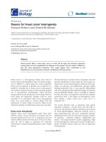
Fig 1 Sentinel node biopsy following neoadjuvant chemotherapy study design. ALN, axillary lymph nodes; FNA, fine-needle aspiration; NAC,
neoadjuvant chemotherapy; PD, progression of disease; SNB, sentinel node biopsy; ALND, axillary lymph node dissection
Yu et al. BMC Cancer (2016) 16:808
0.6 g/m2) NAC. If cancer progress was detected, NAC
was ceased and mastectomy and ALND were performed.
If ALN remained positive after NAC, mastectomy/
breast-conserving surgery and ALND were performed
within 2 weeks after NAC. For ALN-negative patients
after NAC, we performed mastectomy/breast-conserving
surgery with SLNB and ALND. All patients received
another two sessions of TEC chemotherapy after surgery. Post-surgery assistant therapy (local radiotherapy +
assistant endocrinotherapy/molecular targeted therapy)
was provided if necessary.
Evaluation of NAC efficacy
The tumor and lymph node responses to NAC were
evaluated via physical examination and imaging (mainly
ultrasound). The response of the primary tumor was
assessed using the Solid Tumors System, version 1.1 [8].
Post-NAC ALN-negative breast cancer was defined as
the lack of an enlarged lymph node detected on either
physical examination or imaging.
Surgical technique
A radioactive sulfur colloid tracer was not used;
therefore, a single tracer technique was employed for
all patients. Dye tracing was used for SLNB. SLN was
defined as a blue-stained lymph node or lymph node
directed by a blue-dyed lymph vessel. Any clinically suspicious or enlarged solid lymph node was also defined as
SLN even without blue staining. SLNs were separately
submitted for pathological examination after surgery.
After removing all SLNs, routine breast surgery and
complete level I and II ALN dissection were performed.
SLN pathology
All SLNs were paraffin embedded for hematoxylin-eosin
staining and IHC to assess metastasis. A tumor cell mass
larger than 2 mm in diameter was defined as macrometastasis or as positive. IHC was used to evaluate ER, PR
and Her-2 expression in primary tumors. When Her-2
showed 2+, FISH was performed for further evaluation.
Parameters
This study investigated mainly the post-NAC SLN identification rate (IR) and false negative rate (FNR) of breast
cancer patients with biopsy-proven ALN metastasis. The
following methods were applied to calculate the IR and
FNR respectively: IR (%) = cases with successful SLNB/
all cases with SLNB × 100 %; FNR (%) = false negative
cases/all cases with ALN metastasis × 100 %.
Statistical analysis
All statistical analyses in this study were performed by
the Department of Statistics, Second Military Medical
University. The χ2 test and Fisher's exact test were used
Page 3 of 7
to compare IR and FNR, with α < 0.05 indicating statistical significance. The χ2 test and fourfold table exact
test were used for univariate analysis. The statistics
software program used was SAS 9.3.
Results
General case information
Sixty-six operable breast cancer patients with initial
biopsy-proven ALN metastasis were enrolled in this
study. After four sessions of NAC, 48 patients (Group
A) became clinically ALN negative and underwent
mastectomy/breast-conserving surgery with SLNB and
ALND. Eighteen patients remained ALN positive after
NAC and underwent mastectomy/breast-conservation
surgery and ALND. Additionally, 56 operable breast
cancer patients with negative ALN proven by clinical
examination and biopsy were also included. One of them
presented with ALN progression after NAC and underwent
mastectomy and ALND. The other 55 patients (Group B)
underwent mastectomy/breast-conservation surgery with
SLNB and ALND (Fig. 1).
The average age of the patients in Group A was
50 years. Invasive ductal carcinoma and invasive lobular
carcinoma were confirmed in 44 and 4 cases, respectively. Nineteen cases were the luminal A and eight the
luminal B molecular subtypes. Ten cases were Her-2
positive. Eleven cases were triple-negative. Twelve
(12.5 %) of the 48 patients who completed NAC showed
a complete clinical response of the primary tumor.
Other general information is shown in Table 1.
In Group A, 46 of the 48 patients had at least one SLN
successfully dissected, with a total IR of 95.8 %. For the
other two cases, no blue-stained lymph vessel or lymph
node was observed or palpated during surgery. A total of
68 SLNs were dissected, with an average of 1.48 SLNs per
patient. Of the 46 patients, 32 (66.7 %), 8 (16.7 %), 4
(12.5 %) and 2 (4.2 %) had 1, 2, 3 or 4 dissected SLNs,
respectively. A total of 374 lymph nodes were dissected,
with an average of 15.6 lymph nodes per patient, as shown
in Table 2. Eight cases were SLN positive and non-sentinel
lymph node (NSLN) positive. Twenty-four patients were
SLN negative and NSLN negative. Eight were SLN negative but NSLN positive. Six were SLN positive but NSLN
negative. Fifty-four of the 55 patients in Group B had at
least one SLN dissected successfully, with a total IR of
98.2 %. Data on the lymph node status of these 54 cases
are shown in Table 3.
According to the data in Tables 2 and 3, post-NAC
SLN-positive patients comprised 30.4 % of all cases with
a detected SLN in Group A. Eight of the 46 cases had a
negative SLN and at least one metastatic NSLN confirmed pathologically after surgery. As a result, the FNR
was 36 % (8/(14 + 8)), with a 95 % CI of 17–59 %. Three
of the 54 patients in Group B had a negative SLN and at
Yu et al. BMC Cancer (2016) 16:808
Page 4 of 7
Table 1 Clinical characteristics of the patients in group A
Clinical characteristics
No. of cases
Total
48
Average age
50.2
%
Table 2 The status of axillary lymph node (ALN) after
neoadjuvant chemotherapy in group A
ALN
Non-sentinel node
Total
Positive
Negative
Sentinel node
Age at diagnosis
< 35 yr
18
37.5 %
Positive
8
6
14
≥ 35 yr
30
62.5 %
Negative
8
24
32
Total
16
30
46
BMI
≤ 25
38
79.2 %
> 25
10
20.8 %
≤ 5 cm
34
70.8 %
> 5 cm
14
29.2 %
Clinical tumor volume
Tumor location
Upper-outer quadrant
30
62.5 %
Lower-outer quadrant
6
12.5 %
Upper-inner quadrant
6
12.5 %
Lower-inner quadrant
4
8.3 %
Nipple area
2
4.2 %
Invasive ductal carcinoma
44
91.7 %
Invasive lobular carcinoma
4
8.3 %
Positive
16
33.3 %
Negative
32
66.7 %
Positive
34
70.8 %
Negative
14
29.2 %
Luminal A
19
39.6 %
Luminal B
8
16.7 %
HER2 positive
10
20.8 %
Triple negtive
11
22.9 %
Pathology
HER2 status
ER status
Molecular subtype
Tumor response to neoadjuvant chemotherapy
cPR
26
54.2 %
cCR
12
25 %
cSD
10
20.8 %
least one metastatic NSLN confirmed pathologically
after surgery, with an FNR of 10.7 % (3/(25 + 3)) and
95 % CI of 2–28 %. No significant correlations were
observed between clinical features and the FNR of postNAC SLNB for breast cancer with ALN metastasis
confirmed via biopsy (Table 4).
Discussion
Operable breast cancer patients with ALN metastasis
confirmed by biopsy may become ALN negative after
False negative rate 36.4 % (8/(14 + 8);95 % CI, 17–59 %),Overall accuracy
82.6 % ((14 + 24)/46;95 % CI,71–93 %),Sensitivity rate 64 % (14/(14 + 8);95 %
CI, 41–83 %)
NAC. For those patients, this study showed that the IR
of post-NAC SLNB could reach 95.8 %, which met the
recommended IR standard for SLNB by the ASCO
guidelines for early-stage breast cancer [9]. However, the
FNR of SLNB for patients in Group A was 36 %, much
higher than that recommended by ASCO for early-stage
breast cancer. For operable breast cancer cases indicated
as negative ALN by clinical examination and biopsy, the
IR and FNR of SLNB could reach 98.2 % and 10.7 %,
respectively, if the ALN remained clinically negative
after NAC. These results are consistent with the clinical
indications of SLNB.
We performed a literature review of studies using
post-NAC SLNB to detect ALN metastasis for clinically
positive breast cancer patients. As a result, we found
only six studies that included breast cancer patients with
ALN metastasis confirmed by biopsy [10–15]. As shown
in Table 5, the post-NAC IR ranged from 85.3–96 % and
the FNR of SLNB from 8–25 % in these studies. In five
previous studies, patients (including both ALN-positive
and -negative subjects) underwent SLNB after NAC.
The IR and FNR were calculated by summing the cases
in the above two groups. In all cases, 50 % of patients
still presented as ALN positive after NAC. An expanded
study sample without layering could result in discrepancies in the IR and FNR. Kim et al. limited their study
population to patients with negative ALN after NAC
[15]. Thirty-one of their 120 cases had a negative SLNB
result. Nevertheless, Kim et al. did not validate their
results by ALND. Kim et al. included only 89 cases (20
Table 3 The status of axillary lymph node (ALN) after
neoadjuvant chemotherapy in group B
ALN
Non-sentinel node
Positive
Total
Negative
Sentinel node
Positive
14
11
25
Negative
3
26
29
Total
17
37
54
False negative rate 10.7 % (3/(25 + 3);95 % CI, 2–28 %),Overall accuracy 94.4 %
((25 + 26)/54;95 % CI, 88–99 %),Sensitivity rate 89.3 % (25/(25 + 3);95 %
CI, 72–98 %)
Yu et al. BMC Cancer (2016) 16:808
Page 5 of 7
Table 4 False negative rate(FNR) of sentinel node biopsy
according to clinicopathological factors
Characteristics
ALN positive SN negative ALN
FNR
No. of cases positive No. of cases
Total
22
8
P
36.4 %
Age
0.402
< 50 yr
8
2
25 %
≥ 50 yr
14
6
42.9 %
BMI
0.531
Table 5 Studies of sentinel node biopsy after neoadjuvant
chemotherapy in patients with FNA proved node-positive
breast cancer
Authors
Time
No. of cases
Detection rate
FNR
Shen [7]
2007
69
92.8 %
25 %
Newman [8]
2007
54
98 %
8%
Yagata [9]
2013
95
85.3 %
15.7 %
Park [10]
2013
178
94.9 %
22 %
Boileau [11]
2015
153
87.6 %
8.4 %
2015
120
96 %
10 %
48
95.8 %
36 %
≤ 30
18
6
33.3 %
Kim [12]
> 30
4
2
50 %
Present study
Clinical tumor
volume
0.856
≤ 5 cm
16
6
37.5 %
> 5 cm
6
2
33.3 %
Tumor location
0.149
Upper-outer
quadrant
14
6
42.9 %
Others
8
2
25 %
Invasive ductal
carcinoma
22
8
36.4 %
Invasive lobular
carcinoma
0
0
Yes
8
2
25 %
No
14
6
42.9 %
Pathology
-
Vascular invasion
0.402
HER-2 status
0.07
Positive
6
4
66.7 %
Negative
16
4
25 %
Positive
14
4
28.6 %
Negative
8
4
50 %
ER status
0.315
Tumor response to
neoadjuvant
chemotherapy
0.145
CR
7
2
28.6 %
PR
13
4
30 %
ST
2
2
100 %
8
2
25 %
No. of SN
1
0.862
2
6
3
50 %
More than 3
8
3
37.5 %
SLN-negative cases validated by ALND and 69 SLNpositive cases) to calculate the FNR, which inevitably
resulted in a decreased FNR.
The IR of post-NAC SLNB in prior studies ranged
from 69–94.9 % [13, 16, 17], lower than that of SLNB
for early-stage breast cancer. Some researchers believe
that NAC could alleviate ALN lymphadenectasis but also
injure the axillary lymph vessels, resulting in lymphatic
obstruction. Therefore, tracer and dye usage could not
confirm the SLN [18, 19]. The IR of the SLN in our
study was higher, close to that for early-stage breast
cancer. We attribute this result to our patient selection
strategy. All patients included in our study showed an
ALN response after NAC. The tumor contained few
lymph vessels in these patients, which resulted in limited
injury of lymph vessels after NAC. From this point of
view, these patients were similar to early-stage breast
cancer patients. Thus, it was easier to detect the SLN
using a tracer. Meanwhile, we expanded the definition
of SLN from a blue-stained lymph node/lymph node directed by a blue-stained lymph vessel to any clinically
suspicious lymph node or any enlarged solid lymph
node detected during surgery even without blue staining. As a result, we increased the IR of the SLN to
95.8 %, a high value in studies.
The FNR in this study reached the highest value of
36 % compared with other studies. We believed that
three factors besides surgical skills could contribute to
the elevation of the FNR. First, this result could be associated with the sequence of the ALN response after
NAC. Approximately 20 % of patients with a tumor PCR
were reported to have a positive ALN, indicating that
the tumor CR and lymph CR were not in synchrony
[20]. The SLN and NSLN responses to chemotherapy
were also not in synchrony. Namely, the SLN and NSLN
could have different response sequences after NAC. If
the SLN shows PCR but the NSLN does not after NAC,
false negative results may result. Under these conditions,
the pathology of SLN cannot reflect the reality of the
ALN. Second, a high FNR of the SLNB could be related
to changes in the lymphatic drainage pathway caused by
NAC [18]. The SLN as well as the NSLN may not respond, but lymphatic drainage is altered after NAC. In
this setting, SLNB cannot reflect the actual state of the
ALN. Third, the high FNR could be associated with
patient selection. All patients enrolled were lymph node
negative after NAC. Therefore, our FNR was relatively
Yu et al. BMC Cancer (2016) 16:808
high compared with that found in studies including
post-NAC ALN-positive patients [10–14].
In addition, we investigated other factors such as
clinical and oncologic features to explain the high
FNR of 36.4 % in the present study. No clinicopathologic factors except for Her-2 expression tended to
influence the FNR of SLN. Positive results may have
been seen if the sample size was increased. We
reviewed the literature and found that different molecular subtypes of breast cancer seem to influence
the FNR. Yagata et al. reported that Her-2 expression
was the major determining factor of the FNR of the
SLN among all clinicopathologic factors [12]. Nevertheless, Park et al. stated that triple-negative breast
cancer patients had the lowest post-NAC FNR [13].
Molecular subtypes determine the NAC protocol for
breast cancer, but whether different protocols lead to
a more diverse FNR remains to be confirmed.
Many studies found that the number of lymph nodes
identified by SLNB had a strong correlation with the IR
and FNR of post-NAC SLNB. One study of SLN FNAC
reported an FNR of 18.2 % if one SLN was identified,
much higher than that of 4.9 % if two or more SLN
were detected [14]. A study by Boughey et al. for
lymph-node-positive breast cancer suggested that the
FNR of patients with three or more identified SLNs will
decrease compared with that of patients with two or
fewer identified SLNs (9.1 % vs 21.1 %) [4]. An NSABP
B-32 study found that for post-NAC breast cancer, the
FNR of SLNB decreased with an increasing number of
identified lymph nodes. The FNR was 18 % in patients
with one identified SLN, 10 % in patients with two
identified SLNs, and 7 % in patients with three identified SLNs [21]. Similarly, Hunt et al. reported that for
clinical ALN negative breast cancer, the FNR of postNAC SLNB was higher in patients in whom two or
fewer SLNs were identified [22]. As early as 2005,
Martin et al. validated that only a single identified SLN
could contribute to the elevation of the post-NAC FNR
[23]. In our study, the number of identified SLNs was
not significantly associated with the FNR, which may
have been due to the relatively small sample size in
each group. The average number of identified SLNs
was relatively small. This may be one reason to account
for the increase in FNR.
Patients were classified according to their response to
NAC in our study. Post-NAC ALN-positive patients
immediately underwent ALND, but the ALN-negative
patients underwent SLNB first. This method is more
clinically practical and can yield more accurate conclusions. Nevertheless, this was a single-center clinical
trial with fewer cases than those in other studies.
Multi-center trials with large sample sizes are necessary for a more reliable conclusion.
Page 6 of 7
Conclusions
In general, in this study, we determined whether postNAC SLNB was feasible in breast cancer patients with
ALN metastasis confirmed by biopsy. Due to the high
FNR, breast cancer patients with biopsy-proven ALN
metastasis are not recommended to undergo SLNB even
if their ALNs became clinically negative after NAC.
However, for ALN-negative patients confirmed via clinical examination and biopsy, SLNB is practical if their
ALNs remain negative after NAC.
Abbreviations
ALN: Axillary lymph node; ALND: Axillary lymph node dissection; FNA: Fine
needle aspiration; FNR: False negative rate; IR: Identification rate;
NAC: Neoadjuvant chemotherapy; NSLN: Non-sentinel lymph node;
PCR: Pathological complete response; SLN: Sentinel lymph node;
SLNB: Sentinel lymph node biopsy
Acknowledgements
None of the authors has any relationship with other individuals,
organizations, and companies that could inappropriately influence the work
reported in this study.
Funding
This original study was supported by Changhai hospital “1255” fund
(No.CH125540800).
Availability of data and materials
Our hospital is a military hospital for soldiers and civilians. In this present
study, there are more than 1/3 cases of military personnel. For reasons of
confidentiality, this part of the case data is not allowed to publish. So that
data will not be shared.
Authors’ contributions
Y.Y. and N.C. contributed equally to this work. Y.Y., N.C., and Y.S. participated
in the conception and design of the study. H.Y.L. and Y.M.W. participated in
article selection and data extraction and provided statistical expertise. L.X.
did the studies selection, data extraction, statistical analyses and the writing
of report. M.F. contributed to the literature search and figures. Y.Y., N.C., and
Y.S. participated in the critical revision of the manuscript and interpretation
of data. All authors revised the manuscript and approved the final version.
Authors’ information
Not applicable.
Competing interests
The authors declare that they have no competing interest.
Consent for publication
Not applicable.
Ethics approval and consent to participate
The study was conducted in accordance with local regulations and was
approved by the Ethics Committee of Second Military Medical University.
Participants gave consent to be a part of the study.
Endnotes
Not applicable.
Received: 5 November 2015 Accepted: 3 October 2016
References
1. Papa MZ, Zippel D, Kaufman B, Shimon-Paluch S, Yosepovich A,
Oberman B, et al. Timing of sentinel lymph node biopsy in patients
receiving neoadjuvant chemotherapy for breast cancer. J Surg Oncol.
2008;98(6):403–6.
Yu et al. BMC Cancer (2016) 16:808
2.
3.
4.
5.
6.
7.
8.
9.
10.
11.
12.
13.
14.
15.
16.
17.
18.
19.
20.
Kuehn T, Bauerfeind I, Fehm T, Fleige B, Hausschild M, Helms G, et al.
Sentinel-lymph-node biopsy in patients with breast cancer before and after
neoadjuvant chemotherapy (SENTINA): a prospective, multicentre cohort
study. Lancet Oncol. 2013;14:609–18.
Fu JF, Chen HL, Yang J, Yi CH, Zheng S. Feasibility and accuracy of sentinel
lymph node biopsy in clinically node-positive breast cancer after
neoadjuvant chemotherapy: a meta-analysis. PLoS One. 2014;9(9), e105316.
Pinero-Madrona A, Escudero-Barea MJ, Fernandez-Robayna F, Alberro-Adúriz
JA, García-Fernández A, Vicente-García F, et al. Selective sentinel lymph
node biopsy after neoadjuvant chemotherapy in breast cancer: results of
the GEICAM 2005–07 study[J]. Cir Esp. 2015;93:23–9.
Boughey JC, Suman VJ, Mittendorf EA, Ahrendt GM, Wilke LG, Taback B,
et al. Sentinel lymph node surgery after neoadjuvant chemotherapy in
patients with node-positive breast cancer: the ACOSOG Z1071 (Alliance)
clinical trial. JAMA. 2013;310:1455–61.
Hennessy BT, Hortobagyi GN, Rouzier R, Kuerer H, Sneige N, Buzdar AU,
et al. Outcome after pathologic complete eradication of cytologically
proven breast cancer axillary node metastases following primary
chemotherapy. J Clin Oncol. 2005;23:9304–11.
Dominici LS, Negron Gonzalez VM, Buzdar AU, Lucci A, Mittendorf EA,
Le-Petross HT, et al. Cytologically proven axillary lymph node
metastases are eradicated in patients receiving preoperative
chemotherapy with concurrent trastuzumab for HER2-positive breast
cancer. Cancer. 2010;116:2884–9.
Eisenhauer EA, Therasse P, Bogaerts J, Schwartz LH, Sargent D, Ford R, et al.
New response evaluation criteria in solid tumours: revised RECIST guideline
(version 1.1). Eur J Cancer. 2009;45:228–47.
Lyman GH, Giuliano AE, Somerfield MR, Benson 3rd AB, Bodurka DC,
Burstein HJ, et al. American Society of Clinical Oncology guideline
recommendations for sentinel lymph node biopsy in early-stage breast
cancer. J Clin Oncol. 2005;23:7703–20.
Shen J, Gilcrease MZ, Babiera GV, Ross MI, Meric-Bernstam F, Feig BW, et al.
Feasibility and accuracy of sentinel lymph node biopsy after preoperative
chemotherapy in breast cancer patients with documented axillary
metastases. Cancer. 2007;109:1255–63.
Newman EA, Sabel MS, Nees AV, Schott A, Diehl KM, Cimmino VM, et al.
Sentinel lymph node biopsy performed after neoadjuvant chemotherapy is
accurate in patients with documented node-positive breast cancer at
presentation. Ann Surg Oncol. 2007;14:2946–52.
Yagata H, Yamauchi H, Tsugawa K, Hayashi N, Yoshida A, Kajiura Y, et al.
Sentinel node biopsy after neoadjuvant chemotherapy in cytologically
proven node-positive breast cancer. Clin Breast Cancer. 2013;13:471–7.
Park S, Park JM, Cho JH, Park HS, Kim SI, Park BW, et al. Sentinel lymph
node biopsy after neoadjuvant chemotherapy in patients with
cytologically proven node-positive breast cancer at diagnosis. Ann Surg
Oncol. 2013;20:2858–65.
Boileau JF, Poirier B, Basik M, Holloway CM, Gaboury L, Sideris L, et al.
Sentinel node biopsy after neoadjuvant chemotherapy in biopsy-proven
node-positive breast cancer: the SN FNAC study. J Clin Oncol.
2015;33:258–64.
Kim JY, Kim MK, Lee JE, Jung Y, Bae SY, Lee SK, et al. Sentinel lymph node
biopsy alone after neoadjuvant chemotherapy in patients with initial
cytology-proven axillary node metastasis. J Breast Cancer. 2015;18:22–8.
Jones JL, Zabicki K, Christian RL, et al. A comparison of sentinel node biopsy
before and after neoadjuvant chemotherapy: timing is important. Am J
Surg. 2005;190:517–20.
Takei H, Yoshida T, Kurosumi M, Gadd MA, Hughes KS, Lesnikoski BA, et al.
Sentinel lymph node biopsy after neoadjuvant chemotherapy predicts
pathological axillary lymph node status in breast cancer patients with
clinically positive axillary lymph nodes at presentation. Int J Clin Oncol.
2013;18:547–53.
Brown AS, Hunt KK, Shen J, Babiera GV, Ross MI, Meric-Bernstam F, et al.
Histologic changes associated with false-negative sentinel lymph nodes
after preoperative chemotherapy in patients with confirmed lymph nodepositive breast cancer before treatment. Cancer. 2010;116:2878–83.
Charfare H, Limongelli S, Purushotham AD. Neoadjuvant chemotherapy in
breast cancer. Br J Surg. 2005;92:14–23.
Mamounas EP, Brown A, Anderson S, Julian T, Miller B, Bear HD, et al.
Sentinel node biopsy after neoadjuvant chemotherapy in breast cancer:
results from National Surgical Adjuvant Breast and Bowel Project Protocol B27. J Clin Oncol. 2005;23:2694–702.
Page 7 of 7
21. Krag DN, Anderson SJ, Julian TB, Brown AM, Harlow SP, Ashikaga T, et al.
Technical outcomes of sentinel-lymph-node resection and conventional
axillary-lymph-node dissection in patients with clinically node-negative
breast cancer: results from the NSABP B-32 randomised phase III trial. Lancet
Oncol. 2007;8:881–8.
22. Hunt KK, Yi M, Mittendorf EA, Guerrero C, Babiera GV, Bedrosian I, et al.
Sentinel lymph node surgery after neoadjuvant chemotherapy is accurate
and reduces the need for axillary dissection in breast cancer patients. Ann
Surg. 2009;250:558–66.
23. Martin 2nd RC, Chagpar A, Scoggins CR, Edwards MJ, Hagendoorn L,
Stromberg AJ, et al. Clinicopathologic factors associated with false-negative
sentinel lymph-node biopsy in breast cancer. Ann Surg. 2005;241:1005–12.
discussion 12–5.
Submit your next manuscript to BioMed Central
and we will help you at every step:
• We accept pre-submission inquiries
• Our selector tool helps you to find the most relevant journal
• We provide round the clock customer support
• Convenient online submission
• Thorough peer review
• Inclusion in PubMed and all major indexing services
• Maximum visibility for your research
Submit your manuscript at
www.biomedcentral.com/submit