Oral perfluorooctane sulfonate (PFOS) lessens tumor development in the APCmin mouse model of spontaneous familial adenomatous polyposis
Bạn đang xem bản rút gọn của tài liệu. Xem và tải ngay bản đầy đủ của tài liệu tại đây (667.38 KB, 10 trang )
Wimsatt et al. BMC Cancer (2016) 16:942
DOI 10.1186/s12885-016-2861-5
RESEARCH ARTICLE
Open Access
Oral perfluorooctane sulfonate (PFOS)
lessens tumor development in the APCmin
mouse model of spontaneous familial
adenomatous polyposis
Jeffrey Wimsatt1,2,5* , Meghan Villers1, Laurel Thomas1, Stacey Kamarec1, Caitlin Montgomery1, Leo W. Y. Yeung3,
Yanqing Hu4 and Kim Innes2
Abstract
Background: Colorectal cancer is the second most common cause of cancer deaths for both men and women, and
the third most common cause of cancer in the U.S. Toxicity of current chemotherapeutic agents for colorectal cancer,
and emergence of drug resistance underscore the need to develop new, potentially less toxic alternatives. Our recent
cross-sectional study in a large Appalachian population, showed a strong, inverse, dose–response association of serum
perfluorooctane sulfonate (PFOS) levels to prevalent colorectal cancer, suggesting PFOS may have therapeutic potential
in the prevention and/or treatment of colorectal cancer. In these preliminary studies using a mouse model of familial
colorectal cancer, the APCmin mouse, and exposures comparable to those reported in human populations, we assess
the efficacy of PFOS for reducing tumor burden, and evaluate potential dose–response effects.
Methods: At 5–6 weeks of age, APCmin mice were randomized to receive 0, 20, 250 mg PFOS/kg (females) or 0, 10, 50
and 200 mg PFOS/kg (males) via their drinking water. At 15 weeks of age, gastrointestinal tumors were counted and
scored and blood PFOS levels measured.
Results: PFOS exposure was associated with a significant, dose–response reduction in total tumor number in both
male and female mice. This inverse dose–response effect of PFOS exposure was particularly pronounced for larger
tumors (r2 for linear trend = 0.44 for males, p’s <0.001).
Conclusions: The current study in a mouse model of familial adenomatous polyposis offers the first experimental
evidence that chronic exposure to PFOS in drinking water can reduce formation of gastrointestinal tumors, and that
these reductions are both significant and dose-dependent. If confirmed in further studies, these promising findings
could lead to new therapeutic strategies for familial colorectal cancer, and suggest that PFOS testing in both
preventive and therapeutic models for human colorectal cancer is warranted.
Keywords: APCmin mouse, Perfluorooctane sulfonate, PFOS, Colorectal cancer, Dose–response, Gender
* Correspondence:
1
Department of Medicine, School of Medicine, West Virginia University,
Morgantown, WV 26506, USA
2
Department of Epidemiology, School of Public Health, West Virginia
University, Morgantown, WV 26506, USA
Full list of author information is available at the end of the article
© The Author(s). 2016 Open Access This article is distributed under the terms of the Creative Commons Attribution 4.0
International License ( which permits unrestricted use, distribution, and
reproduction in any medium, provided you give appropriate credit to the original author(s) and the source, provide a link to
the Creative Commons license, and indicate if changes were made. The Creative Commons Public Domain Dedication waiver
( applies to the data made available in this article, unless otherwise stated.
Wimsatt et al. BMC Cancer (2016) 16:942
Background
Colorectal cancer (colorectal cancer) is the second most
common cause of cancer deaths in both men and
women, and the third most common cause of cancer in
the US [1]. Toxicity of current chemotherapeutic agents
for colorectal cancer, and ongoing challenges with drug
resistance suggest that new drug approaches continue to
have value [2, 3].
Perfluoroalkyls and polyfluoroalkyls have been manufactured for over five decades; their unique-oilrepellence and high surface activity make them
excellent surface protectants and surfactants. Some of
these compounds are potent peroxisome proliferatoractivated receptor (PPAR) ligands, and have demonstrated anti-inflammatory effects in vitro [4] and in
animal studies [5]; these effects are thought to operate
via both PPAR-dependent and independent pathways
[6]. Perfluorooctane sulfonate, (PFOS, C8HF17O3S), a
well-studied perfluoro-surfactant, is a widespread environmental contaminant, has been detected in the plasma
of virtually every human population worldwide [7–9].
PFOS is extremely stable in the environment, readily
accumulates in people and animals, and has toxic properties; as a result several countries have voluntarily
joined the Stockholm Convention to stop its use [10].
Lifetime exposure studies in rodents suggest PFOS can
cause liver adenomas, whereas the evidence for cancer
induction in humans remains equivocal, perhaps in part
because most exposures levels are so low [11]. Hence, if
shown of benefit, particularly at low doses, PFOS could
suggest a novel mechanism for treating colorectal
cancer.
Recent research suggests that PFOS may also have
value as a chemopreventive and/or chemotherapeutic
agent for colorectal cancer. In a cross-sectional study in
a large Ohio Valley cohort (the C8 Health Project), we
investigated the potential link between prevalent colorectal cancer and serum PFOS [12]. PFOS levels in this
population were similar to those reported in the general
U.S. population [12, 13], were comparable to or lower
than those reported from non-occupational settings in
other countries (e.g. ≤ 30 ng/ml) [14, 15], and were well
below levels reported in fluorochemical workers [16].
We found a strong, inverse, dose–response association
between serum levels of PFOS and prevalent colorectal
cancer that remained robust after adjustment for multiple possible confounders and persisted even at very
low exposure levels [12]. However, while these findings
suggest that PFOS may be protective against colorectal
cancer, the cross-sectional nature of the data preclude
determination of causality.
Here the potential chemotherapeutic value of PFOS is
tested in APCmin mice, a genetic model for familial adenomatous polyposis) in humans [15, 16].
Page 2 of 10
Methods
Chemicals and reagents
For animal studies, potassium salt of PFOS (Sigma
#77282; heptadecafluorooctanesulfonic acid potassium
salt) was purchased and dissolved in Millipore® water containing 0.5% Tween 20 (Sigma #P2287). Bottles were
made fresh weekly by addition from a stock solution. For
analytical purposes, perfluorohexanoate, perfluoroheptanoate, perfluorooctanoate, potassium salts of perfluorohexanesulfonate, perfluorooctanesulfonate, and 13C4
PFOS were obtained from Wellington Laboratories
(Guelph, Ontario, Canada). The purity of all standards
was over 98%. Tetrabutylammonium hydrogen sulfate
(99%), ammonium acetate (>99%), and ammonia (NH3,
30%) were obtained from Sigma-Aldrich. LCMS grade
methanol and methyl-tert-butyl ether (MTBE, > 99%)
were acquired from EMD Chemicals Inc. (Mississauga,
ON). Oasis® weak anion exchange (WAX; 6 cm3, 150 mg,
30 μm) solid phase extraction (SPE) cartridges were purchased from Waters (Milford, MA).
All studies were approved by the Institutional Animal
Care and Use Committee at West Virginia University.
Animals were housed individually in standard ventilated
barrier caging and fed standard mouse chow, and maintained on a 12 : 12 (L : D) hour light cycle. In two separate studies, female and male APCmin mice (C57BL/6 JApcMin/J) were acquired from JAX at 6 and 5 weeks respectively, acclimated for 1 week, and randomized by
treatment group. Animals in each group received
Tween-20 vehicle or PFOS dissolved in Tween-20 in
their drinking water. In an initial pilot study, female
mice (n = 8/group) were exposed to 0.5% Tween-20
vehicle or Tween-20 with 20, or 250 mg/kg PFOS target doses in their drinking water from 7–15 weeks of
age based on estimated daily water consumption.
Similarly, in the second study, male mice were exposed to vehicle or PFOS target doses of 10, 50 and
200 mg/kg (all groups, n = 6) provided at 6–15 weeks
of age. Animals were weighed twice weekly throughout the study period.
In both studies, animals were humanely euthanized with
CO2, and the complete gastrointestinal tract from the
stomach to the rectum was opened lengthwise and tumors
were counted and categorized using direct visualization
under 3 times magnification. Tumors were recognized by
their characteristic gross morphology and categorized by
location (small intestine, large intestine, cecum), size
(using the average surface dimensions, they were scored
as < 1 mm or ≥ 1 mm), and if bleeding or not.
Blood sampling
In the male study, at 15 weeks of age, cardiac blood
samples were collected under inhalant anesthesia into
EDTA powdered tubes and the plasma collected and
Wimsatt et al. BMC Cancer (2016) 16:942
stored at −80 °C until assayed. Plasma samples were
shipped on ice to the University of Toronto, Department
of Chemistry for PFOS measurement.
PFOS assay
Sample extractions
Mouse plasma samples were extracted using an ion-pair
extraction method [17, 18]. Before extraction, mouse
plasma samples were diluted with Milli-Q water (i.e., 10fold for control group and 1000 to 10000-fold for treatment groups). In brief, in a 15 mL polypropylene tube,
1 mL of TBAS solution (adjusted to pH 10 using 30%
aqueous NH3) was added to 1 mL of the diluted plasma
sample; after the mixture was vortex-mixed for 30 s,
5 mL of MTBE was added and was shaken on a horizontal shaker at 250 RPM for 20 min; then the organic and
aqueous layers were separated by centrifugation at 6000
RPM for 10 min. The organic layer was decanted to a
new tube. The sample was then extracted with another
5 mL aliquot of MTBE and the entire extraction procedure repeated. The MTBE aliquots were combined, evaporated to dryness under a gentle stream of nitrogen, and
reconstituted in 1 mL of methanol for analysis.
Drinking water stock solution with 0.5% Tween-20 was
extracted using a SPE-WAX cartridge [19]. The cartridge
was first conditioned by passing a series of 4 mL of 0.1%
NH4OH in methanol, 4 mL of methanol, and 4 mL of
Milli-Q water; after that, 0.5 mL of the mouse drinking
water stock was loaded onto the cartridge. After loading
the sample, the cartridge was washed with 4 mL of
25 mM ammonium acetate and dried under vacuum.
Target fraction was eluted with 4 mL 0.1% NH4OH in
methanol and evaporated to dryness, and then reconstituted in 0.1 mL of methanol for analysis.
Instrumental analysis
Apart from PFOS, a suite of target Per- and polyfluorinated alkyl substances, C6-C8 perfluorinated carboxylic acids and perfluorohexane sulfonate were analyzed using an Acquity UPLC (Waters Corporation) and
a API 4000 MS/MS (Applied Biosystem/MDS Sciex); an
atmospheric electrospray interface operated in negative
ionization mode was used. Chromatographic separation
was performed on a Kinetex XB-C18 column (50 ×
4.6 mm, 2.6 um 100A), the column temperature was
kept at 40 °C, and 10 mM ammonium acetate in both
Milli-Q water and methanol were the mobile phases.
An internal calibration method using mass-labelled
standard was used to quantify PFOS. The calibration
curve was constructed with standard concentrations ranging from 0.5, 1.0, 2.5, 5.0, 10.0, 20.0, 50.0, and 100.0 ng/
mL. Standard deviations at each data point were < 20%,
with an r2 > 0.99 for the calibration curve. The PFOS
standard used in the present study was the linear isomer,
Page 3 of 10
while samples contained both branched and linear isomers; the concentrations reported for the present study
included both linear and branched isomers, and were estimated based on the linear isomer standard. The limit
of quantification (0.5 ng/mL) was evaluated based on the
lowest concentration of standard on the calibration
curve that could be accurately measured within ± 20% of
its theoretical value and a signal-to-noise ratio ≥ 10.
Quality assurance and quality control
Three procedural blanks (Milli-Q water) were run for
every twelve sera samples to check for possible interference. Matrix recoveries (n = 3) using control mouse sera
were performed prior to real sample analysis to ensure
the reliability of the method. All target chemicals were
spiked (10 ng) into the control samples, and the samples
were extracted and analyzed following the same procedures as described above; matrix recoveries ranged 87–
117% (supporting information, SI). Samples were analyzed in duplicate and the variability of the analysis was
less than 10% as evaluated using 13C4-PFOS recoveries,
which likewise ranged from 88–110%. In response to coeluting interferences at PFOS transition 499 > 80, the
499 > 99 transition was used for quantification. Recoveries (spiked level: 0.5 ng) for water samples ranged 90–
107% (SI) using the same method. A quality control
standard (10 ng/mL) was injected every ten samples to
evaluate intensity change of the MS; samples were reanalyzed if the intensity of the standard varied ± 20% or
more compared to those of the previous one.
Statistical analyses
Animals from the 2 studies represented different total
doses, exposure periods and genders, so each study was
analyzed separately. Both datasets were normality tested
to assure parametric testing was appropriate. Initial
ANOVA analysis was performed looking for treatment
effects. Based on these results, summary statistics were
calculated (means and standard errors), and assuming
unequal variances, pairwise comparisons of treatment
groups were made using two-sample T tests corrected
using Tukey’s criterion for multiple comparisons. Cecum
masses were not included in analyses due to low tumor
numbers. Slopes of mean body weights through time
from the males exposed to vehicle or 200 mg/kg PFOS
from 12–15 weeks were tested to determine if body
weight trajectories are significantly different between the
two groups.
Results
Of the original 24 female APCmin mice, one from the
20 mg/kg dose group developed breast cancer, and 4
from the 250 mg/kg dose group lost > 10% body weight,
so they were lost to follow-up before 15 weeks. Findings
Wimsatt et al. BMC Cancer (2016) 16:942
reported for this pilot study, illustrated in Fig. 1a, are
based on the remaining animals. Initially, ANOVA analysis using treatment as the independent variable and
total tumor number as the dependent variable, revealed
p-values of 0.010 and 0.019 for females and males respectively. At 15 weeks in females, total tumor numbers
in both treatment groups averaged significantly lower
than those in the control group (p = 0.02 and 0.009 for
20 mg/kg and 250 mg/kg, respectively). Likewise, animals in the 250 mg/kg group averaged significantly
fewer tumors than in the 20 mg/kg group (p = 0.04).
Collectively, these findings indicate increasing tumor reductions with rising PFOS dose.
Findings of the study in male mice are shown in
Fig. 1b, and suggest a similar dose–response relationship
overall.
Again, total tumor load in all treatment groups averaged
lower than that in the control group at 15 weeks, with the
largest effect in the highest dose group (p’s = 0.051, 0.068,
and 0.003 for vehicle vs. 10, 50, and 200 mg/kg, respectively). Likewise, while response in the lowest PFOS dose
group did not differ significantly from that in the moderate dose group (p > 0.1), total tumor count in animals receiving PFOS doses of 200 mg/kg was significantly
reduced relative to both the 10 mg/kg (p = 0.006) and
50 mg/kg dose groups (p = 0.02), again suggesting greater
tumor reduction at higher PFOS doses.
As indicated in Fig. 1c, the inverse, dose–response effect of PFOS exposure was particularly pronounced for
tumors ≥ 1 mm in size. For males, PFOS showed a
strong inverse linear association to large tumor numbers
Page 4 of 10
(r2 = 0.44) with a p < 0.001. For tumors < 1 mm, this association did not hold (p = 0.76). In both studies, bleeding tumors were rare, appeared to be evenly distributed
across dose groups, and their occurrence appeared unrelated to either tumor size or location.
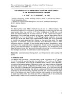
Figure 2 shows the effect of PFOS dose on body
weight in males. Weight gain decreased with dose in the
final weeks of the study, with the between group differences increasingly prominent after 11 weeks. As expected, weight gain reductions were particularly
pronounced in the 200 mg/kg group, with the slope of
body weight change in this group becoming negative by
12 weeks, with body weight trending lower than for the
vehicle controls from 12–15 weeks (p = 0.06). Weight
loss in this high dose group late in the study likely reflects a developing toxic effect of PFOS, and is consistent with the weight loss observed at higher doses in the
female study. As illustrated in Fig. 3, plasma PFOS levels
at 15 weeks of age indicate that PFOS accumulation increased with dose, including estimated levels of both linear and branched isoforms.
Discussion
In these two preliminary studies of male and female
APCmin mice, total tumor number decreased significantly with increasing PFOS dose, with the highest dose
groups showing the largest effects. The observed dose–
response relationship was particularly evident in larger
tumors, suggesting a possible inhibitory effect of PFOS
on tumor formation. Since PFOS administration in our
mouse model was initiated prior to tumor development,
Fig. 1 a-c Fig. 1a shows the number of tumors (mean and s. e.: Total, SI-small intestine, LI-large intestine) counted at 15 weeks in female APCmin
mice exposed to varying target doses from 7–15 weeks of age, and receiving up to 250 mg/kg PFOS in their drinking water. Controls received
0.5% Tween-20 vehicle only. Figure 1b shows the number of tumors counted by region (mean and s. e.: Total, SI-small intestine, LI-large intestine)
in male APCmin mice exposed to target doses of up to 200 mg/kg PFOS in their drinking water. Figure 1c. The number of large tumors 1–3 mm
in diameter plotted against PFOS is depicted. These results suggest PFOS may cause tumor regression, and not just prevent tumor development
(mean and s. e.: Total, SI-small intestine, LI-large intestine). For the female study, animal numbers were 7, 8, and 4 for vehicle, 20, and 250 mg/kg
dose groups respectively. For the male study, animal numbers were n = 6 for each group. Values represent total tumors counted in the vehicle
controls as compared to each treatment group
Wimsatt et al. BMC Cancer (2016) 16:942
Page 5 of 10
Fig. 2 Shown are plasma PFOS levels (mean and s. e.) from male APCmin mice at 15 weeks of age by dose administered. Total linear and
branched PFOS levels are shown. As expected, levels increased with PFOS dose
the observed reduction in tumor burden may reflect effects on both tumor initiation and tumor progression.
Notably, the number of 1–3 mm tumors decreased significantly with increasing PFOS dose (Fig. 1c), suggesting
that PFOS may not only inhibit development, but may
halt progression and even possibly induce tumor regression. If PFOS induces tumor regression, reduction of larger tumors may lead to a corresponding increase in the
number of tumors < 1 mm diameter, thus potentially attenuating the observed effect of PFOS on total tumor
number and helping to explain the stronger effects observed for larger tumors. Collectively, these findings suggest that PFOS has a significant, dose-dependent
inhibitory effect on gastrointestinal tumor formation in
this established genetic mouse model of familial adenomatous polyposis.
Results of these preliminary experimental studies are
broadly consistent with findings from our recent epidemiological investigation in a large population of Appalachian adults exposed to PFOA-contaminated drinking
water. In this cross-sectional study, serum PFOS levels
showed a strong, inverse dose–response association with
prevalent colorectal cancer that remained robust after adjustment for multiple potential confounders [12]. However, while findings of this epidemiological investigation
likewise suggest a possible protective effect of PFOS on
colorectal cancer, the cross-sectional nature of the data
limit causal inference. Although implications for nonfamilial colorectal cancer remain unclear, the current
study in a mouse model of familial adenomatous polyposis
offers the first experimental evidence that chronic exposure to PFOS in drinking water can reduce formation of
gastrointestinal tumors, and that these reductions are both
significant and dose-dependent.
In our present animal study, we provided PFOS in the
drinking water to simulate chronic human PFOS exposure
[20, 21]. Liver enzymes can be induced by PFOS exposure
in mice [22], and toxicity indicated by weight loss [23] was
observed here. In other studies, higher PFOS doses have
been administered over shorter periods by oral bolus [5, 24]
without evident toxic effects; however, our data suggest toxicity likely develops at a lower overall dose when PFOS is
delivered slowly over time [5, 24]. Here progressive weight
loss was observed at doses of 200 mg/kg or higher, indicating this dose is near the maximum tolerated dose for this
mouse strain and delivery method over this time frame.
Fortunately, measurement of plasma PFOS levels in male
mice at 15 weeks indicated that drinking water administration at all doses resulted in plasma levels substantially
higher than those associated colorectal cancer reduction in
humans [12]. In common, PFOS appeared beneficial in human colorectal cancer and in APCmin mice. However, the
Wimsatt et al. BMC Cancer (2016) 16:942
Page 6 of 10
Fig. 3 Shown is the average body weight (mean and s. e.) by dose group from 6–15 weeks of age weeks in male mice. As can be seen, body
weight appeared to slow in proportion to PFOS dose. At approximately 12 weeks of age, the 200 mg/kg group stopped exhibiting weight
increases altogether. Table 1 depicts the gender, dose groups, animal numbers and duration of exposure for the animals completing each study
former effect was observed in humans which metabolize
PFOS differently from rodents, and where the effect was
substantially based on the acquired non-familial form of
colorectal cancer. In the mouse model, PFOS undergoes a
greater degree of metabolism, has a shorter half-life, and
counters a genetic predisposition to colorectal cancer. Further studies should investigate the adverse consequences of
this agent under therapeutic conditions; even so, this study
provides a possible direction to pursue in regard to familial
colorectal cancer.
Although PFOS is widely distributed in the environment [25, 26] and has been detected in human populations worldwide [9, 27–30], non-occupational blood
levels in humans are well below those reported toxic in
lab animals [31–33]. The half-life of PFOS is reported to
be < 40 days in mice [34], and contrasts dramatically
with the estimated 4–5 year half-life documented in
humans [35]. It appears that PFOS in rodents is handled
in a manner similar to fatty acids, and consequently induces hormonal, peroxisomal and P450 enzyme gene activation [36]. The increased PFOS half–life in humans
compared to rodents may be in part due to PFOS inhibition of human cytochrome activity [37]; cytochrome activity inhibition could also reduce the influence of toxic
metabolites which may explain higher degrees of toxicity
as commonly reported in rodents. In addition, PFOS
renal reabsorption and recycling have also been shown
to contribute to a long half-life in humans and monkeys
[38]. Cancer risk with prolonged chronic exposure was
suggested at high doses in rats [11], although consistent
evidence for elevated tumor risk with PFOS exposure in
humans is lacking [11].
Each PFAS has a unique biological and toxicological
profile that limits extrapolation across compounds or
Table 1 Depicts the gender, dose groups, animal numbers and duration of exposure for the animals completing each study
Trial
Dose Groups in mg/kg
(N)
Age at First Exposure
Treatment Duration
Age at Tumor Counts
Females
0 (8), 20 (7a), 250 (4b)
7 weeks
8 weeks
15 weeks
Males
0 (6), 10 (6), 50 (6), 200 (6)
6 weeks
9 weeks
15 weeks
One animal developed breast cancer and was dropped from study; bfour animals had > 10% weight loss before 15 weeks and were lost to follow-up. “0” dose
animals received vehicle only
a
Wimsatt et al. BMC Cancer (2016) 16:942
model species [11]. Potential mechanisms of PFOS action relevant to its effect on tumor development and
progression are still ill-defined, but are not surprising
given the large number of genes (e.g. ~400 in rats) PFOS
appears to influence [36]. Possibilities include antiinflammatory effects via prostanoid pathways, PPAR receptor mediated actions, immune effects, or other as yet
unrecognized mechanisms. PFOS may serve an antiinflammatory role via its influence on downstream transcriptional regulators such as NF-κB [6]. Phospholipase
A2 is inhibited by PFOS in rats; this could, in turn, block
the production of arachidonic acid as a substrate for
prostaglandindin H synthase elaboration of prostanoids
[36]. Similarly, PGE2 has been shown to be a potent inducer of adenoma formation in APCmin mice [39], and
tumor growth [40] was similarly increased by an agonist,
where both were mediated through the PPARδ receptor.
Adenoma formation by PGE2 was removed in mice
missing this receptor [41]. PFOS also significantly stimulates both PPARα and PPARγ [42], which could also
modulate tumor growth [43, 44]. PFOS serves as a partial agonist and induces PPARα mediated effects at high
doses [45], other effects via other PPAR receptor isotypes [46], and produces significant immunomodulatory
effects in mice [47]. In PPARα KO mice, PPARα independent nuclear receptor mediated pathways and downstream effects were noted [6], including suppression of
T-cell dependent antibody production, and modulation
of immune cell and cytokine synthesis (e.g. TNFα and
IL-6) [6]. While PFOS appears to stimulate mouse and
human PPAR receptors [42, 46] when screened in cell
lines, robust in vivo evidence for direct PPAR receptor
mediation is lacking, and human PPARα expression is
considerably reduced compared to in rodents; if so, this
may thus lessen the importance of this pathway in human familial adenomatous polyposis or acquired colorectal cancer [6]. Even so, PPARα stimulation in human
colorectal cancer lines is moderately pro-inflammatory
and stimulates prostaglandin H synthase-2 expression
[48, 49]. The potential impact of PFOS directly on the
Wnt-β-catenin signal transduction pathway also warrants
closer examination [50]. The “min” defect causes catenin
retention and ultimately the Wnt gene group to become
canonically activated; gastrointestinal polyp formation is
one direct consequence [51]. The putative role of PFOS in
blocking this process would be a plausible mechanism to
explain its efficacy in this model system. Alternative
pathways could also be affected [52]. Other influences of
PFOS, by interacting with dietary constituents [53, 54],
or steroidogenic enzyme and hormone disruptive
effects cannot be ruled in or out [55]. Here, both
mouse genders benefited from PFOS exposure, arguing
against a differential role as pertains to specific sex
hormones.
Page 7 of 10
In recent human cross-sectional studies, chronic PFOS
exposure has been associated with modest, adverse
changes in serum lipid profiles [56–58]. Similarly, elevated
blood levels of PFOS have been associated with increased
likelihood of early onset menopause [59] and altered thyroid function [59]. However, in contrast to findings from
animal studies, including our study in APCmin mice, significant adverse effects of PFOS exposure have been rarely
been documented in humans, even in pregnant women
and highly exposed fluorochemical plant workers [16, 58,
60, 61]. Even with its complex disposition in humans,
PFOS appears well tolerated at environmental levels (e.g.
Danish National Birth Cohort Study [62]: PFOS mean
35.3 ng/ml; range 6.4–106.7 ng/ml; elsewhere 0–30 ng/ml
[15, 63–65],) levels also associated with colorectal cancer
protection [66]. Occupational exposures in chemical factory workers are up to 40 times higher, yet adverse outcomes at these levels, even among at risk pregnant
women, are rare [16, 60]. At high occupational exposure
levels, an association between PFOS and bladder cancer
was reported [65]; however, this association was questioned more recently [16, 58, 61]. Therefore, although
PFOS has been decried as an environmental contaminant,
it might still have therapeutic value at low levels. If shown
to be effective for colorectal cancer prevention and/or
treatment in humans, PFOS may offer an option that is
significantly safer, lower cost, and less toxic than alternative therapies [67].
One potential limitation relates to reverse causality,
i.e., the possibility that tumor formation may reduce
PFOS absorption, and thus, blood PFOS levels. However,
this is unlikely given the massive surface area of the
gastrointestinal tract and the inherent lipid solubility of
PFOS. Moreover, serum PFOS generally correlates well
with liver concentrations [61], suggesting that serum is a
reasonably good systemic indicator of PFOS exposure in
humans [68]. Similarly in a mouse model of familial adenomatous polyposis, blood levels corresponded to target dosing levels and predicted tumor reduction, again
indicating that PFOS absorption reflects oral exposure.
Another theoretical argument is that a parent compound
is broken down to make PFOS, and it may be this compound, rather than PFOS itself that led to the reduction
in colorectal cancer observed in our previous epidemiological study [12]. However, PFOS exposure had a clear
benefit here.
Strengths of this study include identifying a beneficial
role for PFOS as a chemo-preventive or therapy in a familial model of colorectal cancer, irrespective of gender.
In addition, the development of a slow delivery method
and estimates of an effective working dose range by this
delivery method were determined. Finally, toxic effects
were easily identified using simple body weight monitoring. Limitations include the absence of more dose
Wimsatt et al. BMC Cancer (2016) 16:942
groups to better define the dose–response relationship,
with the eventual goal of developing a reliable PFOS
therapeutic profile.
While effective against this animal model of familial
adenomatous polyposis, PFOS efficacy in the more common acquired form of colorectal cancer is another important focus for future investigations. Planned studies
will seek to determine a mechanism, such as might be
derived from isolated cell cultures, an optimal dose in
vivo using xenograft models. A mechanistic understanding of PFOS action, from the impetus provided here,
may lead to successful new colorectal cancer treatment
approaches.
Conclusions
Using APCmin mice of both genders, we show that
perfluorooctane sulfonate (PFOS) reduces gastrointestinal tumor burden in this well-established general model of human colorectal cancer, and also a
specific model for familial adenomatous polyposis in
a dose-related manner. Our results represent a proof
of concept based on our previously published epidemiological study that found a protective dose–response relationship between PFOS levels and
reduced likelihood of colorectal cancer from a human population.
Abbreviations
ANOVA: Analysis of variance; C57BL/6 J-ApcMin/J mice: APCmin mice;
LCMS: Liquid chromatography mass spectrometry; MTBE: Methyl-tert-butyl
ether; PFOA: Perfluorooctanoic acid; PFOS, C8HF17O3S: Perfluorooctane
sulfonate; PPAR: Peroxisome proliferator-activated receptor; SPE: Solid phase
extraction; WAX: Oasis® weak anion exchange;
Acknowledgements
The Authors would like to thank A. Forrisi and Dr. V. Rajendran for technical
assistance and Dr. Patrick Callery for reading and commenting on the
analytical methods.
Funding
NIH grant to JW (U54GM104942).
Authors’ contributions
JW provided the conceptual framework, paper writing, and sought and
provided funding support. MV, LT, KK, and CM collected data, and managed
the project day to day. LY-performed assay validations and assayed samples.
YH provided power analyses and statistical expertise. KI provided critical
background leading to the discovery and it relevance to human cancer. All
authors have read and approved the manuscript as written.
Competing interests
Coi certifications are on file with WVU. The authors declare that they have
no competing interests.
Consent for publication
All authors have consented to publish the present manuscript s presented
here.
Ethics approval
IACUC approval from West Virginia University was secured to cover this
animal work.
Page 8 of 10
Primary data
Cannot be posted publicly due to proprietary limitations and patent
infringement-“Not Applicable”.
Author details
1
Department of Medicine, School of Medicine, West Virginia University,
Morgantown, WV 26506, USA. 2Department of Epidemiology, School of
Public Health, West Virginia University, Morgantown, WV 26506, USA.
3
Man-Technology-Environment (MTM) Research Centre, School of Science
and Technology, Örebro University, Fakultetsgatan 1, Örebro SE-70182,
Sweden. 4Department of Statistics, West Virginia University, Morgantown, WV
26506, USA. 5West Virginia University, 186 HSCN, 1 Medical Center Drive,
Morgantown, WV 26508, USA.
Received: 17 February 2016 Accepted: 11 October 2016
References
1. Colorectal (Colon) Cancer Statistics [ />statistics/index.htm]. Accessed 2 Nov 2016.
2. Sartore-Bianchi A, Bencardino K, Cassingena A, Venturini F, Funaioli C, Cipani
T, Amatu A, Pietrogiovanna L, Schiavo R, Di Nicolantonio F, et al.
Therapeutic implications of resistance to molecular therapies in metastatic
colorectal cancer. Cancer Treat Rev. 2010;36 Suppl 3:S1–5.
3. Arber N, Spicak J, Racz I, Zavoral M, Breazna A, Gerletti P, Lechuga MJ,
Collins N, Rosenstein RB, Eagle CJ, et al. Five-year analysis of the prevention
of colorectal sporadic adenomatous polyps trial. Am J Gastroenterol. 2011;
106(6):1135–46.
4. Corsini E, Sangiovanni E, Avogadro A, Galbiati V, Viviani B, Marinovich M,
Galli CL, Dell’Agli M, Germolec DR. In vitro characterization of the
immunotoxic potential of several perfluorinated compounds (PFCs). Toxicol
Appl Pharmacol. 2012;258(2):248–55.
5. Mollenhauer MA, Bradshaw SG, Fair PA, McGuinn WD, Peden-Adams MM.
Effects of perfluorooctane sulfonate (PFOS) exposure on markers of
inflammation in female B6C3F1 mice. J Environ Sci Health A Tox Hazard
Subst Environ Eng. 2011;46(2):97–108.
6. DeWitt JC, Shnyra A, Badr MZ, Loveless SE, Hoban D, Frame SR, Cunard R,
Anderson SE, Meade BJ, Peden-Adams MM, et al. Immunotoxicity of
perfluorooctanoic acid and perfluorooctane sulfonate and the role of
peroxisome proliferator-activated receptor alpha. Crit Rev Toxicol. 2009;
39(1):76–94.
7. Calafat AM, Wong L-Y, Kuklenyik Z, Reidy JA, Needham LL. Polyfluoroalkyl
Chemicals in the U.S. Population: Data from the National Health and
Nutrition Examination Survey (NHANES) 2003–2004 and Comparisons with
NHANES 1999–2000. Environ Health Perspect. 2007;115(11):1596–602.
8. Hanssen L, Rollin H, Odland JO, Moe MK, Sandanger TM. Perfluorinated
compounds in maternal serum and cord blood from selected areas of
South Africa: results of a pilot study. J Environ Monit. 2010;12(6):1355–61.
9. Chunli C, Yonglong L, Xiang Z, Jing G, Tieyu W, Yajuan S, Wenyou H, Jing L.
A review of spatial and temporal assessment of PFOS and PFOA
contamination in China. Chem Ecol. 2009;25(3):163–77.
10. Stockholm Convention on Persistent Organic Pollutants (POPs)[PFOS, its
salts, and perfluorooctane sulfonyl fluoride (PFOSF) -restrictions on
production and use] [ Accessed 2 Nov 2016.
11. Kennedy G, Symons M. Chapter 12. Carcinogencity of perfluoroalkyl
compounds. In: Dewitt J, ed. Toxicological effects of perfluoroalkyl and
polyfluoroalkyl substances, molecular and integrative toxicology. Basel:
Springer; 2015. p. 265–304.
12. Innes KE, Wimsatt JH, Frisbee S, Ducatman AM. Inverse association of
colorectal cancer prevalence to serum levels of perfluorooctane sulfonate
(PFOS) and perfluorooctanoate (PFOA) in a large Appalachian population.
BMC Cancer. 2014;14:45.
13. Calafat AM, Kuklenyik Z, Reidy JA, Caudill SP, Tully JS, Needham LL. Serum
concentrations of 11 polyfluoroalkyl compounds in the u.s. population: data
from the national health and nutrition examination survey (NHANES).
Environ Sci Technol. 2007;41(7):2237–42.
14. Kannan K, Corsolini S, Falandysz J, Fillmann G, Kumar KS, Loganathan BG,
Mohd MA, Olivero J, Van Wouwe N, Yang JH, et al. Perfluorooctanesulfonate
and related fluorochemicals in human blood from several countries. Environ
Sci Technol. 2004;38(17):4489–95.
Wimsatt et al. BMC Cancer (2016) 16:942
15. Yeung LW, Robinson SJ, Koschorreck J, Mabury SA. Part II. A temporal study
of PFOS and its precursors in human plasma from two German cities in
1982–2009. Environ Sci Technol. 2013;47(8):3875–82.
16. Alexander BH, Olsen GW. Bladder cancer in perfluorooctanesulfonyl fluoride
manufacturing workers. Ann Epidemiol. 2007;17(6):471–8.
17. D’Eon JC, Crozier PW, Furdui VI, Reiner EJ, Libelo EL, Mabury SA. Observation
of a commercial fluorinated material, the polyfluoroalkyl phosphoric acid
diesters, in human sera, wastewater treatment plant sludge, and paper
fibers. Environ Sci Technol. 2009;43(12):4589–94.
18. Hansen KJ, Clemen LA, Ellefson ME, Johnson HO. Compound-specific,
quantitative characterization of organic fluorochemicals in biological
matrices. Environ Sci Technol. 2001;35(4):766–70.
19. Taniyasu S, Kannan K, So MK, Gulkowska A, Sinclair E, Okazawa T, Yamashita N.
Analysis of fluorotelomer alcohols, fluorotelomer acids, and short- and long-chain
perfluorinated acids in water and biota. J Chromatogr A. 2005;1093(1–2):89–97.
20. Hu XC, Andrews DQ, Lindstrom AB, Bruton TA, Schaider LA, Grandjean P,
Lohmann R, Carignan CC, Blum A, Balan SA, Higgins CP, Sunderland EM.
Detection of Poly- and Perfluoroalkyl Substances (PFASs) in U.S. Drinking
Water Linked to Industrial Sites, Military Fire Training Areas, and Wastewater
Treatment Plants. Environ Sci Technol Lett. 2016 Oct 11;3(10):344-350.
Epub 2016 Aug 9.
21. Knox SS, Jackson T, Javins B, Frisbee SJ, Shankar A, Ducatman AM.
Implications of early menopause in women exposed to perfluorocarbons. J
Clin Endocrinol Metab. 2011;96(6):1747–53.
22. Tan F, Jin Y, Liu W, Quan X, Chen J, Liang Z. Global liver proteome analysis
using iTRAQ labeling quantitative proteomic technology to reveal
biomarkers in mice exposed to perfluorooctane sulfonate (PFOS). Environ
Sci Technol. 2012;46(21):12170–7.
23. Qazi MR, Nelson BD, DePierre JW, Abedi-Valugerdi M. High-dose dietary
exposure of mice to perfluorooctanoate or perfluorooctane sulfonate exerts
toxic effects on myeloid and B-lymphoid cells in the bone marrow and
these effects are partially dependent on reduced food consumption. Food
Chem Toxicol. 2012;50(9):2955–63.
24. Dong GH, Zhang YH, Zheng L, Liang ZF, Jin YH, He QC. Subchronic effects
of perfluorooctanesulfonate exposure on inflammation in adult male
C57BL/6 mice. Environ Toxicol. 2012;27:285–96.
25. Giesy JP, Kannan K. Global distribution of perfluorooctane sulfonate in
wildlife. Environ Sci Technol. 2001;35(7):1339–42.
26. Kunacheva C, Fujii S, Tanaka S, Seneviratne ST, Lien NP, Nozoe M, Kimura K,
Shivakoti BR, Harada H. Worldwide surveys of perfluorooctane sulfonate
(PFOS) and perfluorooctanoic acid (PFOA) in water environment in recent
years. Water Sci Technol. 2012;66(12):2764–71.
27. Eom J, Choi J, Kim J, Kim Y. A survey of exposure level and lifestyle factors for
perfluorooctanoate and perfluorooctane sulfonate in human plasma from
selected residents in Korea. Int J Environ Res Public Health. 2014;11(7):7231–41.
28. Eriksen KT, Sorensen M, McLaughlin JK, Tjonneland A, Overvad K, RaaschouNielsen O. Determinants of plasma PFOA and PFOS levels among 652
Danish men. Environ Sci Technol. 2011;45(19):8137–43.
29. Harada KH, Yang HR, Moon CS, Hung NN, Hitomi T, Inoue K, Niisoe T,
Watanabe T, Kamiyama S, Takenaka K, et al. Levels of perfluorooctane
sulfonate and perfluorooctanoic acid in female serum samples from Japan
in 2008, Korea in 1994–2008 and Vietnam in 2007–2008. Chemosphere.
2010;79(3):314–9.
30. Lindstrom AB, Strynar MJ, Libelo EL. Polyfluorinated compounds: past,
present, and future. Environ Sci Technol. 2011;45(19):7954–61.
31. Grasty RC, Bjork JA, Wallace KB, Wolf DC, Lau CS, Rogers JM. Effects of
prenatal perfluorooctane sulfonate (PFOS) exposure on lung maturation in
the perinatal rat. Birth Defects Res B Dev Reprod Toxicol. 2005;74(5):405–16.
32. Luebker DJ, Case MT, York RG, Moore JA, Hansen KJ, Butenhoff JL. Twogeneration reproduction and cross-foster studies of
perfluorooctanesulfonate (PFOS) in rats. Toxicology. 2005;215(1–2):126–48.
33. DeWitt JC, Peden-Adams MM, Keller JM, Germolec DR. Immunotoxicity
of perfluorinated compounds: recent developments. Toxicol Pathol.
2012;40(2):300–11.
34. Chang SC, Noker PE, Gorman GS, Gibson SJ, Hart JA, Ehresman DJ,
Butenhoff JL. Comparative pharmacokinetics of perfluorooctanesulfonate
(PFOS) in rats, mice, and monkeys. Reprod Toxicol. 2012;33(4):428–40.
35. Olsen GW, Burris JM, Ehresman DJ, Froehlich JW, Seacat AM, Butenhoff JL,
Zobel LR. Half-life serum elimination of perfluorooctanesulfonate,
perfluorohexanesulfonate, and perfluorooctanoate in retired fluorochemical
production workers. Environ Health Perspect. 2007;115(9):1298–305.
Page 9 of 10
36. Hu W, Jones PD, Celius T, Giesy JP. Identification of genes responsive to
PFOS using gene expression profiling. Environ Toxicol Pharmacol. 2005;
19(1):57–70.
37. Narimatsu S, Nakanishi R, Hanioka N, Saito K, Kataoka H. Characterization of
inhibitory effects of perfluorooctane sulfonate on human hepatic
cytochrome P450 isoenzymes: focusing on CYP2A6. Chem Biol Interact.
2011;194(2–3):120–6.
38. Andersen ME, Clewell 3rd HJ, Tan YM, Butenhoff JL, Olsen GW.
Pharmacokinetic modeling of saturable, renal resorption of
perfluoroalkylacids in monkeys–probing the determinants of long plasma
half-lives. Toxicology. 2006;227(1–2):156–64.
39. Hansen-Petrik MB, McEntee MF, Jull B, Shi H, Zemel MB, Whelan J. Prostaglandin
E(2) protects intestinal tumors from nonsteroidal anti-inflammatory druginduced regression in Apc(Min/+) mice. Cancer Res. 2002;62(2):403–8.
40. Gupta RA, Wang D, Katkuri S, Wang H, Dey SK, DuBois RN. Activation of
nuclear hormone receptor peroxisome proliferator-activated receptor-delta
accelerates intestinal adenoma growth. Nat Med. 2004;10(3):245–7.
41. Wang D, Wang H, Shi Q, Katkuri S, Walhi W, Desvergne B, Das SK, Dey SK,
DuBois RN. Prostaglandin E(2) promotes colorectal adenoma growth via
transactivation of the nuclear peroxisome proliferator-activated receptor
delta. Cancer Cell. 2004;6(3):285–95.
42. Vanden Heuvel J, Thompson J, Frame S, Gillies P. Differential activation of
nuclear receptors by perfluorinated fatty acid analogs and natural fatty
acids: a comparison of human, mouse, and rat peroxisome proliferatoractivated receptor-alpha, −beta, and -gamma, liver X receptor-beta, and
retinoid X receptor-alpha. Toxicol Sci. 2006;92(2):476–89.
43. Niho N, Takahashi M, Kitamura T, Shoji Y, Itoh M, Noda T, Sugimura T,
Wakabayashi K. Concomitant suppression of hyperlipidemia and intestinal
polyp formation in Apc-deficient mice by peroxisome proliferator-activated
receptor ligands. Cancer Res. 2003;63(18):6090–5.
44. Shipley JM, Hurst CH, Tanaka SS, DeRoos FL, Butenhoff JL, Seacat AM, Waxman
DJ. trans-activation of PPARalpha and induction of PPARalpha target genes by
perfluorooctane-based chemicals. Toxicol Sci. 2004;80(1):151–60.
45. Ye L, Zhao B, Yuan K, Chu Y, Li C, Zhao C, Lian QQ, Ge RS. Gene expression
profiling in fetal rat lung during gestational perfluorooctane sulfonate
exposure. Toxicol Lett. 2012;209(3):270–6.
46. Takacs ML, Abbott BD. Activation of mouse and human peroxisome proliferatoractivated receptors (alpha, beta/delta, gamma) by perfluorooctanoic acid and
perfluorooctane sulfonate. Toxicol Sci. 2007;95(1):108–17.
47. Fair PA, Driscoll E, Mollenhauer MAM, Bradshaw SG, Yun SH, Kannan K,
Bossart GD, Keil DE, Peden-Adams MM. Effects of environmentally relevant
levels of perfluorooctane sulfonate on clinical parameters and
immunological functions in B6C3F1 mice. J Immunotoxicol. 2011;8(1):17–29.
Epub 2011 Jan 24 doi:103109/1547691X2010527868 2011, 8(1):17–29.
48. Oshio H, Abe T, Onogawa T, Ohtsuka H, Sato T, Ii T, Fukase K, Muto M,
Katayose Y, Oikawa M, et al. Peroxisome proliferator-activated receptor alpha
activates cyclooxygenase-2 gene transcription through bile acid transport in
human colorectal cancer cell lines. J Gastroenterol. 2008;43(7):538–49.
49. Liao Y, Wang J, Huang QS, Fang C, Kiyama R, Shen H, Dong S. Evaluation of
cellular response to perfluorooctane sulfonate in human umbilical vein
endothelial cells. Toxicol In Vitro. 2012;26(3):421–8.
50. Brembeck FH, Wiese M, Zatula N, Grigoryan T, Dai Y, Fritzmann J, Birchmeier
W. BCL9-2 promotes early stages of intestinal tumor progression.
Gastroenterology. 2011;141(4):1359–70.
51. Taketo MM. Wnt signaling and gastrointestinal tumorigenesis in mouse
models. Oncogene. 2006;25(57):7522–30.
52. Cai J, Maitra A, Anders RA, Taketo MM, Pan D. beta-Catenin destruction
complex-independent regulation of Hippo-YAP signaling by APC in
intestinal tumorigenesis. Genes Dev. 2015;29(14):1493–506.
53. Paulsen JE, Alexander J. Growth stimulation of intestinal tumours in
Apc(Min/+) mice by dietary L-methionine supplementation. Anticancer Res.
2001;21(5):3281–4.
54. Song J, Medline A, Mason JB, Gallinger S, Kim YI. Effects of dietary
folate on intestinal tumorigenesis in the apcMin mouse. Cancer Res.
2000;60(19):5434–40.
55. Du G, Hu J, Huang H, Qin Y, Han X, Wu D, Song L, Xia Y, Wang X.
Perfluorooctane sulfonate (PFOS) affects hormone receptor activity,
steroidogenesis, and expression of endocrine-related genes in vitro and in
vivo. Environ Toxicol Chem. 2013;32(2):353–60.
56. Frisbee S, Shankar A, Knox S, Steenland K, Savitz D, Fletcher T, Ducatman A.
Perfluorooctanoic Acid, perfluorooctanesulfonate, and serum lipids in
Wimsatt et al. BMC Cancer (2016) 16:942
57.
58.
59.
60.
61.
62.
63.
64.
65.
66.
67.
68.
Page 10 of 10
children and adolescents: results from the c8 health project. Arch Pediatr
Adolesc Med. 2010;164(9):860–9.
Steenland K, Tinker S, Frisbee S, Ducatman A, Vaccarino V. Association
of perfluorooctanoic acid and perfluorooctane sulfonate with serum
lipids among adults living near a chemical plant. Am J Epidemiol. 2009;
170(10):1268–78.
Butenhoff JL, Olsen GW, Pfahles-Hutchens A. The applicability of
biomonitoring data for perfluorooctanesulfonate to the environmental
public health continuum. Environ Health Perspect. 2006;114(11):1776–82.
Knox SS, Jackson T, Frisbee SJ, Javins B, Ducatman AM. Perfluorocarbon
exposure, gender and thyroid function in the C8 Health Project. J Toxicol
Sci. 2011;36(4):403–10.
Grice MM, Alexander BH, Hoffbeck R, Kampa DM. Self-reported medical
conditions in perfluorooctanesulfonyl fluoride manufacturing workers. J
Occup Environ Med. 2007;49(7):722–9.
Alexander BH, Olsen GW, Burris JM, Mandel JH, Mandel JS. Mortality of
employees of a perfluorooctanesulphonyl fluoride manufacturing facility.
Occup Environ Med. 2003;60(10):722–9.
Chun Yuan F, McLaughlin JK, Tarone RE, Olsen J. Perfluorinated chemicals
and fetal growth: a study within the Danish national birth cohort. Environ
Health Perspect. 2007;115(11):1677–82.
Vassiliadou I, Costopoulou D, Ferderigou A, Leondiadis L. Levels of
perfluorooctanesulfonate (PFOS) and perfluorooctanoate (PFOA) in blood
samples from different groups of adults living in Greece. Chemosphere.
2010;80(10):1199–206.
Jin Y, Saito N, Harada KH, Inoue K, Koizumi A. Historical trends in human
serum levels of perfluorooctanoate and perfluorooctane sulfonate in
Shenyang, China. Tohoku J Exp Med. 2007;212(1):63–70.
Nakayama S, Harada K, Inoue K, Sasaki K, Seery B, Saito N, Koizumi A.
Distributions of perfluorooctanoic acid (PFOA) and perfluorooctane sulfonate
(PFOS) in Japan and their toxicities. Environ Sci. 2005;12(6):293–313.
Appleman TD, Higgins CP, Quinones O, Vanderford BJ, Kolstad C, ZeiglerHolady JC, Dickenson ER. Treatment of poly- and perfluoroalkyl substances
in U.S. full-scale water treatment systems. Water Res. 2014;51:246–55.
Numico G, Longo V, Courthod G, Silvestris N. Cancer survivorship: long-term
side-effects of anticancer treatments of gastrointestinal cancer. Curr Opin
Oncol. 2015;27(4):351–7.
Karrman A, Domingo JL, Llebaria X, Nadal M, Bigas E, van Bavel B, Lindstrom
G. Biomonitoring perfluorinated compounds in Catalonia, Spain:
concentrations and trends in human liver and milk samples. Environ Sci
Pollut Res Int. 2009;17(3):750–8.
Submit your next manuscript to BioMed Central
and we will help you at every step:
• We accept pre-submission inquiries
• Our selector tool helps you to find the most relevant journal
• We provide round the clock customer support
• Convenient online submission
• Thorough peer review
• Inclusion in PubMed and all major indexing services
• Maximum visibility for your research
Submit your manuscript at
www.biomedcentral.com/submit