Association between adipokines and thyroid carcinoma: A meta-analysis of casecontrol studies
Bạn đang xem bản rút gọn của tài liệu. Xem và tải ngay bản đầy đủ của tài liệu tại đây (1.7 MB, 13 trang )
Zhao et al. BMC Cancer
(2020) 20:788
/>
RESEARCH ARTICLE
Open Access
Association between adipokines and
thyroid carcinoma: a meta-analysis of casecontrol studies
Junyu Zhao1,2†, Jing Wen3†, Shengnan Wang4, Jinming Yao1,2, Lin Liao1,2* and Jianjun Dong5*
Abstract
Background: The incidence of thyroid carcinoma is increasing all over the world. Some studies have suggested
that the change of adipokines expression can induce thyroid carcinoma. However, other studies have come to the
opposite conclusion. Therefore, we studied the relationship between adipokines and thyroid carcinoma.
Methods: Databases—PubMed, Cochrane Library, SinoMed, CNKI, Wanfang, and clinical trial registries were
searched. A meta-analysis was then performed through a fixed or random-effects model to calculate I values for
heterogeneity analysis.
Results: Twenty-nine articles were finally included for analysis. The level of serum tumor necrosis factor-alpha (TNFα) [standardized mean difference (SMD) =1.31, 95% confidence interval (95% CI): 0.35 to 2.28, I2 = 98%, P = 0.008]
and the ratio of TNF-α immunoreactivity in tissues [odds ratios (OR) =6.36, 95% CI: 1.92 to 21.05, I2 = 66%, P = 0.002]
in thyroid carcinoma are significantly higher than those in control. The serum interleukin-6 (IL-6) in patients with
thyroid carcinoma is higher than that in control (SMD = 1.04, 95% CI: 0.40 to 1.67, I2 = 96%, P = 0.001). There is no
significant difference of the ratio of IL-6 immunoreactivity in tissues between carcinoma and control (OR = 1.23, 95%
CI: 0.62 to 2.43, I2 = 86%, P = 0.55). The ratio of leptin immunoreactivity in tissues is significantly associated with the
risk of thyroid carcinoma (OR = 12.21, 95% CI: 3.36 to 44.40, I2 = 85%, P < 0.00001). However, after analyzing the
expression level of serum adiponectin in three studies, no significant difference is found between thyroid
carcinoma and the control (P = 0.81).
Conclusions: Adipokines (TNF-α, IL-6 and leptin) show a strong relationship between elevated concentrations (in
serum and/or tissue) and thyroid carcinoma. However, the association between adiponectin and thyroid carcinoma
needs further research.
Keywords: Thyroid carcinoma, Adipokines, TNF-α, IL-6, Leptin, Meta-analysis
* Correspondence: ;
†
Junyu Zhao and Jing Wen contributed equally to this work.
1
Department of Endocrinology and Metabology, The First Affiliated Hospital
of Shandong First Medical University & Shandong Provincial Qianfoshan
Hospital, Ji-nan 250014, China
5
Department of Endocrinology and Metabology, Qilu Hospital of Shandong
University, Cheeloo College of Medicine, Shandong University, Ji-nan 250012,
China
Full list of author information is available at the end of the article
© The Author(s). 2020 Open Access This article is licensed under a Creative Commons Attribution 4.0 International License,
which permits use, sharing, adaptation, distribution and reproduction in any medium or format, as long as you give
appropriate credit to the original author(s) and the source, provide a link to the Creative Commons licence, and indicate if
changes were made. The images or other third party material in this article are included in the article's Creative Commons
licence, unless indicated otherwise in a credit line to the material. If material is not included in the article's Creative Commons
licence and your intended use is not permitted by statutory regulation or exceeds the permitted use, you will need to obtain
permission directly from the copyright holder. To view a copy of this licence, visit />The Creative Commons Public Domain Dedication waiver ( applies to the
data made available in this article, unless otherwise stated in a credit line to the data.
Zhao et al. BMC Cancer
(2020) 20:788
Background
Thyroid carcinoma is the most common endocrine malignancy but mostly has good prognosis. During the past
decades, a rising incidence of thyroid carcinoma worldwide has aroused the widespread attention of researchers
[1, 2]. Someone supposed that the growing use of diagnostic imaging and fine-needle aspiration biopsy may be
the main reason [3]. But this may be only partial and
can not totally explain the increased incidence of microcarcinoma. Changes in the incidence of a cancer are not
only associated with increased detection and other unknown risk factors need further explore. Recently, some
scientists found that the incidence of thyroid carcinoma
has increased along with a marked rise in obesity rate,
and accumulating evidence of an association between
obesity and increased thyroid carcinoma risk has been
proposed [4–6]. Various hypotheses have been supposed
to interpret the relaitonship between obesity and thyroid
carcinoma, including hyperinsulinemia, up-regulation of
aromatase activity, chronic “low grade” inflammation, altered immune response, and DNA damage caused by
oxidative stress [6]. Furthermore, recent data supporting
the notion that a changed expression of adipokines
caused by obesity can affect the cell proliferation and
even induce a thyroid tumorigenesis [7–10]. Adipose tissue is a specialized connective tissue composed of fat
cells which releases a number of biologically active molecules called adipokines (or adipocytokines), including
leptin, adiponectin, resistin and many cytokines of the
immune system, such as tumor necrosis factor-alpha
(TNF-α), interleukin-6 (IL-6) and complement factor D
(also known as adipsin). Adipokines refer to various enzymes, hormones, cytokines, growth factors, proteins
and other biological active substances secreted by adipocytes, including adiponectin, leptin, resistin and interleukin. The concentration of adipokines, such as TNF-α,
IL-6 and leptin, were significantly higher in obese subjects and the elevated levels was linked to obesity, and
even positively correlated with body mass index [11–15].
It is reported that adipokines took part in the biological
processes of insulin sensitivity, inflammation and proliferation [16, 17], which the proliferation have been recognized as an important factor leading to the
tumorigenesis and development. At present, many kinds
of adipokines have been reported to be associated with
thyroid carcinoma. Rehem RA et al. [18] suggested that
serum leptin levels were higher in well-deffierentiated
thyroid carcinoma patients and a significant drop after
surgery. Another envidence showed that adiponectin related with tumor size [19]. However, the opposite results
were also found in other studies [20]. Some researches
reported the expression of adipokines is lower in tumor
tissue than normal control [21–23]. It is clearly that certain confounders, such as age, sex, ethnicity, and also
Page 2 of 13
heterogeneity in study size, methodology and original of
sample, should be considered when trying to analyze the
association between adipokines and thyroid carcinoma.
These confunding factors above may be the cause of inconsistency results from different researches. Additionaly, the association between adipokines and thyroid
carcinoma are still not well documented. Therfore, the
aim of this meta-analysis was to investigate the association between adipokines and thyroid carcinoma, and
propose that adipokine as a risk factor for thyroid
carcinoma.
Methods
Searching progress
We conducted a search of all studies published until
27th July 2019, regarding the association between adipokine and thyroid carcinoma. Eligible case-control studies
were found by searching the database of PubMed,
Cochrane library, Sinomed, CNKI and Wanfang, and restricted to published results. Clinical trial register centers () were also searched.
The following search terms: (“Adipokine” or “Leptin” or
“adiponectin” or “resistin” or “tumor necrosis factoralpha” or “Interleukin-6” or “Complement factor D” or
“Adipocytokines” or “tumor necrosis factor-α” or “TNFα” or “IL-6” or “adipsin”) and (“thyroid cancer*” or “thyroid neoplasm*” or “thyroid tumor” or “thyroid carcinoma*” or “differentiated thyroid carcinoma” or “DTC” or
“Papillary thyroid carcinoma” or “Thyroid carcinoma,
papillary” or “PTC” or “Thyroid cancer, follicular” or
“FTC” or “Thyroid Carcinoma, Anaplastic” or “ATC” or
“Thyroid cancer, medullary” or “MTC”). Hand searching
was used to identify appropriate studies including reference lists of eligible articles and related previous review
articles. Eligible studies met the following criteria: (1)
published in English or Chinese language; (2) study
assessed the association between adipokine and thyroid
carcinoma; (3) study designed as the case-control study;
(4) study reported the expression of at least one adipokine either in blood or tissue. Studies were excluded if
any of the followings were identified: (1) insufficient information concerning adipokine or thyroid carcinoma:
outcome cannot directly extract or calculate OR and
95%CI, the type of study was not a case-control design,
have not full-text; (2) animal trials.
Study selection and data extraction
Two reviewers screened the studies and extracted data
independently. Any disagreement was resolved by discussion or consensus with a third senior reviewer. Data
included the following: first author, publication year,
country; participant characteristics (i.e., mean age, sample size, sex ration, pathological type of thyroid carcinoma, source of controls); measured outcomes or the
Zhao et al. BMC Cancer
(2020) 20:788
percentage of samples show immunoreactivity for adipokines antibody both in the case and control groups. The
calculation method is shown below (take thyroid cancer
for example): the number of samples obtained from thyroid carcinoma that show immunoreactivity for adipokines antibody divided by the total number of thyroid
carcinoma samples).
Statistical analysis
For meta-analysis, dichotomous outcomes were analyzed
by using the odds ratios (OR) computed using the Mantel
Haenszel method (fixed or random models). Continuous
variables measured on the same scale, expressed as a mean
value and standard deviation, were analyzed by using
weighted mean differences (WMD). Otherwise, standardized mean difference (SMD) were used for different scale.
All results were reported with 95% confidence interval
(95% CI). I2 was used to assess heterogeneity between studies, and I2 values of 0, 25, 50 and 75% representing no, low,
moderate and high heterogeneity, respectively. Visual inspection of the funnel plot was done to assess publication
bias. The analyses were performed by Review Manager 5.3
(Cochrane Collaboration, United Kingdom, http://www.
cochrane.org).
Quality assessment and risk of bias
The methodological quality of case-control study was
assessed by the Newcastle-Ottawa Scale (NOS) (Supplement Table 1), which consists of the three parameters
(eight questions with nine possible scores): Selection, Exposure and Comparability. A study can be awarded a
maximum of one score for each numbered item within
the Selection an Exposure categories. A maximum of
two scores can be got for Comparability. A higher score
means better quality in methodology and five or more
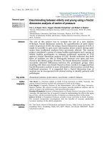
Fig. 1 Flow chart of the systematic search process
Page 3 of 13
scores were considered to be of high quality. Disagreements were resolved by reevaluating and discussing between two reviewers.
Results
Search results and characteristics of included studies
1298 articles, regarding the association between adipokine and thyroid carcinoma, were searched in the
related database and clinical trial websites. After
screening the title and abstracts, 69 articles were selected for full-text review. Finally, 30 studies were eligible in this meta-analysis. Searching progress,
included and excluded details are all shown in Fig. 1.
Eighteen of these 30 studies are published in Chinese
[21, 22, 24–39] and the rest are published in English
[40–49]. Nineteen studies were conducted in China,
two in India and two in Turkey. Brazil, Greece, Iran,
Italy, Denmark and Serbia each had one study. Totally, there are 2174 patients with thyroid carcinoma
in the case group and 1807 controls including healthy
subjects, patients with benign thyroid diseases or normal thyroid tissue near carcinoma were included in
the control group. The sample size ranges from 10 to
236 in the case group while 13 to 131 in the control
group. All the thyroid carcinoma patients were confirmed by pathologically. Among these 30 studies,
fourteen studies reported papillary thyroid carcinoma
(PTC), eight studies reported differentiated thyroid
carcinoma (DTC), three studies reported different
pathological types in one paper, one study reported
medullary thyroid carcinoma (MTC), and the rest
four studies did not show the pathological details.
The detailed characteristics of included studies are
summarized in Table 1.
Zhao et al. BMC Cancer
(2020) 20:788
Page 4 of 13
Table 1 Characteristic of 30 included studies
First author,
Year
Country
L. Kayser, 1996
[33]
Cao Guangyao,
1998 [24]
Pathological
type of thyroid
cancer
Source of controls
Number of
Mean age, year
participants, n
Denmark PTC and FTC
multinodular goiters,
adenomas, Hashimoto’s
thyroiditis, hyperplastic
glands
10
29
Unknown
Unknown
TNF-α (+)
%--tissue
China
Unknown
thyroid adenoma and
nodular goiter
44
27
Unknown
Unknown
TNF-α (+)
%--tissue
M.Trovato, 2003 Italy
[23]
DTC and
undifferentiated
carcinoma
normal thyroid tissues and
benign nodules
28
46
Unknown
Unknown
IL-6 (+)
%--tissue
Zhao Jianqiang, China
2007 [25]
PTC, FTC, ATC
and MTC
thyroid adenoma and
normal health
236
131
Unknown
Unknown
IL-6、TNF-α-blood
Melih Akinci,
2009 [41]
Turkey
PTC
healthy volunteers
43
30
42.8 ±
13.2
100%
leptin--blood
Wang Jingxia,
2009 [26]
China
PTC and FTC
normal thyroid tissues
62
18
Unknown
87.10% Unknown TNF-α (+)
%--tissue
Zhuang
China
Xiaoming, 2010
[27]
PTC, FTC and
MTC
thyroid adenoma and
normal health
38
100
46
73.70% Unknown IL-6、TNF-α-blood
Yu Xiao, 2011
[28]
China
PTC
thyroid adenoma and
normal thyroid tissue near
carcinoma
58
26
Unknown
Unknown
Hou Sen, 2013
[29]
China
PTC
thyroid adenoma
76
16
Unknown
73.70% Unknown leptin(+)%-tissue
Snezana
ZivancevicSimonovic,
2014 [42]
Serbia
WDTC
healthy subjects
13
13
51.23 ± 45.75 ± 84.60% 84.60%
14.9
12.89
TNF-α--blood
Xu Xiaocheng,
2014 [30]
China
thyroid
carcinoma
thyroid adenoma
44
36
54.3 ±
18.6
58.4 ±
17.4
36.40% 55.60%
IL-6--blood
Xeni
Provatopoulou,
2014 [43]
Greece
PTC
benign thyroid disease and
healthy controls
20
38 +
50
49.2 ±
13.7
48.9 ±
14.5 /
49.5 ±
13.2
80%
IL-6--blood
Sun Qinnuan,
2014 [31]
China
PTC
normal thyroid tissue near
carcinoma and healthy
controls
74
74 +
26
40.3 ±
3.6
40.3 ±
3.6 /
37.9 ±
2.4
60.81% 60.81% /
53.85%
Zhang Zijie,
2014 [32]
China
PTC
thyroid adenoma
60
20
Unknown
73.33% Unknown leptin(+)%-tissue
Zhong Xiuxiu,
2014 [33]
China
PTC
thyroid adenoma
78
12
Unknown
Unknown
Zhang Bo, 2014 China
[34]
DTC
normal thyroid tissue near
carcinoma
167
40
Unknown
82.63% Unknown adiponectin-tissue
Hu Jinhua,
2015 [35]
China
DTC
thyroid adenoma and
healthy controls
64
42 +
40
49.8 ±
9.1
75%
Snezana
ZivancevicSimonovic,
2015 [44]
Serbia
PTC
control subjects
16
24
Unknown
Unknown
IL-6--blood
Yan-Lan Fan,
2015 [45]
China
thyroid
carcinoma
nodular goitre, Hashimoto’s
173
thyroiditis, follicular adenoma
and adjacent non-neoplastic
thyroid tissue samples
162
Unknown
Unknown
leptin(+)%-tissue
cases control cases
Female (%)
control cases
54.6 ±
8.9
46/48
36.8 ±
11.3 /
45.3 ±
8.1
Outcome index
control
100%
81.6% /
86.0%
69.04% /
70%
leptin(+)%-tissue
TNF-α--blood
and
tissue
adiponectin(+)%
--tissue
IL-6、
TNF-α--blood
Zhao et al. BMC Cancer
(2020) 20:788
Page 5 of 13
Table 1 Characteristic of 30 included studies (Continued)
First author,
Year
Country
Pathological
type of thyroid
cancer
Source of controls
Number of
Mean age, year
participants, n
Wang
China
Xinzheng, 2015
[36]
thyroid
carcinoma
benign thyroid disease and
normal thyroid tissue near
benign thyroid disease
40
40 +
40
72.35 ± 72.83 ± 40%
7.44
7.73
35% /
35%
TNF-α--tissue
Song Runbo,
2015 [37]
China
PTC
thyroid adenoma
60
60
40.5 ±
8.4
46.7 ±
5.6
60%
53.33%
TNF-α (+)
%--tissue
Kemal Beksac,
2016 [46]
Turkey
PTC
healthy volunteers
31
39
44
41
100%
100%
IL-6--blood
Toral P.
Kobawala,
2016–1 [47]
India
PTC
benign thyroid diseases and
healthy individuals
83
67 +
67
Unknown
67.47% Unknown TNF-α--blood
Toral P.
Kobawala,
2016–2 [48]
India
PTC
benign thyroid diseases and
healthy individuals
84
67 +
67
Unknown
67.47% Unknown IL-6--blood
Raziyeh
Abooshahab,
2016 [20]
Iran
MTC
healthy subjects
45
45
29.46 ± 27.53 ± 53.33% 46.67%
13.97
13.66
Zhang Bo, 2016 China
[38]
DTC
normal thyroid tissue near
carcinoma
167
40
Unknown
China
Zhou
Xiaodong, 2016
[39]
DTC
healthy subjects
50
50
43.82 ± 42.96 ± 56%
12.58
13.29
Ma Xiaokai,
2016 [22]
China
PTC
thyroid adenoma
60
45
Unknown
Mariana
Bonjiorno
Martins, 2017
[49]
Brazil
DTC
benign thyroid nodules and
healthy controls
200
60 +
100
40.73 ± 47.95 ± 86.50% 91.67% /
82%
13.88
14.17 /
40.35 ±
13.34
IL-6--blood
Sun Zhenhua,
2017 [21]
China
PTC
nodular goiter
50
20
41.2
IL-6 (+)
%--tissue
cases control cases
Female (%)
control cases
43.1
Outcome index
control
leptin、
adiponectin-blood
82.63% Unknown leptin--tissue
52%
IL-6、TNF-α-blood
58.33% Unknown leptin(+)%-tissue
64%
70%
TNF-α tumor necrosis factor-a, DTC differentiated thyroid carcinoma, IL-6 interleukin-6, PTC papillary thyroid carcinoma, FTC follicular thyroid carcinoma, ATC
anaplastic thyroid carcinoma, MTC medullary thyroid carcinoma, WDTC well-differentiated thyroid carcinoma, FNAC fine needle aspiration cytology
Quality of included studies
The quality assessment of these 30 studies is assessed by
the NOS and the result is shown in Supplemental
Table 2. Five or more scores are determined as high
quality. Two studies conducted by Cao G et al. in 1998
[24] and L. Kayser et al. in 1996 [40] only get two scores
showing a poor quality in methodology. The rest 28
studies are assessed as high quality.
TNF-α and thyroid carcinoma
Twelve studies reported the expression of TNF-α both
in patients with thyroid carcinoma and control subjects
[24–27, 31, 35–37, 39, 40, 42, 47]. Among these, seven
studies [25, 27, 31, 35, 39, 41, 46] had tested the level of
serum TNF-α, two studies [31, 36] had tested the expression of TNF-α in tissues, and the ratio of TNF-α immunoreactivity was tested in four studies [24, 26, 37,
40]. Firstly, fixed-effect model is used to merge the SMD
values of serum TNF-α level, however, a large heterogeneity is found by the heterogeneity analysis (heterogeneity test, Chi2 = 494.13, P < 0.00001, I2 = 98%) and it
may be due to the different units, different testing
methods in different researches, or other unknown factors. Then, random-effect model to merge the SMD is
used and pooled effect size in favor of control group is
1.31 (95% CI 0.35 to 2.28, P = 0.008) (Fig. 2a). SMD
values of the expression of TNF-α in tissues is merged
by fixed-effected model and the heterogeneity analysis
show a considerable heterogeneity (heterogeneity test,
Chi2 = 305.77, P < 0.00001, I2 = 99%). The different units
and limited numbers of research may be the original of
heterogeneity. So, the pooled SMD with random-effect
model of the expression of TNF-α in tissues is 2.84 (95%
CI − 3.72 to 9.39, P < 0.00001) (Fig. 2b). The pooled OR
with fixed-effect model of the ratio of TNF-α immunoreactivity in thyroid carcinoma tissues is 7.67 (95% CI
4.11 to 14.31, P < 0.00001). However, a significant heterogeneity is detected (heterogeneity test, Chi2 = 8.71,
P = 0.03, I2 = 66%). The article published by L. Kayser in
1996 with a poor quality in methodology may attribute
to this high heterogeneity. Then, random-effect model of
pooled OR is used and pooled effect size in favor of
Zhao et al. BMC Cancer
(2020) 20:788
Page 6 of 13
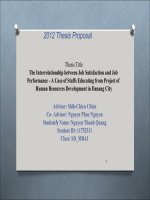
Fig. 2 Forest plot of the TNF-α level and the ratio of TNF-α immunoreactivity in tissues in patients with thyroid carcinoma. a Level of serum TNFα. b Expression of TNF-α in tissue. c Ratio of TNF-α immunoreactivity in tissue
control group is 6.36 (95% CI 1.92 to 21.05, P = 0.002)
(Fig. 2c). In conclusion, level of serum TNF-α and the
ratio of TNF-α immunoreactivity in tissues of thyroid
carcinoma patients are significantly higher than control
subjects which are without thyroid carcinoma.
IL-6 and thyroid carcinoma
Among the 30 included studies, 9 reported the level of
serum IL-6 in patients with thyroid carcinoma and control subjects [27, 30, 35, 39, 43, 44, 46–49]. Due to the
large heterogeneity of the merged SMD values of serum
IL-6 level by the heterogeneity analysis (heterogeneity
test, Chi2 = 334.36, P < 0.00001, I2 = 96%), random-effect
model was used to pooled the SMD values, and the
pooled effect size in favor of control subjects is 1.04
(95% CI 0.40 to 1.67, P = 0.001) (Fig. 3a), which means
that patients with thyroid carcinoma have a significantly
higher level of serum IL-6 than control subjects. Two
studies reported the ratio of IL-6 immunoreactivity both
in thyroid carcinoma tissue and non-carcinoma tissue
[21, 23]. The pooled OR of the limited two studies do
not show an increased ratio of IL-6 immunoreactivity in
thyroid carcinoma tissues (OR = 1.23 (95% CI 0.62 to
2.43, P = 0.55)) and a large heterogeneity always exists
(heterogeneity test, Chi2 = 7.16, P = 0.007, I2 = 86%) (Fig.
3b). Thus, the level of serum IL-6 is higher in patients
with thyroid carcinoma. However, it needs more clinical
data to verify the relationship between the expression of
IL-6 and thyroid carcinoma tissue.
Leptin and thyroid carcinoma
Two studies reported the level of serum leptin [20, 40]
and another five studies reported the ratio of leptin immunoreactivity in tissues [22, 28, 29, 32, 45]. Because of
the considerable heterogeneity of the pooled WMD of
serum leptin level (heterogeneity test, Chi2 = 32.30, P <
0.00001, I2 = 94%) and pooled OR of the ratio of leptin
immunoreactivity in tissues (heterogeneity test, Chi2 =
32.39, P < 0.00001, I2 = 85%) by the heterogeneity analysis with fixed-effect model, random-effect model is further used to merge the values and analysis. However,
there is no association of higher level of serum leptin
Zhao et al. BMC Cancer
(2020) 20:788
Page 7 of 13
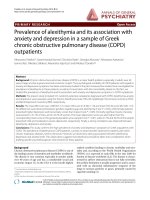
Fig. 3 Forest plot of the IL-6 level and ratio of IL-6 immunoreactivity in tissue in patients with thyroid carcinoma. a Level of serum IL-6. b Ratio of
IL-6 immunoreactivity in tissue
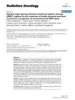
Fig. 4 Forest plot of the leptin level and ratio of leptin immunoreactivity in tissuein patients with thyroid carcinoma. a Level of serum leptin. b
Ratio of leptin immunoreactivity in tissue
Zhao et al. BMC Cancer
(2020) 20:788
Page 8 of 13
with risk of thyroid carcinoma (WMD = 0.51, 95%CI (−
0.38 to 1.40)) (Fig. 4a). Moreover, the pooled OR of the
ratio of leptin immunoreactivity in tissues from five
studies is 12.21 (95%CI 3.36 to 44.40) (Fig. 4b), which
means a high ratio of leptin immunoreactivity in tissue
is significantly related to thyroid carcinoma.
Adiponectin and thyroid carcinoma
Three studies reported the expression of adiponectin in
thyroid carcinoma, including serum and tissue [20, 33,
34], and the result is summarized in Table 2. It could be
found that the level of serum adiponectin is not statically
different comparing thyroid carcinoma patients with
control subjects (P = 0.81). Interestingly, it was found
that the expression of adiponectin in thyroid carcinoma
tissue is significantly lower than control tissue, while the
opposite result is found when comparing the ratio of
adiponectin immunoreactivity. However, there was only
one study for each result and this may be the reason
why the two results are diametrically opposed. Thus, it
needs more clinical studies to confirm in the future.
Publication bias
The funnel plot was applied for assessing publication
bias of studies included in the three results, including
TNF-α (Fig. 5a), IL-6 (Fig. 5b) and leptin (Fig. 5c). In
Fig. 5a and Fig. 5b, almost all studies lies inside the
95%CIs, with an even distribution around the vertical,
indicating no evident publication bias was obtained
through the visual distribution of funnel plot. However,
a potential publication bias was found in Fig. 5c when
comparing the ratio of leptin immunoreactivity in tissues, and that might influence the result of this metaanalysis.
Discussion
Currently, obesity affects one third of population among
US adults [50], and China has become a big country of
obesity with the incidence ranking first worldwide in the
year of 2014 [51]. Nowadays, increasing clinical and experimental studies and documented the closely relationship between malignancies (including colon, esophagus,
kidney, liver, breast, endometrium, pancreas and prostate as well as non-Hodgkin’s lymphoma and multiple
myeloma) and obesity/overweight, which affect its occurrence, development and prognosis [52–54]. Because
of the increasing incidence of thyroid carcinoma during
the past decades, lots of scientists focus on studying the
risk factors of thyroid carcinoma. It was found that the
incidence of thyroid carcinoma has increased along with
a marked rising rate of obesity [4–6]. Furthermore, obesity is an independent risk factor for thyroid carcinoma
[55]. Increased insulin resistance, elevated serum cholesterol level and upregulated COX2 expression may be the
target of the correlation between obesity and thyroid
carcinoma [56]. It is reported that people with higher
body mass index have a higher concentration of adipokines [12–16]. Adipokines take part in the following
pathological and physiological processes, such as, insulin
sensitivity, inflammation and proliferation [17, 57], and
these are important in the process of tumorigenesis and
developing. So adipokines may be one of the targets
linking obesity with thyroid cancer. The meta-analysis
was based on previous published studies. In previous
studies, the analysis of adiponectin and thyroid cancer
mostly focused on TNF-, IL-6, Leptin and Adiponectin.
While few studies focused on other molecules (including
IL-1 and IL-8) and we failed to combine statistics.
Therefore, in this meta-analysis, only TNF-, IL-6, Leptin
and Adiponectin, which are the most published adiponectin, were analyzed.
TNF-α, produced by adipose tissue and inflammatory
cells, can lead to inflammatory response, necrocytosis,
and assist other cytokines to kill tumor cells, and improve the anti-tumor ability. Meanwhile, TNF-α plays an
important role in the process of inflammation, insulin
resistance, diabetes and obesity. A moderate amount of
TNF-α has a protective effect, while an excessive amount
will cause damage, which may lead to a resistant of
tumor cells to TNF-associated apoptosis-induced ligands
when the microenvironment of apoptosis is maladjusted.
TNF-α has the ability to promote the production of
granulocyte-colony stimulating factor by thyroid fibroblasts [58], which may play an important role in thyroid
cancer. Moreover, TNF-α can stimulate the vasoactive
mediators such as interleukin and prostaglandin [59],
and these mediators can promote the proliferation of
tumor cells and significantly reduce the immune function. TNF-α can also induce an increased expression of
vascular endothelial growth factor (VEGF) [60], the later
of that can promote the proliferation of tumor cells and
provide conditions for tumors metastasis.
Table 2 Summary of adiponectin expression in thyroid carcinoma
Effect size
95%CI
P
I2
serum adiponectin [20]
WMD = 0.01
−0.05, 0.07
0.81
0%
ratio of adiponectin immunoreactivity [33]
OR = 6.00
1.39, 25.86
0.02
Not applicable
adiponectin in tissue [34]
WMD = -4.35
−4.64, −4.05
< 0.00001
99%
95% CI 95% confidence interval, WMD weighted mean differences, OR odds ratios
Zhao et al. BMC Cancer
(2020) 20:788
Page 9 of 13
Fig. 5 Funnel plots of a TNF-α, b IL-6 and c leptin revealed no significant publication bias. SE (SMD) standard error of standardized
mean difference
In conclusion, surprisingly, the results of clinical studies provide evidence for basic research. Simonovic SZ
et al. [42] evaluated cytokine profiles (determined in supernatants obtained from whole blood cultures) in 13
patients with DTC before and 7 days after radioactive
iodine (131-I) therapy and 13 control subjects, and
found that the expression of TNF-α in DTC patients is
higher than control subjects, and it showed a decreased
level after 131-I therapy than those before therapy. However, no statistical difference found for the limited sample size. Another study conducted by Kobawala TP et al.
[47], with more patients (67 patients with benign thyroid
disease, 83 PTC patients and 67 healthy individuals), determined the circulating levels of TNF-α, and it was
found that the serum level of TNF-α was significantly
higher in PTC patients than benign thyroid disease patients, and the later was also significantly higher than
healthy individuals. Furthermore, serum TNF-α was reported to be a significant prognosticator for overall survival in PTC patients. It is a pity thatopposite result was
reported in a case-control study that included 475 DTC
cases and 1016 matched cancer-free cohort participants,
which found that TNF-a was not associated with thyroid
risk in either gender [61].
Based on current evidence, our meta-analysis suggests
that TNF-α exhibit a strong association with thyroid carcinoma. It may because that elevated TNF-α may involved in the tumorigenesis and development of thyroid
cancer. Another possible reason is that the TNF-α decreased with tumor cells less resulted the activation of
the immune system by thyroid carcinomaTherefore,
more clincal studies and basic reseaches should be conducted in the future.
IL-6, a multifunctional cytokine, plays important roles
in different types of cells including tumor cells. It is reported that elevated serum IL-6 level is closely related to
the tumorigenesis and development of a variety of tumors [62]. A strong positive association between the
serum IL-6 and the progression and poor prognosis of
tumors in patients with several types of tumor was
already found [63–65]. Serum IL-6 level in thyroid cancer has been evaluated in numerous studies including
Zhao et al. BMC Cancer
(2020) 20:788
in vivo and in vitro studies. Provatopoulou X et al. [43]
found that serum IL-6 were significantly higher in malignant and benign thyroid diseases compared to healthy
controls. However, other studies show a different result
that no significance different of IL-6 was found between
thyroid cancer and non-thyroid cancer [16, 23, 43, 44,
49]. A limited sample size, different inclusion criteria,
different population characteristics, or different pathological type of thyroid cancer may explain such a difference. For in vitro research, IL-6 was also found to be
expressed in thyroid cancer cell lines and a potential role
of IL-6 in PTC was confirmed indirectly [66].
The underlying mechanism may be the followings
below. Tumor cells including esophageal cancer, lung
cancer, colorectal cancer and melanoma were found
have the function of autocrine IL-6, which can affect the
growth and proliferation of tumor cells and participate
in the tumor growth and metastasis by acting on the
membrane receptors [67]. Also, IL-6R was found associated with the characterization of thyroid nodules’ malignancy and tumor aggressiveness [49]. In addition,
Iliopoulos D et al. [68] found that Src (non-somatic tyrosine kinase family oncogene) can induce the normal epithelial cell transformation by activating NF-κB, and this
transformation contributes to tumorigenesis. IL-6 is considered as an important regulatory factor in this process.
Another possibility is that the activation of the immune
system of patients with thyroid cancer leads to an increase in adikopines level.
In general, the data above support that IL-6 is important for thyroid cancer, but the detail mechanism remain
to be further study.
Leptin, a circulating hormone secreted by adipocytes,
exerts its biological effect by combing with its receptor,
which is mainly presented in the hypothalamus. Meanwhile, gene of leptin receptor is also expressed in many
other tissues, such as lung, liver and kidney. It is reported that obesity and overweight can lead to a high
level of serum leptin, which may because that obesity always accompanies with insulin resistance and hyperinsulinemia, and insulin further enhance the expression of
leptin. Moreover, leptin acts as a growth factor in a variety of human cells, including both normal cells and
tumor cells, which regulates the process of differentiation, proliferation and apoptosis thus stimulate the
tumorigenesis and development of tumors through mediating JAK/STAT3 pathway, RhoA/LIMK1/Cofilin
pathway, and MAPK/ERK pathway, [69]. Kim WG et al.
[70] evaluated the effect of diet-induced obesity on thyroid carcinogenesis in a mouse model that spontaneously develops thyroid cancer (Thrb (PV/PV) Pten
(+/−) mice) and found that obesity increases the frequency of anaplasia of thyroid cancer and exacerbates
thyroid cancer progression that were mediated by
Page 10 of 13
increased activation of the JAK2 signaling transducer
and activator of STAT3 signaling pathway and induction
of STAT3 target gene expression. Leptin is always reported a high expression on solid tumors [71], and it is
confirmed that serum leptin level is significantly increased in thyroid cancer (mainly PTC), while other
studies showed a same results in cancer tissues [11, 15,
21, 41, 45]. Yu Xiao et al. [21] conducted a clinical study
comparing the level of serum leptin in 58 PTC patients
(including 29 patients with lymph node metastasis) and
26 thyroid adenoma patients in Dalian, China, and found
that patients with lymph node metastasis have a higher
level of leptin than those without lymph node metastasis.
Leptin can induce the expression of vascular endothelial
growth factor and promote neovascularization in tumor
tissue [72]. In addition, it can also inhibit the apoptosis
through Bcl-2 dependent mechanism. Meanwhile, leptin
receptor exists in all thyroid cancer cells. It is overexpressed in PTC and is involved in tumor invasion and
lymph node metastasis [73, 74]. Thus, leptin may be involved in the tumorigenesis and metastasis of thyroid
cancer through a complex pathway and a monitoring
may have some significance. Due to the absence of direct
evidence, elevated leptin levels can also be caused by
thyroid carcinoma. The cause and effect relationship between leptin and thyroid carcinoma are unclear now and
need further studies.
Compared to lean women, overweight/obese women
had lower serum adiponectin levels and this difference
has statistical significance [75]. In addition, adiponectin
is negatively associated with a variety of benign and malignant tumors, especially those associated with obesity
and insulin resistance, such as leukemia [76], renal carcinoma [77], gastric carcinoma [78] and colon cancer
[79]. Moreover,, the association of adiponectin with potential tumor-limiting functions has been widely proposed [80].
Otvos L Jr. et al. [81] tried in vitro experiments and
proved that adiponectin can inhibit the metastasis of
cancer cells. Mitsiades N et al. [82] measured circulating
adiponectin levels in ptaients with PTC and found that
it is independently and inversely associated with the risk
of thyroid cancer. As the receptor that binds to adiponectin for biological effects, adiponectin receptor had
been reported closely correlated with the development
of PTC. Adiponectin receptor-1 and 2 are higher expression in PTC tissues than that in the surrounding normal
tissues and this is thought to be associated with a better
prognosis [83].
However, other studies have shown different results
[13, 27] and more studies should be done furtherly to
support the anti-tumor effect of adiponectin, and the
positive correlation between the increased level of adiponectin in circulating blood and the prognosis of thyroid
Zhao et al. BMC Cancer
(2020) 20:788
neoplasms and provide new ideas for the prevention and
treatment of thyroid neoplasms.
From the above, a strong relationship between elevated
concentrations of adipokines (in serum and/or tissue)
and thyroid cancer can be concluded. And this may explain why increased incidence of obesity and thyroid
cancer are consistent. Thus, targeted drugs for adipokine
may be useful for the treatment of thyroid cancer in the
future.
However, some limitations in our meta-analysis should
be taken into account. First, some data were not normally distributed and were reported in the form of median and quartile, and therefore these data were
calculated by formulas. Second, due to the insufficient
database access, six articles are not available in full, and
therefore could not be included in this meta-analysis.
Third, all the included studies were cross-sectional casecontrol study and the dynamic changes of these adipokines in preoperative and postoperative were not provided. The last but not the least, most of the included
studies (18 of these 30 studies) are published in Chinese,
thus a considerable but may inevitable bias can result of
this meta-analysis. All these limitations above should be
improved in the future study, thus a strong conclusion
could be get.
Conclusions
In summary, our meta-analysis suggests that adipokines,
including TNF-α, IL-6 and leptin are associated with
thyroid carcinoma. Nevertheless, it is not conclusive for
adiponectin due to the limited number of the clinical
studies. Therefore, larger sample sizes of different ethnic
population are required to confirm and update our
findings.
Supplementary information
Supplementary information accompanies this paper at />1186/s12885-020-07299-x.
Additional file 1 Supplemental Table 1 Newcastle-Ottawa Quality Assessment Scale—Case-control Studies.
Additional file 2 Supplemental Table 2 Quality assessment according
to the Newcastle-Ottawa Scale.
Abbreviations
TNF-α: Tumor necrosis factor-alpha; IL-6: Interleukin-6; OR: Odds ratios;
WMD: Weighted mean differences; SMD: Standardized mean difference; 95%
CI: 95% confidence interval; NOS: Newcastle-Ottawa Scale;
DTC: Differentiated thyroid carcinoma; PTC: Papillary thyroid carcinoma;
FTC: Follicular thyroid carcinoma; ATC: Anaplastic thyroid carcinoma;
MTC: Medullary thyroid carcinoma; WDTC: Well-differentiated thyroid
carcinoma; FNAC: Fine needle aspiration cytology; SE (SMD): Standard error
of standardized mean difference; VEGF: Vascular endothelial growth factor;
131-I: Radioactive iodine
Acknowledgements
Not applicable.
Page 11 of 13
Authors’ contributions
JZ, JW and LL designed the study and wrote the manuscript. JZ, SW and JY
performed the literature searches and collected the data. JZ, JW and JY
performed the statistical analysis. All authors read and approved the final
manuscript.
Funding
This study was funded by Projects of medical and health technology
development program in Shandong province [grant number 2016WS0499],
Shandong Provincial Natural Science Foundation of China Grants [grant
number ZR2019PH025]. They support the study design; the data collection,
analysis and interpretation of data; the writing of the report; and the
decision to submit the article for publication.
Availability of data and materials
The datasets used and/or analyzed during the current study are available
from the corresponding author on reasonable request.
Ethics approval and consent to participate
Not applicable.
Consent for publication
All the authors agreed this article be published.
Competing interests
The authors declare that they have no competing interests.
Author details
Department of Endocrinology and Metabology, The First Affiliated Hospital
of Shandong First Medical University & Shandong Provincial Qianfoshan
Hospital, Ji-nan 250014, China. 2Department of Endocrinology and
Metabology, Shandong Provincial Qianfoshan Hospital, Cheeloo College of
Medicine, Shandong University, Ji-nan 250014, China. 3College of Traditional
Chinese Medicine, Shandong University of Traditional Chinese Medicine,
Ji-nan 250000, China. 4Department of Endocrinology and Metabology,
Shandong First Medical University & Shandong Academy of Medical
Sciences, Ji-nan 250014, China. 5Department of Endocrinology and
Metabology, Qilu Hospital of Shandong University, Cheeloo College of
Medicine, Shandong University, Ji-nan 250012, China.
1
Received: 29 March 2020 Accepted: 13 August 2020
References
1. Davies L, Welch HG. Current thyroid cancer trends in the United States.
JAMA Otolaryngol Head Neck Surg. 2014;140:317–22.
2. Zheng R, Zeng H, Zhang S, Chen W. Estimates of cancer incidence and
motality in China, 2013. Chin J Cancer. 2017;36(1):66.
3. Kitahara CM, Sosa JA. The changing incidence of thyroid cancer. Nat Rev
Endocrinol. 2016;12(11):646–53.
4. NCD Risk Factor Collaboration (NCD-RisC). Trends in adult body-mass index
in 200 countries from 1975 to 2014: a pooled analysis of 1698 populationbased measurement studies with 19.2 million participants. Lancet. 2016;
387(10026):1377–96.
5. Lauby-Secretan B, Scoccianti C, Loomis D, Grosse Y, Bianchini F, Straif K.
International Agency for Research on Cancer handbook working group.
Body fatness and Cancer--viewpoint of the IARC working group. N Engl J
Med. 2016;375(8):794–8.
6. Pappa T, Alevizaki M. Obesity and thyroid Cancer: a clinical update. Thyroid.
2014;24(2):190–9.
7. Hedayati M, Yaghmaei P, Pooyamanesh Z, Zarif Yeganeh M, Hoghooghi RL.
Leptin: a correlated peptide to papillary thyroid carcinoma? J Thyroid Res.
2011;2011:832163.
8. Cheng SP, Liu CL, Hsu YC, Chang YC, Huang SY, Lee JJ. Regulation of leptin
receptor expression in human papillary thyroid cancer cells. Biomed
Pharmacother. 2012;66:469–73.
9. Cheng SP, Liu CL, Hsu YC, Chang YC, Huang SY, Lee JJ. Expression and
biologic significance of adiponectin receptors in papillary thyroid
carcinoma. Cell Biochem Biophys. 2013;65:203–10.
10. Mitsiades N, Pazaitou-Panayiotou K, Aronis KN, Moon HS, Chamberland JP,
Liu X, Diakopoulos KN, Kyttaris V, Panagiotou V, Mylvaganam G, Tseleni-
Zhao et al. BMC Cancer
11.
12.
13.
14.
15.
16.
17.
18.
19.
20.
21.
22.
23.
24.
25.
26.
27.
28.
29.
30.
31.
32.
33.
(2020) 20:788
Balafouta S, Mantzoros CS. Circulating adiponectin is inversely associated
with risk of thyroid cancer: in vivo and in vitro studies. J Clin Endocrinol
Metab. 2011;96:E2023–8.
Pomp D, Mohlke KL. Obesity genes: so close and yet so far. Sci J Biol. 2008;
7:36.
Popko K, Gorska E, Stelmaszczyk-Emmel A, Plywaczewski R, Stoklosa A,
Gorecka D, Pyrzak B, Demkow U. Proinflammatory cytokines IL-6 and TNF-α
and the development of inflammation in obese subjects. Eur J Med Res.
2010;15(Suppl 2):120–2.
Mishra S, Gupta V, Mishra S, Sachan R, Asthana A. Serum level of orexin-a,
leptin, adiponectin and insulin in north Indian obese women. Diabetes
Metab Syndr. 2017 Dec;11(Suppl 2):S1041–3.
Saghizadeh M, Ong JM, Garvey WT, Henry RR, Kern PA. The expression of
TNF a by human muscle. Relationship to insulin resistance. J Clin Investig.
1996;97:1111–6.
Kern PA, Saghizadeh M, Ong JM, Bosch RJ, Deem R, Simsolo RB. The
expression of tumor necrosis factor in human adipose tissue. Regulation by
obesity, weight loss, and relationship to lipoprotein lipase. J Clin Investig.
1995;95:2111–9.
Mohamed-Ali V, Pinkney JH, Coppack SW. Adipose tissue as an endocrine and
paracrine organ. Int J Obes Relat Metab Disord. 1998 Dec;22(12):1145–58.
Rega-Kaun G, Kaun C, Wojta J. More than a simple storage organ: adipose
tissue as a source of adipokines involved in cardiovascular disease. Thromb
Haemost. 2013 Oct;110(4):641–50.
Rehem RA, Elwafa WA, Elwafa RA, Abdel-Aziz TE. Study of serum leptin in
well-differentiated thyroid carcinoma: correlation with patient and tumor
characteristics. World J Surg. 2014;38(10):2621–7.
Pazaitou-Panayiotou K, Panagiotou G, Polyzos SA, Mantzoros CS. Serum
adiponectin and insulin-like growth factor 1 in predominantly female
patients with thyroid cancer: association with histologic characteristics of
the tumor. Endocr Pract. 2016;22(1):68–75.
Abooshahab R, Yaghmaei P, Ghadaksaz HG, Hedayati M. Lack of association
between serum Adiponectin/Leptin levels and medullary thyroid Cancer.
Asian Pac J Cancer Prev. 2016;17(8):3861–4.
Sun Z, Zhao Z, Jiang H, Xu L, Sun Y, Long W, Wang K. Expressions of tumor
microenvironment-associated factors IL-6, IL-10 and chemokine receptor 7
in papillary thyroid carcinoma and their clinical significance. Chin J Gen
Surg. 2017;26(5):578–82.
Ma X, Xie C, Wang S, Zhen X, Huang J, Zhu Z, Yao Y, Zhang H, Feng Z. High
level co-expression of leptin and leptin receptor in papillary thyroid
carcinoma. Chin J Histochem Cytochem. 2016;25(5):442–6.
Trovato M, Grosso M, Vitarelli E, Ruggeri RM, Alesci S, Trimarchi F, Barresi G,
Benvenga S. Distinctive expression of STAT3 in papillary thyroid carcinomas
and a subset of follicular adenomas. Histol Histopathol. 2003;18(2):393–9.
Cao G, Wang C, Li Q, Wang W, Ma F. Expression of Fas/FasL and TNF-α in
human thyroid carcinoma. Chin J Oncol. 1998;20(5):336.
Zhao J, Feng J, Guo L, Wang K. Clinical study of the effect of Th1/Th2 drifts
on natural killer cells in patients with thyroid tumor. Clin Med China. 2007;
23(3):241–3.
Wang J, Xu H, Deng W, Qi Y, Li Y, Zhang H, Wang S, Li Y, Li W. Expression
and significance of VEGF-C and TNF-α in papillary and follicular thyroid
cancer. Chin J Gerontol. 2009;29(13):1617–8.
Zhuang X. Study on the correlation between hypothyroid tumors and the
levels of TNF-α, IL-6 and sIL-2R. Chin J Pract Diagnosis Treat. 2010;24(1):32–6.
Yu X, Zhao J, Wang B, Li L, Li P, Tang J, Sun L. The expression of leptin in
thyroid papillary carcinoma and its effect on intratumor angiogenesis and
metastasis. China Oncol. 2011;21(4):283–6.
Hou S, Cui W, Zhang G, Zhou J, Zhong X, Ma L. Expression of leptin and
leptin receptor in papillary thyroid carcinoma and relationship with clinical
features. J Clin Exp Pathol. 2013;29(3):284–6.
Xu X, Wang K. The value of detection of EB virus in patients with thyroid
tumor. Chin J Gen Pract. 2014;12(7):1063–5.
Sun Q, Li D, Wu G, Tu J, Yun F, Xu X, Yu H. Expression of P53, Fas, TNF-α
and Cyclin E with carcinoma papillary thyroid cacner patients in serum and
its clinical significance. Chin J Immunol. 2014;30(10):1383–7.
Zhang Z. Relationship between expression and pathological characteristics
of leptin and its receptor in thyroid papillary carcinoma. J Med Res. 2014;
43(6):162–4.
Zhong X, Cui W, Zhang G, Hou S, Wang S, Zhou J. Expression and
clnicopathological correlation studies of adiponectin in papillary thyroid
carcinoma. J Jining Med Univ. 2014;37(1):24–6.
Page 12 of 13
34. Zhang B, Yang W, Su J, Sun F, Chen L, Li J, Cui G, Dai D. Relationship
between body mass index and serum adiponectin, vascular endothelial
growth factor lavels in patients with differentiated thyroid carcinoma. J Clin
Exp Med. 2014;13(21):1775–7.
35. Hu J, Zhang Y, Ai R, Zhu B. Correlation of serum TNF-α, IL-6 and VEGF of
patients with differentiated thyroid carcinoma and its invasion and
metastasis. Modern Oncol. 2015;23(6):771–4.
36. Wang X, Liu J, Hou Y, Wang N, Wang M. Expression of HSP70 and TNF in
differentiated thyroid carcinoma and its correlation with clinicopathology.
Chin J Gerontol. 2015;35(1):112–3.
37. Song R, Ma J, Meng Z, Jia Q, Dong Y, Liu W. Expression and significance of
PTEN protein and tumor necrosis factor in papillary thyroid carcinoma.
Hebei Med J. 2015;37(18):2751–3.
38. Zhang B, Yang W, Zhao C, Sun F, Chen L, Li J, Cui G, Dai D. Study on the
relationship between body mass index and leptin expression in patients
with differentiated thyroid carcinoma. Modern Oncol. 2016;24(1):44–6.
39. Zhou X, Huang X, Lu K, Zhang H, Tan Z. The influence of total laparoscopic
surgery on T cell subsets and its cytokines in patients with early
differentiated thyroid cancer. Chin J Gen Pract. 2016;14(12):2011–48.
40. Kayser L, Broholm H, Francis D, Perrild H, Olsen BE, Bendtzen K, Høyer PE.
Immunocytochemical localisation of tumor necrosis factor alpha in thyroid
tissues from patients with neoplastic or autoimmune thyroid disorders.
Autoimmunity. 1996;23(2):91–7.
41. Akinci M, Kosova F, Cetin B, Aslan S, Ari Z, Cetin A. Leptin levels in thyroid
cancer. Asian J Surg. 2009;32(4):216–23.
42. Simonovic SZ, Mihaljevic O, Majstorovic I, Djurdjevic P, Kostic I, Djordjevic OM,
Teodorovic LM. Cytokine production in peripheral blood cells of patients with
differentiated thyroid cancer: elevated Th2/Th9 cytokine production before
and reduced Th2 cytokine production after radioactive iodine therapy. Cancer
Immunol Immunother. 2015;64(1):75–82 Epub 2014 Oct 9.
43. Provatopoulou X, Georgiadou D, Sergentanis TN, Kalogera E, Spyridakis J,
Gounaris A, Zografos GN. Interleukins as markers of inflammation in
malignant and benign thyroid disease. Inflamm Res. 2014;63(8):667–74.
44. Zivancevic-Simonovic S, Mihaljevic O, Majstorovic I, Popovic S, Markovic S,
Milosevic-Djordjevic O, Jovanovic Z, Mijatovic-Teodorovic L, Mihajlovic D,
Colic M. Cytokine production in patients with papillary thyroid cancer and
associated autoimmune Hashimoto thyroiditis. Cancer Immunol
Immunother. 2015;64(8):1011–9.
45. Fan YL, Li XQ. Expression of leptin and its receptor in thyroid carcinoma:
distinctive prognostic significance in different subtypes. Clin Endocrinol.
2015;83(2):261–7.
46. Beksac K, Sonmez C, Cetin B, Kismali G, Sel T, Tuncer Y, Kosova F. Evaluation
of proinflammatory cytokine and neopterin levels in women with papillary
thyroid carcinoma. Int J Biol Markers. 2016;31(4):e446–50.
47. Kobawala TP, Trivedi TI, Gajjar KK, Patel DH, Patel GH, Ghosh NR.
Significance of TNF-α and the adhesion molecules: L-Selectin and VCAM-1
in papillary thyroid carcinoma. J Thyroid Res. 2016;2016:8143695.
48. Kobawala TP, Trivedi TI, Gajjar KK, Patel DH, Patel GH, Ghosh NR.
Significance of Interleukin-6 in papillary thyroid carcinoma. J Thyroid Res.
2016;2016:6178921.
49. Martins MB, Marcello MA, Batista FA, Peres KC, Meneghetti M, Ward MAL,
Etchebehere ECSC, da Assumpção LVM, Ward LS. Serum interleukin
measurement may help identify thyroid cancer patients with active disease.
Clin Biochem. 2018;52:1–7 Epub 2017 Oct 5.
50. Flegal KM, Carroll MD, Kit BK, Ogden CL. Prevalence of obesity and trends in
the distribution of body mass index among US adults. 1999–2010. JAMA.
2012;307:491–7.
51. NCD Risk Factor Collaboration (NCD-RisC). Trends in adult body-mass index
in 200countries from 1975 to 2014: a pooled analysis of 1968 populationbased measurement studies with 19.2 million participants. Lancet. 2016;
387(10026):1377–96.
52. De Pergola G, Nardecchina A, Giagulli VA, et al. Obesity and heart failure.
Endocr Metab Immune Disord Drug Targets. 2013;13(1):51–7.
53. Calle EE, Rodriguez C, Walker-Thurmond K, Thun MJ. Overweight, obesity,
and mortality from cancer in a prospectively studied cohort of U.S. adults. N
Engl J Med. 2003;348:1625–38.
54. Renehan AG, Tyson M, Egger M, Heller RF, Zwahlen M. Body mass index
and incidence of cancer: a systematic review and meta-analysis of
prospective observational studies. Lancet. 2008;371:569–78.
55. Xu L, Port M, Landi S, Gemignani F, Cipollini M, Elisei R, Goudeva L, Müller
JA, Nerlich K, Pellegrini G, Reiners C, Romei C, Schwab R, Abend M, Sturgis
Zhao et al. BMC Cancer
56.
57.
58.
59.
60.
61.
62.
63.
64.
65.
66.
67.
68.
69.
70.
71.
72.
73.
74.
75.
76.
77.
(2020) 20:788
EM. Obesity and the risk of papillary thyroid cancer: a pooled analysis of
three case-control studies. Thyroid. 2014 Jun;24(6):966–74.
Allott EH, Hursting SD. Obesity and cancer: mechanistic insights from
transdisciplinary studies. Endocr Relat Cancer. 2015 Dec;22(6):R365–86.
Zhang Y, Proenca R, Maffei M, Barone M, Leopold L, Friedman JM. Positional
cloning of the mouse obese gene and its human homologue. Nature. 1994
Dec 1;372(6505):425–32.
Aust G, Hofmann A, Laue S, Ode-Hakim S, Scherbaum WA. Differential
regulation of granulocyte-macrophage colony-stimulating factor mRNA and
protein expression in human thyrocytes and thyroid-derived fibroblasts by
interleukin-1 alpha and tumor necrosis factor-alpha. J Endocrinol. 1996 Nov;
151(2):277–85.
Jana B, Kozłowska A, Andronowska A, Jedlińska-Krakowska M. The effect of
tumor necrosis factor-alpha (TNF-alpha), interleukin (IL)-1 beta and IL-6 on
chorioamnion secretion of prostaglandins (PG) F 2 alpha and E2 in pigs.
Reprod Biol. 2008;8(1):57–68.
Wang H, Han X, Wittchen ES, Hartnett ME. TNF-α mediates choroidal
neovascularization by upregulating VEGF expression in RPE through ROSdependent β-catenin activation. Mol Vis. 2016 Feb 3;22:116–28.
Dossus L, Franceschi S, Biessy C, Navionis AS, Travis RC, Weiderpass E,
Scalbert A, Romieu I, Tjønneland A, Olsen A, Overvad K, Boutron-Ruault MC,
Bonnet F, Fournier A, Fortner RT, Kaaks R, Aleksandrova K, Trichopoulou A,
La Vecchia C, Peppa E, Tumino R, Panico S, Palli D, Agnoli C, Vineis P, HBA
B-d-M, Peeters PH, Skeie G, Zamora-Ros R, Chirlaque MD, Ardanaz E,
Sánchez MJ, Ramón Quirós J, Dorronsoro M, Sandström M, Nilsson LM,
Schmidt JA, Khaw KT, Tsilidis KK, Aune D, Riboli E, Rinaldi S. Adipokines and
inflammation markers and risk of differentiated thyroid carcinoma: The EPIC
study. Int J Cancer. 2018;142(7):1332–42.
Lippitz BE. Cytokine patterns in patients with cancer: a systematic review.
Lancet Oncol. 2013;14(6):218–28.
Ahmed AM, Adel DR, Diab NS. Gobran, prognostic value of serum level of
interleukin-6 and interleukin-8 in metastatic breast cancer patients. Egypt J
Immunol. 2006;13(2):61–8.
Hsia CY, Huo TI, Chiang SY, Lu MF, Sun CL, Wu JC, et al. Evaluation of
interleukin-6, interleukin-10 and human hepatocyte growth factor as tumor
markers for hepatocellular carcinoma. Eur J Surg Oncol. 2007;33(2):208–12.
Ikeguchi M, Hatada T, Yamamoto M, Miyake T, Matsunaga T, Fukumoto Y,
et al. Serum interleukin-6 and -10 levels in patients with gastric cancer.
Gastric Cancer. 2009;12(2):95–100.
Lumachi F, Basso SM, Orlando R. Cytokines, thyroid diseases and thyroid
cancer. Cytokine. 2010;50(3):229–33.
Fujii Y, Takeuchi S, Sasaki O, et al. Multivariate analysis of predictors of
hematoma enlargement in spontaneous intracerebral hemorrhage. Stroke.
1998;29(6):1160–6.
Iliopoulos D, Hirsch HA, Struhl K. An epigenetic switch involving NF-kappa
B, Lin 28, Let-7 microRNA, and IL-6 links inflammation to cell transformation.
Cell. 2009;139(4):693–706.
Vasselli JR. Behavioraland biological determinants of leptin resistance.
Appetite. 2001;37(2):115–7.
Kim WG, Park JW, Willingham MC, Cheng SY. Diet-induced obesity increases
tumor growth and promotes anaplastic change in thyroid cancerin a
mouse model. Endocrinology. 2013;154(8):2936–47.
Garofalo C, Surmacz E. Leptin and cancer. J Cell Physiol. 2006;207(1):12–22.
Lanier V, Gillespie C, Leffers M, et al. Leptin-induced transphosphorylation of
vascular endothelial growth factor receptor increases notch and stimulates
endothelial cell angiogenic transformation. Int J Biochem Cell Biol. 2016;79:
139–50.
Cheng SP, Liu CL, Hsu YC, et al. Regulation of leptin receptor expression in
human papillary thyroid cancer cells. Biomed Pharmacother. 2012;66(6):469–73.
Cheng SP, Yin PH, Hsu YC, et al. Leptin enhances migration of human
papillary thyroid cancer cells through the PI3K/AKT and MEK/ERK signaling
pathways. Oncol Rep. 2011;26(5):1265–71.
Chen C, Hsu M, Lin SH. Adiponectin and leptin in overweight/obese and
lean women with polycystic ovary syndrome. Gynecol Endocrinol. 2015;
31(4):264–8.
Ozturk K, Avcu F, Ural AU. Aberrant expressions of leptin and adiponectin
receptor isoforms in chronic myeloid leukemia patients. Cytokine. 2012;
57(1):61–7.
Shin E, Park DJ, Kim HH, et al. Adiponectin receptor expression in gastric
carcinoma: implications in tumor development and progression. J Cancer
Res Clin Oncol. 2013;139(4):709–18.
Page 13 of 13
78. Wang H, Wu J, Gu W, et al. Serum Adiponectin level may be an
independent predictor of clear cell renal cell carcinoma. J Cancer. 2016;
7(10):1340–6.
79. Saxena A, Baliga MS, Ponemone V, et al. Mucus and adiponectin deficiency:
role in chronic inflammation-induced colon cancer. Int J Color Dis. 2013;
28(9):1267–79.
80. Hebbard L, Ranscht B. Multifaceted roles of Adiponectin in cancer. Best
Pract Res Clin Endocrinol Metab. 2014;28(1):59–69.
81. Otvos L Jr, Kovalszky I, Olah J, Coroniti R, Knappe D, Nollmann FI, Hoffmann
R, Wade JD, Lovas S, Surmacz E. Optimization of adiponectinderived
peptides for inhibition of cancer cell growth and signaling. Pept Sci. 2015;
104(3):156–66.
82. Mitsiades N, Pazaitou-Panayiotou K, Aronis KN, Moon HS, Chamberland JP,
Liu X, Diakopoulos KN, Kyttaris V, Panagiotou V, Mylvaganam G, TseleniBalafouta S, Mantzoros CS. Circulating adiponectin is inversely associated
with risk of thyroid cancer: in vivo and in vitro studies. J Clin Endocrinol
Metab. 2011;96(12):E2023–8.
83. Cheng SP, Liu CL, Hsu YC, et al. Expression and biologic significance of
adiponectin receptors in papillary thyroid carcinoma. Cell Biochem Biophys.
2013;65(2):203–10.
Publisher’s Note
Springer Nature remains neutral with regard to jurisdictional claims in
published maps and institutional affiliations.