Stimulation of triple negative breast cancer cell migration and metastases formation is prevented by chloroquine in a preirradiated mouse model
Bạn đang xem bản rút gọn của tài liệu. Xem và tải ngay bản đầy đủ của tài liệu tại đây (2.08 MB, 14 trang )
Bouchard et al. BMC Cancer (2016) 16:361
DOI 10.1186/s12885-016-2393-z
RESEARCH ARTICLE
Open Access
Stimulation of triple negative breast cancer
cell migration and metastases formation is
prevented by chloroquine in a preirradiated mouse model
Gina Bouchard1, Hélène Therriault1, Sameh Geha4, Yves Bérubé-Lauzière5, Rachel Bujold1,3, Caroline Saucier2
and Benoit Paquette1*
Abstract
Background: Some triple negative breast cancer (TNBC) patients are at higher risk of recurrence in the first three
years after treatment. This rapid relapse has been suggested to be associated with inflammatory mediators induced
by radiation in healthy tissues that stimulate cancer cell migration and metastasis formation. In this study, the ability
of chloroquine (CQ) to inhibit radiation-stimulated development of metastasis was assessed.
Methods: The capacity of CQ to prevent radiation-enhancement of cancer cell invasion was assessed in vitro with
the TNBC cell lines D2A1, 4T1 and MDA-MB-231 and the non-TNBC cell lines MC7-L1, and MCF-7. In Balb/c mice, a
single mammary gland was irradiated with four daily doses of 6 Gy. After the last irradiation, irradiated and control
mammary glands were implanted with D2A1 cells. Mice were treated with CQ (vehicle, 40 or 60 mg/kg) 3 h before
each irradiation and then every 72 h for 3 weeks. Migration of D2A1 cells in the mammary gland, the number of
circulating tumor cells and lung metastasis were quantified, and also the expression of some inflammatory mediators.
Results: Irradiated fibroblasts have increased the invasiveness of the TNBC cell lines only, a stimulation that was
prevented by CQ. On the other hand, invasiveness of the non-TNBC cell lines, which was not enhanced by irradiated
fibroblasts, was also not significantly modified by CQ. In Balb/c mice, treatment with CQ prevented the stimulation of
D2A1 TNBC cell migration in the pre-irradiated mammary gland, and reduced the number of circulating tumor cells
and lung metastases. This protective effect of CQ was associated with a reduced expression of the inflammatory
mediators interleukin-1β, interleukin-6, and cyclooxygenase-2, while the levels of matrix metalloproteinases-2 and −9
were not modified. CQ also promoted a blocking of autophagy.
Conclusion: CQ prevented radiation-enhancement of TNBC cell invasion and reduced the number of lung metastases
in a mouse model.
Keywords: Chloroquine, Inflammation, Invasion, Metastasis, Radiation, Triple negative breast cancer
* Correspondence:
1
Centre for Research in Radiotherapy, Department of Nuclear Medicine and
Radiobiology, Université de Sherbrooke, 3001, 12e Avenue Nord, Sherbrooke,
Québec J1H 5 N4, Canada
Full list of author information is available at the end of the article
© 2016 The Author(s). Open Access This article is distributed under the terms of the Creative Commons Attribution 4.0
International License ( which permits unrestricted use, distribution, and
reproduction in any medium, provided you give appropriate credit to the original author(s) and the source, provide a link to
the Creative Commons license, and indicate if changes were made. The Creative Commons Public Domain Dedication waiver
( applies to the data made available in this article, unless otherwise stated.
Bouchard et al. BMC Cancer (2016) 16:361
Background
Breast cancer is a heterogeneous disease, encompassing
a number of distinct biological entities that are associated with specific morphological features and clinical behaviors. Triple negative breast cancer (TNBC) accounts
for 10–20 % of all breast carcinomas and is characterized by the absence of estrogen receptor (ER), progesterone receptor (PR) and human epidermal growth factor
receptor 2 (HER-2) [1]. Recurrence within 3 years of initial treatment is more likely for this aggressive form of
breast cancer and results in a mortality risk two times
higher than for non-TNBC patients [2]. Without any targeted therapies for TNBC, a better understanding and
optimization of adjuvant treatment as radiotherapy remains essential.
Although radiotherapy is recommended to prevent
locoregional relapse, the early recurrence found in some
TNBC patients suggests that the formation of metastasis
is favored in a subgroup of these patients who respond
poorly to ionizing radiation. This stimulation of metastasis development could be related to the ability of radiotherapy to trigger an inflammatory response [3]. This
inflammation is characterized by an increase of some cytokines and matrix metalloproteinases (MMP) that are
known to favor metastasis development [4]. Further supporting this role of inflammatory cytokines, the association between a chronic inflammation and an increased
risk of developing several types of cancer, including
breast cancer, have been demonstrated [5]. But it is only
recently that an acute inflammation induced by radiation
in animal models has been associated with breast cancer
progression [6, 7]. This feature of radiotherapy may be
particularly important since radiation doses used in clinical practice do not always eradicate all cancer cells scattered in the breast. Such doses rather aim at optimizing
long-term results with minimal adverse effects. It is
therefore important to understand how an inflammation
induced by radiation could accelerate the progression of
breast cancer.
Enhancement of cancer cell invasion after their irradiation or exposure to free radicals has been reported for
pancreatic cancer cells [8], as well as glioma [9], melanoma [10], colon carcinoma [11] and breast cancer cells
[12]. These studies were designed to measure the invasiveness of irradiated cancer cells surviving radiation
treatment. On the other hand, irradiating healthy tissues
surrounding the tumor can also enhance cancer cell invasion. For instance, we showed that pre-irradiation of
mouse mammary glands increased the migration of the
mouse TNBC cell line D2A1, the number of circulating
tumor cells, and favored the development of lung metastases [7]. Similarly, stimulation of cancer cell migration
associated with inflammatory mediators has been reported after irradiation of a mouse thigh and a rat brain
Page 2 of 14
[6, 13], demonstrating that certain inflammatory mediators stimulate the invasion of cancer cells which enter
into the bloodstream and metastasize. These opposite effects of radiation, i.e. kill cancer cells or stimulate their invasiveness, could be particularly important for the TNBC
subgroup that is at higher risk of early recurrence [14].
In the present study, we have determined whether administration of chloroquine (CQ) could prevent radiationstimulated metastasis development in Balb/c mice. CQ is
a large spectrum inhibitor used as antimalarial, antiangiogenesis, autophagy inhibitor and anti-cancer drug
[15]. It is also widely used as an anti-inflammatory agent
for the treatment of rheumatoid arthritis and lupus erythematous [16, 17]. Because of the importance of inflammation in radiation-enhancement of breast cancer cell
invasion, D2A1 mouse mammary carcinoma cell line was
chosen instead of human xenografts tumors which require
immunodeficient animals. The right third mammary gland
of the mouse was irradiated prior the implantation of
TNBC cells in order to better isolate the protective effect
of CQ against radiation-induced inflammation in healthy
tissue. Our study shows that CQ prevented the radiationstimulated migration of D2A cancer cells in pre-irradiated
mammary glands and reduced the development of lung
metastases. As regular nonsteroidal anti-inflammatory
drugs are usually prohibited during radiation therapy
because of potential bleedings [18], CQ could be an interesting option as anti-inflammatory drug, to optimize
the effects of this adjuvant treatment.
Methods
Cell culture
The TNBC cell lines D2A1, 4T1 and MDA-MB-231 and
the non-TNBC cell lines MC7-L1, and MCF-7 were
studied. The mouse breast carcinoma D2A1 cells, kindly
provided by Dr. Ann F. Chambers (University of Western
Ontario, London, ON, Canada), were derived from a
spontaneous mammary tumor in a Balb/c mouse [19].
The mouse mammary carcinoma cell line MC7-L1 was
generously provided by Dr Alfredo A. Molinolo of the
Instituto de Biologia y Medicina Experimental, Concejo
Nacional de Investigaciones Cientificas y Técnicas en
Facultad de Medicina, Universidad de Buenos Aires,
Buenos Aires, Argentina [20]. Other cell lines were
purchases from American Type Culture Collection
(ATCC, Manassas, VA, USA). We confirmed the
TNBC status of the D2A1 cells in collaboration with a
pathologist of our institution pathology service using
the clinical standard for immunohistochemistry protocols. Antibodies against ER and PR were used as well
as Herceptest™ for HER-2, all purchased from Dako
(Burlington, ON, Canada). The receptor status for the
4 T1, MDA-MB-231, MC7-L1 and MCF-7 cell lines
were already reported (Table 1).
Bouchard et al. BMC Cancer (2016) 16:361
Page 3 of 14
Table 1 TNBC status of the breast cancer cell lines
Cell lines
Species
Triple negative
References
MC7-L1
Mouse
No
[20]
4T1
Mouse
Yes
[44]
D2A1
Mouse
a
MCF-7
Human
No
[45, 46]
MDA-MB-231
Human
Yes
[46]
Yes
Additional file 5: Figure S5
a
TNBC status for the cell line D2A1 was determined as described in Materials
and Methods
All cell lines were maintained in a 5 % CO2 humidified
incubator at 37 °C in Dulbecco modified Eagle’s medium
(DMEM) (Sigma-Aldrich, Oakville, ON, Canada) supplemented with 10 % fetal bovine serum (Wisent, St. Bruno,
QC, Canada), 2 mM glutamine, 1 mM sodium pyruvate,
100 units/ml penicillin and 100 μM streptomycin.
Stable cell population of D2A1 encoding for the fluorescent ubiquitinated-based cell cycle indicator (FUCCI)
proteins33 were generated as previously described [7].
In vitro effect of CQ on cell growth and invasion
capabilities
Effect of CQ on growth of the MC7-L1, 4T1, D2A1,
MCF-7 and MDA-MB-231 cell lines was assessed. Cells
(2.5 × 10⁴) plated in 35 mm Petri dishes were either
treated with medium (vehicle), 2.5 μM or 5 μM CQ, and
their number was determined with a haemocytometer
24, 48 and 72 h later. The experiment was realized in
triplicate and repeated 3 times.
For the invasion assay, conditioned media from irradiated Balb/c 3T3 fibroblasts were used as chemoattractant as previously described [7, 12]. Briefly, Balb/c 3T3
fibroblasts seeded in 24-well plates were irradiated using
a 60Co source (Gammacell 220, Nordion, Canada) at a
dose of 5 Gy. Media were immediately removed after irradiation and replaced with DMEM supplemented with
0.1 % BSA and CQ. Twenty-four hrs later, the conditioned media were isolated and used as chemoattractant
in the lower compartment of invasion chambers (Becton
Dickinson Biosciences, Bedford, MA, USA). Cancer cells
were added to the upper compartment in DMEM 0.1 %
BSA supplemented with CQ. Cancer cells that crossed
the layer of Matrigel™ were fixed 6 h (D2A1, 4T1) or
24 h later (MDA-MB-231, MCF-7, MC7-L1), stained
with crystal violet and counted under the microscope.
Results were reported as radiation-enhancement ratio.
Each experiment was performed in triplicate and repeated two times.
Mammary gland pre-irradiation and implantation of D2A1
FUCCI cells
The experimental protocols were approved by the Université de Sherbrooke Ethics Committee for Animal
Care and Use in accordance with guidelines established
by the Canadian Council on Animal Care (Protocol ID
number 013–14). An immunocompetent mouse model
was preferred to human tumor xenografts implanted
in nude mice in order to preserve the inflammatory response induced by radiation. Female retired breeder
Balb/c mice (18 to 24 week-old) were obtained from
Charles River (Raleigh, NA, USA). Animals were anesthetized with 3 % isoflurane and then immobilized
with a stereotactic mice frame adapted to dock on to a
Leskell Gamma Knife® Perfexion™ (Elekta, Stockholm,
Sweden). The third right mammary gland was irradiated daily with 4 fractions of 6 Gy (dose rate of
1.33 Gy/min) as previously described [7]. To determine whether pre-irradiation of the mammary gland
stimulated the migration of mouse mammary cancer
cells, D2A1 FUCCI-expressing cells (1 × 10⁶/100 μl
PBS) were implanted 3 h after the last irradiation into
the pre-irradiated (right side) and non-irradiated (control, left side) mammary glands. Mouse mammary carcinoma cells were also implanted into the mammary
glands of sham-irradiated mice to analyze circulating
tumor cells and lung metastases that were compared
with pre-irradiated animals. Tumor volumes were
measured every 3 days according to Balin-Gauthier
et al. method [21]. Each experiment was performed in
triplicate and repeated at least two times. In another
group of animals, mice were euthanized to quantify
pro-invasive molecules in mammary glands at different
times post-irradiation.
CQ treatment
CQ purchased from Sigma-Aldrich (C6628, Oakville,
Ontario, Canada) was injected intraperitoneally (i.p.) in
Balb/c mice at 40 or 60 mg/kg suspended in 0.9 % saline 3 h before each irradiation. Treatment was then
administered every 72 h, which corresponds to the halflife of CQ, until euthanasia on day 21. Another group
of mice were injected with saline 0.9 % and used as
non-treated control.
Quantification of circulating tumor cells
Blood samples were collected from the lateral saphenous
vein of the sham and pre-irradiated mice, treated with
vehicle or CQ at day 7 after the injection of D2A1
FUCCI-labeled cells into the mammary glands. Samples
diluted 1:10 in PBS were spread in a Petri dish and covered with a glass cover slip. The presence of circulating
tumor cells in each blood sample was quantified by
fluorescence microscopy from 5 images of representative
areas (magnification × 100). Fluorescence microscopy
method was chosen instead of FACS analysis because repeated quantifications with small blood samples can be
performed in the same animals.
Bouchard et al. BMC Cancer (2016) 16:361
In vivo and in situ optical imaging
Migration of D2A1 FUCCI-expressing cancer cells in the
mammary gland was monitored with an animal optical
imager (QOS® Imager, Quidd S.A.S., Val de Reuil,
France). Mice were anesthetized with ketamine/xylazine
(87 : 13 mg/ml at 1 mg/kg). Bright field images of the
mice were taken and then the appropriate filters were
selected for red and green fluorescent image acquisition
(mKO2, λex = 472/30, λem = 536/40; mAG, λex = 531/40,
λem = 593/40). The three images were merged for future
analysis. Distances of D2A1 cell migration in irradiated
and non-irradiated mammary glands were measured to
determine the radiation-enhancement ratio, and the protective effect of CQ. Migration was quantified with ImageJ (NIH, USA) as the distance from the nipple (physical
landmark for the injection site) to the end of fluorescent
smear. Animals were sacrificed on day 21 and tumor
and lung specimens were removed. Fluorescence images
of the lungs were acquired and the number of metastases was quantified. The diameter of the metastases was
also measure using ImageJ. All quantifications were done
for sham and irradiated mice, treated with vehicle,
40 mg/kg or 60 mg/kg CQ. Results are from 2 to 3 independent experiments, each realized in triplicate.
Histology
Mammary tumors and lung specimens containing D2A1
FUCCI-expressing cancer cells were collected and immediately frozen in a solution of Optimum Cutting
Temperature (OCT; Electron Microscopy Sciences, Hatfield, PA, USA) or fixed with 4 % paraformaldehyde for
pathological examination using H&E staining by the
Histology, Electron Microscopy and Phenotyping Services of Université de Sherbrooke. Invasion ratios were
quantified on H&E staining using Nanozoomer Digital
Pathology software. Cryosections of 3 or 7 μm were
made using a Leica CM3050 Microsystems cryostat
(Leica Microsystems Inc., Concord, ON, Canada). Slides
were dried for 30 min at 37 °C and then stored at −80 °
C until further use. The fluorescence emitted by the
D2A1 cells was recorded using a FSX100® Bio Imaging
Navigator microscope (Olympus, Center Valley, PA,
USA) equipped with band pass filters (Chroma Technology Corp, Bellows Falls, VT, USA) for fluorescein isothiocyanate (FITC; λex = 480/30, λem = 535/40) or
tetramethylrhodamine isothiocyanate (TRITC; λex = 560/
40, λem = 630/60). To calculate the ratio of red and green
fluorescence intensity of tumors cells, the entire slide
was scanned (magnification × 42) and every image was
quantified for red and green signals.
Immunohistochemistry
Immunohistochemistry assays were performed on tumor
frozen sections (7 μm) to detect the CD31 blood vessel
Page 4 of 14
marker (dilution 1:100; Santa Cruz Biotechnology, Santa
Cruz, CA, USA). An anti-goat secondary antibody conjugated with horseradish peroxidase was used for revelation (dilution 1:3000; Cedarlane, Burlington, ON,
Canada) combined with the Dako EnVision HRP system
(Burlington, ON, Canada). Tissues were counterstained
with methyl-green. For each tissue, images of 10 representative areas were taken (magnification × 200) for signal quantification. The number of stained pixels were
quantified using Pham et al. method [22] adapted by the
Plateforme d’Analyse et de Visualization d’Images (PAVI)
of the Université de Sherbrooke. The CD31 area (%) was
calculated as the sum of CD31 stained pixels on the total
pixels of each image × 100 and reported as radiationenhancement ratios. Apoptosis in frozen tumor sections
(3 μm) was quantified with an ApopTag® peroxidase in
situ apoptosis detection kit (EMD Millipore, MA, USA)
according to manufacturer’s instruction. The percentage
of positive cells was quantified in 10 representative areas
(magnification × 200) for each tumor section. The results
were reported as percentage of apoptotic cells.
Cell proliferation was measured by Ki67 marker in
tumor paraffin-embedded sections. Tissues were deparaffinized with 3 consecutive baths of xylene and dehydrated with ETOH 95 % and 70 %. Tissues were boiled
3 min in citrate buffer pH 6.0 using a pressure cooker.
Slides were incubated overnight at 4 °C in a humid
chamber with primary antibody (1:100, ab15580, Abcam,
Toronto, ON, Canada) and then for 1 h at room
temperature with secondary antibody (1:1000, LSC181152, LifeSpan BioSciences, Seattle, WA, USA). Tissues were counterstained with methyl-green, washed
with xylene and sealed with Cytoseal™ 60 mounting
medium (18006, Electron Microscopy Sciences, Hatfield,
PA, USA). The percentage of positive cells was quantified in 10 representative areas (magnification × 200) for
each tumor section using Image-based Tool for Counting Nuclei plugin in imageJ software. The results were
reported as percentage of positive cells.
Quantification of inflammatory and pro-migratory factors
The mRNA levels of cyclooxygenase-2 (COX-2),
interleukin-1 beta (IL-1β), interleukin-6 (IL-6) and
cytosolic phospholipase A2 (cPLA2) were determined
by quantitative real-time polymerase chain reaction
(qPCR) in irradiated and contralateral non-irradiated
mammary glands (n = 3) 6 h after the last session of irradiation as previously described [7].
Tissues were homogenized in 150 mM NaCl, 50 mM
Tris pH 7.5, 1 % triton, 0.5 % sodium deoxycholate and
0.1 % sodium dodecyl sulfate. MMP-2 and MMP-9 were
quantified by zymography, as previously described [6].
Autophagy markers LC3B1, LC3B2 and p62 were quantified by Western blot. Proteins were resolved in 15 %
Bouchard et al. BMC Cancer (2016) 16:361
acrylamide gel and transferred to PVDF membrane,
which were probed with LC3B1 + LC3B2 primary antibody (1:10 000, PA5-32254, Thermo Scientific, Rockford,
IL, USA), p62 (1:1000, ab56416, Abcam, Toronto, ON,
Canada) and secondary antibody (1:10 000, LS-C181152,
LifeSpan BioSciences, Seattle, WA, USA). The proteins
were revealed by ECL Plus detection kit (PerkinElmer,
Waltham, MA, USA). Relative intensity of the bands
were normalized to beta-actin internal standard using
ImageJ Gel Analyze function.
Statistical analysis
Experimental data are shown as mean ± standard error
mean (SEM). Statistical analyses were performed using
one-way analysis of variance (ANOVA) with multiple
comparisons test. A P value of less than 0.05 was considered to be statistically significant. *P < 0.05, **P < 0.01,
***P < 0.001 and ****P < 0.0001.
Results
Radiation-stimulated invasion in TNBC cells was blocked
by CQ
The ability of irradiated fibroblasts to increase the invasion of cancer cells was assessed in the TNBC cell lines
D2A1, 4T1 (mouse) and MDA-MB-231 (human) and in
the non-TNBC cell lines MC7-L1 (mouse) and MCF-7
Page 5 of 14
(human). Used as chemoattractant, conditioned media
from irradiated (5 Gy) 3 T3 fibroblasts increased the
invasiveness of all TNBC cell lines: D2A1; 1.7-fold
(****P < 0.0001), 4T1; 1.8-fold (***P < 0.001) and MDAMB-231; 5.8-fold (****P < 0.0001), compared to nonirradiated controls. On the other hand, no increase was
measured with the non-TNBC cell lines MC7-L1 and
MCF-7 (Fig. 1a).
The ability of CQ to prevent this adverse effect of radiation was then assessed; but first, the concentration of
CQ that does not modify the growth of these cancer
cells was determined. Breast cancer cells were incubated
with vehicle, 2.5 or 5 μM CQ and then counted 24, 48
and 72 h later (Fig. 1b). CQ did not significantly decrease the cell proliferation, except for the 4 T1 cell line
for which a slower growth was measured for CQ but
only after 72 h of incubation (CQ 2.5 μM; ****P < 0.0001,
CQ 5 μM; ****P < 0.0001). This late effect was not a constraint since the invasion assays were completed in 6 h
for this cell line. A concentration of 5 μM of CQ was
therefore chosen.
For all the TNBC cell lines, treatment with CQ completely blocked the stimulation of their invasion induced
by radiation (Fig. 1a). It is noteworthy that CQ did not
significantly reduce their basal invasion level measured
without radiation. On the other hand, invasiveness of
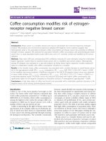
Fig. 1 Effect of CQ on breast cancer cell invasion and growth. a Conditioned media from irradiated 3T3 fibroblasts was added in the lower
compartment of invasion chamber and used as chemoattractant for breast cancer cells added in the upper compartment. Treatment with 5 μM
CQ completely blocked radiation-enhancement of invasion in TNBC cell lines. Invasiveness of the non-TBNC cell lines were not modified by the
irradiated 3T3 fibroblasts. CTL; Control, IRR; Irradiated, CQ; Chloroquine b Effect of CQ at 0, 2.5 or 5 μM on breast cancer cell growth measured
24, 48 and 72 h post treatment. Error bars indicate SEM. The experiment was realized in triplicate and repeated 3 times
Bouchard et al. BMC Cancer (2016) 16:361
the non-TNBC cell lines MCF-7 and MC7-L1, which
was not enhanced by irradiated fibroblasts, was also not
significantly modified by CQ.
Inhibition of D2A1 TNBC cell migration in mouse
mammary gland
As previously reported, D2A1 tumors implanted in preirradiated mammary glands were significantly smaller
compared to those in sham-irradiated mammary glands
[7]. Treatment with CQ at 40 mg/kg before each session
of irradiation, and thereafter at every 72 h, did not further affect tumor growth. The dose of CQ had to be increased to 60 mg/kg to measure a reduction in tumor
volume that was significant from day 18 in nonirradiated animals, and from day 21 in tumors implanted
in pre-irradiated mammary glands (Fig. 2a). To exclude
systemic effect of radiation on tumor growth, tumor volumes of sham-irradiated animals (sham tumors) were
compared to control tumors (left side) of pre-irradiated
animals as a validation of the mice as its own control in
following experiments (Additional file 1: Figure S1).
Page 6 of 14
The effect of CQ on radiation-stimulated migration of
D2A1 cells was then assessed. As measured with an animal optical imager, pre-irradiation of the mouse mammary gland increased by 1.7-fold (**P < 0.01) the
distance of D2A1 cell migration. This stimulation was
completely prevented by treating the animals with CQ at
40 mg/kg (*P < 0.05) or 60 mg/kg (**P < 0.01) (Fig. 2b
and c). These results were then confirmed by H&E staining (Fig. 2d and e).
Reduction of tumor vascularization
Since the anti-angiogenic ability of CQ was previously
reported [16], we determined whether this effect of CQ
was associated with the inhibition of radiationenhancement of TNBC cell migration. Pre-irradiation of
the mammary gland before implantation of D2A1 tumors did not modify the tumor vascularization compared to tumors implanted in non-irradiated mammary
glands, as measured with blood vessel marker CD31. On
the other hand, CQ treatment significantly decreased the
level of CD31 in tumors implanted in the pre-irradiated
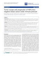
Fig. 2 Effect of CQ on D2A1 tumor growth and migration. a D2A1 tumor volumes measured after implantation in pre-irradiated or non-irradiated
mammary glands of animals treated with vehicle or CQ. Treatment with CQ at 60 mg/kg significantly reduced the tumor volume from day 18 in
non-irradiated animals, and from day 21 in tumors implanted in pre-irradiated mammary glands. b and c in vivo optical imaging of D2A1 cells in
mice mammary glands. White arrows = injection site of D2A1 cells. Cell migration in pre-irradiated mammary glands was enhanced by 1.7-fold
(**P < 0.01) compared to control side. Treatment with CQ at 40 mg/kg (*P < 0.05) or 60 mg/kg (**P < 0.01) completely blocked radiation-stimulation of
cell migration in mammary glands. d H&E staining from tumor sections confirming results observed in B and C. T = D2A1 tumor, MG = mammary
gland. e Quantification of tumor invasion using H&E staining. Invasion was calculated as follow: Invasion area (mm2)/Primary tumor area (mm2). Results
were reported as radiation-enhancement ratio. H&E quantification of tumor sections show a 3.2-fold increase of invasion (***P = 0.004) for tumors
implanted in pre-irradiated mammary glands that was completely prevented using CQ
Bouchard et al. BMC Cancer (2016) 16:361
and non-irradiated mammary glands (Fig. 3). This reduction was similar for the two doses of CQ studied.
Effect on cell cycle distribution
In our model, the FUCCI colorimetric vectors expressed
by the D2A1 cells generate a green fluorescence when
cells are in the S/G2/M phases and red fluorescence for
the G1/GO phases. Using these fluorescent makers, distribution of S/G2/M and G1/GO phases was determined
in frozen sections of tumors implanted in control or
pre-irradiated mammary glands. Stimulation of cancer
cell migration in pre-irradiated mammary gland was associated with an enrichment of D2A1 cells in G1/GO
phases (red fluorescence) by 36.4 % and a decrease in S/
G2/M phases (green fluorescence) by 11.7 %. Treatment
with CQ has completely prevented this enrichment in
the G1/GO phases, as well as the decrease of cells in S/
G2/M (Fig. 4a and b).
The cell proliferation marker Ki67 was then used to
further assess the effect of radiation and CQ on D2A1
cell proliferation. Treatment with CQ at 40 and 60 mg/
kg increased by 2-fold the levels of Ki67 expressed in
D2A1 tumors (Fig. 4c). Since the Ki67 marker is absent
from cells in G0 phase, this suggests that CQ has induced a transfer from quiescent to cycling cell state.
Control tumors were also compared with sham tumors
to exclude possible radiation-induced systemic bias on
proliferation (Additional file 2: Figure S2).
Reduction of lung metastasis development induced by
radiation
The preventive effect of CQ on the development of lung
metastasis stimulated by radiation was first assessed by
quantifying the number of circulating tumor cells
(CTC). In the first group of mice, the right mammary
Page 7 of 14
gland was pre-irradiated before implantation of D2A1
cells on both sides, while in the second group, the D2A1
cells were also implanted in both mammary glands but
in sham-irradiated animals. As we previously reported,
pre-irradiation of the mammary gland before the implantation of D2A1 cells increased the number of CTC
as well as the number of lungs metastases by 2.4-fold
compared to sham-irradiated mice [7]. CQ treatment
with 40 mg/kg and 60 mg/kg completely prevented the
radiation-enhancement of CTC which came back to the
basal level found in sham-irradiated animals (Fig. 5a).
Consequently, CQ also prevented the development of
lung metastasis induced by radiation (Fig. 5b and c), but
did not affect their diameter (Fig. 5d). Interestingly, CQ
did not decrease the basal number of lung metastases
compared to sham-irradiated animals that received the
vehicle. These results suggest that CQ selectively targeted a pathway associated with the radiation-stimulated
development of lung metastasis.
Effect of CQ on apoptosis and autophagy in D2A1 tumors
To further assess how CQ prevented the formation of
new metastases, apoptosis and autophagy were measured in D2A1 tumors. Treatment with 40 mg/kg of CQ
did not significantly modify the percentage of apoptotic
cells. An increase by 3-fold compared to vehicle was observed at 60 mg/kg CQ, but only in tumors implanted in
pre-irradiated mammary glands (****P < 0.0001) (Fig. 6a).
Quantification of autophagy markers LC3B1 and 2 by
Western blot was then performed in tumor homogenates. As expected, the expression of LC3B2 was increased by radiation, supporting an accumulation of
autophagosomes. This accumulation was then confirmed
to be an increase of autophagy since there is no accumulation of the p62 marker. On the other hand, the
Fig. 3 Effect of CQ on tumor vascularization. a Immunohistochemistry against CD31 endothelial marker in frozen tumor sections (magnification × 200).
b Quantification of CD31 signal plotted as percentage of stained area between control (sham) vs control + CQ, or irradiated vs irradiated + CQ.
***P < 0.001, ****P < 0.0001. Error bars indicate SEM for n = 3 to 14 independent experiments for each group
Bouchard et al. BMC Cancer (2016) 16:361
Page 8 of 14
Fig. 4 Effect of CQ on cell cycle distribution in D2A1 FUCCI tumors. a Representative fluorescence images of frozen sections of mammary tumors
used to quantify cancer cells in S/G2/M (green) or G1/G0 (red) phases. b Effect of radiation on cell cycle distribution plotted as radiation-enhancement
ratio of red and green cells in percentage. c Quantification of Ki67 by immunohistochemistry on D2A1 tumor frozen sections. *P < 0.05, **P < 0.01. Error
bars indicate SEM for n = 4 to 11 independent experiments for each group
blockage of autophagy, preferentially in tumors implanted in pre-irradiated mammary glands, was supported by the accumulation of p62 in CQ-treated
tumors, which is usually degraded when autophagy is
activated (Fig. 6b and Additional file 3: Figure S3).
Radiation-induced systemic bias on autophagy were
excluded by comparing autophagy marker in sham
and control tumors (Additional file 3: Figure S3 and
Additional file 4: Figure S4). Overall, autophagy was
preferentially induced in tumors implanted in preirradiated mammary glands underlying the importance
of tumor microenvironment affecting the tumor.
Assessment of pro-migratory and inflammatory factors
To characterize these adverse effects of radiation, some
pro-migratory and inflammatory factors were quantified
in pre-irradiated and control mammary glands. A CQ
dose of 40 mg/kg was chosen to exclude the induction
of cell death occurring at higher doses.
The proteases MMP-2 and MMP-9 are known to favor
the migration and invasion of cancer cells. Their levels
were determined by zymography in mammary glands
6 h after the last irradiation and 21 days after D2A1
tumor implantation (Fig. 7a and b). Radiation did not increase the levels of MMP-2 and −9 in the mammary
glands that were implanted/not implanted with the
D2A1 tumor. The level of either of these proteases was
not reduced after treatment with CQ at 40 mg/kg.
Expression of some inflammatory mediators potentially involved in cancer cell invasion were then quantified (Fig. 7c). The relative mRNA levels of IL-1β and
IL-6 were significantly increased 6 h post-irradiation, as
measured by qPCR. Regarding the pathway of prostaglandins, a higher expression of COX-2 and cPLA2
were also measured in irradiated mammary glands.
Bouchard et al. BMC Cancer (2016) 16:361
Page 9 of 14
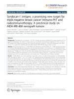
Fig. 5 Inhibition of radiation-enhancement of lung metastases with chloroquine. a Quantification of circulating tumor cells in blood samples of
sham and irradiated mice. b Optical imaging of lung metastases. ****P < 0.0001. c Quantification of the number of lung metastases. *P < 0.05,
**P < 0.01. Sham: Non-irradiated animals with tumor implantation on both sides. Irradiation: Pre-irradiation of the right mammary gland following by
tumors implantation on both sides. d Quantification of the diameter of lung metastases from optical imaging results. No significant difference was
observed for sham or irradiated mice, as for chloroquine treatment. Error bars indicate standard error of the mean (SEM) for n = 4 to 15 animals for
each group
Bouchard et al. BMC Cancer (2016) 16:361
Fig. 6 Apoptosis and autophagy analyses of D2A1 tumors. a TUNEL
assay quantification of the percentage of apoptotic cells in tumor
sections of each groups of mice. ****P < 0.0001. Error bars indicate
SEM for n = 3 to 6 independent experiments. b Immunoblot of protein
lysates from D2A1 tumors for autophagy markers. Experiment was
realized in triplicate
Treatment with CQ significantly decreased the expression
of IL-1β and IL-6 in both irradiated and non-irradiated
mammary glands, and completely inhibited the stimulation COX-2 and cPLA2 induced by radiation.
Discussion
For the subgroup of TNBC patients that responds poorly
to radiotherapy, the risk of recurrence is very high during the first three years after treatment and cure is unlikely [23]. The concept of radiation-stimulated cancer
cell migration and invasion is well accepted [24], but the
hypothesis suggesting that formation of metastasis could
be stimulated by radiation in some TNBC patients still
need to be validated. Meanwhile, it has been shown in
our previous pre-clinical study that pre-irradiation of a
Balb/c mouse mammary gland increased the migration
of murine TNBC cells, the number of CTC and favored
the development of lung metastases [7]. By irradiating
Page 10 of 14
the mammary gland prior to implantation of TNBC
cells, this previous study properly demonstrated the contribution of inflammatory mediators released from
healthy tissues on metastasis development.
In the present study, we first showed that these adverse effects of radiation were observed in vitro only in
the TNBC cell lines and that they can be prevented by
CQ. It should be noted that fibroblasts were used to
mimic the stroma in invasion chambers but the role of
other stromal components in radiation-enhancement of
breast cancer cells should not be excluded and requires
further investigation. Also, it remains to be determined
why radiation did not stimulate the invasion of nonTNBC cancer cells. Also, it is noteworthy that the protective effect of CQ in vitro was not related to inhibition
of cancer cell proliferation since no significant effect on
cell growth was measured.
Accumulation of CQ in the trans-Golgi network leads
to its alkalinization which deregulates the maturation of
many proteins, including MMP. MMP-2 and–9 play an
important role in cancer cell migration and invasion by
cleaving proteins of the extracellular matrix [25, 26]. In
the present study, no increase of MMP-2 and −9 was
found in irradiated Balb/c mouse mammary gland, and
treatment with CQ did not reduce their basal levels.
However, a possible involvement of these MMP in
breast cancer cell invasion cannot be ruled out since
an increased activity of these MMP and a stimulation
of cancer cell invasion was observed in other preclinical models such as irradiated mouse thigh and rat
brain [6, 13]. In breast cancer patients, radiotherapy
can increase the plasma level of MMP-9 [27] and the
level of MMP-2 was also significantly higher in skin biopsies of women after radiotherapy, relative to nonirradiated skin [28]. On the other hand, reduction of
MMP-2 and–9 expression in vitro in the MDA-MB231 cells was reported at higher doses of CQ than used
in our study [29]. Therefore, it remains to be determined in TNBC patients whether radiation can increase the expression of MMP-2 and–9, and whether
this can be prevented by CQ.
It was reported that the development of radiationstimulated lung metastasis after the irradiation of the
mammary gland was correlated with inflammatory pathways involving COX-2 as well as IL-1β and IL-6 cytokines [7]. As CQ is also used as an anti-inflammatory
agent for the treatment of rheumatoid arthritis and
lupus erythematous [16, 17], we determined whether its
anti-cancer effect could be associated with a downregulation of these inflammatory pathways.
In irradiated mouse mammary glands, the stimulation
of cPLA2 (the first enzyme in the production of prostaglandins) and COX-2 expression were completely prevented by CQ treatment. This inhibitory effect of CQ
Bouchard et al. BMC Cancer (2016) 16:361
Page 11 of 14
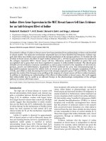
Fig. 7 Quantification of pro-migratory and pro-inflammatory factors after chloroquine treatment. a Zymogram of MMP-2 and −9 levels after
chloroquine treatment performed on protein lysates of both irradiated and non-irradiated mammary glands collected 6 h after irradiation.
b Zymogram of MMP-2 and −9 levels after chloroquine treatment performed on protein lysates of D2A1 tumors implanted in pre- irradiated
and non-irradiated mammary glands collected on sacrifice day (day 21). c Effect of chloroquine 40 mg/kg on the relative expression of
pro-inflammatory genes potentially in mammary quantified by qPCR 6 h after the last session of irradiation. Relative mRNA expressions are
plotted as a radiation enhancement ratio. *P < 0.05, **P < 0.01, ***P < 0.001, ****P < 0.0001. d Summary of the proposed mechanism of
chloroquine in the prevention of TNBC invasion stimulated by radiotherapy. Error bars indicate SEM. Experiments were realized in triplicate
may have a major impact on breast cancer patient survival. Indeed, elevated expression of COX-2 was associated with poor prognosis and distant metastases in
TNBC patients [30, 31], while radiation-enhancement of
cancer cell invasion as assessed in vitro can be completely prevented by adding a COX-2 inhibitor [12].
These results support the hypothesis that the inhibition
of COX-2 may increase the disease free-survival of
TNBC patients, as previously observed for early stage
non-TNBC patients [32].
It is noteworthy that CQ did not reduced the basal
levels of cPLA2 and COX-2 measured in non-irradiated
mammy glands. Since COX-2 is inducible only under
pathological or inflammatory conditions, this may suggest that the effect of CQ would be specific to irradiated
tissues, resulting in fewer adverse effects for nonirradiated healthy tissues.
We previously reported that the inflammatory cytokine IL-1β was increased in the conditioned media of fibroblasts following radiation. In the same study, IL-1β
stimulated the invasiveness of MDA-MB-231 TNBC
cells, and this invasive effect was prevented by adding an
anti-IL-1β antibody [33]. The resulting enhancement of
the invasion appears to be related to an increased expression of COX-2, since the addition of a COX-2 inhibitor
completely prevented the stimulation of cancer cell invasion induced by IL-1β [12, 33]. In our mouse model of
TNBC, the protective effect of CQ on metastasis development was also associated with a reduction of IL-1β expression, suggesting that this cytokine is a primary target
of CQ in the development of lungs metastases.
Regarding IL-6, it is the most important cytokine associated with poor prognosis for breast cancer, and it is
known for controlling breast cancer cell growth and
Bouchard et al. BMC Cancer (2016) 16:361
regulating cancer stem cell renewal [34]. IL-6 has also
been reported to stimulate the proliferation and migration of breast cancer cells in vitro as well as tumor
progression [35], but its potential connection with radiotherapy was less studied [34]. Nevertheless, Yu et al. reported that radiation-induced IL-6 in MDA-MB-231cells
promoted the invasion and migration of non-irradiated
neighboring cells [36]. In our mouse model, CQ reduced
the expression of IL-6 in irradiated and non-irradiated
mammary glands in the same manner observed with IL1β suggesting that this cytokine could also be associated
with induction of lung metastasis.
Irradiation of healthy tissues surrounding a tumor can
modify the balance between proliferation and migration
of cancer cells [7, 13]. This migration/proliferation dichotomy was described as mutually exclusive or as a
«Go or Grow» phenomenon [37]. Using the FUCCI cell
cycle reporter system [38], irradiation of a rat brain or a
mouse mammary gland favored the migration of cancer
cells and their accumulation in the G1/G0 phases [7, 13].
This suggests that cytokines released from irradiated tissues could stimulate the migration/invasion of cancer
cells through a reduction of their proliferation. Treatment with CQ has successfully reduced the radiationenhanced accumulation of D2A1 cells in the G1/G0
phases (red fluorescence), supporting the inhibition of
radiation-induced migration in mammary glands. These
results are consistent with the decrease of G1/G0 cells
after CQ treatment previously observed in human
TNBC cell lines by Jiang et al.[39]. The authors reported
the induction of cell cycle arrest in G2/M which may
affect the interpretation of cell proliferation with the
marker Ki67. This marker of cell proliferation is present
in both G2 and M phases. Consequently, an arrest in
G2/M may increase the number of Ki67 positive cells,
giving the false indication that more tumor cells are proliferating. Indeed, the increased number of Ki67 positive
cells measured in our study is expected to be associated
with a cell blockage in G2/M rather than an increase of
cell proliferation.
The reduction of CTC and the number of lung metastases was not caused by a reduction of tumor blood supply since the presence of CD31 blood vessel marker was
not affected by radiation. It was then impossible to associate the protective effect of CQ with the reduction of
tumor vascularization.
Our results showed that stimulation of metastasis development stimulated by radiation was inhibited by CQ without affecting the tumor volume. Our results also showed
that a low level of apoptosis was only promoted in D2A1
tumors with high dose of CQ (60 mg/kg) in presence of
radiation but not with 40 mg/kg of CQ. This suggests that
the adverse effect of radiation on the development of metastasis can be prevented by low doses of CQ that would
Page 12 of 14
not induce apoptosis in healthy tissues. Consequently, a
low systemic toxicity after treatment with CQ could be
expected.
CQ is also described as an inhibitor of autophagy. Autophagy is a survival pathway activated in response to stress
whereby cellular components are degraded to recycle energy, promote cell survival and cancer resistance. However,
if the cells cannot recover from the damage, autophagy will
ultimately lead to cell death. Therefore, autophagy could
also exert a significant control over the progression of cancer and tissue homeostasis [40]. Our results showed that
treatment with CQ blocked autophagy. These findings are
consistent with those of Jensen et al. [41], who reported
that CQ was highly effective in preventing autophagy.
These authors also reported that CQ preferentially accumulated in acidic tumor environment than in normal tissue,
suggesting that CQ could be less non-toxic for normal
tissues. The increase of autophagy observed in tumor
implanted in pre-irradiated tissue could be directly associated to this previous observation. Overall, according to the experimental conditions, autophagy can be
either cytotoxic (prolonged autophagy will eventually
lead to cell death) or cytoprotective (survival mechanism for the cell). Autophagy is clearly a complex
process and its role in TNBC patients remains to be
further explored. Without knowing how exactly autophagy was regulated, the preferential blocking of autophagy associated with the accumulation of LC3B2
observed for tumors implanted in pre-irradiated mammary glands seems to be associated with the prevention
of the radiation-stimulated of breast cancer cell
migration.
Combined with radiation, CQ successfully induced cell
death in several human TNBC cell lines [42, 43]. Zhao
et al. have shown the radiosensitivity potential of CQ in
MDA-MB-231 TNBC cells, by reporting enhanced apoptosis and necrosis [42]. In our study, the mammary gland
was irradiated before its implantation with D2A1 cells.
Therefore, the anti-cancer effect of CQ cannot be related to
a direct radiosensitization but rather to an indirect effect
on cancer cells that is mediated by irradiated stroma. The
experimental protocol used in this study has provided to
confirm that CQ prevents the stimulation of the metastasis
development induced by the irradiated stroma. Taken together, these results suggest that treating TNBC patients
with CQ could further increase the anti-tumor effect of
radiotherapy and reduce the potential adverse effects of
radiation-induced inflammation on the stimulation of metastasis development.
Conclusion
In conclusion, the ability of radiation to stimulate the invasion of cancer cells was observed in vitro only in
TNBC cell lines. In our mouse model of TNBC,
Bouchard et al. BMC Cancer (2016) 16:361
radiation stimulates the cancer cell migration and development of metastasis which seems to involve multiple
inflammatory pathways including those of COX-2, IL-1β
and IL-6. These adverse effects of radiation were prevented by treating the animals with CQ. A proposed
mechanism is presented in Fig. 7d. Based on these results, a clinical trial to determine whether treatment
with CQ could increase the disease-free survival of the
TNBC patients that poorly respond to radiation treatment could be undertaken.
Additional files
Additional file 1: Figure S1. Validation of the mice as its own control
in mice pre-irradiated at the right mammary gland. D2A1 tumor volumes
of sham irradiated animals (sham tumors) were compared to control
tumors (left side) of pre-irradiated animals. Error bars indicate s.e.m. for n
= 6 to 15 animals for each group. (TIF 224 kb)
Additional file 2: Figure S2. Ki67 immunohistochemistry in sham (nonirradiated animals) and control tumors (left side of pre-irradiated animals)
were realized to exclude possible systemic effect of radiation on tumor
proliferation. The experiment was realized in triplicate. Sham-VH vs ShamCQ 40; P < 0.0001, Sham-VH vs CTL-CQ 40; P = 0.0002, Sham-VH vs ShamCQ 60; P = 0,0001, Sham-VH vs CTL-CQ 60; P < 0,0001, CTL-VH vs ShamCQ 40; P < 0,0001, CTL-VH vs CTL-CQ 40; P = 0,0002, CTL-VH vs Sham-CQ
60; P = 0,0001, CTL-VH vs CTL-CQ 60; P < 0,000.
Additional file 3: Figure S3. Immunoblot of autophagy markers were
realized in sham (non-irradiated animals) and control tumors (left side of
pre-irradiated animals) to exclude possible systemic effect of radiation on
tumor autophagy. The experiment was realized in triplicate. (TIF 281 kb)
Additional file 4: Figure S4. Quantitative densitometry from Western
blots of the expression of (A) LCB3I, (B) LCB3II (Sham-CQ 60 vs IRR-CQ 60;
P = 0.0024, CTL-CQ 60 vs IRR-CQ 60; P = 0.0182, IRR-VH vs IRR-CQ 60; P =
0.0009) and (C) p62 autophagy markers calculated using ImageJ Gel
Analyze function.
Additional file 5: Figure S5. Hormonal status of D2A1 cell line was
confirmed by immunohistochemistry as described in Materials and
Methods. No nuclear (ER and PR) as well as membrane (HER2) staining
were observed. D2A1 cells were then revealed to be triple negative by a
pathologist of our institution. (TIF 9131 kb)
Abbreviations
BSA, bovine serum albumin; CD31, cluster of differentiation 31; Co, cobalt;
COX-2, cyclooxygenase-2; cPLA2, cytosolic Phospholipase A2; CQ, chloroquine;
CTC, circulating tumor cell; CTL, control; DMEM, Dulbecco modified Eagle’s
medium; ER, estrogen receptor; FITC, fluorescein isothiocyanate; FUCCI,
fluorescent ubiquitinated-based cell cycle indicator; H&E, haematoxylin and
eosin; HER2, human epidermal growth factor receptor 2; HRP, horse radish
peroxidase; i.p., intraperitoneal; IL-1β, interleukin-1 beta; IL-6, interleukin-6; IRR,
irradiated; LC3, light chain 3; mAG, monomeric Azami Green; mKO2, monomeric
Kusabira Orange 2; MMP, matrix metalloproteinase; OCT, optimum cutting
temperature; PR, progesterone receptor; qPCR, quantitative polymerase chain
reaction; TNBC, triple negative breast cancer; TRITC, tetramethylrhodamine
isothiocyanate; VH, vehicle.
Acknowledgements
BP, RB, CS and YBL are members of the Fonds de la Recherche en Santé du
Québec (FRSQ)-funded Centre de recherche CHUS. CS is a FRSQ scholar and
is also funded by the Canadian Foundation for Innovation. GB held a
scholarship from FRSQ (grant # 27479). We thank Réjean Lebel for his
graceful help for the in vivo imaging implementation techniques. The
medical physicists, Patrick Delage and Vincent-Hubert Tremblay, are thanked
for their very helpful dosimetry calculations for mice irradiation. The authors
thank the Electron Microscopy & Histology Research Core of the FMSS at the
Université de Sherbrooke for their histology, electron microscopy and
Page 13 of 14
phenotyping services. This research project was supported by the Canadian
Institutes of Health Research (grant # 184671).
Availability of data and materials
Not applicable.
Authors’ contributions
GB performed all animal experiments, analyses, results interpretation and
drafted the manuscript. HT contributed to in vitro experiments and generated
FUCCI cells. GB, CS and BP conceptualized the study. SG contributed to
pathological analysis. YBL contributed to in vivo imaging experiments. BP, CS,
RB, YBL and SG contributed to writing and revising the manuscript. All authors
contributed to critical analysis and approval of the final manuscript. All authors
read and approved the final manuscript.
Authors’ information
1
Centre for Research in Radiotherapy, Department of Nuclear Medicine and
Radiobiology, 2Department of Anatomy and Cellular Biology, Faculty of
Medicine and Health Sciences, Université de Sherbrooke, 3Service of
Radiation Oncology, 4Department of Pathology, Centre Hospitalier
Universitaire de Sherbrooke, 5Centre d’imagerie moléculaire de Sherbrooke
and Department of Electrical and Computer Engineering, Université de
Sherbrooke, 3001 12e avenue Nord, Sherbooke (Québec), J1H 5 N4, Canada.
Competing interests
The authors report no conflicts of interest. The authors alone are responsible
for the content and writing of the paper.
Consent for publication
Not applicable.
Ethics approval and consent to participate
The experimental protocols were approved by the Ethics Committee for
Animal Care and Use of the Université de Sherbrooke in accordance with
guidelines established by the Canadian Council on Animal Care (Protocol ID
number 013–14).
Author details
1
Centre for Research in Radiotherapy, Department of Nuclear Medicine and
Radiobiology, Université de Sherbrooke, 3001, 12e Avenue Nord, Sherbrooke,
Québec J1H 5 N4, Canada. 2Department of Anatomy and Cellular Biology,
Faculty of Medicine and Health Sciences, Université de Sherbrooke,
Sherbrooke, Canada. 3Service of Radiation Oncology, Université de
Sherbrooke, Sherbrooke, Canada. 4Department of Pathology, Centre
Hospitalier Universitaire de Sherbrooke, Sherbrooke, Canada. 5Department of
Electrical and Computer Engineering, Centre d’imagerie moléculaire de
Sherbrooke, Sherbrooke, Québec, Canada.
Received: 15 October 2015 Accepted: 1 June 2016
References
1. Irvin WJ, Carey LA. What is triple-negative breast cancer? Eur J Cancer. 2008;
44:2799–805.
2. Gluz O, Liedtke C, Gottschalk N, Pusztai L, Nitz U, Harbeck N. Triple-negative
breast cancer - current status and future directions. Ann Oncol. 2009;20:
1913–27.
3. Gallet P, Phulpin B, Merlin JL, Leroux A, Bravetti P, Mecellem H, et al. Longterm alterations of cytokines and growth factors expression in irradiated
tissues and relation with histological severity scoring. PLoS One. 2011;6,
e29399.
4. Rodemann HP, Blaese MA. Responses of normal cells to ionizing radiation.
Semin Radiat Oncol. 2007;17:81–8.
5. Mantovani A, Allavena P, Sica A, Balkwill F. Cancer-related inflammation.
Nature. 2008;454:436–44.
6. Lemay R, Archambault M, Tremblay L, Bujold R, Lepage M, Paquette B.
Irradiation of normal mouse tissue increases the invasiveness of mammary
cancer cells. Int J Radiat Biol. 2011;87:472–82.
7. Bouchard G, Bouvette G, Therriault H, Bujold R, Saucier C, Paquette B. Preirradiation of mouse mammary gland stimulates cancer cell migration and
development of lung metastases. Br J Cancer. 2013;109:1829–38.
Bouchard et al. BMC Cancer (2016) 16:361
8.
9.
10.
11.
12.
13.
14.
15.
16.
17.
18.
19.
20.
21.
22.
23.
24.
25.
26.
27.
28.
29.
Qian L, Mizumoto K, Urashima T. Radiation-induced increase in invasive
potential of human pancreatic cancer cells and its blockade by a matrix
metalloproteinase inhibitor, CGS27023. Clin Cancer Res. 2002;4:1223–7.
Christine W-B, Weller M, Gangwick W. Molecular determinants of glioma cell
migration and invasion. J Neurosurg. 2001;94:978–84.
Rofstad EK, Mathiesen B, Galappathi K. Increased metastatic dissemination in
human melanoma xenografts after subcurative radiation treatment :
radiation-induced increase in fraction of hypoxic cells and hypoxia-induced
up-regulation of urokinase-type plasminogen activator Receptor. Cancer
Res. 2004;64:13–8.
Wang JL, Sun Y, Wu S. Gamma-irradiation induces matrix metalloproteinase
II expression in a p53-dependent manner. Mol Carcinog. 2000;27:252–8.
Paquette B, Therriault H, Desmarais G, Wagner R, Royer R, Bujold R.
Radiation-enhancement of MDA-MB-231 breast cancer cell invasion
prevented by a cyclooxygenase-2 inhibitor. Br J Cancer. 2011;105:534–41.
Desmarais G, Fortin D, Bujold R, Wagner R, Mathieu D, Paquette B.
Infiltration of glioma cells in brain parenchyma stimulated by radiation in
the F98/Fischer rat model. Int J Radiat Biol. 2012;88:565–74.
Thomas S, Sharma N, Golden EB, Cho H, Agarwal P, Gaffney KJ, et al.
Preferential killing of triple-negative breast cancer cells in vitro and in vivo
when pharmacological aggravators of endoplasmic reticulum stress are
combined with autophagy inhibitors. Cancer Lett. 2012;325:63–71.
Solomon VR, Lee H. Chloroquine and its analogs: a new promise of an old
drug for effective and safe cancer therapies. Eur J Pharmacol. 2009;625:220–33.
Lesiak A, Narbutt J, Sysa-Jedrzejowska A, Lukamowicz J, McCauliffe DP,
Wózniacka A. Effect of chloroquine phosphate treatment on serum MMP-9
and TIMP-1 levels in patients with systemic lupus erythematosus. Lupus.
2010;19:683–8.
Lesiak A, Narbutt J, Kobos J, Kordek R, Sysa-Jedrzejowska A, Norval M, et al.
Systematic administration of chloroquine in discoid lupus erythematosus
reduces skin lesions via inhibition of angiogenesis. Clin Exp Dermatol. 2009;
34:570–5.
Ulrich CM, Bigler J, Potter JD. Non-steroidal anti-inflammatory drugs for
cancer prevention: promise, perils and pharmacogenetics. Nat Rev Cancer.
2006;6:130–40.
Rak JW, McEachern D, Miller FR. Sequential alteration of peanut agglutinin
binding-glycoprotein expression during progression of murine mammary
neoplasia. Br J Cancer. 1992;65:641–8.
Lanari C, Luthy I, Lamb CA, Fabris V, Pagano E, Helguero LA,Sanjuan N,
Merani S, Molinolo AA. Five novel hormone-responsive cell lines derived
from murine mammary ductal carcinomas: in vivo and in vitro effects of
estrogens and progestins. Cancer Res. 2001;61:293–302.
Balin-Gauthier D, Delord JP, Rochaix P, Mallard V, Thomas F, Hennebelle I,
Bugat R, Canal P, Allal C: In vivo and in vitro antitumor activity of oxaliplatin in
combination with cetuximab in human colorectal tumor cell lines expressing
different level of EGFR. Cancer Chemother Pharmacol. 2006;57:709–18.
Pham NA, Morrison A, Schwock J, Aviel-Ronen S, Iakovlev V, Tsao MS, Ho J,
Hedley DW. Quantitative image analysis of immunohistochemical stains
using a CMYK color model. Diagn Pathol. 2007;2:8.
Langlands FE, Horgan K, Dodwell DD, Smith L. Breast cancer subtypes: response
to radiotherapy and potential radiosensitisation. Br J Radiol. 2013;86:20120601.
Moncharmont C, Levy A, Guy JB, Falk AT, Guilbert M, Trone JC, Alphonse G,
Gilormini M, Ardail D, Toillon RA, Rodriguez-Lafrasse C, Magne N. Radiationenhanced cell migration/invasion process: A review. Crit Rev Oncol
Hematol. 2014;92:133–42.
Basque J, Martel M, Leduc R, Cantin AM. Lysosomotropic drugs inhibit
maturation of transforming growth factor-beta. Can J Physiol Pharmacol.
2008;86:606–12.
Overall CM, Wrana JL, Sodek J. Independent regulation of collagenase, 72-kDa
progelatinase, and metalloendoproteinase inhibitor expression in human
fibroblasts by transforming growth factor-beta. J Biol Chem. 1989;264:1860–9.
Susskind H, Hymowitz MH, Lau YH, Atkins HL, Hurewitz AN, Valentine ES,
Meek AG, Zucker S. Increased plasma levels of matrix metalloproteinase-9
and tissue inhibitor of metalloproteinase-1 in lung and breast cancer are
altered during chest radiotherapy. Int J Radiat Oncol. 2003;56:1161–9.
Riekki R, Jukkola A, Sassi ML, Hoyhtya M, Kallioinen M, Risteli JOikarinen A.
Modulation of skin collagen metabolism by irradiation: collagen synthesis is
increased in irradiated human skin. Br J Dermatol. 2000;142:874–80.
Tuomela J, Sandholm J, Kauppila JH, Lehenkari P, Harris KW, Selander KS.
Chloroquine has tumor-inhibitory and tumor-promoting effects in triplenegative breast cancer. Oncol Lett. 2013;6:1665–72.
Page 14 of 14
30. Hsia TC, Tu CY, Chen YJ, Wei YL, Yu MC, Hsu SC, Tsai SL, Chen WS, Yeh MH, Yen
CJ, Yu YL, Huang TC, Huang CY, Hung MC, Huang WC. Lapatinib-mediated
cyclooxygenase-2 expression via epidermal growth factor receptor/HuR
interaction enhances the aggressiveness of triple-negative breast cancer cells.
Mol Pharmacol. 2013;83:857–69.
31. Zhou L, Li K, Luo Y, Tian L, Wang M, Li C, Huang Q. Novel prognostic markers
for patients with triple-negative breast cancer. Hum Pathol. 2013;44:2180–7.
32. Litzenburger BC, Brown PH. Advances in preventive therapy for estrogenreceptor-negative breast cancer. Curr Breast Cancer Rep. 2014;6:96–109.
33. Paquette B, Therriault H, Wagner JR. Role of interleukin-1β in radiationenhancement of MDA-MB-231 breast cancer cell invasion. Radiat Res. 2013;
180:292–8.
34. Dethlefsen C, Hojfeldt G, Hojman P. The role of intratumoral and systemic
IL-6 in breast cancer. Breast Cancer Res Treat. 2013;138:657–64.
35. Di G-H, Liu Y, Lu Y, Liu J, Wu C, Duan H-F. IL-6 secreted from senescent
mesenchymal stem cells promotes proliferation and migration of breast
cancer cells. PLoS One. 2014;9, e113572.
36. Yu Y-C, Yang P-M, Chuah Q-Y, Huang Y-H, Peng C-W, Lee Y-J, Chiu S-J.
Radiation-induced senescence in securin-deficient cancer cells promotes
cell invasion involving the IL-6/STAT3 and PDGF-BB/PDGFR pathways. Sci
Rep. 2013;3:1–11.
37. Giese A, Loo MA, Tran N, Haskett D, Coons SW, Berens ME. Dichotomy of
astrocytoma migration and proliferation. Int J cancerJournal Int du cancer.
1996;67:275–82.
38. Sakaue-Sawano A, Kurokawa H, Morimura T, Hanyu A, Hama H, Osawa H,
Kashiwagi S, Fukami K, Miyata T, Miyoshi H, Imamura T, Ogawa M, Masai H,
Miyawaki A. Visualizing spatiotemporal dynamics of multicellular cell-cycle
progression. Cell. 2008;132:487–98.
39. Jiang P, Zhao Y, Shi W, Deng X, Xie G, Mao Y, Li Z, Zheng Y, Yang S. Cellular
physiology and biochemistry biochemistry cell growth inhibition, G2/M cell
cycle arrest, and apoptosis induced by chloroquine in human breast cancer
cell line Bcap-37. Cell Physiol Biochem. 2008;22:431–40.
40. Chen N, Karantza-Wadsworth V. Role and regulation of autophagy in cancer.
Biochim Biophys Acta. 2009;1793:1516–23.
41. Jensen PB, Sandahi B, Sehested M, Grue P, Demant EJF, Hansen HH. Tumor
Cells in Acidic Environments’. Cancer Res. 1994;54(11):2959–63.
42. Zhao H, Cai Y, Santi S, Lafrenie R, Lee H. Chloroquine-mediated
radiosensitization is due to the destabilization of the lysosomal membrane and
subsequent induction of cell death by necrosis. Radiat Res. 2005;164:250–7.
43. Chaachouay H, Ohneseit P, Toulany M, Kehlbach R, Multhoff G, Rodemann
HP. Autophagy contributes to resistance of tumor cells to ionizing radiation.
Radiother Oncol. 2011;99:287–92.
44. Kau P, Nagaraja GM, Zheng H, Gizachew D, Galukande M, Krishnan S, Asea
A. A mouse model for triple-negative breast cancer tumor-initiating cells
(TNBC-TICs) exhibits similar aggressive phenotype to the human disease.
BMC Cancer. 2012;12:120.
45. Soule HD, Vazguez J, Long A, Albert S, Brennan M. A human cell line from a
pleural effusion derived from a breast carcinoma. J Natl Cancer Inst. 1973;51:
1409–16.
46. Holliday DL, Speirs V. Choosing the right cell line for breast cancer research.
Breast Cancer Res. 2011;13:215.
Submit your next manuscript to BioMed Central
and we will help you at every step:
• We accept pre-submission inquiries
• Our selector tool helps you to find the most relevant journal
• We provide round the clock customer support
• Convenient online submission
• Thorough peer review
• Inclusion in PubMed and all major indexing services
• Maximum visibility for your research
Submit your manuscript at
www.biomedcentral.com/submit