Patterns of recurrence after selective postoperative radiation therapy for patients with head and neck squamous cell carcinoma
Bạn đang xem bản rút gọn của tài liệu. Xem và tải ngay bản đầy đủ của tài liệu tại đây (629.1 KB, 10 trang )
Murakami et al. BMC Cancer (2016) 16:192
DOI 10.1186/s12885-016-2229-x
RESEARCH ARTICLE
Open Access
Patterns of recurrence after selective
postoperative radiation therapy for patients
with head and neck squamous cell
carcinoma
Naoya Murakami1*, Fumihiko Matsumoto2, Seiichi Yoshimoto2, Yoshinori Ito1, Taisuke Mori3, Takao Ueno4,
Keisuke Tuchida1, Tairo Kashihara1, Kazuma Kobayashi1, Ken Harada1, Mayuka Kitaguchi1, Shuhei Sekii1,
Rei Umezawa1, Kana Takahashi1, Koji Inaba1, Hiroshi Igaki1 and Jun Itami1
Abstract
Background: The radiation field for patients with postoperative head and neck squamous cell carcinoma is
narrower in our institution than in Western countries to reduce late radiation related toxicities. This strategy is
at a risk of loco-regional or distant metastasis. However, because patients are more closely checked than in
Western countries by every 1 to 2 months intervals and it is supposed that regional recurrences are identified
and salvage surgeries are performed more quickly. Therefore, it is considered that patient survival would not
be compromised with this strategy. The aim of this study was to investigate the feasibility of this strategy retrospectively.
Methods: Patients who underwent neck dissection with close or positive margin, extra-capsular spread (ECS), multiple
regional lymph node metastasis, pT4, with or without primary tumor resection were treated with postoperative radiation
therapy. The volume of radiation field, especially the coverage of prophylactic regional lymph node area, was discussed
among head and neck surgeons and radiation oncologists taking into account the clinical factors including patient’s age,
performance status, number of positive lymph nodes, size of metastatic lymph nodes, extension of primary
tumor beyond the midline, and existence of ECS.
Results: Seventy-two patients were identified who were treated with postoperative radiation therapy for head
and neck squamous cell carcinoma between November 2005 and December 2014. There were 20 patients
with oropharynx, 19 with hypopharynx, 7 with larynx, 23 with oral cavity, and 3 with other sites. Thirty eight
patients had their neck irradiated bilaterally and 34 unilaterally. Median follow-up period for patients without
relapse was 20.7 months (5.1–100.7). Thirty two patients had disease relapse after treatment including 22 loco-regional
recurrence and 14 distant metastases. Among 22 loco-regional recurrence, seven patients underwent salvage surgery
and one of them was no relapse at the time of the analysis. Among patients without bilateral neck lymph
node metastasis who were treated with unilateral neck irradiation, patients with oral cavity or recurrent
disease had significantly lower DFS compared with those without (2-y DFS 41.7 % vs 88.2 %, p = 0.017).
Conclusions: In patients without bilateral neck lymph node involvement, the postoperative unilateral neck
irradiation is a reasonable treatment strategy for patients with the exception of oral cavity or recurrent disease.
Keywords: Head and neck squamous cell carcinoma, Postoperative radiation therapy, Patterns of recurrence, Selective
neck irradiation
* Correspondence:
1
Department of Radiation Oncology, National Cancer Center Hospital, 5-1-1
Tsukiji, Chuo-ku, Tokyo 104-0045, Japan
Full list of author information is available at the end of the article
© 2016 Murakami et al. Open Access This article is distributed under the terms of the Creative Commons Attribution 4.0
International License ( which permits unrestricted use, distribution, and
reproduction in any medium, provided you give appropriate credit to the original author(s) and the source, provide a link to
the Creative Commons license, and indicate if changes were made. The Creative Commons Public Domain Dedication waiver
( applies to the data made available in this article, unless otherwise stated.
Murakami et al. BMC Cancer (2016) 16:192
Background
According to statistics from Cancer Information Service
in Japan, death from head and neck malignant tumors
in Japan (malignant tumors arising from oral cavity,
pharynx, and larynx) was 8142 in 2013 and this figure
accounts for 2.2 % of all the death from malignant
tumors [1]. Although the percentage is decreasing,
the smoking rate in 2013 was 32.2 % in male and
8.2 % in female and still many people smoke in our
country [2].
In 1970’s, Radiation Therapy Oncology Group (RTOG)
73–03 trial was carried out to compare preoperative
with postoperative radiation therapy combined with
surgical resection for patients with advanced operable
squamous cell carcinoma of the supraglottic larynx or
hypopharynx in the context of a phase III study [3].
Loco-regional control was significantly favorable for
patients assigned to postoperative radiation therapy
compared with those assigned to preoperative radiation therapy (65 % vs 48 %, p = 0.04), and the postoperative radiation therapy has been a standard of
care for patients with advanced resectable head and
neck squamous cell carcinoma (HNSCC). Nevertheless, development of distant metastasis was frequently
observed in both arms and the addition of chemotherapy to surgery and adjuvant radiation therapy was
considered as a next important issue. In the Intergroup study 0034 (or RTOG 85–03), a randomized
clinical trial was conducted by cooperative groups
which was consisted of RTOG, Southwest Oncology
Group (SWOG), Eastern Cooperative Oncology Group
(ECOG), Cancer and Leukemia Group B (CALGB),
Northern California Oncology Group (NCOG), and
Southwest Group (SEG), patients with advanced HNSCC
were randomly assigned either to postoperative radiation
alone or sequential three cycles of cis-platinum and 5-FU
followed by postoperative radiotherapy [4]. While distant
metastasis-free survival was significantly improved in sequential CT/RT arm (23 % vs 15 %, p = 0.02), both locoregional relapse-free survival and overall survival did not
differ between the two arms and the concomitant use of
chemotherapy and radiation therapy was awaited. In 2004,
the European Organization for Research and Treatment
of Cancer (EORTC) and RTOG published simultaneously
the results of two phase III trials (the EORTC 22931 and
the RTOG 95–01) which compared concurrent postoperative chemoradiation using tri-weekly 100 mg/m2 of cisplatinum with postoperative radiotherapy alone [5, 6].
There were slight differences in settings between these
two phase III clinical trials. In the RTOG 95–01, the primary endpoint was the rates of local and regional control
whereas, in the EORCT 22931 it was chosen to be the
progression-free survival. The definition of the high-risk
characteristics also differed between these two trials. In
Page 2 of 10
the RTOG 95–01, the following characteristics were defined as high-risk; histologic evidence of invasion of two
or more regional lymph nodes, extra-capsular spread
(ECS) of nodal disease, and microscopically involved mucosal margins of resection. On the other hand, in the
EORTC 22931, the following characteristics were defined
as high-risk; ECS, positive resection margins, perineural
involvement, vascular tumor embolism, or tumors with
involved lymph nodes at level IV or V from carcinomas
arising in the oral cavity or oropharynx. While primary
endpoint of these two phase III clinical trials were both
met and overall survival benefit was demonstrated in the
EORTC 22931 trial (p = 0.02), the RTOG 95–01 showed
only a trend in the same direction in overall survival
(p = 0.19). Bernier et al. conducted a comparative analysis using data pooled from the EORTC 22931 and
the RTOG 95–01 to identify which patients require
adjuvant concomitant chemoradiation following surgery and they concluded that microscopically involved
resection margins and ECS of tumor from neck nodes
were the most significant adverse factors for poor
outcome [7]. Therefore, concurrent chemoradiation
(cCRT) is a standard therapy for postoperative highrisk HNSCC patients.
Originally the radical neck dissection (RND) consists
of removal of all the lymphatic as well as non-lymphatic
structures from the mastoid process down to the clavicle
except the carotid artery, brachial plexus, hypoglossal,
lingual, vagus, and phrenic nerves [8, 9], which demands
heavy burden to patients. Later on, selective neck dissection (SND) was introduced which preserved one or more
lymph node levels [10] and the development of common
terminology of discriminating neck levels which was
well-known as the classification of American Head and
Neck Society (AHNS) followed [11, 12]. However, the
applicability of the concept of the selective nodal irradiation in postoperative setting is controversial [13–15].
Gregoire et al. [13] and Chao et al. [14] proposed the
clinical target volume (CTV) guidelines for postoperative
neck region, but the authors admitted the paucity of
data on which one could create a specific guideline for
postoperative CTV. According to the guideline of Chao
et al., only patients with buccal T1-2 N0 and tonsil
T1-2 N0 were allowed for hemi-neck postoperative
radiation. After extensive neck irradiation patients
usually are suffered from late radiation toxicities, in
especially chemotherapy was administered concurrently with radiotherapy. In our institution radiation
field for patients with postoperative head and neck
squamous cell carcinoma is narrower than in Western
countries to reduce late radiation related toxicities.
This strategy is at a risk of loco-regional recurrence
and/or distant metastasis. However, because patients
are checked closely by every 1 to 2 months intervals
Murakami et al. BMC Cancer (2016) 16:192
and the salvage surgery would be performed immediately after the identification of the regional recurrences, therefore, it is considered that the patient’s
survival would not be compromised with this treatment strategy. This retrospective study was conducted
to investigate the feasibility of this strategy.
Methods
All consecutive patients with HNSCC who underwent
neck dissection and received postoperative radiation
therapy were recruited for this study. In our institution,
patients with HNSCC who underwent neck dissection
with pathologic findings of close or positive resection
margin, ECS, multiple regional lymph node metastasis,
or pT4, with or without primary tumor resection were
treated with postoperative radiation therapy. Surgical
margin status was defined as follows; close margins were
defined as ≤ 3 mm and positive margins defined as
tumor touching an inked surface.
From April 2011 concurrent chemoradiation (cCRT)
with tri-weekly CDDP 80 mg/m2, and from March 2013
Cetuximab-radiation according to the Bonner protocol
[16] was introduced in our institution for patients with
positive resection margin or ECS. Because there is no evidence supporting the superiority of Cetuximab-radiation
over platinum-based cCRT in the management of advanced HNSCC, our first choice was cCRT. However, if
patients did not have enough kidney function with favorable performance status, Cetuximab-radiation was chosen.
From June 2009 neoadjuvant chemotherapy (NAC)
was started as chemoselection for patients with advanced HNSCC who required total laryngectomy or who
expected severe postoperative pharyngeal dysfunction. If
favorable response was achieved after two to three cycles
of induction chemotherapy, subsequent cCRT was
followed with or without neck dissection. If not, total
laryngectomy or appropriate surgery was applied. NAC
was also applied as induction chemotherapy for patients
with far-advanced disease for whom it was impossible to
separate metastatic lymph nodes from carotid artery
which precludes radical operation or patients with N2c
and/or lower neck metastasis who’s possibility of developing distant metastasis soon after surgery was expected
to be very high. Agents used for NAC was either the
combination of CDDP and 5FU or CDDP, 5FU, and
docetaxel.
The extent of prophylactic neck resection was determined by the status of primary lesion. If primary lesion
extended midline, prophylactic contralateral neck dissection was applied. Otherwise, unilateral prophylactic neck
dissection was performed.
Patients with distant metastasis, treated for palliative
intention, or for salvage intention after recurrence without surgical resection were excluded from this study.
Page 3 of 10
Radiotherapy
Radiotherapy was prescribed in 2-Gy fractions with 4
or 6-MV photons in either three-dimensional conformal radiotherapy (3DCRT) or intensity-modulated
radiotherapy (IMRT). From September 2008 a simultaneous integrated boost intensity-modulated radiotherapy (SIB-IMRT) using sliding window technique
or volumetric modulated arc therapy (VMAT) by dynamic MLC system (Varian Medical Systems, Palo
Alto, CA) was introduced in our institution, in case
of the CTV contained large volume of major salivary
gland, oral cavity, larynx, or pharynx. Our IMRT procedure for head and neck cancer patients is described
in a previous report [17]. Patients were immobilized
from head to shoulders with thermoplastic masks in
the supine position. Target volumes were defined as
follows: no gross tumor volume (GTV) was defined
except patients without primary lesion resection because all gross tumor was resected during operation.
The high-risk CTV 60–66 Gy (CTV60-66Gy) was defined as areas considered as high risk for having
microscopic disease such as positive surgical margin
or metastatic lymph node with ECS based on preclinical imaging, preoperative physical exam/endoscopy,
operative findings, and final pathologic findings. The
intermediate-risk CTV (CTV44Gy for 3DCRT and
CTV54Gy for IMRT) included the cervical lymphatic
pathways which are considered to be at risk for having potential microscopic disease. The extent of the
CTV44Gy or the CTV54Gy was discussed among head
and neck surgeons and radiation oncologists taking into
account of clinical factors including patient’s age, performance status, number and distribution of positive lymph
nodes, size of metastatic lymph nodes, extension of primary tumor beyond the midline, pathological resection
status, and existence of ECS. For example, patients with
ipsilateral multiple neck lymph node metastases with large
nodes more than 3 cm in diameter and/or multiple ECSs
generally received prophylactic contralateral neck irradiation. However, if patients were elderly and fragile,
prophylactic contralateral neck irradiation was often omitted. If patients received total laryngectomy, the risk of acquiring aspiration pneumonia was reduced, therefore,
threshold of providing prophylactic contralateral neck irradiation would be lowered.
After completion of radiotherapy, patients were
closely followed by every 1 to 2 months for the initial
2 years, every 3 to 4 months for years 3–5, and once
or twice a year thereafter. When surgically resectable
recurrent lymph nodes were identified in the regional
neck area without distant metastasis during the
follow-up visits and patients had favorable performance status, a salvage surgery would be performed
immediately after the identification.
Murakami et al. BMC Cancer (2016) 16:192
Statistics
Survival curves were estimated by using the KaplanMeier method and the differences were assessed by the
log-rank test. The relationships between clinical and
treatment variables and disease-free survival (DFS)
were analyzed by the univariate analysis. Student’s unpaired t test was used to compare the continuous variables and Pearson’s chi-square test to compare the
categorical variables. A P value of ≤0.05 was considered
statistically significant. Factors with p value ≤0.05 were
further analyzed in the multivariate analysis by the Cox
regression analysis. This analysis was intended to find
out a most appropriate population suitable for postoperative unilateral-RT, so the promising factors were
combined and analyzed by the multivariate analysis.
However, to eliminate the statistics confounding, factors were used in the multivariate analysis only once.
The Statistical analysis was performed using SPSS Statistics (version 18.0; SPSS, Inc., Chicago, IL).
This retrospective study was approved by the institutional ethical review board of the National Cancer Center Hospital. This retrospective study was performed in
accordance with the ethical standards laid down in the
1964 Declaration of Helsinki and its later amendments.
Results
From November 2005 and December 2014, 72 patients
were identified who underwent neck dissection and
postoperative radiation therapy for HNSCC. Pretreatment patient and tumor characteristics are summarized
in Table 1. There were 20 patients with oropharynx, 19
with hypopharynx, 7 with larynx, 23 with oral cavity,
and 3 with other sites. Because HPV infectious status
has been routinely assessed since 2011, only 6 out of 20
patients of oropharyngeal cancer patients were assessed
for HPV and 5 of them (83.3 %) were positive for HPV.
Two patient were stage III, 48 IVA, 3 IVB, and 19 after
salvage surgery for recurrent disease. In recurrent cases,
they were classified into either stage rIII or rIVA. Thirty
eight patients had their neck irradiated bilaterally and 34
unilaterally. There was no difference between bilateral
and unilateral neck irradiated cohorts except number of
lymph node metastases. Statistically more patients had
more than two lymph node metastases in bilateral neck
cohort than unilateral neck cohort (p = 0.031), suggesting that more advanced patients were treated by bilateral
neck irradiation. There was one patient with N2c who
received unilateral neck irradiation. Because this 71 years
old patient had past history of subtotal esophagectomy,
left upper lobe segmentectomy, and major depression, and
his contralateral side of neck lymph node was without
ECS, therefore, it was decided that contralateral neck
should be omitted for prophylactic irradiation to reduce
toxicity. Pathological characteristics and treatment details
Page 4 of 10
are summarized in Table 2. Eight patients received NAC
before surgery. Seven patients received the combination of
CDDP and 5FU and one the combination of docetaxcel,
CDDP, and 5FU. Three patients had their primary lesion
treated by brachytherapy and nine patients by external
beam radiation therapy. The others had their primary lesion
as well as regional neck area surgically resected. Among 60
patients whose primary tumor had surgically resected, 38
patients had their primary site irradiated mainly because of
positive/close resection margin or T4 disease. Statistically
fewer patients were treated by 3DCRT in unilateral neck
cohort than in bilateral neck cohort (p = 0.031).
Median follow-up period for patients without failure
was 20.7 months (range, 5.1–100.7 months). 2-year
Overall survival (OS), DFS, and Loco-regional control
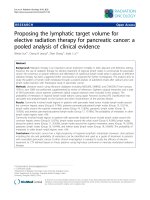
(LRC) were 66.0, 53.4, and 66.0 %, respectively (Fig. 1).
Pattern of the first relapse is summarized in Table 3.
Thirty two patients had disease relapse after treatment
including 22 loco-regional recurrence and 14 distant
metastases. Significantly more patients with failure were
identified in bilateral neck cohort (p = 0.015). Ten patients developed in-field recurrence. Nodal failure was
found within the high-risk CTV in seven patients and
within the intermediate-risk CTV in three patients.
Twelve patients were identified with extra-field locoregional failure: three recurrences were found in ipsilateral retropharyngeal lymph node, one in ipsilateral level
Ib and IV simultaneously, two in ipsilateral level V, two
in tumor bed region, one in the nasopharynx, two in
neck skin, and one in non-irradiated contralateral neck
region. The most frequently affected site as a distant metastasis was lung, following bone and mediastinal lymph
node. Among 22 loco-regional recurrence, seven patients underwent salvage surgery although only one patient remains without relapse at the time of the analysis.
Potential factors influencing DFS were summarized in
Table 4. In the univariate analysis, IMRT was found out
to be factors for favorable DFS. On the other hand, more
than two lymph node metastasis, oral cavity primary or
recurrent disease, T4 or recurrent disease, and oral cavity primary or recurrent disease were identified to be
factors for unfavorable DFS. The aim of this study was
to find a group of patients who could be safely applied
unilateral neck irradiation and generally it is natural to
irradiate bilateral neck for patients with bilateral neck
lymph node involvement. Consequently, all 10 patients
with bilateral neck lymph node metastasis were excluded
and uni- and multivariate analysis was performed
(Table 5). In the multivariate analysis, it was found that
inferior DFS correlated with oral cavity or recurrent disease (Hazard Ratio 1.696; 95 % confidence interval 1.29–
1.87, Fig. 2). Among patients with unilateral lymph node
metastasis treated with unilateral neck irradiation, oral
cavity or recurrent disease were adverse factors for DFS
Murakami et al. BMC Cancer (2016) 16:192
Page 5 of 10
Table 1 Patient and tumor characteristics
Bilateral RT
Unilateral RT
(n = 38)
(n = 34)
Table 1 Patient and tumor characteristics (Continued)
Maximum diameter of LN (cm)
p value
Primary site
Oral cavity
10
13
Oropharynx
12
8
Hypopharynx
10
9
Larynx
5
2
Others
1
2
0.556
4
4
T2
13
11
T3
5
4
T4
8
4
Rec
8
11
rT0
6
8
rT1
0
0
rT2
2
2
rT3
0
0
rT4
0
1
N0
0
2
N1
0
0
N2a
3
2
N2b
18
17
N2c
8
1
N3
1
1
Rec
8
11
rN1
1
1
rN2
7
10
rN3
0
0
III
1
1
IVA
28
20
IVB
1
2
Rec
8
11
rIII
1
1
rIVA
7
10
2.5
2.7
Range
0.4–7.5
0.8–4.1
0.783
Number of LN metastasis
0–1
6
13
≥2
32
21
Male
32
26
Female
6
8
Median
63
63
Range
38–80
34–84
0.031*
Sex
T-classification
T1
Median
0.673
0.217
Age
0.270
RT radiation therapy, Rec recurrence, LN lymph node. *A P value of ≤0.05 was
considered statistically significant
N-classification
0.124
(2-y DFS 41.7 % vs 88.2 %, p = 0.017, Fig. 3). On the
other hand, among patients who had unilateral lymph
node metastasis treated with bilateral neck irradiation,
no statistically significant difference was found but a tendency towards inferior DFS for patients with oral cavity
or recurrent disease compared to that of those without
(2-y DFS 22.2 % vs 60.9 %, p = 0.056). Out of 40 relapsefree patients, one patient was para-enteral nutrition
dependent, two patients developed hypothyroidism requiring the hormone replacement treatment, one patient developed ulcer at tonsil which resolved conservatively, one
patient developed severe dry mouth which always required
water to moisten the mouth, two patients developed
metachronous malignancy in head and neck region,
and two patients died of intercurrent disease (one
died of subarachnoid hemorrhage and one liver cirrhosis). The one who remained para-enteral nutrition
dependent was treated by bilateral neck irradiation.
Stage
0.637
Bilateral neck LN metastasis
Yes
9
1
No
29
33
Yes
21
21
No
14
9
Unknown
3
4
0.011*
Necrosis in LN
0.401
Discussion
The relapse rate was significantly higher in bilateral neck
cohort compared with unilateral neck cohort although
larger volume being irradiated (Table 3, p = 0.015). The
possible explanation of this unfavorable results in bilateral neck cohort was that statistically more patients with
two or more lymph node metastases were treated by bilateral radiation therapy (Table 1).
Eisbruch et al. reported that there existed dose-volume
relationship between the pharyngeal constrictors, the
glottic, and supraglottic larynx and late radiation complications such as dysphagia and aspiration [18]. Because
our study was only a retrospective study and it was not
possible to collect reliable data concerning late neck toxicities. Therefore, it was not possible to show inferior
quality of life for patients who were treated by bilateral
neck irradiation compared with those who were treated
Murakami et al. BMC Cancer (2016) 16:192
Page 6 of 10
Table 2 Pathological characteristics and treatment details
NAC
Yes
8
No
64
Bi-lateral ND
Yes
26
No
46
Treatment for primary lesion
Surgery
60
EBRT
9
BT
3
Degree of differentiation
Poorly differentiated
20
Moderately differentiated
23
Well differentiated
15
Unknown
14
Extracapsular spread
Yes
46
No
10
Unknown
16
Positive/close margin
Yes
40
No
32
Concurrent systemic therapy
CDDP
17
Cetuximab
2
TS-1
2
None
51
RT total dose (Gy)
Median
66
Range
60–74
Radiation technique
3DCRT
14
IMRT
58
RT radiation therapy, NAC neoadjuvant chemotherapy, ND neck dissection,
EBRT external beam radiation therapy
BT brachytherapy, LN lymph node, 3DCRT three-dimensional conformal radiation therapy, IMRT intensity modulated radiation therapy
by unilateral neck irradiation. However, it is common in
daily practice to see patients with bilateral neck irradiation who are suffered from neck stiffness or shoulder
discomfort which not merely worsen patient’s quality of
life but also hinders early detection of neck lymph node
recurrence or make it difficult to perform possible salvage surgery. Therefore, if it is feasible, to reduce acute
and late complications related to irradiation it is obviously desirable to irradiate as smaller volume as possible.
There were only two patients with N0 in this study
(Table 1), but large T classification T3 with positive margin and T4. Hence, if the guideline for postoperative radiation therapy created by Chao et al. [14] would have
been applied to our patients, theoretically all the patients
should have been treated by bilateral neck irradiation.
This patient with T3N0 and positive margin eventually
developed local and regional recurrence. Because there
were only two patients with postoperative N0 in our
study, it is difficult to make any recommendations of
postoperative radiation therapy for postoperative N0 patients. However, because one among the two N0 patients
developed regional recurrence, prophylactic postoperative radiation seems to be also important for postoperative N0 patients with high risk pathological features.
It was observed in this study that seven out of 22 patients with loco-regional recurrence could undergo salvage surgery and only one of them eventually achieved
no relapse at the time of the analysis. Therefore, salvage
surgery had only minor impact on patient’s overall survival. This finding was in line with a recent randomized
phase III trial comparing elective neck dissection or
watchful wait with close follow up for early-stage oral
cancer. The latter strategy was significantly inferior in
overall survival rate despite the protocol mandated close
follow-up for neck examination [19]. In this study, patients who developed nodal relapse presented with a
more advanced nodal stage and a higher prevalence of
ECS than initial presentation, which possibly made it
more difficult to control disease by salvage interventions.
Accordingly, finding patients who are unlikely to develop loco-regional recurrence after unilateral neck irradiation seems to be a better treatment strategy.
HNSCC with a positive human papilloma virus (HPV)
has been recently reported to be radiosensitive [20, 21].
Ki-67 and p53 were also reported to be prognostic
markers for HNSCC postoperative radiotherapy [22].
The prognostic impact of these markers on survival for
patients with HNSCC who were treated with postoperative radiation therapy could not be assessed because only
part of patients were examined for p16, Ki-67, and p53
status in this study. Similarly, although ECS of lymph
node is a well-known major adverse pathological factor
among patients of HNSCC [5–7] and description concerning ECS of lymph node has been found since 2005,
it was only from 2011 that documentation about ECS of
lymph node has been made without exception. Therefore, there were as many as 16 missing data and the
prognostic impact of ECS of lymph node could not be
found in our study. Resection margin status is also a
well-known major adverse pathological factor [5–7].
However, in our study adverse prognostic feature of resection margin status could not be shown presumably
because patients with positive/close margin received
postoperative radiation therapy appropriately. On the
Murakami et al. BMC Cancer (2016) 16:192
Page 7 of 10
Fig. 1 Kaplan-Meyer curves of overall survival (OS), disease-free survival (DFS), and loco-regional control (LRC)
Table 4 Potential predictors influencing DFS
2-y DFS (%)
no
p value
OC
38.9
59.8
0.067
Necrosis in LN
63.8
49.1
0.245
Bi-lateral ND
50.3
54.7
0.654
NAC
87.5
48.6
0.076
Rec
30.6
59.6
0.062
Table 3 Pattern of first failures
Bilateral RT
Unilateral RT
(n = 38)
(n = 34)
p value
Any failure
Yes
No
22
16
10
0.015*
24
Loco-regional failure
Yes
15
7
No
23
27
0.082
In-field failure
Yes
No
8
30
2
0.062
32
Extra-field loco-regional failure
Yes
7
5
No
31
29
0.572
Distant failure
Yes
No
10
28
4
0.119
30
RT radiation therapy. *A P value of ≤0.05 was considered statistically significant
DFS
yes
T4
37.5
56.2
0.21
LN+ ≥2LN+ ≥2
44.9
76.5
0.019*
Extracapsular extention
50.7
62.5
0.177
Positive/close margin
48.5
57.2
0.82
Bilateral RT
42
68.4
0.057
IMRT
69.5
35.7
0.047*
Systemic therapy
38.7
57.1
0.449
OC or rec
33.9
66.4
0.013*
T4 or rec
36.7
65.7
0.012*
OC or T4
37.5
65.1
0.012*
OC or T4 or rec
37.9
72.4
0.006*
DFS disease free survival
OC oral cavity, LN lymph node, ND neck dissection, Rec recurrence. *A P value
of ≤0.05 was considered statistically significant
Murakami et al. BMC Cancer (2016) 16:192
Page 8 of 10
Table 5 Potential predictors influencing DFS for patients excluding bilateral neck lymph node metastasis
2-y DFS (%)
DFS
yes
no
p value in uni.
OC
36.9
64.4
0.034*
Necrosis in LN
64.9
48.4
0.423
Bi-lateral ND
53.0
55.9
0.998
NAC
87.5
48.6
0.076
Rec
30.6
64.2
0.029*
T4
46.7
56.6
0.398
LN+ ≥2LN+ ≥2
45.3
76.5
0.032*
Extracapsular extention
52.2
57.1
0.442
Positive/close margin
56.1
54.2
0.824
Bilateral RT
44.4
67.3
0.146
IMRT
60.8
33.3
0.050*
Systemic therapy
43.3
56.5
0.619
OC or rec
31.9
74.0
0.004*
p value in multi.
HR
95 % CI
1.696
1.29–1.87
0.062
0.162
0.006
DFS disease free survival, uni. univariate analysis, multi. multivariate analysis, HR hazard ration, CI confidence interval, OC oral cavity, LN lymph node, ND neck
dissection, Rec recurrence. *A P value of ≤0.05 was considered statistically significant
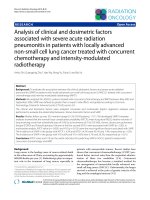
other hand, multivariate analysis in this study revealed
that patients with oral cavity or recurrent disease were significantly worse DFS compared with those without and its
disease-free survival disadvantage was 69.6 % (Table 5,
Fig. 2). Radiation resistance of tumors from oral cavity
[23] has been reported previously, therefore, current
findings were clinically comprehensible. Among patients
without bilateral neck node metastasis and treated with
unilateral neck irradiation, patients with oral cavity or recurrent disease had significantly inferior DFS compared
with those without (2-y DFS 41.7 % vs 88.2 %, p = 0.017,
Fig. 3). Therefore, in patients without bilateral neck lymph
Fig. 2 Disease-free survival (DFS) stratified by the group of patients with oral cavity or recurrent disease or those without
Murakami et al. BMC Cancer (2016) 16:192
Page 9 of 10
Fig. 3 Disease-free survival (DFS) for patients treated by unilateral neck cohort. Survival curves were stratified by the group of patients with oral
cavity or recurrent disease or those without
node involvement, the postoperative unilateral neck irradiation is a reasonable treatment strategy for patients
with the exception of oral cavity or recurrent disease.
On the contrary, for patients with oral cavity origin
or recurrent disease, bilateral neck irradiation did not
seem to be a promising solution. If bilateral neck irradiation was a favorable solution for patients with
oral cavity or recurrent disease, DFS should have
been superior for patients with bilateral neck irradiation to those with unilateral neck irradiation. However, among patients with oral cavity or recurrent
disease, 2-y DFS for patients with bilateral and unilateral neck irradiation were 22.2 % and 41.7 %, respectively (p = 0.412). Thus, different approaches should be
made to improve the clinical results for patients with
oral cavity or recurrent disease. In this study, the
most frequent site of regional recurrence was the
high-risk CTV (70 %), similar results showed with
Carrillo et al. [15]. Out of field regional recurrence
was observed more frequently in ipsilateral neck than
contralateral neck whereas only one patient developed
contralateral-neck failure. Because concurrent CDDP
administration was only started since 2008 in our institution, the majority patients did not received cCRT
in this analysis, which could be a possible explanation
for many loco-regional recurrences. Therefore, dose
escalation for the high-risk CTV or application of
cCRT or widening the intermediate-risk CTV in ipsilateral neck region to submandibular or posterior
neck would possibly decrease the rate of loco-regional
recurrence in the future.
There were several limitations in this study. Treatment strategy and radiation field was not uniformed
according to several patient’s clinical backgrounds.
For example, treatment plans were heterogeneous including bioradiation, chemoradiation, or radiation
alone. And the chemotherapy agents used were not
unified. Also, this study was a retrospective study
consisted of a small number of patients from single
institution. In spite of these drawbacks, several insights were derived from this analysis which would
possible improve treatment in the future.
Murakami et al. BMC Cancer (2016) 16:192
Conclusions
In patients without bilateral neck lymph node involvement, the postoperative unilateral neck irradiation is a
reasonable treatment strategy for patients with the exception of oral cavity or recurrent disease.
Abbreviations
3DCRT: three-dimensional conformal radiotherapy; AHNS: American Head
and Neck Society; CALGB: Cancer and Leukemia Group B; cCRT: concurrent
chemoradiation; CTV: clinical target volume; DFS: disease-free survival;
ECOG: Eastern Cooperative Oncology Group; ECS: extra-capsular spread;
EORTC: European Organization for Research and Treatment of Cancer;
GTV: gross tumor volume; HNSCC: head and neck squamous cell carcinoma;
HPV: human papilloma virus; IMRT: intensity-modulated radiotherapy; LRC:
loco-regional control; NAC: neoadjuvant chemotherapy; NCOG: Northern
California Oncology Group; OS: overall survival; RND: radical neck dissection;
RTOG: Radiation Therapy Oncology Group; SEG: Southwest Group; SIBIMRT: simultaneous integrated boost intensity-modulated radiotherapy;
SND: selective neck dissection; SWOG: Southwest Oncology Group;
VMAT: volumetric modulated arc therapy.
Competing interests
The authors declare that they have no competing interests.
Authors’ contributions
NM, FM, and SY have made substantial contributions to conception and
design of this study. NM and FM have been involved in drafting the
manuscript or revising it critically for important intellectual content. YI, TM,
TU, KT, TK, KK, KH, MK, SS, RU, KT, KI, HI, and JI Participated in acquisition and
interpretation of data. All authors read and approved the final manuscript.
Acknowledgement
Part of this study was financially supported by JSPS KAKENHI Grant Number
15 K19836, the Practical Research for Innovative Cancer Control from Japan
Agency for Medical Research and development, AMED, and the National
Cancer Center Research and Development Fund (26-A-18 and 26-A-28).
Author details
1
Department of Radiation Oncology, National Cancer Center Hospital, 5-1-1
Tsukiji, Chuo-ku, Tokyo 104-0045, Japan. 2Department of Head and Neck
Surgery, National Cancer Center Hospital, 5-1-1 Tsukiji, Chuo-ku, Tokyo
104-0045, Japan. 3Department of Clinical Laboratory and Pathology, National
Cancer Center Hospital, 5-1-1 Tsukiji, Chuo-ku, Tokyo 104-0045, Japan.
4
Department of Oral Health and Diagnostic Sciences, National Cancer Center
Hospital, 5-1-1 Tsukiji, Chuo-ku, Tokyo 104-0045, Japan.
Page 10 of 10
8.
9.
10.
11.
12.
13.
14.
15.
16.
17.
18.
19.
20.
21.
Received: 13 August 2015 Accepted: 29 February 2016
References
1. Cancer Registry and Statistics. Cancer Information Service, National Cancer
Center, Japan. />2. Japan Health Promotion & Fitness Foundation. Ministry of Health, Labour and
Welfare. 2008. />3. Kramer S, Gelber RD, Snow JB, Marcial VA, Lowry LD, Davis LW, et al.
Combined radiation and surgery in the management of advanced head
and neck cancer: Final report of study 73–03 of the Radiation Therapy
Oncology Group. Head Neck Surg. 1987;10:19–30.
4. Laramore GE, Scott CB, Al-Sarraf M, Haselow RE, Ervin TJ, Wheeler R, et al. Adjuvant
chemotherapy for resectable squamous cell carcinomas of the head and neck:
report on Intergroup Study 0034. Int J Radiat Oncol Biol Phys. 1992;23:705–13.
5. Bernier J, Domenge C, Ozsahin M, Matuszewska K, Lefebvre JL, Greiner
RH, et al. Postoperative irradiation with or without concomitant
chemotherapy for locally advanced head and neck cancer. N Engl J
Med. 2004;350:1945–52.
6. Cooper JS, Pajak TF, Forastiere AA, Jacobs J, Campbell BH, Saxman SB, et al.
Postoperative concurrent radiotherapy and chemotherapy for high-risk
squamous cell carcinoma of the head and neck. N Engl J Med. 2004;350:1937–44.
7. Bernier J, Cooper JS, Pajak TF, van Glabbeke M, Bourhis J, Forastiere A,
et al. Defining risk levels in locally advanced head and neck cancers: a
22.
23.
comparative analysis of concurrent postoperative radiation plus
chemotherapy trials of the EORTC (#22931) and RTOG (#9501). Head
Neck. 2005;27:843–50.
Hamoir M, Silver CE, Schmitz S, Takes RP, Rinaldo A, Rodrigo JP, et al. Radical
neck dissection: is it still indicated? Eur Arch Otorhinolaryngol. 2013;270:1–4.
Ferlito A, Johnson JT, Rinaldo A, Pratt LW, Fagan JJ, Weir N, et al. European
surgeons were the first to perform neck dissection. Laryngoscope. 2007;117:
797–802.
Schmitz S, Machiels JP, Weynand B, Gregoire V, Hamoir M. Results of
selective neck dissection in the primary management of head and neck
squamous cell carcinoma. Eur Arch Otorhinolaryngol. 2009;266:437–43.
Robbins KT, Medina JE, Wolfe GT, Levine PA, Sessions RB, Pruet CW.
Standardizing neck dissection terminology. Official report of the Academy’s
committee for head and neck surgery and oncology. Arch Otlolaryngol
Head Neck Surg. 1991;117:601–5.
Robbins KT, Clayman G, Levine PA, Medina J, Sessions R, Shaha A, et al.
Neck dissection classification update. Revisions proposed by American Head
and Neck Society and the American Academy of Otolaryngology Head and
Neck Surgery. Arch Otlolaryngol Head Neck Surg. 2002;128:751–8.
Gregoire V, Eisbruch A, Hamoir M, Levendag P. Proposal for the delineation
of the nodal CTV in the node positive and the postoperative neck.
Radiother Oncol. 2006;79:15–20.
Chao KS, Wippold FJ, Ozyigit G, Tran BN, Dempsey JF. Determination and
delineation of nodal target volumes for head-and-neck cancer based on
patterns of failure in patients receiving definitive and postoperative IMRT.
Int J Radiat Oncol Biol Phys. 2002;53:1174–84.
Carrillo MM, Martin IT, Lara IM, Almodovar RRJM, Moral DAR. Selective use of
postoperative neck radiotherapy in oral cavity and oropharynx cancer: a
prospective clinical study. Radiat Oncol. 2013;8:103–10.
Bonner JA, Harari PM, Giralt J, Azarnia N, Shin DM, Cohen RB, et al.
Radiotherapy plus cetuximab for squamous-cell carcinoma of the head and
neck. N Engl J Med. 2006;354:567–78.
Murakami N, Yoshimoto S, Matsumoto F, Ueno T, Ito Y, Watanabe S, et al.
Severe gastrointestinal bleeding in patients with locally advanced head and
neck squamous cell carcinoma treated by concurrent radiotherapy and
cetuximab. J Cancer Res Clin Oncol. 2015;141:177–84.
Eisbruch A, Schwartx M, Rasch C, Vineberg K, Damen E, Van As CJ, et al.
Dysphagia and aspiration after chemoradiotherapy for head-and-neck
cancer: which anatomic structures are affected and can they be spared by
IMRT? Int J Radiat Oncol Biol phys. 2004;60:1425–39.
D’Cruz AK, Vaish R, Kapre N, Dandekar M, Gupta S, Hawaldar R, et al. Elective
versus therapeutic neck dissection in node-negative oral cancer. N Engl J
Med. 2015;373:521–9.
Nadiaye C, Mena M, Alemany L, Arbyn M, Castellsaque X, Laporte L, et al.
HPV DNA, E6/E7 mRNA, and p16INK4a detection in head and neck cancers:
a systematic review and meta-analysis. Lancet Oncol. 2014;15:1319–31.
Chung CH, Zhang Q, Kong CS, Harris J, Fertig EJ, Harari PM, et al. p16
protein expression and human papillomavirus status as prognostic
biomarkers of nonoropharyngeal head and neck squamous cell carcinoma.
J Clin Oncol. 2014;32:3930–8.
Grabenbauer G, Mühlfriedel C, Rödel F, Niedobitek G, Hornung J, Rödel C,
et al. Squamous cell carcinoma of the oropharynx: Ki-67 and p53 can
identify patients at high risk for local recurrence after surgery and
postoperative radiotherapy. Int J Radiat Oncol Biol Phys. 2000;48:1041–50.
Biau J, Chautard E, Miroir J, Lapeyre M. Radioresistance parameters in head
and neck cancers and methods to radiosensitize. Cancer Radiother. 2015.
Epub ahead of print.