Characterising timing and pattern of relapse following surgery for localised oesophagogastric adenocarcinoma: A retrospective study
Bạn đang xem bản rút gọn của tài liệu. Xem và tải ngay bản đầy đủ của tài liệu tại đây (511.76 KB, 10 trang )
Moorcraft et al. BMC Cancer (2016) 16:112
DOI 10.1186/s12885-016-2145-0
RESEARCH ARTICLE
Open Access
Characterising timing and pattern of
relapse following surgery for localised
oesophagogastric adenocarcinoma: a
retrospective study
Sing Yu Moorcraft, Elisa Fontana, David Cunningham, Clare Peckitt, Tom Waddell, Elizabeth C. Smyth,
William Allum, Jeremy Thompson, Sheela Rao, David Watkins, Naureen Starling and Ian Chau*
Abstract
Background: Oesophagogastric adenocarcinoma (OGA) has a poor prognosis, even for patients with operable
disease. However, the optimal surveillance strategy following surgery is unknown.
Methods: We performed a retrospective review of all patients with OGA who had undergone surgery with radical intent
at the Royal Marsden between January 2001 and December 2010.
Results: Of the 360 patients with OGA who underwent potentially curative surgery, 100/214 patients (47 %) with
oesophageal/gastro-oesophageal junction (GOJ) adenocarcinoma and 47/146 patients (32 %) with gastric
adenocarcinoma developed recurrent disease. 51, 79 and 92 % of relapses occurred within 1, 2 and 3 years respectively
and the majority of patients relapsed at distant sites. Of the patients who relapsed, 67 % (67/100) with oesophageal/GOJ
adenocarcinoma and 72 % of patients with gastric cancer (34/47) were symptomatic at the time of relapse. The majority
of asymptomatic relapses were first detected by a rise in tumour markers. There was no difference in disease-free survival
between asymptomatic and symptomatic patients, but asymptomatic patients were more likely to receive further
treatment and had a longer survival beyond relapse.
Conclusion: The majority of relapses occur within the first 3 years and at distant sites. Monitoring of tumour markers
should be considered as part of a surveillance program.
Keywords: Follow-up, Gastric cancer, Oesophageal cancer, Recurrence, Surveillance
Background
Oesophagogastric adenocarcinoma (OGA) has a poor
prognosis, even in patients who present with localised
disease. Over time, staging has become more accurate,
leading to improvements in the selection of patients for
surgery, and treatment has improved, with perioperative chemotherapy becoming a standard of care in
the United Kingdom, based on a 5–year overall survival
(OS) of 36 - 38 % compared to 23–24 % for surgery
alone [1, 2]. Worldwide, other treatment options include
neoadjuvant or adjuvant chemoradiotherapy or chemotherapy. Extended lymph node dissection (D2
* Correspondence:
The Royal Marsden NHS Foundation Trust, London and Surrey, United Kingdom
lymphadenectomy) has also become a standard of care
due to evidence that this leads to a reduced rate of gastric cancer-related deaths [3]. In addition, the treatment
of metastatic OGA has improved, with the addition of
new treatment options. For example, trastuzumab is
used in the first-line treatment of HER2 positive gastric
cancer [4], second-line chemotherapy is now a standard
of care [5] and benefit has also been seen with the antiangiogenic agent ramucirumab [6].
In theory, early detection of disease relapse could lead
to improved outcomes for patients. However, the optimal follow-up schedule for patients after potentially
curative resection for OGA is not yet determined and
there are significant variations between guidelines. For
example, the National Comprehensive Cancer Network
© 2016 Moorcraft et al. Open Access This article is distributed under the terms of the Creative Commons Attribution 4.0
International License ( which permits unrestricted use, distribution, and
reproduction in any medium, provided you give appropriate credit to the original author(s) and the source, provide a link to
the Creative Commons license, and indicate if changes were made. The Creative Commons Public Domain Dedication waiver
( applies to the data made available in this article, unless otherwise stated.
Moorcraft et al. BMC Cancer (2016) 16:112
guidelines recommend performing a history and physical
examination every 3–6 months for 1–2 years, then every
6–12 months for 3–5 years and then annually, with
other investigations being done as clinically indicated
[7], whereas other guidelines state that there is no
evidence that intensive follow-up impacts on outcomes [8–10]. This leaves clinicians with uncertainty
regarding the optimal management of these patients.
We conducted a retrospective analysis to investigate patterns of relapse following resection for OGA to assist in formulating an optimal surveillance strategy for these patients.
Methods
This project was classified as a service evaluation by our
institution’s Committee for Clinical Research as the aim
of the project was to evaluate our institution’s follow-up
strategy for patients undergoing surgery for OGA.
Therefore, in accordance with guidance from the National Health Service (NHS) Health Research Authority,
specific patient consent and ethical approval was not required. After approval from our institution’s Committee
for Clinical Research (SE3407), we searched the Royal
Marsden (RM) electronic medical record system for patients with a diagnosis of oesophageal, gastrooesophageal junction (GOJ) or gastric adenocarcinoma
who had undergone surgery with radical intent between
January 2001 and December 2010. Patients who were
followed up in another hospital, patients for whom no
data was available apart from the date of surgery and patients who were found to have unresectable metastatic
disease at the time of surgery were excluded.
Prior to 2006, our institution’s policy for patients with
oesophageal/type I/II GOJ cancer was 2 cycles of neoadjuvant chemotherapy with cisplatin and 5-fluorouracil.
The follow-up schedule involved clinical assessment and
tumour markers 3 monthly for the first year, then 6
monthly, with endoscopies or CT scans performed as
clinically indicated. Patients with operable type III GOJ/
gastric cancer underwent surgery alone, unless they were
participating in a clinical trial, and there were no specific
Page 2 of 10
follow-up recommendations. From 2006, our institution’s
policy changed to 3 cycles of neoadjuvant chemotherapy
with epirubicin, cisplatin and 5-fluorouracil/capecitabine
(ECF/X) followed by surgery and a further 3 cycles of
ECF/X for oesophageal, GOJ and gastric adenocarcinoma.
Follow-up continued as per our previous standard practice
for oesophageal cancer. The treatment and surveillance
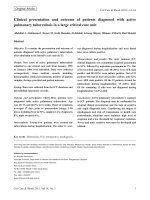
paradigms are summarised in Fig. 1. Patients with
oesophageal or type I/II GOJ adenocarcinoma underwent
oesophagogastrectomy and patients with gastric cancer
underwent total or subtotal gastrectomy. Nodal dissection
tended to be D2 throughout the study period.
Clinical information, including patient demographics,
clinical characteristics, outcomes and details of first relapse (including date, site, symptoms, method of relapse
detection, CEA and CA19-9) were retrospectively collected from patient records. Patients were categorised as
having local relapse (recurrence at the anastomosis) or
distant relapse (recurrence at distant sites or regional
lymph nodes). Symptomatic relapse was defined as the
presence of patient-reported symptoms triggering further investigations, whereas asymptomatic relapse was
defined as relapse detected by a routine radiological, laboratory or endoscopic investigation that was not
prompted by any clinical concerns.
Statistical analysis
Disease-free survival (DFS) was calculated from the date
of surgery to the date of death or relapse at any site. OS
was calculated from the date of surgery to the date of
death. Survival beyond relapse (SBR) was calculated
from the date of relapse at any site to the date of death
from any cause. Patients who were still alive and event
free were censored at the time of last follow-up.
Survival rates were calculated using Kaplan Meier
methods. Association of survival outcomes with baseline
prognostic factors was determined by Cox regression
univariate analysis, with hazard ratios being presented
with 95 % confidence intervals. Factors included in the
univariate analysis were peri-operative treatment (pre-
Fig. 1 Changes in the treatment and surveillance paradigms for oesophageal, GOJ and gastric adenocarcinomas. CF = cisplatin and 5-fluorouracil,
ECF/X = epirubicin, cisplatin and 5-fluorouracil/capecitabine
Moorcraft et al. BMC Cancer (2016) 16:112
Page 3 of 10
Table 1 Baseline characteristics, initial treatment details and pathological characteristics of patients with oesophagogastric
adenocarcinoma who underwent surgery with curative intent
Oesophageal/GOJ (n = 214)
Gastric (n = 146)
N (%)
N (%)
Male
188 (88 %)
98 (67 %)
Female
26 (12 %)
48 (33 %)
64 years (33–83)
70 years (24–89)
0
58 (27 %)
40 (27 %)
1
69 (32 %)
41 (28 %)
2
2 (1 %)
10 (7 %)
Unknown
85 (40 %)
55 (38 %)
Oesophagus
29 (14 %)
-
Type 1 GOJ
77 (36 %)
-
Type 2 GOJ
63 (29 %)
-
Type 3 GOJ
45 (21 %)
-
Gastric
-
146 (100 %)
Yes
61 (29 %)
27 (19 %)
No
122 (57 %)
75 (51 %)
Unknown
31 (14 %)
44 (30 %)
69 (32 %)
24 (16 %)
Gender
Median age (range)
ECOG performance status
Site of primary tumour
Elevated tumour markers pre-operatively
Baseline PET performed
Yes
Treatment
Neoadjuvanta
125 (58 %)
30 (21 %)
Peri-operativeb
51 (24 %)
56 (38 %)
Adjuvant
5 (2 %)
7 (5 %)
Surgery only
33 (15 %)
53 (36 %)
Oesophagogastrectomy
178 (83 %)
3 (2 %)
Total gastrectomy
35 (16 %)
51 (35 %)
Sub-total gastrectomy
1 (1 %)
92 (63 %)
Well
8 (4 %)
4 (3 %)
Moderate
84 (39 %)
43 (30 %)
Poor
107 (50 %)
94 (64 %)
Unknown
15 (7 %)
5 (3 %)
Surgery
Differentiation
T stage
T0
11 (5 %)
7 (5 %)
T1
48 (22 %)
34 (23 %)
T2
53 (25 %)
66 (45 %)
T3
89 (42 %)
27 (19 %)
T4
10 (5 %)
9 (6 %)
Tx
3 (1 %)
3 (2 %)
Moorcraft et al. BMC Cancer (2016) 16:112
Page 4 of 10
Table 1 Baseline characteristics, initial treatment details and pathological characteristics of patients with oesophagogastric
adenocarcinoma who underwent surgery with curative intent (Continued)
N stage
N0
105 (49 %)
72 (49 %)
N1
92 (43 %)
40 (27 %)
N2
10 (5 %)
20 (14 %)
N3
3 (1 %)
11 (8 %)
Nx
4 (2 %)
3 (2 %)
M stagec
M0
204 (95 %)
139 (95 %)
M1
5 (2 %)
4 (3 %)
Mx
5 (2 %)
3 (2 %)
28 (4–76)
24 (3–69)
1 (0–33)
1 (0–35)
Number of lymph nodes resected
Median (range)
Number of positive lymph nodes
Median (range)
Resection margin
R0
161 (75 %)
135 (92 %)
R1
47 (22 %)
7 (5 %)
R2
0 (0 %)
0 (0 %)
unknown
6 (3 %)
4 (3 %)
a
2 patients received pre-operative chemotherapy followed by pre-operative chemoradiotherapy, b 19 patients received pre-operative chemotherapy and postoperative chemoradiotherapy, c M1 = patients with resected metastatic disease (usually peritoneal)
Results
operative, post-operative or both vs surgery alone), pathological T-stage (T0-2 vs T3/4) and N-stage (N0 vs N1-3),
differentiation (well/moderate vs poor), resection margin
(R0 vs R1/2, includes both circumferential and longitudinal margins), type of relapse (local vs distant vs both), elevated tumour markers pre-operatively (yes vs no) and
symptoms at time of recurrence (yes vs no). Significant
variables were included in a multivariate analysis.
Patient characteristics
Between January 2001 and December 2010, 360 patients
with oesophagogastric adenocarcinoma (214 patients
with oesophageal/GOJ tumours and 146 patients with
gastric tumours) underwent surgery with curative intent
at RM. Baseline demographic, clinical and pathological
characteristics are shown in Table 1.
A
B
100
100
Gastric
OG
80
80
Proportion Alive
Proportion Disease Free
Gastric
OG
60
40
60
40
20
20
0
0
0
1
2
3
4
5
Time from date of surgery (years)
6
7
0
1
2
3
4
5
6
7
Time from date of surgery (years)
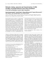
Fig. 2 Disease free survival and overall survival for patients who had radical surgery for oesophageal/GOJ (OG) and gastric adenocarcinoma. a:
Disease –free survival. b: Overall survival. (colour figure)
Moorcraft et al. BMC Cancer (2016) 16:112
Page 5 of 10
Table 2 Patterns of disease recurrence and treatment of recurrent disease
Oesophageal/GOJ (n = 100)
Gastric (n = 47)
N (%)
N (%)
Time to relapse
< 12 months
53 (53 %)
22 (47 %)
12–24 months
29 (29 %)
12 (25 %)
24–36 months
12 (12 %)
7 (15 %)
> 36 months
6 (6 %)
6 (13 %)
Local
7 (7 %)
4 (9 %)
Distant
79 (79 %)
37 (79 %)
Both
14 (14 %)
6 (13 %)
52 (52 %)
14 (30 %)
Relapse type
a
Site of relapse
Lymph nodes
Anastomosis
21 (21 %)
10 (21 %)
Peritoneum
16 (16 %)
18 (38 %)
Liver
18 (18 %)
9 (19 %)
Bone
12 (12 %)
4 (9 %)
Abdominal wall
3 (3 %)
5 (11 %)
Lung
10 (10 %)
2 (4 %)
Brain
10 (10 %)
0 (0 %)
Mediastinum
9 (9 %)
1 (2 %)
Other
8 (8 %)
5 (11 %)
Yes
63 (63 %)
24 (51 %)
No
24 (24 %)
16 (34 %)
Unknown
13 (13 %)
7 (15 %)
Elevated tumour markers at relapse
Symptoms at time of relapse
Yes
How relapse was first detected in asymptomatic patients
67 (67 %)
34 (72 %)
(n = 33)
(n = 12)
Routine tumour markers
22 (67 %)
4 (33 %)
Routine CT
6 (18 %)
4 (33 %)
Concurrent routine CT/ markers
1 (3 %)
3 (25 %)
Endoscopy
2 (6 %)
1 (8 %)
Other
2 (6 %)
0 (0 %)
0
12 (12 %)
3 (6 %)
1
13 (13 %)
7 (15 %)
ECOG performance status at relapse
2
4 (4 %)
2 (4 %)
3–4
8 (8 %)
4 (9 %)
Unknown
63 (63 %)
31 (66 %)
72 (72 %)
22 (47 %)
63 (88 %)
19 (86 %)
Further treatment for recurrent disease
Yes
Type of treatment for recurrent diseaseb
Chemotherapy
Moorcraft et al. BMC Cancer (2016) 16:112
Page 6 of 10
Table 2 Patterns of disease recurrence and treatment of recurrent disease (Continued)
a
b
Radiotherapy
21 (29 %)
3 (14 %)
Chemoradiotherapy
1 (1 %)
0 (0 %)
Surgery
5 (7 %)
1 (5 %)
Relapse may have occurred at more than one site
Patients may have received more than one type of treatment
Survival outcomes
After a median follow-up of 61.7 months, 100 patients
(47 %) with oesophageal/GOJ adenocarcinoma and 47
patients (32 %) with gastric adenocarcinoma had developed local and/or distant recurrence. Patients with
oesophageal/GOJ adenocarcinoma had a median DFS of
26.1 months (95 % CI 17.7–41.9) and median OS of
45.2 months (95 % CI 36.1–76.7); whereas patients with
gastric adenocarcinoma had a median DFS of 65.4 (95 %
CI 34.8–99.2) and median OS of 81.2 months (95 % CI
40.6–99.2) (see Fig. 2). The 5-year OS rate was 47.6 %
(95 % CI 40.5–54.4) for oesophageal/GOJ adenocarcinoma and 52.6 % (95 % CI 43.7–60.8) for gastric adenocarcinoma. Median SBR was 8.1 months (95 % CI 6.1–
13.4) and 5.9 months (95 % CI 3.4–8.2) for oesophageal/
GOJ and gastric adenocarcinoma respectively.
Patterns of relapse
The majority of relapses occurred at distant sites and occurred within the first 3 years following surgery, with 51,
79 and 92 % of relapses occurring within 1, 2 and 3 years
respectively (see Table 2). Sixty-three patients (63 %)
with oesophageal/GOJ adenocarcinoma and 24 patients
(51 %) with gastric cancer had elevated tumour markers
at the time of relapse. Of the 11 patients with anastomotic relapse only, 7 received further treatment (chemotherapy: 3 patients, chemotherapy followed by
radiotherapy: 2 patients, radiotherapy: 1 patient, chemoradiotherapy and surgery: 1 patient).
Sixty-seven patients (67 %) with oesophageal/GOJ
adenocarcinoma and 34 patients with gastric cancer
(72 %) were symptomatic at the time of relapse.
Twenty-six of the asymptomatic patients (58 %) had
relapse initially detected via elevated tumour markers.
Therefore, elevated tumour markers were the first
sign of relapse in 18 % of the 147 patients who relapsed. Occasionally patients had CT scans erroneously arranged as part of routine follow-up and these
scans detected relapse in 10 of the asymptomatic patients (22 %) (see Table 2). There were no differences
in pathological T or N stage at surgical resection between symptomatic and asymptomatic patients. There
was no difference in median DFS between asymptomatic and symptomatic patients with oesophageal/GOJ
cancer (p = 0.793) or gastric cancer (p = 0.259), but
asymptomatic patients were more likely to receive
further
treatment
than
symptomatic
patients
(oesophageal/GOJ: 84.5 % vs 65.6 %, p = 0.045; gastric:
76.9 % vs 35.3 %, p = 0.011) and had a longer SBR
(oesophageal/GOJ: 14.6 months vs 5.8 months, HR
1.75, 95 % CI 1.10–2.76, p = 0.017; gastric: 10.6 months
vs 3.8 months, HR 3.35, 95 % CI 1.55–7.26, p =
0.002). Of the 94 patients who received treatment
after relapse, SBR was longer in asymptomatic patients compared to symptomatic patients (15.9 months
vs 10.7 months, p = 0.032).
Prognostic variables
Univariate analyses (see Table 3), demonstrated that differentiation, pathological T-stage and pathological Nstage were prognostic for DFS and OS for both
oesophageal/GOJ and gastric adenocarcinoma and type
of relapse was prognostic for OS. In addition, resection
margin (R0 vs R1/2) was prognostic for DFS and OS for
oesophageal/GOJ adenocarcinoma and there was a trend
towards positivity for gastric cancer, although this did
not reach statistical significance. The results of a multivariate analysis are shown in Table 4.
Discussion
There are no randomised controlled trials investigating
the optimum follow-up strategy for patients undergoing
curative resection for OGA and strategies vary significantly. For example, some institutions have intensive
surveillance programs involving regular imaging and endoscopy, whereas other institutions have a clinicallybased follow-up strategy or no follow-up at all [11–14].
It is important to remember that follow-up is not only
about the detection of recurrent disease. Other important aspects of follow-up include helping patients to adjust to the social, physical and psychological
consequences of surgery [15], correction of nutritional
deficiencies and anaemia [11, 16], providing reassurance
to patients and providing a forum for patients to mention any new concerns [11].
In keeping with previously published results, 32 % of
patients with gastric adenocarcinoma and 47 % of patients with oesophageal/GOJ adenocarcinoma developed
recurrent disease [13, 17–19], with the majority of relapses occurring within the first 3 years. This pattern is
similar to other studies, which reported that 46–50 % of
relapses occurred within 1 year, 75–80 % within 2 years
Moorcraft et al. BMC Cancer (2016) 16:112
Page 7 of 10
Table 3 Univariate analysis of disease-free and overall survival
Disease-free survival
Oesophageal/GOJ adenocarcinoma
Covariate
Gastric adenocarcinoma
N
Median DFS (months,
95 % CI)
Hazard ratio
(95 % CI)
P -value N
Median DFS (months,
95 % CI)
Hazard ratio
(95 % CI)
P -value
No
24
12.2 (8.8–16.2)
1.0
0.794
16
10.8 (5.0–13.7)
1.0
0.081
Yes
63
11.8 (8.4–13.6)
1.07 (0.66–1.72)
24
15.0 (10.6–24.8)
0.56 (0.29–1.08)
94
37.9 (21.5–71.8)
1.0
47
99.2 (36.2 – NA)
0.54 (0.32–0.91)
Elevated tumour markers
Differentiation
Poor
107 12.3 (8.8–20.7)
Moderate/well
92
85.9 (33.1 – NA)
1.0
<0.001
0.40 (0.27–0.58)
0.020
Pathological T-stage
T0-2
112 111.7 (77.7 – NA)
1.0
T3/4
97
2.89 (2.00–4.18)
12.2 (8.7–18.0)
<0.001
107 86.9 (51.4–99.6)
1.0
36
21.5 (12.7–40.5)
1.91 (1.16–3.12)
72
87.1 (86.9 – NA)
1.0
71
21.1 (12.7 - 38.0)
3.10 (1.89–5.10)
0.010
Pathological N-stage
N0
105 111.7 (77.7 – NA)
1.0
N1-3
105 11.8 (8.4–15.7)
3.38 (2.32–4.94)
R0
161 77.7 (26.1 – NA)
1.0
R1/R2
47
8.7 (7.0–14.8)
2.87 (1.96–4.20)
No
33
10.9 (7.9–14.8)
1.0
Yes
67
11.8 (7.2–12.4)
1.06 (0.70–1.61)
No
33
140.0 (111.7 – NA)
1.0
Yes
181 20.9 (14.3–27.2)
<0.001
<0.001
Resection margin
<0.001
135 71.8 (35.6–99.6)
1.0
7
13.2 (0.3 – NA)
2.13 (0.91–4.98)
13
11.5 (4.8–21.5)
1.0
34
13.2 (8.1–20.6)
0.68 (0.35–1.32)
53
34.1 (13.1–87.1)
1.0
93
86.9 (41.7 – NA)
0.67 (0.42–1.08)
0.080
Presence of symptoms at time of
relapse
0.793
0.259
Neoadjuvant, adjuvant or
perioperative therapy
0.001
3.57 (1.74–7.31)
0.100
Overall survival
Oesophageal/GOJ adenocarcinoma
Covariate
Gastric adenocarcinoma
N
Median OS (months,
95 % CI)
Hazard ratio
(95 % CI)
P -value N
Median OS (months,
95 % CI)
Hazard ratio
(95 % CI)
P -value
No
24
28.8 (15.2–40.7)
1.0
0.343
16
20.0 (9.6–29.1)
1.0
0.842
Yes
63
22.4 (14.9–31.5)
1.28 (0.77–2.11)
24
22.6 (15.9–34.4)
0.91 (0.49–1.80)
94
40.5 (28.5–86.9)
1.0
47
99.2 (53.7 – NA)
0.50 (0.29–0.85)
Elevated tumour markers
Differentiation
Poor
107 21.5 (15.2–33.0)
Moderate/well
92
85.9 (76.7 – NA)
1.0
<0.001
0.37 (0.25–0.55)
0.011
Pathological T-stage
T0-2
112 111.7 (77.7 – NA)
1.0
T3/4
99
2.96 (2.00–4.36)
27.9 (14.9–35.2)
<0.001
107 81.2 (53.9–99.6)
1.0
36
29.1 (17.2–40.5)
2.19 (1.33–3.61)
72
87.1 (86.9 – NA)
1.0
71
28.5 (19.4–48.7)
3.16 (1.90–5.26)
0.002
Pathological N-stage
N0
105 111.7 (77.7 – NA)
1.0
N1-3
105 25.1 (14.7–34.1)
3.33 (2.23–4.97)
R0
161 77.7 (51.9 - NA)
1.0
R1/R2
47
2.83 (1.91–4.19)
<0.001
<0.001
Resection margin
13.8 (8.6–36.1)
<0.001
135 81.2 (48.8–99.6)
1.0
7
2.25 (0.96–5.26)
17.2 (0.3 - NA)
0.062
Moorcraft et al. BMC Cancer (2016) 16:112
Page 8 of 10
Table 3 Univariate analysis of disease-free and overall survival (Continued)
Type of relapse
None
114 140.0 (111.7 – NA)
1.0
(<0.001) 99
99.2 (63.9–110.5)
1.0
(<0.001)
Local
5
38.1 (23.8 – NA)
3.72 (1.43–9.67)
0.007
4
20.0 (17.2 - NA)
5.61 (1.93–16.2)
0.001
Distant
79
19.4 (14.4–27.9)
6.66 (4.25–10.4)
<0.001
37
20.9 (15.4–28.5)
7.13 (4.17–12.2)
<0.001
Both
14
26.3 (12.5 – NA)
5.31 (2.55–11.1)
<0.001
6
23.1 (14.0 – NA)
5.88 (2.39–14.5)
<0.001
No
33
26.3 (22.1–38.9)
1.0
0.071
13
29.1 (15.4–53.7)
1.0
0.137
Yes
66
18.6 (13.2–28.4)
1.52 (0.96–2.41)
34
20.0 (14.7–26.9)
1.68 (0.85–3.36)
No
33
140 (111.7 – NA)
1.0
53
34.4 (18.7–87.1)
1.0
Yes
181 39.8 (28.8–59.6)
93
86.9 (53.6 – NA)
0.59 (0.37–0.94)
Presence of symptoms at time of
relapse
Neoadjuvant, adjuvant or
perioperative therapy
0.006
2.59 (1.31–5.14)
0.028
NA means confidence interval is un-obtainable
and 90–94 % within 4 years [13, 14, 18–22]. The greatest
benefit from a surveillance program is therefore likely to
be in the first few years after surgery, and it may be reasonable to discontinue routine follow-up after this time
due to the low risk of recurrence.
The majority of relapses occur at distant sites and only
7 % of relapses occurred at the anastomotic site alone.
There are variations in the definition of local relapse as
some studies define this as relapse at the anastomosis
and others include relapse at local or locoregional lymph
nodes. However, previous studies demonstrated that 63–
90 % of relapses involve regional or distant sites [1, 14,
17, 18, 20, 21, 23, 24]. This highlights the importance of
systemic chemotherapy as this can reduce the risk of
metastatic disease and improve OS [1, 2]. Although the
univariate analysis did not show an improvement in survival for patients with oesophageal/GOJ adenocarcinoma
who received neoadjuvant/perioperative or adjuvant
treatment, this may be due to patients with less
advanced disease being treated with surgery alone. In
keeping with results reported by other patient series, we
found that differentiation, lymph node involvement,
depth of tumour invasion and resection margin were associated with risk of relapse and OS [13, 17, 19, 21, 23].
Tumour markers can be a useful indicator of relapse.
A nationwide Japanese study demonstrated that in gastric cancer, the sensitivity of CEA, CA19-9 and a combination of both for detection of relapse were 66, 55 and
85 % respectively, and the specificity was 81 % for CEA
and 94 % for CA19-9 [25]. In a large Korean study, 21 %
of relapses detected by regular follow-up were first suspected due to a rise in tumour markers [12], and in our
study, the majority of asymptomatic relapses were first
detected by routine tumour markers. Tumour markers
may rise prior to detection of recurrence by imaging and
are particularly useful if elevated at baseline [25, 26]. In
the future, newer techniques may become available for
the detection of micrometastatic disease. For example,
Table 4 Multivariate analysis of disease-free and overall survival
Disease-free survival
Oesophageal/GOJ
Gastric
Covariate
Hazard ratio (95 % CI)
P-value
Hazard ratio (95%CI)
P-value
Differentiation
0.58 (0.39–0.86)
0.007
-
-
N-stage
1.59 (1.05–2.40)
0.028
-
-
T-stage
-
-
1.9 (1.13–3.13)
0.015
Overall survival
Oesophageal/GOJ
Covariate
Hazard ratio (95 % CI)
P-value
Hazard ratio (95%CI)
Differentiation
0.47 (0.31–0.72)
0.000
0.45 (0.26–0.78)
0.005
N-stage
1.64 (1.06–2.53)
0.027
-
-
Local relapse
2.92 (1.01–8.48)
0.049
3.98 (1.36–11.69)
0.012
Distant relapse
5.40 (3.28–8.90)
0.000
9.10 (5.13–16.14)
0.000
Local and distant relapse
3.61 (1.61–8.10)
0.002
8.75 (3.44–22.24)
0.000
Neoadjuvant, adjuvant or perioperative therapy
-
-
0.31 (0.19–0.52)
0.000
Gastric
P-value
Moorcraft et al. BMC Cancer (2016) 16:112
elevated plasma DNA has a higher sensitivity (but lower
specificity) than CEA for the detection of recurrent disease [27].
Endoscopy is not part of routine follow-up in our institution. Although endoscopy can be helpful for the detection of surgical complications, such as benign
strictures [28] and annual endoscopies following partial
gastrectomy have been suggested due to the risk of second malignancies [16], there is no definitive evidence for
its role as part of a surveillance strategy. Firstly, as previously discussed, the frequency of local relapse only is
low. Secondly, a large study of 1147 patients at Memorial Sloan-Kettering Cancer Centre who underwent regular endoscopies as part of their follow-up schedule
showed that only 1 % of asymptomatic recurrences were
detected by routine endoscopies and 65 % of patients
with peri-anastomotic recurrences were initially suspected by the presence of symptoms [14]. Furthermore,
local curative re-resection is usually only possible in a
small number of patients [14, 29], and of our 11 patients
with anastomotic recurrence, only one subsequently
underwent surgery.
Previous studies have shown that although relapse
may be detected earlier with intensive surveillance, this
does not translate to an OS benefit [20, 30, 31] and earlier diagnosis of recurrent disease could adversely affect
patients’ quality of life due to anxiety associated with the
knowledge of disease relapse. The management of recurrent disease is a major challenge in OGA. Surgery is not
usually appropriate because the majority of patients relapse with metastatic disease, and although small case
series have suggested that some patients with small, solitary liver metastases may derive benefit from hepatic resection [32], the overall outcomes remain poor and
surgery is unlikely to be curative [20].
In our study, 69 % of patients had symptoms at the
time of relapse, which is comparable to that reported by
other studies (range 50–78 %) [18, 20, 33–35]. However,
in agreement with other studies, there was no significant
difference in the median time to recurrence between
symptomatic and asymptomatic patients [12, 29, 33–35],
and therefore the differences in SBR were not due to
lead time bias. It has been suggested that the presence of
symptoms at the time of relapse is an adverse prognostic
factor, as these patients have a shorter SBR and OS than
asymptomatic patients [12, 14, 20, 29, 33–36]. This may
indicate that the presence of symptoms is a marker of
biological aggressiveness, although results are conflicting
as to whether there are any true differences in the sites
of recurrence between symptomatic and asymptomatic
patients [12, 14, 18, 34–36]. On the other hand, asymptomatic patients were more likely to receive chemotherapy at the time of relapse and this has also been shown
in other studies [20, 34, 35], although not in others [36],
Page 9 of 10
thereby potentially resulting in improved outcomes. It is
uncertain as to the reasons why symptomatic patients
were less likely to receive post-recurrence chemotherapy.
Although we can postulate that this may be due to these
patients having a worse performance status, it was not
possible to analyse this due to the number of patients in
whom information on performance status was not available, highlighting the limitations of this retrospective
study. There may also be other potential confounding
variables, patients were not always followed-up exactly
in accordance with our unit guidelines and it can be
challenging to clearly elucidate the sequence of events
from the medical notes.
We suggest that patients are followed up by 3 monthly
clinical review for the first year, followed by 6 monthly
in years 2 and 3 and then consideration of discharge
from follow-up due to the low risk of relapse after
3 years. The role of tumour markers and the benefits of
early relapse detection are uncertain, but as CEA and
CA19-9 monitoring is relatively inexpensive and
straightforward, this could also be performed at the
same timepoints. The benefit of this approach could be
assessed by a prospective trial that randomised patients
to clinical review only versus clinical review plus tumour
marker monitoring, although this may be logistically
challenging.
Conclusions
In conclusion, there is currently no proven survival
benefit from an intensive surveillance strategy following
surgery for OGA. Due to the low frequency of anastomotic relapse alone and the very small proportion of patients with local relapse who are suitable for potentially
curative treatment, we feel that a routine endoscopic
surveillance program is not currently warranted and we
suggest that clinical review is the main component of
any surveillance strategy. Monitoring of tumour markers
may also be useful for the detection of relapse, however
it is unclear whether early detection of relapse is beneficial as curative treatment in this setting is only possible
in a very small proportion of patients. Prospective, randomised clinical trials are needed to determine the most
effective follow-up strategy.
Abbreviations
DFS: disease-free survival; ECF/X: Epirubin, cisplatin and fluorouracil/
capecitabine; GOJ: gastro-oesophageal junction; OGA: oesophagogastric
adenocarcinoma; OS: overall survival; RM: Royal Marsden; SBR: survival
beyond relapse.
Competing interests
The authors declare that they have no competing interests.
Authors’ contributions
SYM analysed/interpreted the data and drafted the manuscript. EF conceived
and designed the study, collected the data and assisted with data analysis/
interpretation. DC, ES, WA, JT, SR, DW and NS assisted with the data analysis/
Moorcraft et al. BMC Cancer (2016) 16:112
interpretation and editing of the manuscript. CP performed the statistical
analysis. TW and IC participated in the study concept, design, data analysis/
interpretation and editing of the manuscript. All authors read and approved
the final manuscript.
Acknowledgements
We acknowledge support from the NIHR RM/ICR Biomedical Research
Centre.
Received: 1 December 2015 Accepted: 8 February 2016
References
1. Cunningham D, Allum WH, Stenning SP, Thompson JN, Van de Velde CJ,
Nicolson M, et al. Perioperative chemotherapy versus surgery alone for
resectable gastroesophageal cancer. N Engl J Med. 2006;355(1):11–20.
2. Ychou M, Boige V, Pignon JP, Conroy T, Bouche O, Lebreton G, et al.
Perioperative chemotherapy compared with surgery alone for resectable
gastroesophageal adenocarcinoma: an FNCLCC and FFCD multicenter
phase III trial. J Clin Oncol. 2011;29(13):1715–21.
3. Songun I, Putter H, Kranenbarg EM, Sasako M, van de Velde CJ. Surgical
treatment of gastric cancer: 15-year follow-up results of the randomised
nationwide Dutch D1D2 trial. Lancet Oncol. 2010;11(5):439–49.
4. Bang Y-J, Van Cutsem E, Feyereislova A, Chung HC, Shen L, Sawaki A, et al.
Trastuzumab in combination with chemotherapy versus chemotherapy
alone for treatment of HER2-positive advanced gastric or gastrooesophageal junction cancer (ToGA): a phase 3, open-label, randomised
controlled trial. Lancet. 2010;376(9742):687–97.
5. Ford HE, Marshall A, Bridgewater JA, Janowitz T, Coxon FY, Wadsley J, et al.
Docetaxel versus active symptom control for refractory oesophagogastric
adenocarcinoma (COUGAR-02): an open-label, phase 3 randomised
controlled trial. Lancet Oncol. 2014;15(1):78–86.
6. Wilke H, Muro K, Van Cutsem E, Oh SC, Bodoky G, Shimada Y, et al.
Ramucirumab plus paclitaxel versus placebo plus paclitaxel in patients with
previously treated advanced gastric or gastro-oesophageal junction
adenocarcinoma (RAINBOW): a double-blind, randomised phase 3 trial.
Lancet Oncol. 2014;15(11):1224–35.
7. National Comprehensive Cancer Network (NCCN). Clinical Practice
Guidelines in Oncology: Gastric Cancer Version 3. 2015. n.
org/professionals/physician_gls/f_guidelines.asp#gastric. Accessed 12.02.16.
8. Allum WH, Blazeby JM, Griffin SM, Cunningham D, Jankowski JA, Wong R,
et al. Guidelines for the management of oesophageal and gastric cancer.
Gut. 2011;60(11):1449–72.
9. Waddell T, Verheij M, Allum W, Cunningham D, Cervantes A, Arnold D, et al.
Gastric cancer: ESMO-ESSO-ESTRO clinical practice guidelines for diagnosis,
treatment and follow-up. Ann Oncol. 2013;24 Suppl 6:vi57–63.
10. Stahl M, Mariette C, Haustermans K, Cervantes A, Arnold D, Group EGW.
Oesophageal cancer: ESMO clinical practice guidelines for diagnosis,
treatment and follow-up. Ann Oncol. 2013;24 Suppl 6:vi51–6.
11. Baiocchi GL, Kodera Y, Marrelli D, Pacelli F, Morgagni P, Roviello F, et al.
Follow-up after gastrectomy for cancer: results of an international web
round table. World J Gastroenterol. 2014;20(34):11966–71.
12. Eom BW, Ryu KW, Lee JH, Choi IJ, Kook MC, Cho SJ, et al. Oncologic
effectiveness of regular follow-up to detect recurrence after curative
resection of gastric cancer. Ann Surg Oncol. 2011;18(2):358–64.
13. Mariette C, Balon JM, Piessen G, Fabre S, Van Seuningen I, Triboulet JP. Pattern
of recurrence following complete resection of esophageal carcinoma and
factors predictive of recurrent disease. Cancer. 2003;97(7):1616–23.
14. Lou F, Sima CS, Adusumilli PS, Bains MS, Sarkaria IS, Rusch VW, et al.
Esophageal cancer recurrence patterns and implications for surveillance.
J Thorac Oncol. 2013;8(12):1558–62.
15. McCorry NK, Dempster M, Clarke C, Doyle R. Adjusting to life after
esophagectomy: the experience of survivors and carers. Qual Health Res.
2009;19(10):1485–94.
16. D’Ugo D, Biondi A, Tufo A, Persiani R. Follow-up: the evidence. Dig Surg.
2013;30(2):159–68.
17. Spolverato G, Ejaz A, Kim Y, Squires MH, Poultsides GA, Fields RC, et al. Rates
and patterns of recurrence after curative intent resection for gastric cancer: a
United States multi-institutional analysis. J Am Coll Surg. 2014;219(4):664–75.
Page 10 of 10
18. D’Angelica M, Gonen M, Brennan MF, Turnbull AD, Bains M, Karpeh MS.
Patterns of initial recurrence in completely resected gastric
adenocarcinoma. Ann Surg. 2004;240(5):808–16.
19. Hulscher JB, van Sandick JW, Tijssen JG, Obertop H, van Lanschot JJ. The
recurrence pattern of esophageal carcinoma after transhiatal resection.
J Am Coll Surg. 2000;191(2):143–8.
20. Kodera Y, Ito S, Yamamura Y, Mochizuki Y, Fujiwara M, Hibi K, et al.
Follow-up surveillance for recurrence after curative gastric cancer surgery
lacks survival benefit. Ann Surg Oncol. 2003;10(8):898–902.
21. de Manzoni G, Pedrazzani C, Pasini F, Durante E, Gabbani M,
Grandinetti A, et al. Pattern of recurrence after surgery in
adenocarcinoma of the gastro-oesophageal junction. Eur J Surg Oncol.
2003;29(6):506–10.
22. Oppedijk V, van der Gaast A, van Lanschot JJ, van Hagen P, van Os R, van
Rij CM, et al. Patterns of recurrence after surgery alone versus preoperative
chemoradiotherapy and surgery in the CROSS trials. J Clin Oncol. 2014.
23. Lopez-Sebastian J, Marti-Obiol R, Lopez-Mozos F, Ortega-Serrano J.
Recurrence of esophageal cancer after R0 surgery. Risk factors and
evolution. Revista Esp Enferm Dig. 2013;105(6):318–25.
24. van Hagen P, Hulshof MCCM, van Lanschot JJB, Steyerberg EW,
Henegouwen MIB, Wijnhoven BPL, et al. Preoperative chemoradiotherapy
for esophageal or junctional cancer. N Engl J Med. 2012;366(22):2074–84.
25. Takahashi Y, Takeuchi T, Sakamoto J, Touge T, Mai M, Ohkura H, et al. The
usefulness of CEA and/or CA19-9 in monitoring for recurrence in gastric
cancer patients: a prospective clinical study. Gastric Cancer. 2003;6(3):142–5.
26. Ikeda Y, Mori M, Kajiyama K, Kamakura T, Maehara Y, Haraguchi Y, et al.
Indicative value of carcinoembryonic antigen (CEA) for liver recurrence
following curative resection of stage II and III gastric cancer.
Hepatogastroenterology. 1996;43(11):1281–7.
27. Banki F, Yacoub WN, Hagen JA, Mason RJ, Ayazi S, DeMeester SR, et al.
Plasma DNA is more reliable than carcinoembryonic antigen for diagnosis
of recurrent esophageal cancer. J Am Coll Surg. 2008;207(1):30–5.
28. Lee SY, Lee JH, Hwang NC, Kim YH, Rhee PL, Kim JJ, et al. The role of
follow-up endoscopy after total gastrectomy for gastric cancer. Eur J Surg
Oncol. 2005;31(3):265–9.
29. Villarreal-Garza C, Rojas-Flores M, Castro-Sanchez A, Villa AR, Garcia-Aceituno
L, Leon-Rodriguez E. Improved outcome in asymptomatic recurrence
following curative surgery for gastric cancer. Med Oncol. 2011;28(4):973–80.
30. Tan IT, So BY. Value of intensive follow-up of patients after curative surgery
for gastric carcinoma. J Surg Oncol. 2007;96(6):503–6.
31. Cardoso R, Coburn NG, Seevaratnam R, Mahar A, Helyer L, Law C, et al. A
systematic review of patient surveillance after curative gastrectomy for
gastric cancer: a brief review. Gastric Cancer. 2012;15 Suppl 1:S164–7.
32. Sakamoto Y, Ohyama S, Yamamoto J, Yamada K, Seki M, Ohta K, et al.
Surgical resection of liver metastases of gastric cancer: an analysis of a
17-year experience with 22 patients. Surgery. 2003;133(5):507–11.
33. Bennett JJ, Gonen M, D’Angelica M, Jaques DP, Brennan MF, Coit DG. Is
detection of asymptomatic recurrence after curative resection associated
with improved survival in patients with gastric cancer? J Am Coll Surg.
2005;201(4):503–10.
34. Bohner H, Zimmer T, Hopfenmuller W, Berger G, Buhr HJ. Detection and
prognosis of recurrent gastric cancer–is routine follow-up after gastrectomy
worthwhile? Hepatogastroenterology. 2000;47(35):1489–94.
35. Kim JH, Jang YJ, Park SS, Park SH, Mok YJ. Benefit of post-operative
surveillance for recurrence after curative resection for gastric cancer. J
Gastrointest Surg. 2010;14(6):969–76.
36. Bilici A, Salman T, Oven Ustaalioglu BB, Unek T, Seker M, Aliustaoglu M, et al.
The prognostic value of detecting symptomatic or asymptomatic
recurrence in patients with gastric cancer after a curative gastrectomy.
J Surg Res. 2013;180(1):e1–9.