A case series of children with adenovirus pneumonia: Three-year experiences in a tertiary PICU
Bạn đang xem bản rút gọn của tài liệu. Xem và tải ngay bản đầy đủ của tài liệu tại đây (412.73 KB, 8 trang )
Shi et al. BMC Pediatrics
(2020) 20:375
/>
RESEARCH ARTICLE
Open Access
A case series of children with adenovirus
pneumonia: three-year experiences in a
tertiary PICU
Jingyi Shi†, Yiping Zhou†, Fei Wang, Chunxia Wang, Huijie Miao, Ting Sun, Yijun Shan, Yun Cui* and
Yucai Zhang*
Abstract
Background: Describe the outcome of adenovirus pneumonia in a pediatric intensive care unit (PICU) over a 3-year
period, to identify the risk factors that may be associated with worse outcome.
Methods: A retrospective observational study was performed in the PICU of children’s hospital in Shanghai from July
2016 to June 2019. Sixty-seven children over 29 days to 14 years old with adenovirus pneumonia who were admitted
to PICU with acute hypoxemic respiratory failure were included in this study. The primary outcome was hospital
mortality, and secondary outcomes were hospital and PICU length of stay (LOS), and risk factors of worse outcome.
Results: Of 67 children with severe adenovirus pneumonia, the hospital mortality was 16.42% (11/67) and 28-day
mortality was 14.93% (10/67). Median Pediatric Risk of Mortality III (PRISM III) score at admission was 13 (interquartile
range [IQR], 10–15). Median PICU LOS stay was 11 days (8-18d) and hospital LOS was 22 days (16-31d). Among children
with extracorporeal membrane oxygenation (n = 9), 6 cases survived and 3 cases died. The patients who need renal
replacement therapy, neuromuscular blockade, parenteral nutrition, and packed red blood cell perfusion had higher
hospital mortality (p < 0.001, p = 0.041, p = < 0.001, p = 0.012, respectively). Multivariate logistic analysis indicated that
liver dysfunction and nosocomial infection were associated with high risk of mortality.
Conclusions: The hospital mortality of adenovirus pneumonia in our PICU was 16.42%. Patients complicated liver
dysfunction and co-infection & nosocomial infection were associated with poor outcome.
Keywords: Adenovirus pneumonia, Outcome, Mortality, Pediatric intensive care unit (PICU)
Background
Adenovirus is a common pathogen of respiratory tract
infection in all age groups. The clinical course of this
virus infection in immunocompetent patients is usually
self-limited. However, adenovirus infection can cause
significant morbidity and mortality in young children or
immunocompromised persons [1, 2]. Moreover,
* Correspondence: ;
†
Jingyi Shi and Yiping Zhou contributed equally to this work.
Department of Critical Care Medicine, Shanghai Children’s Hospital; Institute
of Pediatric Critical Care, Shanghai Jiao Tong University, No.355 Luding Road,
Putuo District, Shanghai 200062, China
adenovirus has been increasingly found to be involved in
sporadic cases and outbreaks of community acquired
pneumonia (CAP) in infants and young children [3–5].
In some patients adenovirus infection cause severe pneumonia, myocarditis, hepatitis, encephalitis, and disseminated disease [2], which may quickly lead to refractory
respiratory failure, acute respiratory distress syndrome
(ARDS), and multiple organ dysfunction syndrome
(MODS). If patients did not receive timely treatment,
the mortality rate is over 50% had been described [3, 6].
Unfortunately, no effective antivirals or vaccines available for the prevention or treatment of adenovirus in
© The Author(s). 2020 Open Access This article is licensed under a Creative Commons Attribution 4.0 International License,
which permits use, sharing, adaptation, distribution and reproduction in any medium or format, as long as you give
appropriate credit to the original author(s) and the source, provide a link to the Creative Commons licence, and indicate if
changes were made. The images or other third party material in this article are included in the article's Creative Commons
licence, unless indicated otherwise in a credit line to the material. If material is not included in the article's Creative Commons
licence and your intended use is not permitted by statutory regulation or exceeds the permitted use, you will need to obtain
permission directly from the copyright holder. To view a copy of this licence, visit />The Creative Commons Public Domain Dedication waiver ( applies to the
data made available in this article, unless otherwise stated in a credit line to the data.
Shi et al. BMC Pediatrics
(2020) 20:375
children and adults either. Although Cidofovir reported
to reduce the adenovirus load and to improve some
series survivals, has not widely used in children yet. So,
severe adenovirus pneumonia continued to provide
pediatric intensive care unit (PICU) challenges.
The management of refractory hypoxic respiratory failure /
ARDS seems to be improving in severe infection [7, 8]. Recently, limited studies reported that blood hemofiltration and
ECMO were potential effective support for severe adenovirus
pneumonia. However, the outcome is still far from satisfactory [9–12]. Furthermore, there is little information available
for identifying risk factors for morbidity and mortality with
severe adenovirus pneumonia in PICU [13].
Based on Lee and colleague’s study, adenovirus accounts
for 5 to 10% of pediatric respiratory tract infection [14].
More recently, the incidence of pediatric adenoviral pneumonia has increased in some parts of China mainland
[15]. The National Health Commission of China has issued the diagnosis and treatment of adenoviral pneumonia
in children (2019) ( />01906/ab8ec27548ea48f793734e8d09c8d42c.shtml) recommended that children with severe illness should apply
broad-spectrum antibiotics, glucocorticoids, bronchoscopy
and mechanical ventilation. The indications of extracorporeal membrane lung (ECMO) and blood purification
need to be carefully evaluated. Therefore, this retrospective
observational study was conducted to better describe the
clusters therapy strategies and outcomes of adenovirus infection in PICU.
Methods
Study design and inclusion criteria
We performed a retrospective analysis of prospectively
collected data of patients with severe adenovirus pneumonia admitted to a 36-bed PICU in a tertiary university
hospital (Shanghai Children’s Hospital, Shanghai Jiao
Tong University, China) between July 2016 and June
2019. All patients with pneumonia were initially
screened with rapid respiratory virus assay including respiratory syncytial virus, adenovirus, influenza virus and
coxsackie virus with nasopharyngeal swab at PICU admission. If rapid assay screen was negative, the deeper
respiratory secretions obtained via endotracheal tube or
bronchoalveolar lavage collected by bronchoscopy were
tested by real-time polymerase chain reaction (RT-PCR).
The inclusion criteria were an age of 29 days to 14 years
old. Adenovirus pneumonia was confirmed by a positive
RT-PCR from respiratory secretions as well as chest Xray. The exclusion included:1) Patient was hospital acquired adenovirus pneumonia;2) Children had been admitted to other hospital within the last 3 days prior to
the present admission; and 3) Children re-admitted to
the PICU without 7 days symptom-free period. The
study was approved by the ethics committee of Hospital
Page 2 of 8
(Approval number: 2016R007-E01). Informed consent
was waived because of its retrospective design.
Observational variables
The clinical course of each patient was obtained through
computerized medical record database at hospital. Patient outcomes were grouped into two categories: survivors and non-survivors. The primary end point was
hospital mortality. Key secondary outcomes included 28day mortality, length of PICU stay and hospital stay, duration of mechanical ventilation and ventilator parameters, the clusters of therapy strategies: extracorporeal
membrane oxygenation [ECMO] applied for refractory
shock or refractory hypoxic respiratory failure, continuous renal replacement therapy or renal replacement
therapy [CRRT/RRT] applied for fluid overload or acute
kidney injury, prone position ventilation applied when
the ratio of PaO2/FiO2 lower than 150 mmHg, and
neuromuscular blockade applied when the ratio of
PaO2/FiO2 lower than 150 mmHg as well as the peak inspiration pressure higher than 27cmH2O. And also, the
vasoactive and steroids use, IV immunoglobulin, packed
red blood cell perfusion, parenteral nutrition and etc.
were recorded respectively.
The parameters including age, gender, pediatric risk of
mortality III (PRISM III), the ratio of PaO2/FiO2, lung
dynamic compliance (Cdyn), cardiac index (CI), mean
arterial pressure (MAP), co-morbidities, secondary infection pathogen were collected. We also collect blood gas
values and transcutaneous saturations. The biochemical
parameters for organ functions (total bilirubin [TBIL];
lactic acid [LA]; serum creatinine [sCr]; etc.), Above laboratory indexes were collected from the first test within
24 h PICU admission. The laboratory indexes include
white blood cell, platelet counts (PLT), natural kill cell
(NK), cytokines and T lymphocytes series at within 24 h
and after 7 days PICU admission.
Statistical analysis
Patient’s characteristics and outcomes were summarized
as median (interquartile range, IQR) for variables and
percentage for categorical variables. Mann-Whitney U
test was used to compare the continuous variables with
abnormally distributed data The Fisher’s exact test or
chi-square test was used to compare the categorical data.
Adjusted odd ratios (ORs) were estimated by multivariate logistic regression models including the variables
with significant difference obtained from group comparison. A value of P < 0.05 was considered statistically significant. Data analyses were performed using Statistical
analyses were performed using STATA 15.0 MP (College Station, Texas, USA).
Shi et al. BMC Pediatrics
(2020) 20:375
Results
Baseline characteristics
Of 842 patients with pneumonia that requires PICU admission during the study period, 671 cases were
community-acquired pneumonia (CAP). Among CAP,
67 with primary adenovirus infection were identified,
and adenovirus accounted for 9.99% for all severe CAP
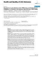
admission. The patient enrolment and study profile were
shown in Fig. 1. Among included patients, the median
age was 18 (10, 38.5) months and 40 patients (59.7%)
were male. Children aged < 24 months accounted for
83.6% (56/67) of all cases. The main characteristics at
initial PICU admission between survivors and nonsurvivors were summarized in Table 1.
All patients were admitted to PICU for the reasons of
fever (100%) and respiratory symptoms consistent with
cough (100%) or whoop (65.7%), tachypnea (100%), acute
respiratory failure (100%) requiring oxygenation support.
At PICU admission, co-infection (defined as pneumonia caused by adeno virus as well as typical bacteria,
mycoplasma pneumoniae and other viruses) was seen in
25.37% (17/67) patients. During the PICU stay, nosocomial infection including VAP and bloodstream infection
were seen in 32.84% (22/67) patients, higher morbidity
in non-survival (63.6%,7/11) than in survival (26.78%,15/
56). Nosocomial infected pathogens were isolated from
different specimens including blood, sputum, bronchoalveolar lavage fluid, and hydrothorax. The most frequently isolated pathogens were Acinetobacter baumanii
Fig. 1 Patient enrolment and Study profile
Page 3 of 8
9 patients (13.4%), Klebsiella pneumoniae in 7(10.5%),
mycoplasma pneumoniae in 6 (9.8%), Stenotrophomonas
maltophilia in 4 (5.9%), and Candida albicans in 3
(4.5%) (Table 2).
Management and outcomes
All management decisions were performed by intensivist
according to the guideline recommendation [8, 16, 17],
experts’ opinion [18], and routine practice in our PICU.
Additional oxygen was utilized in 100% (67cases) patients with 8.96% (6cases) requiring high flow nasal oxygen therapy, and 92.54% (62cases) requiring mechanical
ventilation at some period during hospitalization. The
indications for CRRT/RRT were:1) AKI which was defined according to the KDIGO criteria [19]; 2) Fluid
overload which was defined as the fluid overload > 10%
[fluid overload = (CRRT initial weight-PICU admission
weight)/PICU admission weight× 100%] [20, 21]. The indications for ECMO were:1) severe hypoxemia with a
PaO2/ FiO2 ratio of < 50 mmHg for > 3 h or < 80 mmHg
for > 6 h, or pH < 7.25 and a partial pressure of arterial
CO2 of ≥60 mmHg for > 6 h [22]. 2) hypoxemia complicated with cardio dysfunction when cardiac index (CI)
less than 2.2 L/min.m2; and 3) hypoxemia complicated
with circulatory dysfunction when persistent lactatemia
greater than 4 mmol/L and vasoactive inotropic score
(VIS) greater than 50.VIS was calculated as ([(epinephrine+
norepinephrine) ug/kg.min] × 100 + [(dobutamine + dopamine) ug/kg.min] + [milrinone ug/kg.min] × 15. Intravenous
Shi et al. BMC Pediatrics
(2020) 20:375
Page 4 of 8
Table 1 Baseline characteristics at PICU admission between survivors and non-survivors
p value
Variables at PICU admission
Survivors (n = 56)
Nonsurvivors (n = 11)
Total (n = 67)
Age, mo, median (IQR)
18 (11, 38)
20(7.5, 41.5)
18 (10, 38.5)
0.889
Male gender, n (%)
32 (57.14%)
8 (72.7%)
40 (59.7%)
0.335
PRSM III score, median (IQR)
13 (10, 15)
14 (11, 18)
13 (10, 15)
0.133
days of illness before at PICU admission, median (IQR)
9 (7, 10.5)
6 (5, 12)
9 (6, 11)
0.959
6.55 (4.36,12.23)
6.11 (4.35, 10.87)
6.48 (4.29, 11.96)
0.923
Laboratory values, median (IQR)
White blood cell, 109/L
Platelet, 109/L
254 (179, 349.5)
258 (148.5, 444.5)
258 (178, 353)
0.837
NK cells, %
3.85 (2.23, 6.57)
5 (2, 6.9)
4.06 (2.19, 6.68)
0.787
pH
7.4 (7.33, 7.44)
7.33 (7.28, 7.42)
7.4 (7.33, 7.44)
0.109
PaO2, mmHg
71 (57, 82.5)
73 (65.38, 84.38)
72 (57, 84)
0.853
PaCO2, mm Hg
43 (38, 55)
52 (44.5, 68)
44 (38, 55.5)
0.104
MAP, mmHg
55 (52, 68.25)
49 (42.5, 57)
55 (51.5, 67)
0.171
LA, mmol/L
1.95 (1.38, 2.33)
2 (1.65, 4.25)
2 (1.4, 2.6)
0.968
CI, L/min/m2
4.2 (3.9, 5)
3.6 (3.45, 4.1)
4.2 (3.85, 4.85)
0.011
TBIL,umol/L
4.42 (3.2, 5.87)
10.85 (5.35, 20.22)
5.02 (3.29, 6.61)
0.069
serum creatinine, umol/L
32.5 (25.5, 40.88)
34.5 (29.25, 54)
33 (26.25, 42)
0.291
IQR interquartile range, NK cells natural kill cell; CI: cardiac index, MAP mean arterial pressure, TBIL total bilirubin, LA Lactate
Table 2 PICU therapeutic interventions between survivors and non-survivors
Survivors (n = 56)
Non-survivors (n = 11)
Total (n = 67)
p value
Median of PICU stay, days
11 (7.75, 18)
15 (11, 19.5)
11 (8, 18)
0.861
Median of hospital stay, days
22.5 (16, 34.25)
17 (16, 23.5)
22 (16, 31)
0.124
ARDS
27 (48.21%)
9 (81.82%)
36 (53.73%)
0.041
Liver dysfunction
21 (31.34%)
10 (90.9%)
31 (46.27%)
0.001
AKI
4 (7.14%)
5 (45.45%)
9 (13.43%)
0.001
shock
42 (75%)
10 (90.9%)
52 (77.61%)
0.247
GI dysfunction
31 (55.36%)
11 (100%)
42 (62.69%)
0.005
Invasive Mechanical ventilation
51 (91.07%)
11 (100%)
62 (92.54%)
0.303
CRRT/RRT
11 (19.64%)
8 (72.73%)
19 (28.36%)
<0.001
ECMO
6 (10.71%)
3 (37.5%)
9 (13.43%)
0.141
Prone positioning, n (%)
15 (26.79%)
4 (36.36%)
19 (28.36%)
0.519
Neuromuscular blockade, n (%)
22 (39.29%)
8 (72.73%)
30 (44.78%)
0.041
Vasoactive use
48 (85.71%)
10 (90.9%)
58 (86.57%)
0.644
diuretics use
43 (76.79%)
8 (72.73%)
51 (76.12%)
0.773
Steroids use
55 (98.21%)
11 (100%)
66 (98.51%)
0.655
IV immunoglobulin
50 (89.29%)
10 (90.9%)
60 (89.55%)
0.872
Parenteral nutrition
12 (21.43%)
10 (90.9%)
22 (32.84%)
<0.001
Co-morbidity, n (%)
PICU and hospital therapies, n (%)
Packed red blood cell perfusion
28 (50%)
10 (90.9%)
38 (56.72%)
0.012
15 (26.79%)
7 (63.64%)
22 (32.84%)
0.017
bacterial
15 (26.79%)
7 (63.64%)
22 (32.84%)
fungal
4 (7.14%)
1 (9.09%)
5 (7.46%)
Nosocomial infenction a, n (%)
a
parts of patients complicated with bacteria, mycoplasma pneumoniae or fungi in nosocomial infection or co-infection
0.043
0.687
Shi et al. BMC Pediatrics
(2020) 20:375
Page 5 of 8
neuromuscular blockade was started if the peak inspiratory
pressures approximated 28–30 cmH2O, and the patient
was hypoxemic and continued to show excessive work of
breathing despite adequate sedation [23]. Other cluster
therapies included prone positioning, IV immunoglobulin,
parenteral nutrition, vasoactive drugs, packed red
blood cell perfusion, steroids (methylprednisolone
0.5–2.0 mg/kg.d for 3–5 days), diuretics, and antibiotics if needed (see in Table 2).
Among the 67 patients with severe adenovirus pneumonia, 11(16.42%) children died in PICU and among
them, 10 (14.93%) cases died within 28 day after PICU
admission. The overall PICU mortality was 16.42% (11/
67), and 28-day mortality was14.93% (10/67). Patients
aged less 2-year old accounted for 72.73% (8/11) of nonsurvivors.
The median lengths of stay in the PICU and hospital
were 11 days (8, 18 days) and 22 days (16, 31 days), respectively (Table 2). The median duration of mechanical
ventilation was 5.75 days (3.98, 11.67) in patients required invasive ventilation. In non-survivors, the median
ventilator days was longer than that in survivors but
without statistical significance (8.48 [4.77, 11.82] days vs.
10.75 [8.17, 19.08] days, p = 0.348). The rate of CRRT
and use of neuromuscular blockade, parenteral nutrition,
or packed red blood cell perfusion were significantly
higher in non-survivors than that in survivors (p < 0.001,
p = 0.041, p < 0.001, p = 0.012, respectively; Table 2).
Moreover, the ratio of nosocomial infection was higher
in non-survivors compared with survivors (63.64% vs.
26.79%, p = 0.017; Table 2).
The changes of parameters about ventilator characteristics and blood gas analysis in survivors and non-
survivors were shown in Table 3. There were no significant differences in parameters including peak inspiratory
pressure (PIP), positive expiratory end pressure (PEEP)
and mean airway pressure (MAP) on the initial day, 3rd
day and 7th day of invasive ventilation between survivors
and non -survivors (all p > 0.05, Table 3). However, the
values of Cydn on 3rd day and 7th day of invasive ventilation were significantly lower in non-survivors compared with survivors (p = 0.012, p = 0.045, Table 3). In
addition, the ratio of PaO2/ FiO2 and SaO2 levels displayed a tendency decrease in non-survivors compared
with survivors on the 3rd day after receiving invasive
mechanical ventilation (p = 0.038, p = 0.008, respectively;
Table 3).
Besides the lower cardiac index (CI) in non-survivors
than survivors at PICU admission (Table 1), CD4+ cells
percentage showed a higher tendency in non-survivors
than that in survivors at PICU admission (p = 0.071,
Table 4). There were no significant differences in aspects
of white blood cell and platelet count, NK cell, CD4+,
CD8+, CD19+ percentage between two groups at PICU
admission. During PICU stay, platelet Count was significantly lower in non-survivors at 7 days after PICU admission when compared with survivors (93 [85, 371], vs.
327 [257.75, 443.75] × 109/L, p = 0.039; Table 4). In
addition, the interleukin 6 (IL-6) and IL-10 were significantly higher in non-survivors than those of survivors at
7 days after PICU admission (p = 0.035, p < 0.01, respectively; Table 4).
Multivariate logistic analysis
By univariate logistic analysis, the patients were associated with worse outcome of severe adenovirus
Table 3 The changes of parameters about patients receiving invasive mechanical ventilation
Variables
D1
Survivor
(n = 56)
D3
Nonsurvivor
(n = 11)
D7
p-value Survivors
(n = 56)
Nonsurvivor
(n = 11)
24 (20.5, 28)
PSurvivor
value (n = 56)
Nonsurvivor
(n = 11)
p-value
PIP,cmH2O
21 (19,23.5)
25 (21, 27)
0.145
21.5 (20, 24)
0.377 22 (20, 26)
23 (21.75, 30)
0.27
MAP,cmH2O
11 (10, 12.5)
12 (10.5, 13.5)
0.372
11.5 (9.25, 13) 12 (11, 14.5)
0.412 12 (11.5, 14.5)
13 (11, 17.25)
0.458
PEEP, cm H2O
5 (4, 5)
5 (4, 5)
0.368
5 (5, 6)
5 (5, 5.5)
0.801 5 (5, 6)
6 (5, 6)
0.567
Cdyn, cm H2O/kg 0.4 (0.32, 0.48)
0.33 (0.31,
0.425)
0.286
0.52 (0.43,
0.55)
0.4 (0.31, 0.4)
0.012 0.5 (0.4, 0.56)
0.26 (0.22, 0.34)
0.045
Tidal volume,
ml/kg
8 (7.8, 8)
7.2 (6.65, 8)
0.0029
8.3 (8, 8.5
8 (7.2, 8.1)
0.072 8.2 (8, 8.5)
7.55 (6.03, 8.35)
0.307
PaO2/FiO2 ratio,
mmHg
151 (113, 180.25) 140 (90, 143.5)
0.177
185 (139, 224) 111 (103,174.5) 0.038 170 (110, 212)
137.5 (89.5, 157.75) 0.384
Oxygenation index
PaCO2, mm Hg 44 (35, 50.25)
48 (41.5, 57.5)
0.138
45 (40.5, 48.5) 42 (37.5, 55.5)
0.727 44 (41, 48)
58 (43, 69.5)
0.305
PaO2, mm Hg
72 (57, 86.25)
73 (58.375, 84.5) 0.871
78 (72.5, 89.5) 78 (72.5, 89.5)
0.087 77 (55, 90)
65 (51.5, 75)
0.357
SaO2, %
95 (92, 96)
93 (90.5, 95)
96 (95, 97)
0.008 96.5 (92.25, 98) 91 (89, 95.5)
0.655
0.265
93 (91.5, 95.5)
D1: initial day of invasive mechanical ventilation; D3: 3 days of ventilation; D7: 7 days of ventilation; PIP: Cdyn: lung dynamic compliance; MAP mean
airway pressure
Shi et al. BMC Pediatrics
(2020) 20:375
Page 6 of 8
Table 4 Changes of blood cell and immunological parameters at PICU admission and 7 days after admission
Variables
PICU admission
7 days after admission
Survivors (n = 56)
Non-survivors (n = 11)
P value
Survivors (n = 56)
Non-survivors (n = 11)
P value
101.5 (90, 113.5)
100 (100, 109.5)
0.699
99.5 (95, 106)
95 (90, 110)
0.457
WBC, 109/L
6.55 (4.36, 12.23)
6.11 (4.35, 10.87)
0.923
8.78 (5.26, 10.77)
4.37 (3.39, 11.69)
0.774
platlat,109/L
254 (179, 349.5)
258 (148.5, 444.5)
0.837
327 (257.75, 443.75)
93 (85, 371)
0.039
NK cells, %
3.85 (2.23, 6.57)
5 (2, 6.895)
0.787
3.98 (2.185, 6.01)
2.98 (1.02, 8.08)
0.812
CD19+, %
44.02 (30.91, 52.64)
42.1 (24.47, 53.55)
0.437
34.58 (30.13, 45.36)
38.58 (24.66, 44.04)
0.804
CD4+, %
25.56 (19.5, 31.39)
32.48 (23.2, 41.59)
0.071
27.49 (22.96, 35.48)
33.23 (29.82, 38.79)
0.489
CD8+, %
20.02 (14.96, 26.75)
16.36 (15.34 20.25)
0.555
20.14 (17.76, 28.28)
20.58 (14.23, 22.5)
0.614
IL-6, ng/L
0.1 (0.1, 0.1)
0.1 (0.1,0.1)
0.449
0.1 (0.1, 0.1)
47.77 (0.1, 239.29)
0.035
IL-8, ng/L
0.1 (0.1, 12.09)
0.1 (0.1, 36.39)
0.707
0.1 (0.1, 0.1)
7.15 (3.09, 20.21)
0.144
Hb, g/L
IL-10, ng/L
0.1 (0.1, 4.05)
8.99 (0.1, 32.57)
0.855
0.1 (0.1, 0.1)
18.39 (0.1, 32.74)
0.0005
IL-2R, ug/L
15.31 (4.27, 28.78)
18.92 (11.38, 33.37)
0.459
10.25 (20.81, 24.32)
21.71 (16.37 33.43)
0.089
pneumonia in complicated liver dysfunction due to the
adeno virus infection (16.485 [1.745 ~ 155.705], p =
0.014), AKI (10.833[2.269 ~ 51.706], p = 0.003), gastrointestinal dysfunction (0.355 [0.178 ~ 0.706], p = 0.003)
which was defined according to the European Consensus
Definition of acute gastrointestinal injury (AGI) [24], encephalopathy (5.629 [1.333 ~ 23.774], p = 0.019), coinfection & nosocomial infection (15.455 [1.847 ~
129.326], p = 0.012) (Table 5). By multivariate logistic regression analysis, the independently risk factor associated with mortality was liver dysfunction (21.231 [1.696
~ 265.779], p = 0.018) and nosocomial infection (2.574
[0.986 ~ 15.671], p = 0.05) (Table 5).
Discussion
This is the first REPORT describing overall morbidity
and mortality for pediatric patients with severe adenoviral pneumonia admitted to the PICU in mainland
China. In our PICU 3-year period, the hospital all-cause
hospital mortality of severe community acquired
adenoviral pneumonia was 16.42%, and 28-day mortality
(deaths after discharge from hospital were not included)
was 14.93%. In the 11 non-survivor patients, 3 of them
died from liver dysfunction due to adeno virus infection,
3 died from refractory hypoxic respiratory failure, two of
them died from refractory septic shock caused by nosocomial infection of Klebsiella pneumoniae and Stenotrophomonas maltophili, and one patient died from
intracranial bleeding. We identified the independently
risk factors for mortality including patients complicated
with liver dysfunction and nosocomial infection.
Adenovirus disease is a self-limiting in majority of immunocompetent population, but can cause lifethreatening illness in immunocompromised hosts [25–
27]. Adenovirus accounts for at least 5 to 10% of
pediatric respiratory tract infections in children [1, 2].
The overall PICU hospitalization with severe adenoviral
pneumonia in the present study was 9.99% of CAP.
More importantly, the cases number from 2016 to 2019
was with increased tendency in our PICU, especially with
Table 5 Logistic analysis of variables independently associated with hospital mortality
OR
St.Err.
95%CI
P value
PRISM III
1.079
0.119
0.868 ~ 1.342
0.690
ARDS
0.5
0.185
0.242 ~ 1.032
0.061
Liver dysfunction
16.485
18.887
1.745 ~ 155.705
0.014
AKI
10.833
8.639
2.269 ~ 51.706
0.003
Shock
3.333
3.644
0.391 ~ 28.41
0.271
Outcome
Univariate logistic regression
Gastrointestinal dysfunction
0.355
0.124
0.178 ~ 0.706
0.003
Encephalopathy
5.629
4.138
1.333 ~ 23.774
0.019
Co-infection& nosocomial infection
15.455
16.751
1.847 ~ 129.326
0.012
Liver dysfunction
21.231
27.376
1.696 ~ 265.779
0.018
nosocomial infection
2.574
0.706
0.986 ~ 15.671
0.05
Multivariate logistic regression
Shi et al. BMC Pediatrics
(2020) 20:375
a higher incidence rate between 2018 to 2019. Most patients
with adenovirus infection are younger than 2-year old [56/
67,83.6%]. When severe adenovirus pneumonia progressed
with MODS, the mortality is higher over 50% [5]. In the
present study, children aged < 24 months accounted for
72.73% of total deaths. There are limited antiviral drugs available for adenovirus. Cidofovir is an antiviral drug which use
has been associated with significant reductions of adenovirus
load and, in some series improved survival in reports [10,
28]. Until recently, Cidofovir is not available in China till
2019, and has been neither widely used in children, nor has
it been used in our cases. All these results suggested that
adenovirus pneumonia requires our attention due to the
high mortality involved, especially in China where there have
no specific anti-adenovirus drugs or vaccine for children
until now.
Mechanical ventilation remains the main stay of management. For the hypoxemia respiratory failure/ARDS
ventilated patients caused by adenovirus in this study, the
PaO2/FiO2 ratio at initial presentation was relatively low
in survivors (151[interquartile range:113, 180.25]) and
non-survivors (140[interquartile range:107, 143.5]).The
PaO2/FiO2 was no statistical difference at initial day, 3rd
day and 7th day ventilation between survivors and nonsurvivors. But lower Cdyn at 3rd day and 7th day ventilation in non-survivors (p = 0.012, p = 0.045). In order to ensure the mechanical ventilation and to improve the level
of PaO2/FiO2, we used prone position and neuromuscular
blockers in appropriate patients. There was no difference
in the proportion of prone position between the two
groups (p = 0.519), but the proportion of neuromuscular
blockers was significantly higher in non-survivors than
that in survivors (p = 0.041).
Under 2-year old could partially contribute to the high
incidence of severe adenovirus pneumonia and high
mortality [2, 24–26]. Adenovirus-induced immunosuppression might augment the susceptibility to nosocomial
microbial infections. In this retrospective study, the high
levels of IL-6 and IL-10 in non-survivors were measured,
and we identified that the nosocomial infection after
PICU admission was an independent risk factors for allcause hospital mortality. This indicated that high levels
of IL-6 and IL-10 in non-survivors could provide an
insight for adenovirus-associated nosocomial infection.
IL-6 plays a role in immunosuppression by driving differentiation of myeloid suppressor cells together with
TGF-β in cancer pathogenesis [29]. Otherwise, IL-10 is
produced by Treg cells and Th2-type cells and suppresses the Th1 response [30]. The continued release of
IL-10 contributes to sepsis-induced immunosuppression
resulting in more susceptibility to nosocomial infection
[31, 32]. Whether high levels of IL-6 and IL-10 in patients with adenovirus infection contribute the worse
outcome warrants further investigation.
Page 7 of 8
ECMO support for severe adenoviral infection has been
reported in several studies [10, 33, 34]. Retrospective data
from the extracorporeal life support organization (ELSO)
registry showed that pediatric patients with AV infection
supported with ECMO, had a survival to hospital discharge of 38% which was even lower in neonates [8]. B
More recently, Ramanathan et al. observed over the last
25 years ELSO registry across all age groups who needed
ECMO for severe adenoviral pneumonia in neonatal,
pediatric, and adult patients, the hospital mortality was
58% with no significant improvement from 1992 to 2016
[12]. In our study, 6 patients survived in whom (9cases)
received ECMO support from 2016 to 2019. Our results
suggest that ECMO as the last rescue treatment for severe
adenoviral pneumonia, is worthy of further exploration.
Our study has several limitations. First, it is a retrospective analysis from single PICU, we didn’t include
those adeno virus pneumonia not requiring PICU admission, which contributed to the power of our study is limited by the small size of case series. Second, we didn’t
detect of adenovirus serotype, which might affect the
judgment of the outcomes. Third, long-term follow-up
data was unavailable.
Conclusion
Our study demonstrated that adenovirus pneumonia remains a major cause of morbidity and mortality in the
PICU. We identified several factors with higher mortality,
including complicated with shock, liver dysfunction, AKI,
gastrointestine dysfunction, encephalopathy, and coinfection& nosocomial infection. The patients complicated
with liver dysfunction and associated nosocomial infection
were independent risk factors for mortality.
Abbreviations
PICU: Pediatric intensive care unit; CAP: Community acquired pneumonia;
ARDS: Acute respiratory distress syndrome; MODS: Multiple organ
dysfunction syndrome; RT-PCR: Real-time polymerase chain reaction;
ECMO: Extracorporeal membrane oxygenation; CRRT/RRT: Continuous renal
replacement therapy/ renal replacement therapy; PaO2/FiO2: The ratio of the
partial pressure of oxygen in arterial blood (PaO2) to the inspired oxygen
fraction (FiO2); PRISM III: Pediatric risk of mortality III; Cdyn: Lung dynamic
compliance; CI: Cardiac index; MAP: Mean arterial pressure; TBIL: Total
bilirubin; LA: Lactic acid; sCr: serum creatinine; PLT: Platelet counts;
NK: Natural kill cell; IQR: Interquartile range; ORs: Odd ratios; AKI: Acute
kidney injury; ELSO: Extracorporeal life support organization
Acknowledgements
We acknowledge that our manuscript has been presented as a poster of the
conference: AS16 Sepsis / Infections in the Critical Care Unit / Antimicrobial
Stewardship / Tropical and Parasite Infections.
Authors’ contributions
Conceived and designed the study: CXW and YCZ. Collected and analyzed
data: JYS, YPZ, TS and YJS. Contributed analysis tools: FW and HJM.
Contributed to discussion: JYS, YC and YCZ. Wrote the paper: JYS, YPZ, YC
and YCZ. All authors read and approved the final manuscript.
Funding
This study was supported by the Multicenter Clinical Research Program of
Shanghai Jiao Tong University School of Medicine (DLY201618), funded by
Shi et al. BMC Pediatrics
(2020) 20:375
the Science and Technology Commission of Shanghai Municipality
(18411951000), grant supported by Shanghai Children’s Hospital
(2019XGLC01, 2018YLY004). The funders had no role in the study design,
data collection or analysis, decision to publish, or preparation of the
manuscript.
Availability of data and materials
Our present study was a retrospective observational study. All the data were
obtained from medical records of patients. The datasets used and/or
analysed during the current study are not publically available, but they can
be shared from the corresponding author on reasonable request except for
the identifying/confidential patient data.
Ethics approval and consent to participate
This study was approved by Ethics Review Committee, Children’s Hospital of
Shanghai/Shanghai Children’s Hospital, Shanghai Jiao Tong University and
conducted in accordance with the provisions of the Declaration of Helsinki
(Approval number: 2016R007-E01). Informed content consent was waived because of its retrospective design.
Consent for publication
Not applicable.
Competing interests
The authors have declared that no competing interests exist.
Received: 7 April 2020 Accepted: 2 August 2020
References
1. Lynch JP, Fishbein M, Echavarria M. Adenovirus. Semin Respir Crit Care Med.
2011;32:494–511.
2. Lynch JP 3rd, Kajon AE. Adenovirus: epidemiology, global spread of novel
serotypes, and advances in treatment and prevention. Semin Respir Crit
Care Med. 2016;37(4):586–602.
3. Radke JR, Cook JL. Human adenovirus infections: update and consideration
of mechanisms of viral persistence. Curr Opin Infect Dis. 2018;31:251–6.
4. Lee E, Kim CH, Lee YJ, Kim HB, Kim BS, Kim HY, et al. Annual and seasonal
patterns in etiologies of pediatric community-acquired pneumonia due to
respiratory viruses and mycoplasma pneumoniae requiring hospitalization in
South Korea. BMC Infect Dis. 2020;20(1):132.
5. Chowdhury F, Shahid ASMSB, Ghosh PK, Rahman M, Hassan MZ, Akhtar Z,
et al. Viral etiology of pneumonia among severely malnourished under-five
children in an urban hospital, Bangladesh. PLoS One. 2020;15(2):e0228329.
6. Lion T. Adenovirus infections in immunocompetent and
immunocompromised patients. Clin Microbiol Rev. 2014;27:441–62.
7. Bellani G, Laffey JG, Pham T, Fan E, Brochard L, Esteban A, et al.
Epidemiology, patterns of care, and mortality for patients with acute
respiratory distress syndrome in intensive care units in 50 countries. JAMA.
2016;315:788–800.
8. Khemani RG, Smith LS, Zimmerman JJ, Erickson S, Pediatric Acute Lung
Injury Consensus Conference Group, et al. Pediatric acute respiratory distress
syndrome: definition, incidence, and epidemiology: proceedings from the
pediatric acute lung injury consensus conference. Pediatr Crit Care Med.
2015;16:S23–40.
9. Prodhan P, Bhutta AT, Gossett JM, Stroud MH, Rycus PT, Bratton SL, et al.
Extracorporeal membrane oxygenation support among children with
adenovirus infection: a review of the extracorporeal life support
organization registry. ASAIO J. 2014;60:49–56.
10. Lee M, Kim S, Kwon OJ, Kim JH, Jeong I, Son JW, et al. Treatment of adenoviral
acute respiratory distress syndrome using cidofovir with extracorporeal
membrane oxygenation. J Intensive Care Med. 2017;32:231–8.
11. Gupta P, Tobias JD, Goyal S, Hervie P, Harris JB, Sadot E, et al. Prolonged
mechanical support in children with severe adenoviral infections: a case
series and review of the literature. J Intensive Care Med. 2011;26:267–72.
12. Ramanathan K, Tan CS, Rycus P, MacLaren G. Extracorporeal membrane
oxygenation for severe adenoviral pneumonia in neonatal, pediatric, and
adult patients. Pediatr Crit Care Med. 2019;20(11):1078–84.
13. Binder AM, Biggs HM, Haynes AK, Chommanard C, Lu X, Erdman DD, et al.
Human adenovirus surveillance - United States, 2003-2016. MMWR Morb
Mortal Wkly Rep. 2017;66(39):1039–42.
Page 8 of 8
14. Lee J, Choi EH, Lee HJ. Comprehensive serotyping and epidemiology of
human adenovirus isolated from the respiratory tract of Korean children
over 17 consecutive years (1991-2007). J Med Virol. 2010;82:624–31.
15. Wu PQ, Zeng SQ, Yin GQ, Huang JJ, Xie ZW, Lu G, et al. Clinical
manifestations and risk factors of adenovirus respiratory infection in
hospitalized children in Guangzhou, China during the 2011-2014 period.
Medicine (Baltimore). 2020;99:e18584.
16. Dellinger RP, Levy MM, Rhodes A, Annane D, Gerlach H, Opal SM, et al.
Surviving sepsis campaign guidelines for management of severe sepsis and
septic shock. Intensive Care Med. 2013;34:783–5.
17. Davis AL, Carcillo JA, Aneja RK, Deymann AJ, Lin JC, Nguyen TC, et al.
American College of Critical Care Medicine Clinical Practice Parameters for
hemodynamic support of pediatric and neonatal septic shock. Crit Care
Med. 2017;45:1061–93.
18. Chiumello D, Brochard L, Marini JJ, Slutsky AS, Mancebo J, Ranieri VM,
Thompson BT, Papazian L, Schultz MJ, Amato M, et al. Respiratory support
in patients with acute respiratory distress syndrome: an expert opinion. J
Crit Care. 2017;21(1):240.
19. Sutherland SM, Byrnes JJ, Kothari M, et al. AKI in hospitalized children:
comparing the pRIFLE, AKIN, and KDIGO definitions. Clin J Am Soc Nephrol.
2015;10:554–61.
20. Miao H, Shi J, Wang C, et al. Continuous renal replacement therapy in
pediatric severe Sepsis: a propensity score-matched prospective multicenter
cohort study in the PICU. Crit Care Med. 2019;47:e806–13.
21. Goldstein SL, Currier H, Graf C, et al. Outcome in children receiving
continuous venovenous hemofiltration. Pediatrics. 2001;107:1309–12.
22. Patel B, Chatterjee S, Davignon S, Herlihy JP. Extracorporeal membrane
oxygenation as rescue therapy for severe hypoxemic respiratory failure. J
Thorac Dis. 2019;11(Suppl 14):S1688–97.
23. Wilsterman MEF, de Jager P, Blokpoel R, Frerichs I, Dijkstra SK, Albers MJ,
et al. Short-term effects of neuromuscular blockade on global and regional
lung mechanics, oxygenation and ventilation in pediatric acute hypoxemic
respiratory failure. Ann Intensive Care. 2016;6(1):103–13.
24. Reintam Blaser A, Malbrain ML, Starkopf J, et al. Gastrointestinal function in
intensive care patients: terminology, definitions and management.
Recommendations of the ESICM working group on abdominal problems.
Intensive Care Med. 2012;38(3):384–94.
25. Echavarria M. Adenoviruses in immunocompromised hosts. Clin Microbiol
Rev. 2008;21:704–15.
26. Ghanaiem H, Averbuch D, Koplewitz BZ, Yatsiv I, Braun J, Dehtyar N, Wolf DG,
Mandelboim M, Engelhard D. An outbreak of adenovirus type 7 in a residential
facility for severely disabled children. Pediatr Infect Dis J. 2011;30:948–52.
27. Peled N, Nakar C, Huberman H, Scherf E, Samra Z, Finkelstein Y, Hoffer V,
Garty BZ. Adenovirus infection in hospitalized immunocompetent children.
Clin Pediatr (Phila). 2004;43:223–9.
28. Ganapathi L, Arnold A, Jones S, Patterson A, Graham D, Harper M, Levy O.
Use of cidofovir in pediatric patients with adenovirus infection. F1000Res.
2016;5:758.
29. Eriksson E, Milenova I, Wenthe J, Moreno R, Alemany R, Loskog A. IL-6
signaling blockade during CD40-mediated immune activation
FavorsAntitumor factors by reducing TGF-β, collagen type I, and PD-L1/PD1. J Immunol. 2019;202(3):787–98.
30. Muehlstedt SG, Lyte M, Rodriguez JL, Muehlstedt SG, Lyte M, Rodriguez JL.
Increased il-10 production and hla-dr suppression in the lungs of injured patients
precede the development of nosocomial pneumonia. Shock. 2002;17(6):443–50.
31. Oberholzer A, Oberholzer C, Moldawer LL. Interleukin-10: a complex role in
the pathogenesis of sepsis syndromes and its potential as an antiinflammatory drug. Crit Care Med. 2002;30(Suppl):S58–63.
32. Ono S, Tsujimoto H, Hiraki S, Aosasa S. Mechanisms of sepsis-induced
immunosuppression and immunological modification therapies for sepsis.
Ann Gastroenterol Surg. 2018;2(5):351–8.
33. Allibhai TF, Spinella PC, Meyer MT, Hall BH, Kofos D, DiGeronimo RJ. Survival
after prolonged pediatric extracorporeal membrane oxygenation support
for adenoviral pneumonia. J Pediatr Surg. 2008;43(8):e9–11.
34. Ha SO, Kim HS, Park S, Jung KS, Jang SH, Han SJ, Kim HS, Lee SH. Severe
ARDS caused by adenovirus: early initiation of ECMO plus continuous renal
replacement therapy. SpringerPlus. 2016;5(1):1909.
Publisher’s Note
Springer Nature remains neutral with regard to jurisdictional claims in
published maps and institutional affiliations.