Periosteal new bone formation in KlippelTrénaunay syndrome: A case report
Bạn đang xem bản rút gọn của tài liệu. Xem và tải ngay bản đầy đủ của tài liệu tại đây (901.45 KB, 5 trang )
Fang et al. BMC Pediatrics
(2020) 20:388
/>
CASE REPORT
Open Access
Periosteal new bone formation in KlippelTrénaunay syndrome: a case report
Xiang Fang1†, Wenli Zhang1†, Zeping Yu1, Fuguo Kuang2, Bin Huang3 and Hong Duan1*
Abstract
Background: Klippel-Trénaunay syndrome (KTS) is a complex congenital vascular disorder, typically accompanied
by port-wine stains, varicose veins, and limb hypertrophy. This paper reports a rare and unusual clinical condition of
periosteal reaction in a pediatric case of KTS. Although periosteal new bone formation is not rare in children, as is
KTS, their dual occurrence or the presentation of the former due to KTS has not been previously documented. Our
objective in this study is to highlight the potential association between periosteal new bone formation and KTS, as
well as to help physicians consider this association when bone neoplasm has been ruled out.
Case presentation: A 7-year old girl, initially presented with a persistent mild swelling in her left shank, with no
abnormalities in the X-ray of the tibiofibular. However, after a few consults and examinations, 7 weeks later, a
17 cm-long periosteal new bone formation along the left tibia and diffused dilated vessels in the left shank were
revealed by the radiological examination. Not knowing the true nature of the fast-growing lesion in a typical case
of KTS was worrying. Therefore, a core needle biopsy was performed. The test demonstrated a possible parosteal
hemangioma. Following further investigation through an excisional biopsy, and a pathological analysis, hyperplasia
of the bone tissues with no tumor cells was revealed. Thereafter, an elastic stocking treatment was prescribed.
During the first two-year follow-up, recurrence of the mass or sign of progression of KTS was not observed.
Conclusions: Periosteal new bone formation is a potential manifestation of KTS. Based on the conclusive
pathological results of the excisional biopsy, invasive examinations and surgeries could be avoided in future KTSsubperiosteal lesion manifestations.
Keywords: Bone tumors, Children, Definite diagnosis, Periosteal reaction
Background
Klippel-Trénaunay syndrome (KTS) in childhood is
well-documented and commonly characterized by portwine stains, varicose veins, and the overgrowth of long
bones and soft tissues [1, 2]. In this report, we describe
an atypical pediatric case of KTS, in which a 17 cm-long
periosteal new bone formation of the tibia developed
rapidly within 7 weeks. Periosteal new bone formation is
very common in pediatric bone neoplasm, especially
bone malignancy; however, it rarely occurs in cases of
KTS. Therefore, this presentation has not been described
in extant KTS-related literature. Our objective in this
study is to highlight the potential association between
periosteal new bone formation and KTS, as well as to
help physicians consider this association when bone neoplasm has been ruled out.
* Correspondence:
Xiang Fang and Wenli Zhang contributed equally to this work, and they are
co-first authors.
1
Department of Orthopedics, West China Hospital, Sichuan University, 37
Guo Xue Lane, 610064 Chengdu, Sichuan, People’s Republic of China
Full list of author information is available at the end of the article
Case presentation
A 7-year old girl presented to the local hospital with
mild swelling in her left shank that had persisted for one
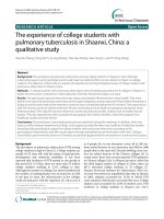
week. A radiograph of the tibiofibular showed no abnormalities (Fig. 1), and a herbal remedy for external use
© The Author(s). 2020 Open Access This article is licensed under a Creative Commons Attribution 4.0 International License,
which permits use, sharing, adaptation, distribution and reproduction in any medium or format, as long as you give
appropriate credit to the original author(s) and the source, provide a link to the Creative Commons licence, and indicate if
changes were made. The images or other third party material in this article are included in the article's Creative Commons
licence, unless indicated otherwise in a credit line to the material. If material is not included in the article's Creative Commons
licence and your intended use is not permitted by statutory regulation or exceeds the permitted use, you will need to obtain
permission directly from the copyright holder. To view a copy of this licence, visit />The Creative Commons Public Domain Dedication waiver ( applies to the
data made available in this article, unless otherwise stated in a credit line to the data.
Fang et al. BMC Pediatrics
(2020) 20:388
Fig. 1 Initial radiograph of the tibiofibular shows no abnormalities
Page 2 of 5
was applied. One week later, the shank swelling aggravated, and she presented to our hospital. On examination, apart from the swollen shank, we also observed
multiple port-wine stains and limb overgrowth (longer
extremity and larger foot). Ultrasonography revealed
great/lesser saphenous vein thrombosis in the lower left
leg and an ill-defined hypoechoic mass containing flaky
hyperechoic foci near the left tibia. Based on the aforementioned findings, a clinical diagnosis of KTS was
established. The use of low-molecular weight heparin
and mucopolysaccharide polysulfate cream was initiated.
The patient re-admitted 6 weeks later, reporting a
palpable hard mass in the anterolateral left shank and
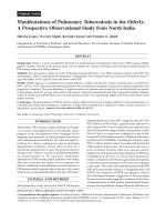
complete resolution of the swelling. Radiological examination revealed a 17-cm periosteal new bone formation
along the left tibia and multiple dilated vascular structures in the left shank (Fig. 2). Coagulation status was
normal. No fever, allergies, severe pain, or a recent history of trauma were presented.
Bone neoplasm was first suspected but subsequently
ruled out due to the regular pattern of periosteal new
bone formation without soft tissue mass, bone destruction, and symptoms. However, concerning the fastgrowing lesion in KTS, we performed a core needle biopsy, which revealed a possible parosteal hemangioma.
Consequently, an excisional biopsy of the lesion was performed and intraoperatively, only regularly thickened
eggshell-like hard tissues and blood clots in the cavity of
the lesion were found, Pathological analysis revealed
Fig. 2 Radiographs and computed tomography images of the tibiofibular (obtained 7 weeks after the initial radiograph) reveal a parosteal highdensity lesion with well-defined borders along the long axis of the left tibia (a, b, c). T2-weighted magnetic resonance imaging scans of the
lower legs show heterogeneous high-signal intensity in the anterolateral aspect of the tibia, with diffused dilated vessels in the left shank ( d, e)
Fang et al. BMC Pediatrics
(2020) 20:388
Page 3 of 5
hyperplasia of the bone tissues with cystic wall-like
structures, old hemorrhage with hemosiderin deposition,
and no tumor cells. Thereafter, an elastic stocking treatment was prescribed. During the first two-year followup, recurrence of the mass or sign of progression of KTS
was not observed.
Discussion and conclusions
First described by Maurice Klippel and Paul Trénaunay
in the year 1900, KTS is estimated to affect approximately one in 30 000–100 000 liveborn neonates [3–5].
It is a complex congenital vascular disorder accompanied by capillary malformation (port-wine stains), venous
malformation (varicose veins), and overgrowth of the
long bones and soft tissues, usually involving a single
lower extremity [1]. Our patient was born with portwine stains from the abdomen to toes and minor limb
discrepancy that became more apparent over time
(Fig. 3).
The actual pathogenesis of KTS is not completely
understood. A somatic mosaic mutation in
phosphatidylinositol-4,5-bisphosphate 3-kinase catalytic
subunit alpha (PIK3CA) is hypothetically a major potential cause of KTS [6]. However, other factors, including
the mutations in the angiogenic factor with G patch and
FHA domains 1 (AGGF1), Ras p21 protein activator 1
(RASA1), and Krev interaction trapped 1 (KRIT1) genes,
and alterations in the fetal mesoderm for intrauterine
damage, may also be involved in disease pathogenesis
[6–12]. However, our patient denied any intrauterine
damage and her parents did not allow her to undergo
gene mutation tests.
Typical KTS cases are diagnosed based on physical
examination findings without imaging, laboratory, or
genetic testing [13]. However, it should be differentiated
from Parkes–Weber syndrome—a high-shunt fast-flow
arteriovenous malformation—by checking for the presence of a significant arteriovenous fistula. In our patient,
the diagnosis was made based on her inborn port-wine
stains, limb overgrowth, vasodilation in her right lower
limb, as detected on MRI, and absence of an arteriovenous fistula in ultrasonography.
There is no cure for KTS; therefore, treatment is symptomatic. Nonoperative modalities play a major role in
most symptomatic KTS patients. Compression and elevation are the fundamental bases for lower extremity
chronic venous disease. Although patients with KTS are at
an increased risk of thromboembolic events, anticoagulation therapy is not indicated in the early clinical course.
However, it should be initiated following the presentation
of deep venous thrombosis, like at the time of the first
hospital presentation of our patient, or for prophylaxis
during the perioperative course [14]. The absolute indications for operative vascular intervention are persistent
Fig. 3 Photograph of the patient (taken 2 years postoperatively)
shows multiple port-wine stains and limb overgrowth
hemorrhage, acute thromboembolism, and refractory ulcerations, while the relative indications are pain, cosmetic,
limb asymmetry, swelling secondary to venous insufficiency, and functional impairment [15]. When limb length
discrepancy is < 1.5 cm, heel inserts or compensatory
shoes can be used to improve the limp and to avoid possible scoliosis. However, when the discrepancy is > 2 cm,
orthopedic osteotomy or epiphysiodesis should be considered [16]. Our patient had a mild form of KTS. She was
mostly asymptomatic, even prior to the use of the elastic
stocking. Despite the discrepancy in the limb length, she
could walk and run normally, without limping, with heel
inserts. However, as her limb overgrowth gradually became more apparent, she was constantly annoyed by her
larger right foot when buying new shoes.
Fang et al. BMC Pediatrics
(2020) 20:388
Atypical clinical manifestations of KTS reportedly include hypersplenism, nephrotic syndrome, cerebral cavernous angioma, and puerperal hemorrhage [17–19].
Bone involvement in KTS is commonly noted and typically manifests as circumferential hypertrophy, longer
extremities, ectrodactyly, polydactyly, syndactyly, camptodactyly, and clinodactyly, and in rare cases, intraosseous
vascular malformation [1, 2, 20, 21]. However, to the best
of our knowledge, this report presents the first case of
KTS with periosteal new bone formation.
Periosteal new bone formation, also called periosteal reaction, is a nonspecific response of the periosteum to
underlying “irritation,” which typically presents not only in
patients with benign and malignant tumors, as well as
osteomyelitis and thalassemia. A nonaggressive periosteal
bone formation is usually slow-growing with thin, solid,
thick and irregular, or septated imaging features, while fastgrowing masses with laminated (onion skin), spiculated
(perpendicular/hair-on-end and sunburst), disorganized,
and Codman triangle images are generally found in the aggressive periosteal reactions [22]. Our patient developed a
rapidly enlarging osseous mass with nonaggressive periosteal new bone formation, thus not implying malignancy.
However, the true nature of the lesion remained unknown
before final excision, as there are reports on tumors detected in patients with KTS, including malignant peripheral
nerve sheath tumors, angiosarcomas, astrocytomas, hemangiopericytomas, hemangiomas, and meningiomas [23–28].
Moreover, isolated hemihypertrophy, a major clinical manifestation of KTS, is a potential risk factor for developing
neoplasms, although the risk of embryonal cancer is reportedly not higher in children with KTS [29–31].
The precise pathophysiology for the large periosteal
new bone formation in KTS remains unknown to date.
However, due to the KTS-related massive vascular malformation, we speculated that the spontaneous rupture
or minor trauma of the diseased capillaries on the periosteum led to subperiosteal bleeding, which further
lifted the periosteum. The new bone was suspected to
generate by the periosteum; accordingly, bone tumor
was suspected. Although a serious malignancy was not
found in radiology, the fast-growing lesion in a typical
KTS case still worried physicians. However, with definitive pathological results, invasive examinations and surgical interventions may be avoided for KTS patients with
subperiosteal lesions in the future.
Abbreviations
KTS: Klippel-Trénaunay syndrome; PIK3CA: Phosphatidylinositol-4:5bisphosphate 3-kinasecatalytic subunit alpha; AGGF1: Angiogenic factor with
G patch and FHA domains 1; RASA1: Ras p21 protein activator 1; KRIT1: Krev
interaction trapped 1
Acknowledgements
Not applicable
Page 4 of 5
Authors' contributions
Conception/Design: XF, HD, WLZ, BH; Provision of study material or patients:
XF, ZPY, FGK; Collection and/or assembly of data: FGK, WLZ; Data analysis
and interpretation: WLZ, BH; Manuscript writing: XF, WLZ; Final approval of
manuscript: HD, ZPY, XF, WLZ, BH, FGK. These authors have reviewed the
final version for the manuscript and approve it for publication.
Funding
The author(s) received no financial support for the research, authorship, and/
or publication of this article.
Availability of data and materials
All data generated or analysed during this study are included in this
published article
Ethics approval and consent to participate
This study was conducted in accordance with approval from the Ethics
Committee of West China Hospital (Chengdu, China). Written informed
consent has been provided by the patient’s father. And the father gave us
full permission for the materials to appear in the print and online, and grant
permission to third parties to reproduce this material.
Consent for publication
Written informed consent has been provided by the patient’s father. And the
father gave us full permission for the materials to appear in the print and
online, and grant permission to third parties to reproduce this material.
Competing interests
The authors declare that they have no conflict of interest.
Author details
Department of Orthopedics, West China Hospital, Sichuan University, 37
Guo Xue Lane, 610064 Chengdu, Sichuan, People’s Republic of China.
2
Department of Orthopedics, People’s Fourth Hospital of Sichuan Province,
Chengdu, Sichuan, People’s Republic of China. 3Department of Vascular
Surgery, West China Hospital, Sichuan University, Chengdu, Sichuan, People’s
Republic of China.
1
Received: 30 June 2020 Accepted: 13 August 2020
References
1. Lee A, Driscoll D, Gloviczki P, Clay R, Shaughnessy W, Stans A. Evaluation
and management of pain in patients with Klippel-Trenaunay syndrome: a
review. Pediatrics. 2005;115(3):744–9.
2. Al-Najjar RM, Fonseca R. An atypical case of Klippel-Trénaunay syndrome
presenting with crossed-bilateral limb hypertrophy and postaxial
polydactyly: a case report. BMC Pediatr. 2019;19(1):95.
3. Horbach SE, Lokhorst MM, Oduber CE, Middeldorp S, van der Post JA, van
der Horst CM. Complications of pregnancy and labour in women with
Klippel-Trénaunay syndrome: a nationwide cross-sectional study. BJOG: an
international journal of obstetrics gynaecology. 2017;124(11):1780–8.
4. Klippel M, Trenaunay P. Naevus variqueux Osteohypertrophique. Arch Gen
Med. 1900;14:65–70.
5. Opdenakker O, Renson T, Walle JV. Vesical Hemangioma in a Patient with
Klippel-Trenaunay-Weber Syndrome. The Journal of Pediatrics. 2019;208:
293–3.e292.
6. Luks VL, Kamitaki N, Vivero MP, Uller W, Rab R, Bovée JV, Rialon KL, Guevara
CJ, Alomari AI, Greene AK, et al. Lymphatic and other vascular malformative/
overgrowth disorders are caused by somatic mutations in PIK3CA. J Pediatr.
2015, 166(4):1048–1054.e1041-1045.
7. Sepulveda A, Soriano H, Espino A. Gastrointestinal tract involvement in
Klippel-Trénaunay syndrome. The lancet Gastroenterology hepatology. 2018;
3(7):518.
8. Oduber CEU, Horst CMAMvd, Hennekam RCM. Klippel-Trenaunay Syndrome:
Diagnostic Criteria and Hypothesis on Etiology. Ann Plast Surg. 2008;60(2):
217–23.
9. Noel AA, Gloviczki P, Cherry KJ, Rooke TW, Stanson AW, Driscoll DJ. Surgical
treatment of venous malformations in Klippel-Trénaunay syndrome. J Vasc
Surg. 2000;32(5):840–7.
Fang et al. BMC Pediatrics
(2020) 20:388
10. Boon LM, Mulliken JB, Vikkula M. RASA1: variable phenotype with capillary
and arteriovenous malformations. Curr Opin Genet Dev. 2005;15(3):265–9.
11. Boutarbouch M, Ben Salem D, Giré L, Giroud M, Béjot Y, Ricolfi F. Multiple
cerebral and spinal cord cavernomas in Klippel-Trenaunay-Weber syndrome.
Journal of clinical neuroscience: official journal of the Neurosurgical Society
of Australasia. 2010;17(8):1073–5.
12. Timur AA, Driscoll DJ, Wang Q. Biomedicine and diseases: the KlippelTrenaunay syndrome, vascular anomalies and vascular morphogenesis. Cell
Mol Life Sci. 2005;62(13):1434–47.
13. John PR. Klippel-Trenaunay Syndrome. Tech Vasc Interv Radiol. 2019;22(4):
100634.
14. Jacob AG, Driscoll DJ, Shaughnessy WJ, Stanson AW, Clay RP, Gloviczki P.
Klippel-Trénaunay syndrome: spectrum and management. Mayo Clin Proc.
1998, 73(1):28–36.
15. Gloviczki P, Driscoll DJ. Klippel-Trenaunay syndrome: current management.
Phlebology. 2007;22(6):291–8.
16. Wang SK, Drucker NA, Gupta AK, Marshalleck FE, Dalsing MC. Diagnosis and
management of the venous malformations of Klippel-Trénaunay syndrome.
J Vasc Surg Venous Lymphat Disord. 2017;5(4):587–95.
17. Kundzina L, Lejniece S. Klippel-Trenaunay-Weber syndrome with atypical
presentation of hypersplenism and nephrotic syndrome: a case report. J
Med Case Rep. 2017;11(1):243.
18. Karadag A, Senoglu M, Sayhan S, Okromelidze L, Middlebrooks EH. KlippelTrenaunay-Weber Syndrome with Atypical Presentation of Cerebral
Cavernous Angioma: A Case Report and Literature Review. World
Neurosurg. 2019;126:354–8.
19. Zhang J, Wang K, Mei J. Late puerperal hemorrhage of a patient with
Klippel-Trenaunay syndrome: A case report. Medicine. 2019;98(50):e18378.
20. Redondo P, Bastarrika G, Aguado L, Martínez-Cuesta A, Sierra A, Cabrera J,
Alonso-Burgos A. Foot or hand malformations related to deep venous
system anomalies of the lower limb in Klippel-Trénaunay syndrome. J Am
Acad Dermatol. 2009;61(4):621–8.
21. McGrory BJ, Amadio PC, Dobyns JH, Stickler GB, Unni KK. Anomalies of the
fingers and toes associated with Klippel-Trenaunay syndrome. The Journal
of bone joint surgery American volume. 1991;73(10):1537–46.
22. Wu JS, Hochman MG. Bone Tumors: A Practical Guide to Imaging. New
York: Springer; 2012.
23. Lezama-del Valle P, Gerald WL, Tsai J, Meyers P, La Quaglia MP. Malignant
vascular tumors in young patients. Cancer. 1998;83(8):1634–9.
24. Mathews MS, Kim RC, Chang GY, Linskey ME. Klippel-Trenaunay syndrome
and cerebral haemangiopericytoma: a potential association. Acta Neurochir
(Wien). 2008, 150(4):399-402.
25. Howitz P, Howitz J, Gjerris F. A variant of the Klippel-Trenaunay-Weber
syndrome with temporal lobe astrocytoma. Acta Paediatr Scand. 1979;68(1):
119–21.
26. Ploegmakers MJM, Pruszczynski M, De Rooy J, Kusters B, Veth RPH.
Angiosarcoma with malignant peripheral nerve sheath tumour developing
in a patient with klippel-trénaunay-weber syndrome. Sarcoma. 2005;9(3–4):
137–40.
27. Spallone A, Tcherekayev VA. Simultaneous occurrence of aneurysm and
multiple meningioma in Klippel-Trenaunay patients: case report. Surg
Neurol. 1996;45(3):241–4.
28. van der Loo LE, Beckervordersandforth J, Colon AJ, Schijns OEMG. Growing
skull hemangioma: first and unique description in a patient with KlippelTrénaunay-Weber syndrome. Acta Neurochir (Wien). 2017;159(2):397–400.
29. Greene AK, Kieran M, Burrows PE, Mulliken JB, Kasser J, Fishman SJ. Wilms
Tumor Screening Is Unnecessary in Klippel-Trenaunay Syndrome. Pediatrics.
2004;113(4):e326.
30. Blatt J, Finger M, Price V, Crary SE, Pandya A, Adams DM. Cancer Risk in
Klippel-Trenaunay Syndrome. Lymphat Res Biol. 2019;17(6):630–6.
31. Rao A, Rothman J, Nichols KE. Genetic testing and tumor surveillance for
children with cancer predisposition syndromes. Curr Opin Pediatr. 2008;
20(1):1–7.
Publisher’s Note
Springer Nature remains neutral with regard to jurisdictional claims in
published maps and institutional affiliations.
Page 5 of 5