Epirubicin: A new entry in the list of fetal cardiotoxic drugs? Intrauterine death of one fetus in a twin pregnancy case report and review of literature
Bạn đang xem bản rút gọn của tài liệu. Xem và tải ngay bản đầy đủ của tài liệu tại đây (1.47 MB, 7 trang )
Framarino-dei-Malatesta et al. BMC Cancer (2015) 15:951
DOI 10.1186/s12885-015-1976-4
CASE REPORT
Open Access
Epirubicin: a new entry in the list of fetal
cardiotoxic drugs? Intrauterine death of
one fetus in a twin pregnancy. Case report
and review of literature
Marialuisa Framarino-dei-Malatesta1*, Giuseppina Perrone1, Antonella Giancotti1, Flavia Ventriglia2, Martina Derme1,
Isabella Iannini1, Valentina Tibaldi1, Paola Galoppi1, Paolo Sammartino3, Gianluca Cascialli1 and Roberto Brunelli1
Abstract
Background: Current knowledge indicate that epirubicin administration in late pregnancy is almost devoid of any
fetal cardiotoxicity. We report a twin pregnancy complicated by breast cancer in which epirubicin administration
was causatively linked to the death of one twin who was small for gestational age (SGA) and in a condition of
oligohydramnios and determined the onset of a transient cardiotoxicity of the surviving fetus/newborn.
Case presentation: A 38-year-old caucasic woman with a dichorionic twin pregnancy was referred to our center at
20 and 1/7 weeks for a suspected breast cancer, later confirmed by the histopathology report. At 31 and 3/7 weeks,
after the second chemotherapy cycle, ultrasound examination evidenced the demise of one twin while cardiac
examination revealed a monophasic diastolic ventricular filling, i.e. a diastolic dysfunction of the surviving fetus who
was delivered the following day due to the occurrence of grade II placental abruption. The role of epirubicin
cardiotoxicity in the death of the first twin was supported by post-mortem cardiac and placental examination and
by the absence of structural or genomic abnormalities that may indicate an alternative etiology of fetal demise. The
occurrence of epirubicin cardiotoxicity in the surviving newborn was confirmed by the report of high levels of
troponin and transient left ventricular septal hypokinesia.
Conclusion: Based on our findings we suggest that epirubicin administration in pregnancy should be preceded by
the screening of some fetal conditions like SGA and oligohydramnios that may increase its cardiotoxicity and that,
during treatment, the diastolic function of the fetal right ventricle should be specifically monitored by a pediatric
cardiologist; also, epirubicin and desamethasone for lung maturation should not be closely administered since
placental effects of glucocorticoids may increase epirubicin toxicity.
Keywords: Epirubicin, Cardiotoxicity, Twin pregnancy, Fetal death, Breast cancer in pregnancy
Background
The rapidly changing sociocultural and epidemiological
scene seems to increase in the near future the incidence
of breast cancer in pregnancy (BCP). Recent years have
witnessed a rising age at childbearing in Western
countries. In Italy the mean age at first delivery
increased from 29.8 years in 1995 to 31.5 years in 2013
* Correspondence:
1
Department of Gynecologic Obstetrics and Urology Sciences, University of
Rome “Sapienza”, Rome, Italy
Full list of author information is available at the end of the article
[1]. At the same time, age at breast cancer onset in Italy
has reportedly decreased, and the incidence rates for
breast cancer in non-pregnant women under 45 years
increased from 20.06 per 100,000 in 1980 to 32.85 per
100,000 in 2015 [2]. The increasing age at childbearing
and younger age at breast cancer onset therefore imply
an increased risk of BCP.
Therapeutic approaches in BCP depend on tumor
stage, tumor biology, gestational age and patient’s
wishes. Systemic chemotherapy may be required before
or after surgery, and benefits for the mother must be
© 2015 Framarino-dei-Malatesta et al. Open Access This article is distributed under the terms of the Creative Commons
Attribution 4.0 International License ( which permits unrestricted use, distribution,
and reproduction in any medium, provided you give appropriate credit to the original author(s) and the source, provide a link
to the Creative Commons license, and indicate if changes were made. The Creative Commons Public Domain Dedication
waiver ( applies to the data made available in this article, unless otherwise
stated.
Framarino-dei-Malatesta et al. BMC Cancer (2015) 15:951
compared with the potential harm to the fetus from in
utero exposure to chemotherapeutics. The adhesion of
patients with BCP to standard protocols based on the
administration of anthracyclines/alkylating agents is
highly recommended [3] as it grants patients with BCP
the same disease-free interval and overall survival rates
observed for non-pregnant patients with the same stage
of disease [4, 5].
All chemotherapeutics are potentially teratogenic or
may induce toxicity and organ dysfunction in the fetus
but current knowledge indicate that anthracyclines
including epirubicin administration in late pregnancy is
almost devoid of any fetal cardiotoxicity.
We report a case of dichorionic pregnancy complicated
by breast cancer, in which epirubicin administration was
associated to the death of one twin and to the contemporary evidence of a reversible cardiotoxicity of the
surviving fetus/newborn.
Case presentation
A 38-year-old caucasic woman, G1P0, with a dichorionic twin pregnancy was referred to our center at 20
and 1/7 weeks for an excisional breast biopsy due to a
suspected breast cancer. The patient underwent right
external quadrantectomy with first level lymphnode
(LN) dissection. Neither family history for breast cancer
nor previous surgical interventions were reported. The
pathology report showed an invasive and poorly differentiated (G3) ductal carcinoma not otherwise specified (NOS)
measuring 2 cm in diameter (pT1). Examined LN (10)
were negative for metastasis. The immunohistochemical evaluation [absent estrogen receptors (ER 0 %) and
C-erb-Neu expression, positive progesteron receptors
(PgR 40 %), and Ki-67 67 %] suggested a high risk of
relapse, prompting the start of adjuvant chemotherapy.
Maternal echocardiogram and laboratory tests were all
within the normal range. A chemotherapy regimen
based on epirubicin 90 mg/ m2 and cyclophosphamide
600 mg/m2 was started at a gestational age of 27 and
0/7 weeks; overall, the patient received 2 cycles of
chemotherapy on a 21 days outpatient basis.
A complete assessment of fetal well-being, including
the combined evaluations of fetal heart rate short term
variation (STV), the largest vertical pocket of amniotic
fluid (LVP-AF), pulsatility indices of the umbilical artery
(UA-PI), middle cerebral artery (MCA-PI) and ductus
venosus (DV-PIV), was performed before the start of
chemotherapy and weekly thereafter; control of fetal
growth pattern was scheduled every 3 weeks.
At baseline fetal ultrasound evaluation (26 and 6/7 weeks),
one twin (A) displayed normal anatomy and was scored as
small for gestational age (SGA), due to an estimated fetal
weight (EFW) < 10 percentile for gestational age (679 gr) in
the absence of signs of chronic placental dysfunction
Page 2 of 7
including fetal circulatory redistribution (UA-PI: 1 and
MCA-PI: 1.5) and/or abnormal Doppler analysis of the
uterine arteries (mean resistence index: 0.4); for twin A a
condition of oligohydramnios was also evidenced (LVPAF: 15 mm).
Fetal surveillance was completely unremarkable for both
twin A and B during the two weeks that followed the first
cycle of chemotherapy. At 31 and 0/7 weeks, immediately
after the second chemotherapy cycle, all parameters of
twin B were scored as normal. Twin A presented an
unaltered growth pattern (EFW <10 percentile) and a persistent oligohydramnios (LVP-AF of 12 mm) along with
normal UA-PI (1.19), MCA-PI (1.6) and STV (6.3 msec);
of note, right cardiac sections appeared dilated and an
abnormally high DV-PIV (1.1) was recorded, although
with no evidence of reverse flow (Fig. 1 Panel a). Antenatal
corticosteroids were administered (desamethasone
12 mg, 24 h apart). The following day (31 and 1/
7 weeks), STV values were normal (5.9 and 6.3 msec
for twins A and B, respectively).
At 31 and 3/7 weeks, ultrasounds evidenced the
demise of twin A; an echocardiogram of twin B showed
a monophasic diastolic filling addressing a diastolic
dysfunction of the right ventricle, together with a mild
hypokinesia of the ventricular septum (Fig. 1 Panel b).
At 31 and 4/7 weeks of gestation, twin B (2028 gr) was
delivered by emergency cesarean section due to grade II
placental abruption (Apgar score 7/9 at 1 and 5 min,
respectively).
The autopsy of twin A (900 g) did not evidence structural abnormalities; cardiac histology showed severe
myocardial interstitial edema with fiber dissociation and
sporadic vacuolar myocyte degeneration (Fig. 1 Panel c).
The only other remarkable findings were found in twin
A extravillous throphoblast (hypertrophic vacuolization
and nuclear pleomorphism) and chorionic villi (interstitial edema and fibrinoid necrosis) (Fig. 1 Panel D).
Comparative genomic hybridization (CGH) analysis, performed on umbilical cord extracted DNA, did not show
any genomic rearrangement.
Routine laboratory tests obtained for twin B during
the first day of life (DOL) were normal; however, a documented significant septal hypokinesia (Fig. 1 Panel e),
confirmed by a pediatric cardiologist with 20 years of
experience in perinatal cardiac assessment, was associated to extremely high concentrations of troponin (Tn1)
(Tn1: 0.21 μg/L; reference < 0.0014 μg/L); Tn1 was still
abnormally elevated on DOL 19 (Tn1: 0.06 μg/L) but
progressively decreased back to normal values on DOL
40. Pediatric cardiology examinations were regularly
performed (every four weeks) up to six months of age
and confirmed regular post-natal cardiac function.
Overall, the causative role of epirubicin cardiotoxicity
in the death of twin A is supported by various evidences
Framarino-dei-Malatesta et al. BMC Cancer (2015) 15:951
Page 3 of 7
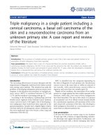
Fig. 1 Panel a: Sonogram showing a significantly elevated DV-PIV in twin A. Panel b: Echocardiogram of twin B. Four chamber view. PW Doppler of
flow through tricuspidal valve. Monofasic diastolic filling of the right ventricle; hallmark of diastolic function. Panel c: Myocardial severe interstitial
edema with fiber dissociation and sporadic vacuolar myocyte degeneration of the twin A fetal heart. Panel d: Hypertrophic vacuolization and nuclear
pleomorphism of extravillous throphoblast, with interstitial edema and areas of fibrinoid necrosis of placenta (✷). Panel e: Neonatal echocardiography
of twin B. M-mode long axis of the left ventricle. Evidence of mild septal hypokinesia with an overall preserved global contractility
including: 1- the post-mortem cardiac examination
revealing the hallmarks of subacute anthracycline toxicity, i.e. massive interstitial edema without cellular infiltrates and myofibrillar damage/vacuolization [6]; 2- the
histologic findings in both chorionic villi and extravillous
throphoblast, well fitting the described placental effects
of anthracycline exposure [7]; 3- the manifestation of
acute myocardial diastolic dysfunction, evidenced by the
enlarged right cardiac chambers and an elevated ductus
venosus pulsatility index, preceding fetal death; 4 – the
absence of structural or genomic abnormalities that may
indicate an alternative etiology of fetal demise. Of note,
neither arterial Doppler nor STV evaluation anticipated
the impending fetal demise.
Conclusions
The first trimester is the most critical time regarding
teratogenic effects. The blastocyst is resistant to teratogenic drugs in the first 2 weeks from conceptions whereas
the administration of chemotherapy during organogenesis
from 4 to 13 weeks of pregnancy is associated with an
increased risk of miscarriage or congenital malformations
[8] as largely documented from case reports, case series
and collected reviews [9–11]. Second and third trimester
chemotherapeutics exposure after the end of organogenesis, does not usually increase the teratogenic risk but may
cause neurocognitive development disorders and increasing risk of intrauterine growth retardation (IUGR),
pre-term labour and low birth weight [12–16].
According to SOGC guidelines, we should administer
the standard regimens based on a combination of
anthracyclines/alkylating agents after the end of the first
trimester [17].
The first prospective collection of data addressing the
issue of antracyclines safety profile in pregnancy was
first reported by the Texas MD Anderson Cancer Center
back in 1999. These authors treated 24 pregnant patients
with primary or recurrent cancer of the breast managed
with a standardized protocol of 5-fluorouracil + doxorubicin + cyclophosphamide (FAC) chemotherapy in the
second and third trimester of pregnancy and did not
report an increased rate of congenital anomalies [18]. In
Framarino-dei-Malatesta et al. BMC Cancer (2015) 15:951
the same year, a French survey reporting 12 patients
with BCP alternatively treated with FAC, 5-fluorouracil +
epirubicin + cyclophosphamide (FEC), epirubicin + cyclophosphamide (EC) or doxorubicin + cyclophosphamide
(AC) evidenced only one case of intrauterine death at
30 weeks of gestation [19]. In a retrospective, cohort study
evaluating the fetal risks involved in the administration
of cancer chemotherapy during gestation, one fetus died
after second trimester exposure to epirubicin, vincristine
and prednisone but no malformation was detected [20].
Hahn et al. extended the previous evidences from the MD
Anderson Cancer Center and confirmed the absence of
congenital birth defects in fetuses exposed to anthracyclines chemotherapy in utero; indeed, only three children
reported congenital malformations in a group of 57
women treated up to 2006, with a median number of four
FAC cycles given during pregnancy; one neonate was born
with Down syndrome, one with ureteral reflux, and a
third with club foot [21]. Ring et al. evaluated 16 out
of 28 BCP patients receiving anthracyclines-based
chemotherapy during pregnancy without reporting any
congenital birth defect [22].
The German Breast Group issued the first International Recommendations on BCP and confirmed the
safety of anthracyclines [23]. An international consensus
meeting held in 2010 confirmed that anthracyclines can
be used in the setting of BCP [24] and RCOG guidelines
assigned an Evidence level 3 to the statement that
anthracycline-based chemotherapy in the second and
third trimesters can be administered with minimal risk
to the developing fetus [25].
Anthacyclines display well-known cardiotoxic effects:
age, cumulative dose and previous radiotherapy increases
the rates of cardiac damage in children and adults [26].
The molecular mechanisms underlying antracyclines
cardiotoxicity are not fully understood, but include alterations of cell membranes fluidity and ion transport
with generation of reactive oxygen species by ironanthracycline complexes, leading to lipid peroxidation
and membrane damage [27] and the impairment of DNA
repair through the interaction with the topoisomerase-IIbeta enzyme in myocytes [28]. Increasing evidences show
that the extracellular matrix plays a complex and diverse
role in some processes initiated by anthracyclines that
finally lead to cardiac damage [29]. Notably, fetal myocardium is theoretically more vulnerable to damage by
chemotherapeutics because fetal myocytes are smaller
than adult ones, and contain fewer sarcomeres and
mitochondria [30].
Fetal safety during the administration of anthracyclinebased chemotherapy in pregnancy is of theoretical
concern because anthracyclines can cross the placenta,
even if their fetal plasma concentrations are lower than
those found in the mother, and have cumulative toxicity
Page 4 of 7
[31]. Available data provide only limited experimental
and clinical data on the transplacental transfer of these
chemotherapeutics in pregnant women; in a baboon
model, fetal plasma concentrations of doxorubicin, epirubicin and paclitaxel were about 7.5 %, 4.0 %, and 1.4 %,
of the respective maternal concentrations [32]. Fetal
blood samples from pregnant rats receiving doxorubicin
showed a plasmatic concentration that was 6.2 % that
of the mother; interestingly, neither Doppler analysis
nor heart microstructure or cellular DNA turnover and
apoptosis were influenced by doxorubicin exposure [33].
Owing to the molecular weight of doxorubicin is 580
dalton, there is an incomplete transfer of the drug across
the placental barrier [8]. However, the transplacental
passage cannot be simply predicted from the physicalchemical properties of the drugs like the molecular
weight. Really, while assessing fetal plasma drug concentrations, the functional expression of many members of
the ATP-binding cassette (ABC) efflux transporters that
are highly expressed in the human placenta, should be
adequately considered; indeed, these transporters prevent the trans-placental transfer of cytotoxic compounds
present in the maternal circulation, therefore protecting
the fetus [34, 35]; specifically, anthracyclines and taxanes
are substrates for ABC-transporters like the major
placental drug-transporting P-glycoprotein, that keeps
low the fetal plasma concentrations of these harmful
compounds [36].
Anthracyclines do not collectively share the same low
rate of transplacental transfer. Indeed, idarubicin, being
more lipophilic than other antracyclines, easily crosses
the placenta; Germann reported one fetal death and one
case of reversible heart dysfunction in a group of
patients affected by acute myeloid leukemia receiving
idarubicin-based chemotherapy during the third trimester of pregnancy [37]. Similarly, Baumgartner reported
one case of reversible fetal cardiomyopathy following the
use of idarubicin during pregnancy [38] while the occurrence of a severe idarubicin-related cardiotoxicity in a
newborn was described in a swiss study [39]. Altogether,
these findings suggest a close fetal monitoring during
idarubicin based chemotherapy; long-term outcomes of
idarubicin exposed children need further investigations.
Unlike idarubicin, doxorubicin and epirubicin, due to
their low levels in fetal plasma, may be administered
during the second and third trimesters without significant risk of fetal myocardial dysfunction. At first, clinicians gained some experience on the safety profile of
these two antracyclines from case reports and small case
series [40]. Further reassuring evidence was granted by
an Italian review reporting that only 13/out of 403 (3 %)
children exposed to these anthracyclines during late
pregnancy developed short-term cardiac complications
[41]. Azim et al. reported that different epirubicin and
Framarino-dei-Malatesta et al. BMC Cancer (2015) 15:951
doxorubicin regimens administered in adjuvant, neoadjuvant and metastatic settings (23 patients and 3 patients,
respectively) did not adversely affect the course of
pregnancy or fetal/neonatal outcome [42]. In a small
cohort of patients, even a dose-dense antracyclines
chemotherapy administered every two weeks did not
involve a higher risk of fetal complications [43].
In a prospective case–control clinical study, Gziri
found that maternal and fetal cardiac functions were not
significantly hampered by anthracyclines exposure in
pregnancy but rather displayed only minor changes of
the myocardial performance index and the tricuspid
inflow devoid of any clinical relevance [44].
Notably, epirubicin in pregnancy has a shorter terminal
half-life than doxorubicin due to its combined glucuronization by the liver and the placenta [45] and therefore
displays a better therapeutic index with fewer systemic
and cardiotoxic effects. In an Austrian study, all three
patients managed at the University Hospital of Vienna
with six courses of FEC neoadjuvant chemotherapy delivered healthy newborns [46]. Others case reports on
multidrugs regimens including epirubicin as adjuvant
treatment for pregnant women with high-risk breast
cancer failed to show any fetal cardiotoxicity [47, 48].
Some authors report that weekly epirubicin schedule
seems particularly safe because it decreases the potential
adverse events and simultaneously facilitates a close
monitoring of pregnancy [49].
Overall, anthracyclines emerge as theoretically safe during the late trimesters of pregnancy, fetal concentrations
being 100/1000-fold lower than adults as a result of the
high molecular weight, the hydrophilic charge leading to a
limited transplacental passage and the active clearance
operated by the placental P-glycoprotein transporter.
Indeed, despite the difficulty of comparing different agents
and schedules used for BCP, fetal cardiotoxicity never
emerged as a major problem of anthracyclines administration; in particular the available evidences indicate that
epirubicin harmful effects on fetal heart are very limited with only one reported case of transient ventricular
hypokinesia [41].
In this otherwise quite reassuring scenario, we provide
evidence that, in a twin pregnancy complicated by breast
cancer, epirubicin administration was causatively linked
to the death of one twin and to the onset of a reversible
cardiotoxicity of the surviving fetus/newborn.
The ultimate cause of twin A great susceptibility to
the cardiotoxic action of epirubicin remains elusive.
Anthracyclines are concentrated up to nine times more
in the amniotic fluid than in fetal plasma [32]; in this
regard, the presence of oligohydramnios and the histologic evidences of altered extravillous throphoblast and
chorionic villi, suggest the a putative contribution of an
abnormal amniotic fluid dynamics to the increased/
Page 5 of 7
prolonged toxicity of epirubicin in twin A. Further
support to the hypothesis of an hampered epirubicin
farmacokynetics is offered by the circumstantial evidences that the succumbing SGA twin A died shortly
after the administration for lung maturation of glucocorticoids; these steroids, among a myriad of actions,
are known to downregulate the throphoblast expression
of the detoxifying P-glycoprotein transporter [50]. Epirubicin cardiotoxicity was also evident, although to a lesser
extent, in Twin B as shown by 1- the prenatal findings of
an isolated mild right ventricular diastolic dysfunction
(reflecting the greater after load of this ventricle in the
fetal circulation) and 2- postnatal recording of increased Tn1, associated to a transient left ventricular
septal hypokinesia [51].
In conclusion, with reference to the above mentioned considerations, we suggest that a precautional
use of epirubicin in pregnancy should include; 1- the
screening of oligohydramnios since this condition may
putatively increase epirubicin cardiotoxicity 2- a
timely surveillance by a pediatric cardiologist of the
diastolic function of the fetal right ventricle, because
the other indices of fetal well being are poorly predictive of an impending fetal cardiac decompensation
3- the avoidance of a close administration of epirubicin and desamethasone since glucocorticoids may
hamper placental metabolism of epirubicin, ultimately
increasing its toxicity.
Consent to publish
Written informed consent was obtained from the patient
for publication of this Case report and any accompanying images. A copy of the written consent is available for
review by the Editor of this journal.
Competing interests
The authors declare that they have no competing interests.
Authors’ contributions
MFdM conceived the study, drafted and revised the manuscript, performed
the analysis of data and was one of the attending physician of the patient.
GP substantially contributed to the design and revision of the manuscript.
AG substantially contributed to the acquisition of data and critically revised
the manuscript. FV substantially contributed to the acquisition of data and
critically revised the manuscript. MD analyzed and interpretated data and
critically revised the manuscript. II analyzed and interpretated data and
critically revised the manuscript. VT analyzed and interpretated data and
drafted the manuscript. PG substantially contributed to the design and
revision of the manuscript. PS analyzed and interpretated data and drafted
the manuscript. GC analyzed and interpretated data and critically revised the
manuscript. RB conceived the study, drafted and revised the manuscript,
performed the analysis of data and was one of the attending physician of
the patient. All Authors read and approved the final manuscript. All Authors
read and approved the final manuscript.
Acknowledgements
There are no acknowledgements to be mentioned.
Framarino-dei-Malatesta et al. BMC Cancer (2015) 15:951
Author details
1
Department of Gynecologic Obstetrics and Urology Sciences, University of
Rome “Sapienza”, Rome, Italy. 2Department of Pediatrics, University of Rome
“Sapienza”, Rome, Italy. 3Department of Surgery “Pietro Valdoni”, University of
Rome “Sapienza”, Rome, Italy.
Received: 23 July 2015 Accepted: 5 December 2015
References
1. Fecondità in ripresa e calendario riproduttivo posticipato. National Institute
of Statistics 2013. . Accessed 30 June 2015.
2. I tumori in Italia. National Institute of Health. .
Accessed 18 May 2015.
3. Kaklamani VG, Gradishar WJ (2003) Epirubicin versus doxorubicin: which is
the anthracycline of choice for the treatment of breast cancer? Clin Breast
Cancer 4(Suppl 1):S26–S33
4. Framarino Dei Malatesta M, Piccioni MG, Brunelli R, Iannini I, Cascialli G,
Sammartino P (2014) Breast cancer during pregnancy: a retrospective study on
obstetrical problems and survival. Eur J Obstet Gynecol Reprod Biol 173:48–52
5. Litton JK, Warneke CL, Hahn KM, Palla SL, Kuerer HM, Perkins GH et al (2013)
Case control study of women treated with chemotherapy for breast cancer
during pregnancy as compared with nonpregnant patients with breast
cancer. Oncologist 18:369–76
6. Hengel CL, Russel PA, Gould PA, Kaye DM (2006) Subacute anthracycline
cardiotoxicity. Heart Lung Circ 15:59–61
7. Abellar RG, Pepperell JR, Greco D, Gundogan F, Kostadinov S, Schwartz
J et al (2009) Effects of chemotherapy during pregnancy on the
placenta. Pediatric Dev Pathol 12:35–41
8. Koren G (2011) Fetal risks of maternal pharmacotherapy: identifying signals.
Handb Exp Pharmacol 205:285–94
9. Cardonick E, Iacobucci A (2004) Use of chemotherapy during human
pregnancy. Lancet Oncol 5:283–91
10. Nurmohamed L, Moretti ME, Schechter T, Einarson A, Johnson D, Lavigne SV
et al (2011) Outcome following high-dose methotrexate in pregnancies
misdiagnosed as ectopic. Am J Obstet Gynecol 205:533–e1-3
11. Leyder M, Laubach M, Breugelmans M, Keymolen K, De Greve J, Foulon W
(2011) Specific congenital malformations after exposure to
cyclophosphamide, epirubicin and 5-fluorouracil during the first trimester of
pregnancy. Gynecol Obstet Invest 71:141–4
12. Berretta M, Di Francia R, Lleshi A, De Paoli P, Li Volti G, Bearz A et al (2012)
Antiblastic treatment, for solid tumors, during pregnancy: a crucial decision.
Int J Immunopathol Pharmacol 25(2 Suppl):1S–19S
13. Abdel-Hady E-S, Hemida RA, Gamal A, El-Zafarany M, Toson E, El-Bayoumi
MA (2012) Cancer during pregnancy: perinatal outcome after in utero
exposure to chemotherapy. Arch Gynecol Obstet 286:283–6
14. Amant F, Loibl S, Neven P, Van Calsteren K (2012) Breast cancer in
pregnancy. Lancet 379:570–9
15. Ko EM, Van Le L (2011) Chemotherapy for gynecologic cancers occurring
during pregnancy. Obstet Gynecol Surv 66:291–8
16. Van Calsteren K, Heyns L, De Smet F, Van Eycken L, Gziri MM, Van Gemert W
et al (2010) Cancer during pregnancy: an analysis of 215 patients
emphasizing the obstetrical and the neonatal outcomes. J Clin Oncol 28:
683–9
17. Koren G, Karey N, Gagnon R, Marxell C, Nulman I, Senikas V (2013) Cancer
chemotherapy and pregnancy. J Obstet Gynaecol Can 35:263–80
18. Berry DL, Theriault RL, Holmes FA, Parisi VM, Booser DJ, Singletary SE et al
(1999) Management of breast cancer during pregnancy using a
standardized protocol. J Clin Oncol 17:855–61
19. Giacalone PL, Laffargue F, Bénos P (1999) Chemotherapy for breast
carcinoma during pregnancy: A French national survey. Cancer 86:2266–72
20. Peres RM, Sanseverino MT, Guimarães JL, Coser V, Giuliani L, Moreira RK et al
(2001) Assessment of fetal risk associated with exposure to cancer chemotherapy
during pregnancy: a multicenter study. Braz J Med Biol Res 34:1551–9
21. Hahn KM, Johnson PH, Gordon N, Kuerer H, Middleton L, Ramirez M et al
(2006) Treatment of pregnant breast cancer patients and outcomes of
children exposed to chemotherapy in utero. Cancer 107:1219–26
22. Ring AE, Shannon C, Galani E, Ellis PA (2005) Chemotherapy for breast
cancer during pregnancy: an 18-year experience from five London teaching
hospitals.Chemotherapy for breast cancer during pregnancy: an 18-year
experience from five London teaching hospitals. J Clin Oncol 23:4192–7
Page 6 of 7
23. Loibl S, von Minckwitz G, Gwyn K, Ellis P, Blohmer JU, Schlegelberger B et al
(2006) Breast carcinoma during pregnancy. International recommendations
from an expert meeting. Cancer 106:237–46
24. Amant F, Deckers S, Van Calsteren K, Loibl S, Halaska M, Brepoels L et al
(2010) Breast cancer in pregnancy: recommendations of an international
consensus meeting. Eur J Cancer 46:3158–68
25. Pregnancy and Breast Cancer. Royal College of Obstetricians and
Gynaecologists. />26. Gianni L, Herman EH, Lipshultz SE, Minotti G, Sarvazyan N, Sawyer DB (2008)
Anthracycline cardiotoxicity: from bench to bedside. J Clin Oncol 26:3777–84
27. Patanè S (2014) Cardiotoxicity: anthracyclines and long term cancer
survivors. Int J Cardiol 176:1326–8
28. Lyu YL, Kerrigan JE, Lin CP, Azarova AM, Tsai YC, Ban Y et al (2007)
Topoisomerase 11beta mediated DNAdouble-strand breaks:
implications in doxorubicin cardiotoxicity and prevention by
dexrazoxane. Cancer Res 67:8839–46
29. Nikitovic D, Juranek I, Wilks MF, Tzardi M, Tsatsakis A, Tzanakakis GN (2014)
Anthracycline-dependent cardiotoxicity and extracellular matrix remodeling.
Chest 146:1123–30
30. Siedner S, Krüger M, Schroeter M, Metzler D, Roell W, Fleischmann BK
et al (2003) Developmental changes in contractility and sarcomeric
proteins from the early embryonic to the adult stage in the mouse
heart. J Physiol 548:493–505
31. Van Calsteren K, Verbesselt R, Van Bree R, Heyns L, de Bruijn E, de
Hoon J et al (2011) Substantialvariation in transplacental transfer of
chemotherapeutic agents in a mouse model. Reprod Sci 18:57–63
32. Van Calsteren K, Verbesselt R, Beijnen J, Devlieger R, De Catte L,
Chai DC et al (2010) Transplacental transfer of anthracyclines,
vinblastine, and hydroxy-cyclophosphamide in a baboon model.
Gynecol Oncol 119:594–600
33. Gziri MM, Pokreisz P, De Voz R, Verbeken E, Debiève F, Mertens L et al
(2013) Fetal rat hearts do not display acute cardiotoxicity in response to
maternal doxorubicin treatment. J Pharmacol Exp Ther 346:362–9
34. Behravan J, Piquette-Miller M (2007) At Drug transport across the placenta, role
of the ABC drug efflux transporters. Expert Opin Drug Metab Toxicol 3:819–30
35. Staud F, Cerveny L, Ceckova M (2012) Pharmacotherapy in pregnancy; effect
of ABC and SLC transporters on drug transport across the placenta and fetal
drug exposure. J Drug Target 20:736–63
36. Syme MR, Paxton JW, Keelan JA (2004) Drug transfer and metabolism by
the human placenta. Clin Pharmacokinet 43:487–514
37. Germann N, Goffinet F, Goldwasser F (2004) Anthracyclines during
pregnancy: embryo-fetal outcome in 160 patients. Ann Oncol 15:146–50
38. Baumgärtner AK, Oberhoffer R, Jacobs VR, Ostermayer E, Menzel H, Voigt M
et al (2009) Reversible foetal cerebral ventriculomegaly and cardiomyopathy
under chemotherapy for maternal AML. Onkologie 32:40–3
39. Achtari C, Hohlfeld P (2000) Cardiotoxic transplacental effect of
idarubicin administered during the second trimester of pregnancy.
Am J Obster Gynecol 183:511–2
40. Meyer-Wittkopf M, Barth H, Emons G, Schmidt S (2001) Fetal cardiac effects
of doxorubicin therapy for carcinoma of the breast during pregnancy: case
report and review of the literature. Ultrasound Obstet Gynecol 18:62–6
41. Parodi E, Alluto A, Moggio G, Liberale V, Frigerio M, Sismondi P
(2012) Transient ventricular hypocinesia after in utero anthracyclines
exposure: a case report and review of the literature. J Matern Fetal
Neonatal Med 25:189–92
42. Azim HA, Peccatori FA, Scarfone G, Acaia B, Rossi P, Cascio R et al (2008)
Anthracyclines for gestational breast cancer: course and outcome of
pregnancy. Ann Oncol 19:1511–2
43. Cardonick E, Gylmandiar D, Somer RA (2012) Maternal and neonatal
outcomes of dose-dense chemotherapy for breast cancer in pregnancy.
Obstet Gynecol 120:1267–72
44. Gziri MM, Debiève F, De Catte L, Mertens L, Barrea C, Van Calsteren K et al
(2012) Chemotherapy during pregnancy: Effects of anthracyclines on fetal
and maternal cardiac function. Acta Obstet Gynecol Scand 91:1465–8
45. Zaya MJ, Hines RN, Stevens JC (2006) Epirubicin glucuronidation and
UGT2B7 developmental expression. Drug Metab Dispos 34:2097–101
46. Bodner-Adler B, Bodner K, Zeisler H (2007) Breast cancer diagnosed during
pregnancy. Anticancer Res 27:1705–7
47. Gadducci A, Cosio S, Fanucchi A, Nardini V, Roncella M, Conte PF et al (2003)
Chemotherapy with epirubicin and paclitaxel for breast cancer during
pregnancy: case report and review of the literature. Anticancer Res 23:5225–9
Framarino-dei-Malatesta et al. BMC Cancer (2015) 15:951
Page 7 of 7
48. Ginopoulos PV, Michail GD, Kourounis GS (2004) Pregnancy associated
breast cancer: a case report. Eur J Gynaecol Oncol 25:261–3
49. Peccatori FA, Azim HA Jr, Scarfone G, Gadducci A, Bonazzi C, Gentilini
O et al (2009) Weekly epirubicin in the treatment of gestational breast
cancer (GBC). Breast Cancer Res Treat 115:591–4
50. Morrison JL, Botting KJ, Soo PS, McGillick EV, Hiscock J, Zhang S et al (2012)
Antenatal steroids and the IUGR fetus: are exposure and physiological
effects on the lung and cardiovascular system the same as in normally
grown fetuses? J Pregnancy 2012:1–15
51. Kilickap S, Barista I, Akgul E, Aytemir K, Aksoyek S, Aksoy S et al (2005) cTnT
can be a useful marker for early detection of anthracycline cardiotoxicity.
Ann Oncol 16:798–804
Submit your next manuscript to BioMed Central
and we will help you at every step:
• We accept pre-submission inquiries
• Our selector tool helps you to find the most relevant journal
• We provide round the clock customer support
• Convenient online submission
• Thorough peer review
• Inclusion in PubMed and all major indexing services
• Maximum visibility for your research
Submit your manuscript at
www.biomedcentral.com/submit