Potential anti-cancer effect of N-hydroxy-7- (2-naphthylthio) heptanomide (HNHA), a novel histone deacetylase inhibitor, for the treatment of thyroid cancer
Bạn đang xem bản rút gọn của tài liệu. Xem và tải ngay bản đầy đủ của tài liệu tại đây (2.34 MB, 11 trang )
Kim et al. BMC Cancer (2015) 15:1003
DOI 10.1186/s12885-015-1982-6
RESEARCH ARTICLE
Open Access
Potential anti-cancer effect of N-hydroxy-7(2-naphthylthio) heptanomide (HNHA), a
novel histone deacetylase inhibitor, for the
treatment of thyroid cancer
Seok-Mo Kim1†, Ki-Cheong Park1†, Jeong-Yong Jeon2, Bup-Woo Kim1, Hyeung-Kyoo Kim1, Ho-Jin Chang1,
Seung-Hoon Choi1, Cheong-Soo Park1 and Hang-Seok Chang1*
Abstract
Background: Thyroid cancer has been indicated to have a higher global proportion of DNA methylation and a
decreased level of histone acetylation. Previous studies showed that histone gene reviser and epigenetic changes
role significant parts in papillary and anaplastic thyroid cancer tumorigenesis. The goal of this research was to
study the endoplasmic reticulum (ER) stress-mediated actions of the dominant histone deacetylase (HDAC)
inhibitor, N-hydroxy-7-(2-naphthylthio) hepatonomide (HNHA), in thyroid cancer and to explore its effects on
apoptotic cell death pathways.
Methods: Experiments were achieved to conclude the effects of HNHA in papillary thyroid cancer (PTC) and
anaplastic thyroid cancer (ATC) cell lines and xenografts, as compared with two other established HDAC inhibitors
(SAHA; suberoylanilide hydroxamic acid and TSA; trichostatin A).
Results: Apoptosis, which was induced by all HDAC inhibitors, was particularly significant in HNHA-treated cells,
where noticeable B-cell lymphoma-2 (Bcl-2) suppression and caspase activation were observed both in vitro and
in vivo. HNHA increased Ca2+ release from the ER to the cytoplasm. ER stress-dependent apoptosis was induced
by HNHA, suggesting that it induced caspase-dependent apoptotic cell death in PTC and ATC. PTC and ATC
xenograft studies demonstrated that the antitumor and pro-apoptotic effects of HNHA were greater than those
of the established HDAC inhibitors. These HNHA activities reflected its induction of caspase-dependent and ER
stress-dependent apoptosis on thyroid cancer cells.
Conclusions: The present study indicated that HNHA possibly provide a new clinical approach to thyroid cancers,
including ATC.
Background
Thyroid cancer is the most commonly occurring endocrine malignancy and its incidence has increased steadily over the past three decades worldwide [1, 2].
Generally, thyroid cancer can be treated effectively with
surgery or radioactive iodine [3]. ATC is the least common, but the most aggressive, of all thyroid cancers [4].
* Correspondence:
†
Equal contributors
1
Department of Surgery, Thyroid Cancer Center, Gangnam Severance
Hospital, Yonsei University College of Medicine, 211 Eonjuro, Gangnam-gu,
Seoul 135-720, South Korea
Full list of author information is available at the end of the article
The mechanisms driving the progress of ATC are not
completely understood. ATCs are currently treated with
chemotherapy, radiotherapy, and/or surgery [4, 5].
Nevertheless, patients with ATC only have a median
survival of 5 months and less than 20 % survive for
1 year after diagnosis [6]. Early tumor dissemination
occurs in this type of cancer, resulting in 40 % of patients showing distant metastases and 90 % showing invasion of adjoining tissue on presentation [7]. The
present study investigated HDAC inhibitors as a novel
chemotherapy for PTC and ATC. HDACs are often
highly expressed in cancer cells [8–10]. These enzymes
© 2015 Kim et al. Open Access This article is distributed under the terms of the Creative Commons Attribution 4.0
International License ( which permits unrestricted use, distribution, and
reproduction in any medium, provided you give appropriate credit to the original author(s) and the source, provide a link to
the Creative Commons license, and indicate if changes were made. The Creative Commons Public Domain Dedication waiver
( applies to the data made available in this article, unless otherwise stated.
Kim et al. BMC Cancer (2015) 15:1003
Page 2 of 11
restrain the transcription of tumor suppressor genes
and so offer bright targets for cancer therapy [11, 12].
HDAC inhibitors are a group of small molecules that
accelerate gene transcription by reducing HDAC activity, inducing chromatin remodeling; these inhibitors
have been extensively studied as potential drugs for
treating cancer [12–15]. HDAC inhibitors affect various
well-known features of cancer cells, involving apoptosis, autophagy, growth inhibition and differentiation
[16–18]. They are extremely specific for cancer cells
over normal cells, owing to their induction of proapoptotic genes and ER stress, in addition to their effects on DNA repair mechanisms [19, 20]. HNHA is a
dominant HDAC inhibitor that was previously shown
to drive histone acetylation and downregulate the expression of HDAC target genes [21, 22]. HNHA showed
powerful anti-cancer activity in breast cancer cells and
fibrosarcoma [21–23]. Here, we researched this dominant HDAC inhibitor and its ER stress-mediated roles in
thyroid cancer and explored the effects of HNHA on
apoptotic cell death pathways in PTC and ATC.
Methods
Cell culture
The patient-derived thyroid cancer cell lines, SNU-80
(ATC) and SNU-790 (PTC), were purchased from the
Korea Cell Line Bank (Seoul National University, Seoul,
Korea) and cultured in RPMI-1640 medium with 10 %
fetal bovine serum. The cells lines were authenticated by
short tandem repeat profiling, karyotyping and isoenzyme analysis. Ethics approval about patient-derived thyroid cancer cell lines was approved by the Institutional
Review Board (IRB) of Seoul National University hospital
(Seoul, Republic of Korea).
Cell viability assay
Cell viability was measured by 3-(4,5-Dimethylthiazol2-yl)-2,5-Diphenyltetrazolium Bromide (MTT) assay.
Cells were cultured and grown to accomplish 70 % confluency. The indicated drugs were added to achieve final
concentrations of 0-100 μM. Cells were then incubated
for the indicated times prior to determination of cell viability by MTT assay. Data were indicated as a proportion
of the signal surveyed in vehicle-treated cells and shown
as the mean ± standard error of the mean (SEM) of triplicate experiments.
Evaluation of apoptotic cell death
Analysis of apoptosis and then identified with a TUNEL
(terminal deoxynucleotidyl transferase dUTP nick end
labeling) kit (Promega, Madison, WI, USA). Images of the
total and apoptotic cells (fluorescent green) were assembled with a confocal microscope (LSM Meta 700; Carl
Zeiss, Oberkochen, Germany) and analyzed with the Zeiss
LSM Image Browser software, version 4.2.0121.
Cytosolic free Ca2+ measurements by
microspectrofluorimetry
The intracellular Ca2+ levels in SNU-80 and SNU-790
cells were imaged using a Ca2+-sensitive fluorescent dye,
Fura-2 AM. Fluorescence intensities (ΔF) were normalized to those recorded in resting cells.
Immunoblot analysis
The antibodies for histone H3 and acetyl-histone H3,
α-tubulin and acetyl-α-tubulin, p53 and p21 were obtained from Abcam (Cambridge, UK). Apaf-1, CDK 4,
CDK 6, cyclin D1, Bcl-2, p-NFκB, caspase-3, caspase9 and β-actin antibodies were purchased from Santa Cruz
Biotechnology (Santa Cruz, CA, USA). Antibodies for
GRP78, ATF4, CHOP, PERK, p-PERK, eIF2α and p-eIF2α
were purchased from Cell Signaling Technology (Danvers,
MA, USA). The Bax antibody was obtained from Novus
Biologicals (Littleton, CO, USA).
Flow cytometry analysis of the cell cycle
Cell cycle dispersion was then analyzed with a FACS
Calibur Flow Cytometer (BD Biosciences, San Jose, CA,
USA). The proportions of cells in the G0/G1, S and G2/M
phases were analyzed by FlowJo v8 software for MacOSX
(Tree Star, Ashland, OR, USA).
Electrophoretic mobility shift assay (EMSA)
The DNA binding effect of NFκB to the Bcl-2 promoter was investigated using a 32P-labeled oligonucleotide
encoding the NFκB transcription factor binding sites
found in the Bcl-2 promoter region. Oligonucleotides
including the consensus-binding site for NFκB (GATCG
AGGGGACTTTCCCTACG) were 5′-end labeled with
Table 1 Half-maximal inhibitory concentration (IC50) values were determined using a cell proliferation assay
Cell line
Histopathology
Animal
Cell proliferation (IC50*) (μM)
HNHA
TSA
SAHA
SNU-80
Thyroid: anaplastic
Human
2.74 (±0.9)*
4.02 (±1.0)
6.74 (±1.1)
SNU-790
Thyroid: papillary
Human
2.32 (±1.0)*
3.91 (±1.2)
5.31 (±1.4)
Each data point denotes the mean of three independent IC50 values calculated from triplicate MTT assays
SD standard deviation
*reflect that were most significantly different between the groups
Kim et al. BMC Cancer (2015) 15:1003
Page 3 of 11
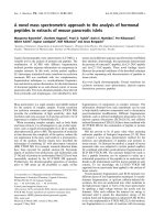
A
B
C
D
E
F
Fig. 1 (See legend on next page.)
Kim et al. BMC Cancer (2015) 15:1003
Page 4 of 11
(See figure on previous page.)
Fig. 1 Histone deacetylase inhibitors suppressed proliferation of anaplastic thyroid cancer (ATC) and papillary thyroid carcinoma (PTC) cell lines.
Cell viability and proliferation assays demonstrated that HNHA caused the greatest inhibition of thyroid cancer cell proliferation in SNU-80 ATC
(a and b) and SNU-790 PTC cells (c and d). TSA, trichostatin A; SAHA, suberoylanilide hydroxamic acid. Data points indicate the mean % of the
value observed in the solvent-treated control. All tests were repeated three times and the data symbolize the mean ± standard deviation. SNU-80
and SNU-790 cells were treated for 24 h with the expressed concentrations of HNHA (e) or with 15 μM HNHA for the indicated time-periods (f)
prior to isolation of total protein and evaluation of histone H3 and α-tubulin acetylation by immunoblotting. *P < 0.05 vs. Control, **P < 0.01 vs.
Control, ***P < 0.005 vs. Control
ɣ-32P-dATP and polynucleotide kinase. Nuclear proteins
(5 μg) were incubated with 1 μl of labeled oligonucleotide
(20,000 c.p.m.) in 20 μl incubation buffer for 20 min at
room temperature.
Human thyroid cancer cell xenograft
Human thyroid cancer cells (2.0 × 10⁷ cells/mouse)
were injected subcutaneously into female BALB/c nude
mice. After 7 ~ 10 days, mice were grouped randomly
(n = 10/group) and injected intraperitoneally with
25 mg/kg SAHA, TSA or HNHA once every 2 days for
a total of ten injections. Tumor size was measured by
calipers. Tumor volume was calculated using the following formula: L × S2/2 (where L was the longest
diameter and S was the shortest diameter). All in vivo
experiments were conducted with the permission of the
Animal Experiment Committee of Yonsei University.
In vivo toxicity study
In vivo toxicity was investigated in female BALB/c nude
mice. Every group of 10 mice was treated intraperitoneally with HNHA, SAHA or TSA at a dose of 25 mg/kg.
Five animals were housed in each cage and they were
observed regularly for external signs of lethality or toxicity. The conditions were controlled to provide 12 h
light and 12 h darkness, at a temperature of 22 °C, with
40–60 % humidity. Membrane-filter purified and autoclaved tap water and standard diet of rodent pellets were
provided ad libitum.
Immunohistochemistry
Immunohistochemical staining was performed using a
standard protocol. Primary monoclonal antibodies directed
Ki-67 (Abcam, Cambridge, UK) were diluted in PBS at a
ratio of 1:100. Mayer’s hematoxylin as a counterstain in all
tissue sections.
Statistical analysis
Statistical analysis was performed with GraphPad Prism
software (GraphPad Software Inc., La Jolla, CA, USA).
One-way ANOVA was performed for the multi-group
analysis, and two-tailed Student’s t-tests were used for
two-group analyses. Values are indicated as means ± SEM.
P values < 0.05 were regarded as statistically significant.
Results
HNHA inhibited the proliferation of ATC and PTC cells
To investigate the anti-cancer activity of HNHA alongside two well-known HDAC inhibitors (TSA and
SAHA), we assayed ATC (SNU-80) and PTC (SNU-790)
cell proliferation in the presence and absence of these
compounds using an MTT assay (Table 1). HNHA had a
lower half-maximal inhibitory concentration (IC50) than
TSA and SAHA in ATC and PTC cells. Further
characterization of the effects of HDAC inhibitors on
ATC and PTC cell viability showed that they all reduced
the viability of ATC and PTC cells, as compared to vehicle control-treated cells. However, HNHA provided
the most significant suppression of cell proliferation
(Fig. 1a and c) and this effect was concentrationdependent (Fig. 1b and d).
HNHA induced histone H3 acetylation in ATC and PTC
We exposed ATC (SNU-80) and PTC (SNU-790) cells to
various concentrations of HNHA and then estimated
histone H3 acetylation by immunoblotting. Acetylation
of α-tubulin and histone H3 were induced by HNHA in
a concentration-dependent manner (Fig. 1e). Histone H3
acetylation climaxed at 1 h after exposure to HNHA and
remained stable for 48 h (Fig. 1f ). These result indicated
that HNHA could induce non-histone proteins, as well
as stable acetylation of histone H3, in ATC and PTC.
Furthermore, HNHA produced concentration-dependent
cytotoxicity and induced greater reductions in cell viability
at low concentrations (2.32 ± 1.0 μM in SNU-790; 2.74 ±
0.9 μM in SNU-80) than did SAHA (5.31 ± 1.4 μM in
SNU-790; 6.74 ± 1.1 μM in SNU-80) or TSA (3.91 ±
1.2 μM in SNU-790; 4.02 ± 1.0 μM in SNU-80).
ER stress-induced release of cytoplasmic free Ca2+ was
increased by HNHA
We measured the change in intracellular Ca2+ levels
using microspectrofluorimetry. As shown in Fig. 2, the
intracellular Ca2+ level increased in HDAC inhibitortreated cells, as compared with control cells (Fig. 2a and
c). The cytoplasmic Ca2+ levels in HDAC inhibitor-treated
cells failed to return to the basal levels observed in control
cells (Fig. 2b and d).
Kim et al. BMC Cancer (2015) 15:1003
Page 5 of 11
A
B
C
D
Fig. 2 Cytosolic free Ca2+ measurements by microspectrofluorimetry in ATC and PTC cells exposed to histone deacetylase inhibitors. Ca2+ response in
Fura 2 AM-loaded ATC (a and b) and PTC (c and d) cells after treatment with SAHA, TSA or HNHA
HNHA induced ER stress-dependent cell cycle arrest in
ATC and PTC
Immunoblot analyses of protein levels in ATC (SNU-80)
and PTC (SNU-790) cell lines indicated that HNHA induced more marked increases in the levels of p53 and
p21, well-known arrestors of the cell cycle, and decreases in the levels of cyclin D1, CDK 4 and CDK 6,
positive regulators of the cell cycle, as compared with
SAHA or TSA (Fig. 3a). We also tested whether these
compounds induced ER stress by treating SNU-80 and
SNU-790 with SAHA, TSA or HNHA for 24 h and analyzing the expression of GRP 78, ATF 4, CHOP, PERK,
p-PERK, eIF2α and p-eIF2α by immunoblotting (Fig. 3b).
The HNHA-treated cells showed the strongest increase
in these markers of ER stress. Flow cytometry was
used to study the influence of these compounds on
Kim et al. BMC Cancer (2015) 15:1003
Page 6 of 11
A
C
B
D
Fig. 3 Histone deacetylase inhibitors induced cell cycle arrest and endoplasmic reticulum stress in ATC and PTC cells. Immunoblot analysis of the
indicated cell lines following exposure to SAHA, TSA or HNHA showed that HNHA potently induced the expression of cell cycle arrest proteins and
reduced expression of positive regulators of the cell cycle (a). SNU-80 and SNU-790 were exposed to the indicated inhibitors for 24 h prior to analyzing
the expression of GRP 78, ATF 4, CHOP, PERK, p-PERK, eIF2α and p-eIF2α (markers of endoplasmic reticulum stress) by immunoblotting (b). Cells were
exposed to the indicated inhibitors, harvested and stained with propidium iodide prior to analysis by flow cytometry and FlowJo v8 software (c and d)
cell cycle progression. The HDAC inhibitors increased
G0/G1 phase arrest and enriched the sub-G0 population
(p < 0.05), indicating cell cycle arrest and apoptosis in
these ATC and PTC cell lines (Fig. 3c and d). These data
suggested that, of the compounds tested, HNHA was the
most potent inducer of ER stress. This resulted in ER
stress-dependent apoptosis, cell cycle arrest and the strongest inhibition of ATC and PTC cell line viability.
HNHA induced caspase-dependent apoptosis of ATC and
PTC cell lines
To research the pro-apoptotic signaling pathways stimulated by exposure of PTC and ATC to HDAC inhibitors,
the expression of pro-apoptotic (Bax and Apaf-1) and
anti-apoptotic (phosphorylated NF-κB p65 and Bcl-2)
members of the Bcl-2 family and the stimulation of
caspase-3 and caspase 9, major executioners of apoptosis,
were investigated by immunoblotting (Fig. 4a). These
results implied that HNHA enhanced the pro-form of
caspase-3 and increased the cleavage of pro-caspase-3 and
-9 more powerfully than did TSA or SAHA (Fig. 4a).
NF-κB is a transcriptional factor and we investigated
the potential p-NF-κB binding sites in the Bcl-2 promoter region. An EMSA (Fig. 4b) identified two bands
corresponding to the labeled NF-κB probe following incubation with nuclear extracts of SNU-80 (Fig. 4b, lanes
7-10) or SNU-790 (Fig. 4b, lanes 3-6) cells. The specificity of the EMSA result was proved by complete inhibition of NF-κB probe-DNA binding by excess unlabeled
NF-κB probe (Fig. 4b, lane 1). In addition, a like amount
of mutated NF-κB probe also foundered to bind (Fig. 4b,
lane 2). HNHA-treated cells showed the strongest decrease in NF-κB binding. Together, these results demonstrated that HNHA inhibited Bcl-2 transcription. The
TUNEL assay proved that HNHA induced apoptosis in
ATC and PTC cell lines more powerfully than did TSA
or SAHA (Fig. 4c and d). These data indicated that
HNHA is a strong inducer of apoptosis in these ATC
and PTC cell lines and that it exerts this effect through
inhibition of the Bcl-2 pathway and caspase activation.
HNHA reduced xenograft growth and improved survival
in vivo
All of the HDAC inhibitors tested showed significant
suppression of SNU-80 and SNU-790 cell xenograft tumors; however, HNHA exhibited greater suppression of
these tumors than SAHA or TSA (Fig. 5a and c). Mouse
survival was extended noticeably by all of the tested
Kim et al. BMC Cancer (2015) 15:1003
Page 7 of 11
A
B
C
D
Fig. 4 Histone deacetylase inhibitors caussed apoptotic cell death in ATC and PTC cells. Immunoblot analyses suggested that the indicated
inhibitors increased the levels of apoptotic proteins and reduced those of anti-apoptotic proteins in ATC and PTC cells (a). An electrophoretic
mobility shift assay was carried out using a 32P-labeled oligonucleotide probe for the NF-κB binding sites on the Bcl-2 promoter (b). TUNEL assay
of ATC and PTC cells; TUNEL-positive (apoptotic) cells are indicated (× 400) (c, d)
HDAC inhibitors, but HNHA produced a greater effect
than SAHA or TSA (Fig. 5b and d). Systemic toxicity
and treatment-related deaths were not observed in any
of the study groups. The body weight of mice treated
with SAHA, TSA or HNHA did not differ significantly
from that of the control group (Fig. 5e and f). The HNHA
treatment group showed significantly smaller tumor
volumes than the SAHA- or TSA-treated groups (Fig. 5g
and h). The HDAC inhibitors also increased the levels of
p21 (cell cycle arrest protein), GRP78 (ER stress protein)
and cleaved caspase, indicating increased cell cycle arrest
and apoptosis due to ER stress in these ATC and PTC
mouse xenografts (Fig. 5i). These data demonstrated that
HNHA produced a powerful suppression of subcutaneous
thyroid cancer xenografts in an animal model.
HNHA inhibits the proliferation of ATC and PTC
xenografts in vivo
Cellular proliferative activity is an important factor in
the assessment of the biological behavior of carcinomas.
At present, Ki-67 is the most useful marker of cell
proliferation because it is expressed in all cells, except
for those in the G0 phase. We detected this marker by
immunohistochemical examination of SNU-80 and
SNU-790 cell xenograft tumors and found that the
HNHA-treated group showed the strongest decrease
in Ki-67 expression (Fig. 6a and b). These data provided further evidence that HNHA had potent antithyroid cancer effects.
Discussion
The present study showed that HNHA had potent cytotoxic effects on PTC and ATC cell lines, both in vitro
and in vivo. HNHA produced a more powerful induction
of apoptosis than did the other HDAC inhibitors tested
in these thyroid cancer cell lines. These other HDAC
inhibitors have previously been used against thyroid
cancer cells [7, 24], and yet HNHA was effective at
lower doses. The mechanisms underlying these cytotoxic
effects of HNHA on ATC and PTC cell lines included
both induction of cell cycle arrest and apoptosis. Apoptosis was demonstrated by the increased proportion of
Kim et al. BMC Cancer (2015) 15:1003
A
B
C
D
Page 8 of 11
G
H
I
E
F
Fig. 5 Histone deacetylase inhibitors produced anti-tumor effects in thyroid cancer cell xenografts in vivo. Athymic nude mice with established tumors
were treated with the indicated inhibitors. Data represent the mean tumor volumes. HNHA caused more powerful inhibition of tumor developement
than did SAHA or TSA and followed in the greatest prolongation of survival in mice with anaplastic thyroid cancer (ATC; a, b) and papillary thyroid
carcinoma (PTC; c, d) xenografts (n = 10 mice/group). ‘No tumor + HNHA’ indicates HNHA-treated mice with no xenograft; no proof of treatment-related
death or systemic toxicity was observed in HNHA-treated groups (b and d). The compounds had no significant effect on mouse body weight, as compared
to the control group (e and f). Photomicrographs of the dissected tumors from the treated and control mice (g). Weights of the dissected tumors (h).
Immunoblot analysis of total proteins isolated from the tumors (i). * P < 0.05 vs. Control, ** P < 0.01 vs. Control, *** P < 0.005 vs. Control
cells in sub G1 and by the activation of caspase 3 [25].
HNHA showed a characteristic effect on cell cycle progression, whereby G1 arrest was already evident in the
presence of lower concentrations of HNHA, as compared to the levels of SAHA and TSA that produced this
effect. This finding was consistent with those of previous
studies showing that HDAC inhibitors usually produce
cytotoxicity and induce G1 arrest at lower concentrations [21, 22]. The major molecular effect of HDAC inhibition is to change the acetylation status of core
histone proteins, consequently facilitating chromatin remodeling and thus altering gene expression and cell differentiation [26–28]. Consistent with this, we found that
HNHA upregulated p21 expression and downregulated
cyclin D1 in the ATC and PTC cell lines. Nevertheless,
although histones are regarded as the canonical acetylation substrate, some research has challenged this minimalist paradigm and indicated that HDAC inhibitors
also modulated acetylation of other proteins required in
an extensive range of cellular processes including protein
transport, apoptosis and cell motility [29]. Histone modifications play an important role in epigenetic regulation
[30] and dysregulated histone deacetylases are indicators
of poor prognosis in numerous cancers. A recent research study showed that HDAC-1, -2 and -3 were
highly expressed in renal cell carcinoma [31] and overexpression of HDAC1 was reported to associate with a
poor prognosis [32–34]. HDAC inhibitors, which can be
grouped into four structural classes, bind to the catalytic
site of the enzyme and can reverse epigenetic silencing
by increasing histone acetylation [35, 36].
ATC is the most aggressive type of thyroid cancer and
is typically lethal, with a 1-year survival rate of just 20 %
[4]. New therapies are needed to improve the prognosis
of patients with this diagnosis. In this study, we have
proved that HDAC inhibitors have the potential to be
used for the treatment of ATC. A previous study also indicated that a different HDAC inhibitor, LBH589, modified cell cycle-controling proteins, especially cyclin D1
and p21, and powerfully inhibited the progress of ATC
in a xenograft model; this was involved by a powerful
decrease in Ki-67 expression in tissues from LBH589treated animals [37].
HNHA is a dominant HDAC inhibitor that has shown
strong anti-tumor activity in breast cancer in vitro and
in vivo [23, 38]. Here, we demonstrated that HNHA produced more powerful anti-tumor effects than SAHA and
TSA in PTC and ATC cells in vitro and in vivo, by causing apoptosis via inhibition of Bcl-2 and modulation of
the cell cycle G1/S checkpoint signaling pathway. HNHA
induced caspase-dependent apoptosis by inducing Ca2+
release from the ER in ATC and PTC cells, thus increasing the levels of cytoplasmic free Ca2+. In our study,
GRP78 was noticeably upregulated in ATC and PTC
cells exposed to all of the tested HDAC inhibitors.
HNHA treatment also resulted in the greatest elevation
of cytoplasmic free Ca2+ levels.
Thyroid carcinomas are generally poorly responsive to
cytotoxic chemotherapy [39–42], which could be attributed to the presence of intracellular inhibitors of apoptotic signaling cascades. The present study showed that
HDAC inhibitors induced pro-apoptotic proteins and
Kim et al. BMC Cancer (2015) 15:1003
A
B
Fig. 6 (See legend on next page.)
Page 9 of 11
Kim et al. BMC Cancer (2015) 15:1003
Page 10 of 11
(See figure on previous page.)
Fig. 6 HNHA reduced tumor Ki-67 expression. Immunohistochemical analysis of the Ki-67 protein levels in paraffin-embedded tumor tissues from
mice with anaplastic thyroid cancer (ATC; SNU-80; a) and papillary thyroid carcinoma (PTC; SNU-790; b) xenografts. HNHA caused more powerful
inhibition of tumor Ki-67 expression than did SAHA or TSA. MetaMorph 4.6 image-analysis software was used to quantify Ki-67 immunostaining.
*P < 0.05; **P < 0.01; ***P < 0.005 for the comparison with the control
reduced anti-apoptotic proteins, producing potent antitumor effects in the two thyroid cancer cell lines studied.
These findings suggest that novel therapies employing
HNHA alone, or in integration with usual chemotherapeutic agents, could improve outcomes in aggressive
thyroid cancer. The contribution of HDAC inhibition as
an anti-cancer therapy in ATC and PTC should be estimated using agents such as HNHA, which are more potent than those tested previously.
In conclusion, the anti-cancer activity of HNHA opens
up a novel therapeutic approach to ATC and PTC, which
do not respond successfully to conventional therapy.
Translational and clinical research efforts will ultimately
determine the clinical benefits and safety of HNHA, used
alone or in integration with other chemotherapeutic
agents, in the treatment of these types of tumor. Our findings led us to propose novel therapeutic approaches for
the treatment of ATC.
Conclusion
The current study suggests that HNHA may offer a new
clinical approach to thyroid cancers, including ATC.
Abbreviations
ATC: anaplastic thyroid cancer; Bcl-2: B-cell lymphoma-2; ER: endoplasmic
reticulum (ER); HNHA: N-hydroxy-7-(2-naphthylthio) hepatonomide;
PTC: papillary thyroid carcinoma; SAHA: suberoylanilide hydroxamic acid);
TSA: trichostatin A.
Competing interests
The authors declare that they have no competing interests.
Authors’ contributions
SMK and KCP carried out most of the in vitro and in vivo studies. KCP and
JYJ were involved in drafting the manuscript. JYJ and BWK performed the
cytosolic free Ca2+ measurements and electrophoretic mobility shift assays.
HKK and HJC carried out the statistical analysis. CSP and SHC were involved
in the study design and in drafting the manuscript. HSC was involved in the
study design, experiments, manuscript drafting and approval of the final
version. All authors read and approved the final manuscript.
Acknowledgments
This work was supported by a faculty research grant from Yonsei University
College of Medicine for 2015-31-0256 and the Brain Korea 21 Project for
Medical Science, Yonsei University.
Author details
1
Department of Surgery, Thyroid Cancer Center, Gangnam Severance
Hospital, Yonsei University College of Medicine, 211 Eonjuro, Gangnam-gu,
Seoul 135-720, South Korea. 2Department of Nuclear Medicine, Yonsei
College of Medicine, Seoul 120-752, South Korea.
Received: 18 August 2015 Accepted: 8 December 2015
References
1. Nikiforov YE. Thyroid carcinoma: molecular pathways and therapeutic
targets. Mod Pathol. 2008;21 Suppl 2:S37–43.
2. Gullu BE, Celik O, Gazioglu N, Kadioglu P. Thyroid cancer is the most
common cancer associated with acromegaly. Pituitary. 2010;13(3):242–8.
3. American Thyroid Association Guidelines Taskforce on Thyroid N,
Differentiated Thyroid C, Cooper DS, Doherty GM, Haugen BR, Kloos RT, Lee
SL, et al. Revised American Thyroid Association management guidelines for
patients with thyroid nodules and differentiated thyroid cancer. Thyroid.
2009;19(11):1167–214.
4. Smallridge RC. Approach to the patient with anaplastic thyroid carcinoma.
J Clin Endocrinol Metab. 2012;97(8):2566–72.
5. Sun C, Li Q, Hu Z, He J, Li C, Li G, et al. Treatment and prognosis of
anaplastic thyroid carcinoma: experience from a single institution in China.
PLoS One. 2013;8(11):e80011.
6. O'Neill JP, Shaha AR. Anaplastic thyroid cancer. Oral Oncol. 2013;49(7):702–6.
7. Baldan F, Mio C, Allegri L, Puppin C, Russo D, Filetti S, et al. Synergy
between HDAC and PARP Inhibitors on Proliferation of a Human Anaplastic
Thyroid Cancer-Derived Cell Line. Int J Endocrinol. 2015;2015:978371.
8. Kwiecinska P, Wrobel A, Tauboll E, Gregoraszczuk EL. Valproic acid, but not
levetiracetam, selectively decreases HDAC7 and HDAC2 expression in
human ovarian cancer cells. Toxicol Lett. 2014;224(2):225–32.
9. Burdelski C, Ruge OM, Melling N, Koop C, Simon R, Steurer S, et al. HDAC1
overexpression independently predicts biochemical recurrence and is
associated with rapid tumor cell proliferation and genomic instability in
prostate cancer. Exp Mol Pathol. 2015;98(3):419–26.
10. Delcuve GP, Khan DH, Davie JR. Targeting class I histone deacetylases in
cancer therapy. Expert Opin Ther Targets. 2013;17(1):29–41.
11. Ropero S, Esteller M. The role of histone deacetylases (HDACs) in human
cancer. Mol Oncol. 2007;1(1):19–25.
12. West AC, Johnstone RW. New and emerging HDAC inhibitors for cancer
treatment. J Clin Invest. 2014;124(1):30–9.
13. De Souza C, Chatterji BP. HDAC inhibitors as novel anti-cancer therapeutics.
Recent Pat Anticancer Drug Discov. 2015;10(2):145–62.
14. Li X, Zhang J, Xie Y, Jiang Y, Yingjie Z, Xu W. Progress of HDAC inhibitor
panobinostat in the treatment of cancer. Curr Drug Targets.
2014;15(6):622–34.
15. Sharma NL, Groselj B, Hamdy FC, Kiltie AE. The emerging role of histone
deacetylase (HDAC) inhibitors in urological cancers. BJU Int.
2013;111(4):537–42.
16. Wagner JM, Hackanson B, Lubbert M, Jung M. Histone deacetylase (HDAC)
inhibitors in recent clinical trials for cancer therapy. Clin Epigenetics.
2010;1(3-4):117–36.
17. Gammoh N, Lam D, Puente C, Ganley I, Marks PA, Jiang X. Role of
autophagy in histone deacetylase inhibitor-induced apoptotic and
nonapoptotic cell death. Proc Natl Acad Sci U S A. 2012;109(17):6561–5.
18. Del Bufalo D, Desideri M, De Luca T, Di Martile M, Gabellini C, Monica V,
et al. Histone deacetylase inhibition synergistically enhances pemetrexed
cytotoxicity through induction of apoptosis and autophagy in non-small
cell lung cancer. Mol Cancer. 2014;13:230.
19. Kikuchi S, Suzuki R, Ohguchi H, Yoshida Y, Lu D, Cottini F, et al. Class IIa
HDAC inhibition enhances ER stress-mediated cell death in multiple
myeloma. Leukemia. 2015;29(9):1918–27.
20. Sato A, Asano T, Isono M, Ito K, Asano T. Panobinostat synergizes with
bortezomib to induce endoplasmic reticulum stress and ubiquitinated
protein accumulation in renal cancer cells. BMC Urol. 2014;14:71.
21. Park KC, Heo JH, Jeon JY, Choi HJ, Jo AR, Kim SW, et al. The novel histone
deacetylase inhibitor, N-hydroxy-7-(2-naphthylthio) hepatonomide, exhibits
potent antitumor activity due to cytochrome-c-release-mediated apoptosis
in renal cell carcinoma cells. BMC Cancer. 2015;15:19.
22. Park KC, Park JH, Jeon JY, Kim SY, Kim JM, Lim CY, et al. A new histone
deacetylase inhibitor improves liver fibrosis in BDL rats through suppression
of hepatic stellate cells. Br J Pharmacol. 2014;171(21):4820–30.
Kim et al. BMC Cancer (2015) 15:1003
Page 11 of 11
23. Park KC, Kim SW, Park JH, Song EH, Yang JW, Chung HJ, et al. Potential
anti-cancer activity of N-hydroxy-7-(2-naphthylthio) heptanomide
(HNHA), a histone deacetylase inhibitor, against breast cancer both in
vitro and in vivo. Cancer Sci. 2011;102(2):343–50.
24. Mitsiades CS, Poulaki V, McMullan C, Negri J, Fanourakis G, Goudopoulou A,
et al. Novel histone deacetylase inhibitors in the treatment of thyroid
cancer. Clin Cancer Res. 2005;11(10):3958–65.
25. Frauenstein K, Sydlik U, Tigges J, Majora M, Wiek C, Hanenberg H, et al.
Evidence for a novel anti-apoptotic pathway in human keratinocytes
involving the aryl hydrocarbon receptor, E2F1, and checkpoint kinase 1. Cell
Death Differ. 2013;20(10):1425–34.
26. Arts J, de Schepper S, Van Emelen K. Histone deacetylase inhibitors: from
chromatin remodeling to experimental cancer therapeutics. Curr Med
Chem. 2003;10(22):2343–50.
27. Lawless MW, O'Byrne KJ, Gray SG. Histone deacetylase inhibitors target
diabetes via chromatin remodeling or as chemical chaperones? Curr
Diabetes Rev. 2009;5(3):201–9.
28. Dey P. Chromatin remodeling, cancer and chemotherapy. Curr Med Chem.
2006;13(24):2909–19.
29. Kanao K, Mikami S, Mizuno R, Shinojima T, Murai M, Oya M. Decreased
acetylation of histone H3 in renal cell carcinoma: a potential target of
histone deacetylase inhibitors. J Urol. 2008;180(3):1131–6.
30. Liu X, Luo M, Wu K. Epigenetic interplay of histone modifications and DNA
methylation mediated by HDA6. Plant Signal Behav. 2012;7(6):633–5.
31. Fritzsche FR, Weichert W, Roske A, Gekeler V, Beckers T, Stephan C, et al.
Class I histone deacetylases 1, 2 and 3 are highly expressed in renal cell
cancer. BMC Cancer. 2008;8:381.
32. Barneda-Zahonero B, Parra M. Histone deacetylases and cancer. Mol Oncol.
2012;6(6):579–89.
33. Mithraprabhu S, Kalff A, Chow A, Khong T, Spencer A. Dysregulated Class I
histone deacetylases are indicators of poor prognosis in multiple myeloma.
Epigenetics. 2014;9(11):1511–20.
34. New M, Olzscha H, La Thangue NB. HDAC inhibitor-based therapies: can we
interpret the code? Mol Oncol. 2012;6(6):637–56.
35. Marks P, Rifkind RA, Richon VM, Breslow R, Miller T, Kelly WK. Histone
deacetylases and cancer: causes and therapies. Nat Rev Cancer.
2001;1(3):194–202.
36. Brown R, Strathdee G. Epigenomics and epigenetic therapy of cancer.
Trends Mol Med. 2002;8(4 Suppl):S43–8.
37. Catalano MG, Pugliese M, Gargantini E, Grange C, Bussolati B, Asioli S, et al.
Cytotoxic activity of the histone deacetylase inhibitor panobinostat
(LBH589) in anaplastic thyroid cancer in vitro and in vivo. Int J Cancer.
2012;130(3):694–704.
38. Kim DH, Lee J, Kim KN, Kim HJ, Jeung HC, Chung HC, et al. Anti-tumor
activity of N-hydroxy-7-(2-naphthylthio) heptanomide, a novel histone
deacetylase inhibitor. Biochem Biophys Res Commun. 2007;356(1):233–8.
39. Busaidy NL, Cabanillas ME. Differentiated thyroid cancer: management
of patients with radioiodine nonresponsive disease. J Thyroid Res.
2012;2012:618985.
40. Smallridge RC, Ain KB, Asa SL, Bible KC, Brierley JD, Burman KD, et al.
American Thyroid Association guidelines for management of patients with
anaplastic thyroid cancer. Thyroid. 2012;22(11):1104–39.
41. Tuttle RM, Grewal RK, Larson SM. Radioactive iodine therapy in poorly
differentiated thyroid cancer. Nat Clin Pract Oncol. 2007;4(11):665–8.
42. Giuffrida D, Prestifilippo A, Scarfia A, Martino D, Marchisotta S. New
treatment in advanced thyroid cancer. J Oncol. 2012;2012:391629.
Submit your next manuscript to BioMed Central
and we will help you at every step:
• We accept pre-submission inquiries
• Our selector tool helps you to find the most relevant journal
• We provide round the clock customer support
• Convenient online submission
• Thorough peer review
• Inclusion in PubMed and all major indexing services
• Maximum visibility for your research
Submit your manuscript at
www.biomedcentral.com/submit