Evaluation of KIF23 variant 1 expression and relevance as a novel prognostic factor in patients with hepatocellular carcinoma
Bạn đang xem bản rút gọn của tài liệu. Xem và tải ngay bản đầy đủ của tài liệu tại đây (3.26 MB, 9 trang )
Sun et al. BMC Cancer (2015) 15:961
DOI 10.1186/s12885-015-1987-1
RESEARCH ARTICLE
Open Access
Evaluation of KIF23 variant 1 expression
and relevance as a novel prognostic factor
in patients with hepatocellular carcinoma
Xiaotong Sun1†, Zhongtian Jin2†, Xiao Song1, Jingjing Wang1, Yan Li1, Xiaoping Qian1, Yu zhang1 and Yanhui Yin1*
Abstract
Background: KIF23 (kinesin family member 23) is a kinesin-like motor protein and plays an important role in
cytokinesis. In search for genes associated with hepatocellular carcinoma (HCC) by cDNA microarray, we found that
KIF23 was upregulated in HCC tissues. At present, much less is known about its expression and functions in tumor
cells. In this work, we aimed to investigate the expression of KIF23 in HCC and the correlation between its
expression and clinical features.
Methods: Total RNA was extracted from 16 HCC and paired adjacent non-cancerous tissues. The expressions of the
two KIF23 splice variants (KIF23 V1 and KIF23 V2) in normal and HCC tissues were determined by reverse transcriptase
polymerase chain reaction (RT-PCR). Polyclonal antibody specific to KIF23 V1 was prepared, and the specificity of the
antibody was confirmed by siRNA knockdown and Western blotting experiments. KIF23 protein expression in HCC was
examined by immunohistochemistry staining with anti-KIF23 V1 or anti-KIF23 (commercially available for recognizing
both KIF23 V1 and V2) antibodies, respectively. Univariate and Multivariate Cox regression analyses were used to
determine the correlation between KIF23 protein expression and overall survival of HCC patients.
Results: The two splicing variants of KIF23 mRNA were not detected in normal liver tissue by RT-PCR, but they were
aberrantly expressed in HCC tissues. Immunohistochemistry staining with anti-KIF23 V1 antibody revealed that KIF23 V1
was mainly distributed in the nucleus, whereas the positive staining signals were predominantly in the cytoplasm
when using anti-KIF23 antibody, suggesting that KIF23 V2 might localize in the cytoplasm of HCC cells. KIF23 V1
protein was detected in 57.6 % (83/144) HCC patients and the mean H-score was 42, while KIF23 V2 was detected in
94.4 % (135/143) HCC samples and the mean H-score was 68. Follow-up study showed that HCC patients with
expression of KIF23 V1 had a longer 5-year survival (p = 0.0052), however, expression of KIF23 V2 protein did not
associate with 3- and 5-year survival.
Conclusion: In this study we show for the first time that KIF23 V1 and V2 have different localizations in HCC cells.
Furthermore, KIF23 V1 protein expression might be a marker of longer overall survival in HCC patients.
Keywords: KIF23, Hepatocellular carcinoma, Immunohistochemisty, Overall survival, Prognostic factor
* Correspondence:
†
Equal contributors
1
Department of Immunology, School of Basic Medical Sciences, Peking
University Health Science Center, 38 Xueyuan Road, Haidian District, Beijing
100191, China
Full list of author information is available at the end of the article
© 2015 Sun et al. Open Access This article is distributed under the terms of the Creative Commons Attribution 4.0
International License ( which permits unrestricted use, distribution, and
reproduction in any medium, provided you give appropriate credit to the original author(s) and the source, provide a link to
the Creative Commons license, and indicate if changes were made. The Creative Commons Public Domain Dedication waiver
( applies to the data made available in this article, unless otherwise stated.
Sun et al. BMC Cancer (2015) 15:961
Background
Hepatocellular carcinoma (HCC), the major type of primary liver cancer, is one of the most prevalent cancers
in the world [1]. China is one of the high-risk areas for
HCC due to the high prevalence of chronic hepatitis B
virus infection [2, 3], representing more than half of the
cases in the entire world [4]. Despite the remarkable advances in diagnostic and therapeutic techniques, prognosis of HCC still remains extremely poor, ranking as the
third leading cause of cancer-related death worldwide
[1]. Therefore, numerous studies have focused on screening for novel diagnostic and prognostic biomarkers as well
as therapeutic targets in HCC [5–7].
We have performed cDNA microarray analysis for
mining differentially expressed genes in HCC in an attempt to identify new HCC biomarkers [5]. Assessing
microarray data, we found that the expression of KIF23
showed a 6-fold increase in HCC tissues compared with
paired non-cancerous tissues. KIF23, also known as
CHO1/MKLP1, was first identified as a motor enzyme
that moves antiparallel microtubules in vitro [8]. Subsequent studies indicated that KIF23 is a key regulator of
cytokinesis [9, 10]. The disfunction of KIF23 resulted
in incomplete cytokinesis and formed binucleated or
multinucleated cells [11, 12], which have been considered as the hallmarks of the cancer cells [13]. However, to date, only few studies have been reported on
the expressions of KIF23 in tumor cells. Valk K et al.
found that KIF23 is upregulated in non-small cell lung
cancer (NSCLC) in screening differentially expressed
genes in NSCLC [14]. Recently, Takahashi S et al. reported a higher level of KIF23 expression in glioma
tissues compared to normal brain tissue [15]. However, the expression of KIF23 in HCC tissues remains
unknown.
Human KIF23 has two splice variants, KIF23 V1 and
KIF23 V2. When we use the term KIF23, it refers to
both KIF23 V1 and V2. KIF23 V1 is different from
KIF23 V2 only in that it contains an extra 312 bp sequence (exon 18) in the COOH-terminal tail [16]. Intriguingly, the sequence encoded by exon 18 is an F-actin
interacting domain, which may be essential for special
functions of KIF23 V1. However, all the commercial
anti-KIF23 antibodies recognize both KIF23V1 and V2,
thus little is known about the expressions and functions
of each individual variant in tumor cells so far. Since the
nucleotide sequence encoding KIF23 V2 is completely
same as the sequence for KIF23 V1 except lacking the
exon 18, it is difficult to investigate KIF23 V2 alone.
Thus we generated anti-KIF23 V1 antibody, and detected the expressions of the two isoforms of KIF23 in
HCC samples with antibodies specific for KIF23 V1 or
for both KIF23 V1 and V2 by immunohistochemistry.
We also investigated the prognostic significance of the
Page 2 of 9
expressions of KIF23 V1 and KIF23 V2 on overall survival of HCC patients.
Methods
Patients and samples
Ninety-eight HCC patients who underwent partial hepatectomy or liver transplantation in the Peking University
People’s Hospital, and commercial microarrays consisting of 46 HCC patients (Shanghai Outdo Biotech, China)
were enrolled in this study for immunohistochemical
analysis. In each case, the HCC diagnosis was confirmed
by post-operative pathological examination. Written informed consent was obtained from all participating patients, according to our university guidelines. The study
included 120 males and 24 females aged between 32 and
85 years with a median age of 55 years. All the patients
were classified according to the 6th edition of the TNM
classification of the International Union Against Cancer,
and there are 42 patients with stage I, 34 patients with
stage II, 46 patients with stage III, and 22 patients with
stage IV. Among the studied 144 patients, there are 80
patients with tumor size more than 5 cm. Follow-up
data were not available for 32 patients leaving 102 patients for final evaluation of survival. Overall survival
(OS) was defined as the time between surgery and death
of any cause or last follow-up.
Fourteen different normal tissue cDNA preparations, including heart, placenta, lung, liver, skeletal muscle, kidney,
pancrease, spleen, thymus, prostate, ovary, small intestine,
colon, and peripheral blood leukocyte, were purchased
from Clontech Laboratories Inc. Sixteen pairs of frozen
tumor tissues and adjacent non-tumor tissues for detection of KIF23 mRNA expression were from the Peking
University People’s Hospital.
The experiment was conducted in compliance with
the Helsinki declaration and was approved by the Ethics
Review Committee of Peking University of Health Science Center.
Cell lines, plasmids and siRNAs
The human cell lines HLE, Huh7, HepG2, SMMC-7721,
BEL-7402 and HEK293T cells were grown in DMEM
containing 10 % fetal calf serum (FCS). Transient transfection of plasmid constructs was performed using
Lipofactamine 2000 (Invitrogen) in HEK293T cells, and
siRNAs were transfected using jetPRIME (Polyplus
transfection) in HLE cells. To construct pRK-FLAGKIF23 V1 and pRK-FLAG-KIF23 V2 expression vectors,
human KIF23 V1 and V2 cDNA fragments amplified by
reverse transcription-PCR (RT-PCR) from HLE cell
RNA were cloned into the pRK-FLAG vectors, respectively. KIF23 V1 siRNA1 (5’-GUACAACACACCUCU
CAAATT-3’) (specific for KIF23 V1), KIF23 V1 siRNA2
(5’- GCAGUCUUCCAGGUCAUCUTT-3’) (target both
Sun et al. BMC Cancer (2015) 15:961
KIF23 V1 and V2), and control siRNA (5’–UUCUCCG
AACGUGUCACGUTT-3’) were synthesized by RiboBio
Co. Ltd (Guangzhou, China).
Page 3 of 9
anti-lamin B1 antibody from Bioworld Technology, and
anti-tubulin antibody from Sino Biological.
Immunofluorescence
Reverse transcription (RT)-PCR
Total RNA of the tumor and paired adjacent noncancerous tissues was isolated using TRIzol reagent (Invitrogen, USA), and first strand cDNA was generated using
random primers and AMV reverse transcriptase (Progema, USA) according to the manufacturer’s instructions. Primer sequences specific for amplifying KIF23 V1
were 5’-CAGATTTCCAACGGCCAGCA-3’ and 5’-TCA
TGGCTTTTTGCGCTTGG-3’, for amplifying KIF23 V2
were 5’-TCCATCACCTGTGCCTTTACT-3’ and 5’-TG
GGACTGTCAGTTCATGGC-3’ (the PCR product is
541 bp for KIF23 V2).
Whole, cytoplasmic, and nuclear protein extraction
Total cellular protein extracts were obtained using RIPA
lysis buffer as described previously [17]. Cytoplasmic
and nuclear extracts were prepared as follows: cells were
lysed in a hypotonic buffer containing 10 mM pH 7.9
Hepes, 10 mM KCl, 0.1 mM EDTA, 1 mM DTT, 0.15 %
NP-40, and protease inhibitor cocktail. After incubating
25 min on ice, samples were centrifuged and supernatants (corresponding to cytoplasmic extracts) were collected. The nuclear pellets were further washed with
PBS and then resuspended in RIPA buffer supplemented
with protease inhibitor cocktail. After vigorously shaking
for 30 min at 4 °C, the nuclear extracts were collected.
Whole, cytoplasmic and nuclear extracts were analyzed
by Western blotting.
Western blotting
Equal amounts of cellular extracts were subjected to
SDS-PAGE for electrophoresis, transferred to a nitrocellulose membrane, followed by incubation with appropriate antibodies. Tubulin antibody was used to
verify equivalent total protein. Immunoreactive bands
were visualized with enhanced chemiluminescence or
infrared imaging working with Odyssey Imager (Li-Cor,
Lincoln, NE).
Antibodies
The polyclonal anti-KIF23 V1 antibody was generated
using a synthesized peptide encompassing the residues
747–761 of the human KIF23 V1 protein (GenBank Accession number: NP_612565), which only presents in
KIF23 V1, but not in KIF23 V2 isoform. The peptide
conjugated to KLH was used to produce antibody in rabbits. The resultant antibody was purified by immunoaffinity chromatography (GE Healthcare). Anti-KIF23
antibody, recognizing both KIF23 V1 and V2 proteins,
was purchased from Santa Cruz (sc867, Santa Cruz),
Cells were grown directly on glass coverslips for 24 h,
and then fixed and permeabilized. After blocking in
PBS-5 % skimmed milk, cells were incubated with antiKIF23 V1, and normal rabbit IgG was used as a negative
control. After washing with PBS, cells were incubated at
room temperature with FITC-conjugated anti-rabbit IgG
(Zhongshan company, China). Cell nuclei were stained
with Hoechst33342. Images were required using confocal microscope.
Immunohistochemistry (IHC)
IHC was performed as previously described [18] with
minor modifications. Briefly, paraffin-embedded tissue
sections were deparaffinized with xylene and rehydrated
with a graded series of ethanol. After antigen retrieval,
inactivation of endogenous peroxidase, and blocking
with normal goat serum, sections were incubated with
anti-KIF23 V1 or anti-KIF23 antibodies at 4 °C overnight, followed by adding dextran carrying anti-rabbit
IgG conjugated to horseradish peroxidase (HRP) and
positive staining was developed using the Dako REAL
EnVision detection system. Images of stained sections
were imported into Olympus CX31 digital microscope
(Olympus, Japan) for quantifying stained cells.
Evaluation of IHC staining
IHC staining was evaluated by taking into account both
the intensity of staining and the percentage of positive
cells [19]. Tumor staining intensity was graded on a
scale from 0 (negative), 1 (weak), 2 (moderate) to 3
(strong) and each intensity category was scored a percentage of tumor cells ranging from 0 to 100 so that the
sum of the percentages adds up to 100. The percentage
score was then multiplied by its intensity category to obtain a final H-score, ranging from 0 to 300.
Statistical methods
Statistical analyses were performed using SAS 9.1.3 Portable for Windows (SAS, SAS Institute Inc, USA) and
GraphPad Prism 5.0 (GraphPad Software, USA).
The relationships between KIF23 V1 expression and
the potential explanatory variables were evaluated with
the Chi-square and cmh (Cochran-Mantel-Haenszel)
Chi-square tests. The survival rate was analyzed using
the Kaplan-Meier method and log-rank test. The univariate examination of the relationship between the
assessed criteria and survival was performed with a Chisquare test. A cox proportional-hazard model was used
for the multivariate analysis. P < 0.05 was considered statistically significant.
Sun et al. BMC Cancer (2015) 15:961
Results
Expressions of KIF23 V1 and V2 mRNA in normal and
HCC tissues
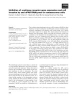
To examine the distribution of mRNA expression of the
two splice variants of KIF23, RT-PCR was performed
using cDNA reversed from mRNA of a variety of human
tissues and human derived cancer cell lines. Both of the
two variants were not detected in normal liver tissues
(Fig. 1a), but they were found to be aberrantly expressed
in HCC tissues (Fig. 1b). KIF23 V1 mRNA was detected
in 81.2 % (13/16) of HCC tissues, while V2 mRNA was
detected in 100 % (16/16) of HCC tissues. The two variants of KIF23 were all detected in the five HCC cell lines
tested (Fig. 1c).
Generation and characterization of polyclonal antibody
specific for KIF23 V1
To characterize the expression of the two isoforms of
KIF23 in HCC, we raised polyclonal antibody directly
against the synthetic peptide derived from the sequence
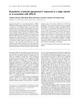
unique to the KIF23 V1 isoform. To test whether the
antibody can discriminate between KIF23 V1 and its
truncated isoform KIF23 V2, plasmids encoding KIF23
Page 4 of 9
V1 or V2 were transiently transfected into human
HEK293T cells and total cellular lysates were analyzed
by SDS-PAGE followed by immunoblotting with antiKIF23 V1 or commercial anti-KIF23 antibodies. The
anti-KIF23 V1 antibody only detected KIF23 V1 protein,
but not KIF23 V2 isoform (Fig. 2a), while both KIF23 V1
and V2 proteins were detected when Western blotting
was performed with anti-KIF23 antibody (Fig. 2b).
To test whether the prepared anti-KIF23 V1 antibody
can recognize endogenous KIF23 V1 protein within
tumor cells, the whole cell extracts of HLE cells was
immunoblotted with anti-KIF23 V1 antibody and a single prominent band with expected size was detected
(Fig. 2c, lane 1). This band was significantly down regulated in cell extracts derived from KIF23 V1 siRNAtreated HLE cells (Fig. 2c, lane 2, 3), indicating this
antibody recognize bona fide KIF 23 V1 protein. This
data was supported by the result using commercial antiKIF23 antibody (Fig. 2d).
Sublocalization of KIF23 V1 protein in HCC cell lines
To determine the subcellular localization of KIF23 V1
protein in HCC cells, immunofluorescence and Western
Fig. 1 Expressions of KIF23 V1 and KIF23 V2 mRNA in normal and malignant tissues. a Expressions of KIF23 V1 and KIF23 V2 mRNAs in normal
tissues. Lane 1: heart; 2: liver; 3: skeletal muscle; 4: pancreas; 5: ovary; 6: colon; 7: PBMC; 8: placenta; 9: lung; 10: kindey; 11: spleen; 12: thymus;
13: prostate; 14: small intestine. b Representative positive expressions of KIF23 V1 and KIF23 V2 mRNA in some HCC tissues. T: cancerous tissues;
A: adjacent noncancerous tissues. c Expressions of KIF23 V1 and KIF23 V2 mRNA in HCC cell lines. Lane 1: HLE; 2: SMMC-7721; 3: BEL-7402; 4: Huh7;
5: HepG2; N: negative control
Sun et al. BMC Cancer (2015) 15:961
Page 5 of 9
Fig. 2 Characterization of anti-KIF23 V1 antibody. a HEK293T cells were transiently transfected with pRK-FLAG-KIF23 V1, pRK-FLAG-KIF23 V2, or
pRK-FLAG plasmids and the protein expression was detected by Western blotting employing anit-KIF23 V1 antibody. b Western blotting analysis
of the same samples as in (a) was performed with commercial anti-KIF23 antibody. c HLE cells were transfected with either control or KIF23 V1
siRNAs and whole cell extracts were processed for immunoblotting with anti-KIF23 V1 antibody. d Western blotting analysis of the same samples
as in (c) was performed with commercial anti-KIF23 antibody. All the membranes were reblotted for the expression of tubulin
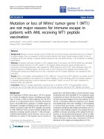
blotting assays were performed. Immunofluorescence
assay with anti-KIF23 V1 antibody demonstrated that
the endogenous KIF23 V1 was located in the nucleus in
both HLE and Huh7 HCC cell lines (Fig. 3a). We further
investigated KIF23 V1 localization in a cell fractionation
assay by Western blotting. The cytoplasmic and nuclear
extracts of HLE cells were immunoblotted with antiKIF23 V1 antibody and the endogenous KIF23 V1 protein was strictly found in nuclear extracts (Fig. 3b).
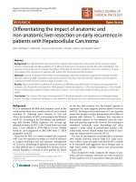
Expression of KIF23 protein in HCC tissues
Expression of KIF23 protein in tumor cells was assessed
by IHC with anti-KIF23 V1 or anti-KIF23 antibodies.
When using anti-KIF23V1 antibody, we found that
KIF23 V1 protein was mainly distributed in the nucleus
of tumor cells, and all the tumor tissues displayed the
heterogeneous pattern, with groups of tumor cells expressing very high level of KIF23 V1 protein, and others
without any detectable expression. Examples of different
expression levels of KIF23 V1 are depicted in Fig. 4a.
KIF23 V1 protein was detectable in 83 of 144 (57.6 %)
HCC tissues and the mean H-score was 42 (range: 0–
290) (Fig. 4c). However, applying anti-KIF23 antibody
for IHC staining, positive staining was predominantly
observed in the cytoplasm of tumor cells, suggesting that
KIF23 V2 localized in the cytoplasm of HCC cells. The
tumor tissues also showed heterogeneous expression
pattern for KIF23 V2, examples of different expression
levels of KIF23 V2 protein are depicted in Fig. 4b. Cytoplasmic expression of KIF23 V2 was detected in 135 of
143 (94.4 %) HCC tissues and the median H-score was
60 (range: 0–290) (Fig. 4d).
For efficacy analyses, the population was divided into
two groups according to the median H-score value.
Expression level of KIF23 V2 was categorized as KIF23
V2 over-expressing tumors (>60) and KIF23 V2 lowexpressing tumors (≤60). Because a small number of
cases showed positive immunostaining, KIF23 V1 cases
were classified into either negative or positive groups.
Clinical relevance of KIF23 V1 expression in HCC tissues
The association between KIF23 V1 expression and overall survival (OS) was evaluated using Kaplan-Meier survival curves with the log-rank test. HCC patients with
tumors expressing KIF23 V1 tended to correlate with
better 3-year survival, but without statistical significance
(P = 0.1604, Fig. 5a), while KIF23 V1-expressing patients
had significantly longer OS (35 months) than the patients
whose tumors did not express KIF23 V1 (15 months)
(P = 0.0052, Fig. 5b). In addition, the association between the expression level of KIF23 V1 and clinical
parameters was analyzed, and no correlation was observed between KIF23 V1 expression and gender, age,
tumor size or TNM stage.
Sun et al. BMC Cancer (2015) 15:961
Page 6 of 9
Fig. 3 Subcellular distribution of endogenous KIF23 V1 protein. a Localization of KIF23 V1 in HLE and Huh7 cells examined by immunofluorescence.
Cells grown on coverslips were fixed and immunostained with polyclonal anti-KIF23 V1 antibody, followed by detection with FITC-conjugated antirabbit IgG secondary antibody. b Subcellular localization of KIF23 V1 examined by cell fractionation and Western blotting. HLE cells were separated into
nuclear and cytoplasmic fractions. Equal amounts of proteins were loaded onto SDS-PAGE gels, and the protein expression was analyzed with antiKIF23 V1, anti-tubulin (cytoplasmic marker), or anti-lamin B1 (nuclear marker) antibodies
In addition, the association between the expression
level of KIF23 V2 and clinical parameters was also analyzed. Based on the cut-off point of KIF23 V2, 67 patients were divided into a high expression group and 76
patients into a low expression group. No significant association between KIF23 V2 expression level and gender,
age, tumor size or TNM stage was found. Furthermore,
KIF23 V2 expression level did not associate with 3-year
and 5-year OS (Fig. 5c, d).
In the univariate survival analysis, we correlated different parameters with the 5-year survival rate. KIF23 V1
expression (negative vs. positive; P = 0.0097), tumor size
(≤5 cm vs. >5 cm; P = 0.0186), TNM stage (I, II vs. III,
IV; P = 0.0040), were identified as parameters significantly influencing survival (Table 1). Multiple Cox regression analysis indicated that TNM stage was an
independent prognostic predictor of the 5-year overall
survival rates in HCC patient after surgery (Table 1).
Discussion
In present study, the expression of the two splice variants of KIF23 mRNA was detected in most clinical HCC
samples and cell lines. Using the prepared antibody specific to KIF23 V1, we found the distinct expression patterns of KIF23 V1 and V2 protein in HCC tumor tissues.
Moreover, the expression of KIF23 V1 protein was associated with prolonged overall survival in the patients
with HCC.
KIF23 is a member of kinesin-like motor protein
families [20] and plays an important role in cytokinesis
[9, 10, 21]. Two splice variants of KIF23 mRNA have
been reported [16]. However, the differences in the
localization, expression, and function for the two splice
variants of KIF23 in tumor cells have remained largely
unknown so far. No commercial antibodies are available
for distinguishing KIF23 V1 from V2 at present. In the
current study, we prepared anti-KIF23 V1 antibody, and
Sun et al. BMC Cancer (2015) 15:961
Page 7 of 9
Fig. 4 Expressions of KIF23 V1 and KIF23 V2 protein in HCC tissues. a Representative images of KIF23 V1 staining in HCC tissues. i: A strong
stained HCC sample (H-score of 290). ii: A moderately stained HCC sample (H-score of 130). iii: A weakly stained HCC sample (H-score of 60).
iiii: A negative stained HCC sample (H-score of 0). Magnification: 1:200. b Representative images of KIF23 V2 staining in HCC tissues. i: A strong
stained HCC sample (H-score of 260). ii: A moderately stained HCC sample (H-score of 125). iii: A weakly stained HCC sample (H-score of 40).
iiii: A negative stained HCC sample (H-score of 0). Magnification: 1:200. c Histograms showing the distribution and frequency for KIF23 V1
expression in HCC tissues. d Histograms showing the distribution and frequency for KIF23 V2 expression in HCC tissues
confirmed the specificity of the antibody by overexpression of KIF23 V1 and V2 as well as knockdown of KIF23
V1. Immunofluorescence staining and cell fraction analysis with the prepared antibody specific to KIF23 V1,
we found that endogenous KIF23 V1 was predominantly
localized in the nucleus of the two HCC cell lines (HLE
and Huh7), which was consistent with the previous report that CHO1 (KIF23 V1) isoform was present in the
nucleus of CHO and HeLa cells [16].
Immunohistochemical staining of HCC tissues with
anti-KIF23 V1 or anti-KIF23 antibodies indicated that
tumor tissues were significant heterogeneity with some
tumor cells expressing high levels of KIF23 V1 or V2
protein while being undetectable in others. Using the
antibody specific to KIF23 V1 for immunohistochemical
staining of HCC tissues, we also found that KIF23 V1
was predominantly localized in nucleus of tumor cells,
which was quite different from the positive staining in
cytoplasm using commercial anti-KIF23 antibody. The
differential expression patterns for the two splice variants of KIF23 suggest that they may have distinct activities in tumor cells. We further found that the
expression of KIF23 V1 protein was significantly associated with prolonged overall survival. The univariate Cox
regression analysis revealed that KIF23 V1 expression is
a factor that significantly influences the outcomes of
HCC patients.
In this study, we observed a favorable effect of KIF23
V1 expression on overall survival. This finding is in contrast with our expectations that KIF23 might promote
tumor development, as KIF23 V1 is upregulated in HCC
tissues and previous report showed that downregulation
of KIF23 decreases proliferation of glioma cells [15]. Furthermore, both KIF23 V1 and V2 have been recently
shown to be down-regulated by tumor suppressor p53 in
a p21-dependent pattern [22]. We speculate that KIF23
Sun et al. BMC Cancer (2015) 15:961
Page 8 of 9
Fig. 5 Association between KIF23 V1 or KIF23 V2 protein expression and overall survival (OS) of HCC patients. Correlation of KIF23 V1 expression
with 3-year (a) or 5-year (b) OS of HCC patients. Correlation of KIF23 V2 expression with 3-year (c) or 5-year (d) OS of HCC patients. The group with
expression of KIF23 V1 showed significantly better survival than the group without expression of KIF23 V1 (p = 0.0052). Patients were categorized by
the median H-scores of KIF23 V2 (60) or by KIF23 V1 expression or not
V1 may be involved in hepatocarcinogenesis, however,
once the tumor is formed, it may play a negative role
during the progression of cancer. Of course, in order to
achieve a better understanding of the mechanism of
KIF23 V1 expression in carcinogenesis and progression
of cancer in patients with HCC, further studies on the
biological functions of KIF23 V1 and V2 as well as their
relationships in tumor cells are necessary.
Table 1 Prognostic factors for survival
Variable
OS
Univariate Multivariate
p-value
HR (95 % CI)
p-value
Gender (Male versus Female)
NS
NA
Age (≤55 vs. >55)
NS
NA
Size (≤5 cm vs. >5 cm)
0.0186
NS
TNM stage (I, II vs. III, IV)
0.0040
KIF23 V2 (≤60 vs. >60)
NS
KIF23 V1 (negative vs. positive) 0.0097
Conclusions
In conclusion, we prepared polyclonal antibody specific
to KIF23 V1 to distinguish KIF23 V1 from KIF23 V2,
and we show for the first time that KIF23 V1 and KIF23
V2 have different localizations in tumor cells. Furthermore, we found that both KIF23 V1 and KIF23 V2 are
up-regulated in HCC patients, and KIF23 V1 expression
might be a marker of longer overall survival in HCC
patients.
Abbreviations
CI: confidence interval; HR: hazard ratio; KIF23: kinesin family member 23;
NA: not adopted; NS: not significant; OS: overall survival; PBMC: peripheral
blood leukocyte; RT-PCR: reverse transcription-polymerase chain reaction;
TNM: tumor- node- metastasis.
Competing interests
There are no financial conflicts of interest.
1.502 (1.121-2.014) 0.0059
NA
NS
Univariate analysis: χ2 test
Multivariate analysis: Cox proportional hazards regression model
Abbreviations: OS Overall survival, HR Hazard Ratio, CI confidence interval,
TNM tumor-node-metastasis, NA not adopted, NS not significant
Authors’ contributions
XS performed the experiments and statistical analyses. ZJ recruited
patients, collected clinical information and analyzed the data. XS and JW
performed part of the experiments. YL, XQ and YZ participated in the
design of the study and analyzed the data. YY designed the experiments
and prepared the manuscript. All authors read and approved the final
manuscript. All authors read and approved the final manuscript.
Sun et al. BMC Cancer (2015) 15:961
Acknowledgements
This work was supported by the National Natural Science Foundation of
China Grants 81171974 and 81071708.
Author details
1
Department of Immunology, School of Basic Medical Sciences, Peking
University Health Science Center, 38 Xueyuan Road, Haidian District, Beijing
100191, China. 2Center of Hepatobiliary Surgery, People’s Hospital, Peking
University Health Science Center, Beijing, China.
Page 9 of 9
20. Miki H, Setou M, Kaneshiro K, Hirokawa N. All kinesin superfamily protein,
KIF, genes in mouse and human. Proc Natl Acad Sci U S A.
2001;98(13):7004–11.
21. Glotzer M. The molecular requirements for cytokinesis. Science.
2005;307(5716):1735–9.
22. Fischer M, Grundke I, Sohr S, Quaas M, Hoffmann S, Knorck A, et al. p53 and
cell cycle dependent transcription of kinesin family member 23 (KIF23) is
controlled via a CHR promoter element bound by DREAM and MMB
complexes. PLoS One. 2013;8(5):e63187.
Received: 24 March 2015 Accepted: 8 December 2015
References
1. Parkin DM, Bray F, Ferlay J, Pisani P. Global cancer statistics, 2002. CA Cancer
J Clin. 2005;55(2):74–108.
2. Xu L, Qian G, Tang L, Su J, Wang JS. Genetic variations of hepatitis B virus
and serum aflatoxin-lysine adduct on high risk of hepatocellular carcinoma
in Southern Guangxi, China. J Hepatol. 2010;53(4):671–6.
3. Kew MC, Yu MC, Kedda MA, Coppin A, Sarkin A, Hodkinson J. The relative
roles of hepatitis B and C viruses in the etiology of hepatocellular
carcinoma in southern African blacks. Gastroenterology. 1997;112(1):184–7.
4. Parkin DM. The global health burden of infection-associated cancers in the
year 2002. Int J Cancer. 2006;118(12):3030–44.
5. Shi YY, Wang HC, Yin YH, Sun WS, Li Y, Zhang CQ, et al. Identification and
analysis of tumour-associated antigens in hepatocellular carcinoma. Br J
Cancer. 2005;92(5):929–34.
6. Yi Y, Wu H, Gao Q, He HW, Li YW, Cai XY, et al. Interferon regulatory factor
(IRF)-1 and IRF-2 are associated with prognosis and tumor invasion in HCC.
Ann Surg Oncol. 2013;20(1):267–76.
7. Chen D, Xing W, Hong J, Wang M, Huang Y, Zhu C, et al. The beta2adrenergic receptor is a potential prognostic biomarker for human
hepatocellular carcinoma after curative resection. Ann Surg Oncol.
2012;19(11):3556–65.
8. Nislow C, Lombillo VA, Kuriyama R, McIntosh JR. A plus-end-directed motor
enzyme that moves antiparallel microtubules in vitro localizes to the
interzone of mitotic spindles. Nature. 1992;359(6395):543–7.
9. Zhu C, Bossy-Wetzel E, Jiang W. Recruitment of MKLP1 to the spindle
midzone/midbody by INCENP is essential for midbody formation and
completion of cytokinesis in human cells. Biochem J. 2005;389(Pt 2):373–81.
10. Chen MC, Zhou Y, Detrich 3rd HW. Zebrafish mitotic kinesin-like protein 1
(Mklp1) functions in embryonic cytokinesis. Physiol Genomics.
2002;8(1):51–66.
11. Zhu C, Zhao J, Bibikova M, Leverson JD, Bossy-Wetzel E, Fan JB, et al.
Functional analysis of human microtubule-based motor proteins, the
kinesins and dyneins, in mitosis/cytokinesis using RNA interference. Mol Biol
Cell. 2005;16(7):3187–99.
12. Liu X, Zhou T, Kuriyama R, Erikson RL. Molecular interactions of Polo-likekinase 1 with the mitotic kinesin-like protein CHO1/MKLP-1. J Cell Sci. 2004;
117(Pt 15):3233–46.
13. Weinberg RA. Cancer Biology and Therapy: the road ahead. Cancer Biol
Ther. 2002;1(1):3.
14. Valk K, Vooder T, Kolde R, Reintam MA, Petzold C, Vilo J, et al. Gene
expression profiles of non-small cell lung cancer: survival prediction and
new biomarkers. Oncology. 2010;79(3–4):283–92.
15. Takahashi S, Fusaki N, Ohta S, Iwahori Y, Iizuka Y, Inagawa K, et al.
Downregulation of KIF23 suppresses glioma proliferation. J Neurooncol.
2012;106(3):519–29.
16. Kuriyama R, Gustus C, Terada Y, Uetake Y, Matuliene J. CHO1, a mammalian
kinesin-like protein, interacts with F-actin and is involved in the terminal
phase of cytokinesis. J Cell Biol. 2002;156(5):783–90.
17. Hao J, Shen R, Li Y, Zhang Y, Yin Y. Cancer-testis antigen HCA587/MAGE-C2
interacts with BS69 and promotes its degradation in the ubiquitinproteasome pathway. Biochem Biophys Res Commun. 2014;449(4):386–91.
18. Dong XY, Peng JR, Ye YJ, Chen HS, Zhang LJ, Pang XW, et al. Plac1 is a
tumor-specific antigen capable of eliciting spontaneous antibody responses
in human cancer patients. Int J Cancer. 2008;122(9):2038–43.
19. Christoph DC, Kasper S, Gauler TC, Loesch C, Engelhard M, Theegarten D,
et al. betaV-tubulin expression is associated with outcome following taxanebased chemotherapy in non-small cell lung cancer. Br J Cancer.
2012;107(5):823–30.
Submit your next manuscript to BioMed Central
and we will help you at every step:
• We accept pre-submission inquiries
• Our selector tool helps you to find the most relevant journal
• We provide round the clock customer support
• Convenient online submission
• Thorough peer review
• Inclusion in PubMed and all major indexing services
• Maximum visibility for your research
Submit your manuscript at
www.biomedcentral.com/submit