Performance of the colorectal cancer screening marker Sept9 is influenced by age, diabetes and arthritis: A nested case - control study
Bạn đang xem bản rút gọn của tài liệu. Xem và tải ngay bản đầy đủ của tài liệu tại đây (583.89 KB, 10 trang )
Ørntoft et al. BMC Cancer (2015) 15:819
DOI 10.1186/s12885-015-1832-6
RESEARCH ARTICLE
Open Access
Performance of the colorectal cancer
screening marker Sept9 is influenced by
age, diabetes and arthritis: a nested
case–control study
Mai-Britt W. Ørntoft1, Hans J. Nielsen2, Torben F. Ørntoft1, Claus L. Andersen1* and On behalf of the Danish Study
Group on Early Detection of Colorectal Cancer
Abstract
Background: Annually, colorectal cancer (CRC) is diagnosed in >1.4 million subjects worldwide and incidence is
increasing. Much effort has therefore been focused on screening, which has proven to reduce cancer-related
mortality. The Sept9 DNA-methylation assay is among the most well studied blood-based screening markers.
However, earlier reported performances may be misleading: the Sept9 test was recently examined in two screening
based cohorts and yielded performances lower than expected. We hypothesize that comorbidities and/or
demographic characteristics affect the results of the Sept9 test.
Methods: Using a retrospective nested case–control study design, we studied plasma from 150 cancer and 150
controls selected from a well-characterized cohort of 4698 subjects referred for diagnostic colonoscopy due to CRCrelated symptoms. The cases and controls were matched on age and gender, and moreover cases were stratified
on tumor-site and tumor-stage. The selected cohort included a wide range of comorbidities. Plasma Sept9 levels
were assessed using a commercially available PCR based assay (Epi-proColon).
Results: Clinical sensitivity for CRC stages I-IV was 37 %, 91 %, 77 %, and 89 %, and the overall sensitivity 73 % (95 % CI,
64–80 %) and specificity 82 % (95 % CI, 75–88 %), respectively. Age >65 was associated with both increased false positive
and false negative results (p < 0.05). Arthritis was associated with a higher false negative rate (p = 0.005) whereas
Arteriosclerosis was associated with a higher false positive rate (p = 0.007). Diabetes was associated with Sept9 positivity
with an OR of 5.2 (95 % CI 1.4–19.1). When the performance of Sept9 was adjusted for these parameters in a final
multivariate regression model, the OR for a positive Sept9 test to be associated with CRC increased from 8.25 (95 % CI
4.83–14.09) to 29.46 (95 % CI 12.58–69.02).
Conclusions: The results indicate that the performance of the Sept9 assay is negatively affected by several factors
commonly associated with CRC screening populations: early-stage disease, age > 65 years, diabetes, arthritis, and
arteriosclerosis. This should be taken into account if the Sept9 assay is used as a single marker for CRC screening, but may
also have a wider impact, as it is likely that such factors may affect other blood based DNA markers as well.
Keywords: 3–10 words: Sept9, Colorectal cancer, Screening, DNA methylation marker, Comorbidities, Epigenetics
* Correspondence:
1
Department of Molecular Medicine, MOMA, Aarhus University Hospital,
Skejby DK-8200Aarhus N, Denmark
Full list of author information is available at the end of the article
© 2015 Ørntoft et al. Open Access This article is distributed under the terms of the Creative Commons Attribution 4.0
International License ( which permits unrestricted use, distribution, and
reproduction in any medium, provided you give appropriate credit to the original author(s) and the source, provide a link to
the Creative Commons license, and indicate if changes were made. The Creative Commons Public Domain Dedication waiver
( applies to the data made available in this article, unless otherwise stated.
Ørntoft et al. BMC Cancer (2015) 15:819
Background
At present, primary colorectal cancer (CRC) is diagnosed in >1.4 million subjects annually and incidence
is increasing [1]. Although improved surgical approaches for especially rectal cancer has improved the
overall survival rates it is mainly early stage cancers
that are curable by surgical intervention. The most
reliable self-reported symptom of CRC cancer is dark
rectal bleeding with a positive predictive value of 20.6
% and an odds ratio (OR) of 7.4 for CRC [2], but
symptoms may be evasive. Therefore, much effort has
been focused on screening and hence earlier detection
of CRC, which has repeatedly proven to reduce the
cancer-related mortality [3, 4].
In recent years several screening markers have
emerged to help diagnosing early stage CRC or even
premalignant lesions. They separate in two different
categories: stool markers, such as FOBT/FIT and bloodbased markers as DNA/RNA and proteins [5]. It is expected that blood-based screening assays may improve
the clinical sensitivity compared with stool tests, because
of an expected higher compliance among screening subjects [6, 7]; however the cost-effectiveness has not yet
been proven [8]. The Sept9 DNA-methylation test is
among the most well studied blood tests; often in case–
control study designs [9–11]. Recently, the Sept9 test
was examined in two medium/large sized asymptomatic
screening based cohorts and yielded overall sensitivities
(range 48–68 %) and specificities (range 78–91 %) lower
than expected based on the earlier case–control studies
[12, 13]. This may indicate that the performance of the
Sept9 test is affected by covariates, such as comorbidities
and/or demographic characteristics of the subjects in
screening populations. However, currently we have only
limited knowledge of which comorbidities or characteristic could be involved as this has not been part of earlier
study objectives.
To address this question we have tested the Sept9
assay on a nested case–control cohort selected from a
large well-characterized cohort of subjects referred to
colonoscopy due to CRC symptoms; the subjects also
had a wide range of co-morbidities.
Methods
Human plasma samples
The retrospective nested case–control cohort in the
present study was selected from the prospective Danish
Endoscopy II study, a multicenter trial, which from April
2010 to November 2012 recruited 4698 subjects referred
to colonoscopy due to CRC related symptoms. All patients gave informed consent to participate and the study
was approved by the The Danish National Ethics Committee (H-3–2009–110) and the Danish Data Protection
Agency (2007–58–0015). Exclusion criteria were
Page 2 of 10
previous colonoscopy, previous CRC or adenoma, diagnosis with HNPCC (Hereditary Nonpolyposis Colorectal
Cancer or Lynch Syndrome) or FAP (Familial adenomatous polyposis), previous or present extracolonic malignant disease, or age under 18. Just prior to colonoscopy
all subjects had a blood sample collected. All clinical information was collected, including surgical/oncological
intervention, as well as cancer TNM stage, and adenoma
histology. Intervention followed the Danish Colorectal
Cancer Group (DCCG) guidelines. Chronic diseases
were divided into large disease groups: Hypertension,
Diabetes (type I and II), manifest Arteriosclerosis (pooling former AMI, stroke due to thrombosis, chronic ischemic diseases in peripheral arteries and chronic
ischemic heart diseases), respiratory diseases and Arthritis (active arthritis in more than one joint).
The nested case–control cohort consisted of 300 participants: 150 cases and 150 controls with no evidence of
CRC disease (NED). Cases consisted of 21 high risk adenomas (size ≥1 cm and/or villous histology >25 % and/
or sessile-serrated polyps and/or high-grade neoplasia
and/or ≥ 3 adenomas), 35 stage I CRC, 35 stage II CRC,
30 stage III CRC and 29 stage IV CRC based on UICC
criteria. To minimize confounding, cases and controls
were matched by age and gender. Cases were selected
with gender evenly distributed according to disease stage
and with 1/3 of tumors localized in the rectum, 1/3 in
the proximal colon (coecum, ascending colon and right
flexure), and 1/3 in the distal colon. Chronic disease and
comorbidities were not included as exclusion criteria.
For all participants, clinical follow up for further 3 years
were obtained; as all Danish patients are registered with
a personal computerized ID-number, and all hospital
treatment is recorded in a national database, there were
no participants lost to follow-up. A survey of the cohort identified the expected associations between comorbidities and life style factors, indicating the cohort
is representative of future CRC screening cohorts
(Additional file 1: Table S1).
For all 300 participants, plasma was isolated from ethylenediaminetetraacetic acid (EDTA) stabilized whole
blood by double centrifugation at 10 min for 3000 g at
room temperature. Plasma was then stored at −80 °C
under 24/7 electronic surveillance until isolation of circulating cell-free DNA (cfDNA).
Sept9 test
cfDNA isolation and analysis for presence of methylated
Sept9 DNA was done using the Epi-ProColon kit as described by Potter et al. [13]: The Epi proColon test comprises the Epi proColon Plasma Quick kit, the Sensitive
PCR kit, and Control kit. All analyses were done blinded
to subject outcome and were performed by Epigenomics
GmbH, Berlin. Samples were processed in batches with
Ørntoft et al. BMC Cancer (2015) 15:819
a random distribution of cases and controls to avoid
analytical bias, and negative and positive processing controls ensured validity of the test result. A minimum of 2
ml of plasma was provided for cfDNA isolation; one participant did not meet this requirement and was excluded. cfDNA was isolated from 3.5 ml plasma for 139
participants, and from between 2.0–3.5 mL plasma from
160 participants; a total of 299 subjects (149 cases and
150 controls).
To isolate cfDNA we used the Plasma Quick kit,
where plasma was mixed with 3.5 ml of lysis buffer and
incubated for 10 min, after which magnetic beads and
absolute ethanol were added; the sample was incubated
on a rotator for 45 min. Impurities were removed from
the magnetic beads in a wash step. The purified DNA
was then released from the beads in elution buffer and
treated at 80 °C with a solution containing ammonium
bisulfite for deamination of cytosine. The converted
cfDNA was captured by use of magnetic beads, passed
through a series of wash steps, and eluted in 60 μL buffer. Samples were then analyzed for presence of methylated Sept9 DNA with the Sensitive PCR kit on a 7500
Fast Dx Real Time PCR device (Life Technologies). The
assay was designed as a duplex real-time PCR for the
methylated Sept9 γ promoter and ACTB (actin, beta) as
an internal reference to assess the integrity of each sample. PCR was performed in triplicate with 15 μL template DNA per well and run for 45 cycles. We recorded
PCR results from the 7500 Fast Dx software for ACTB
and methylated Sept9 for each of the triplicate reactions.
The validity of each sample batch was determined on
the basis of methylated Sept9 and ACTB cycle threshold
(Ct) values for the positive and negative controls. Samples were only deemed valid if the ACTB control was
positive in all three replicates (had amplification curves
detected within 45 cycles). Sept9 test results for individual samples were scored positive, if a Ct value was detected within 45 cycles. Samples were scored negative
when no methylated Sept9 Ct value was reported for any
of the 3 valid PCR replicates.
Statistical methods
We assessed the association in between all variables and
also pair-wise between Sept9 and all other available variables using Fishers exact tests. All tests were two-sided,
and p < 0.05 was considered statistically significant. Test
for trend of increasing true positive results with increasing tumor stage was done using the Wilcoxon rank sum
test. Univariate logistic regression models were used to
assess the diagnostic power of all available variables for
CRC. Bivariate models were used to assess the association between Sept9 and all other available variables, including if any of the available variables modified the
effect of the Sept9 test. Finally a multivariate model was
Page 3 of 10
built to assess the adjusted diagnostic power of the Sept9
assay in the context of all the variables affecting or associated with it. All reported models passed the HosmerLemeshow’s goodness of fit test. All assumptions for the
different analyses were fulfilled. STATA V.12.1 (StataCorp LP, Texas, USA) were used for all statistical
analyses.
Results
Clinical performance
An overview of the demographic and co-morbidity characteristics of the included subjects is provided in Table 1.
In this study the overall sensitivity of the Sept9 test for
detecting CRC was 73 % (95 % CI, 64–80 %) vs 59 %
(95 % CI 50–67 %), using the 1/3 and 2/3 scoring algorithms, respectively. Clinical sensitivity for the individual
CRC stages I-IV, using the 1/3 and 2/3 algorithms was
as follows: 37 % vs 17 %; 91 % vs 74 %; 77 % vs 63 %;
and 89 % vs 86 %. The sensitivity was significantly lower
for stage I than for the higher stage tumors (Wilcoxon
rank sum test, p < 0.001). As expected the two algorithms
yielded significantly different sensitivity and specificity results (p < 0.001, Fisher’s exact test). For high risk adenomas the sensitivity was 14 % (95 % CI, 3–63 %) vs 0 %
(95 % CI, 0–1.6 %), for the two algorithms. The positivity
rates for adenomas were not different from the rates in
the NED group, 18 % (95 % CI, 12–25 %) vs 5 % (95 % CI,
2–9 %), for the two algorithms (Table 2).
The overall test specificity was 82 % (95 % CI, 75–88 %)
vs. 95 % (95 % CI, 91–98 %) for the two algorithms. To investigate if any subjects with NED falsely scored positive
due to an occult cancer, all subsequent instances of cancer
diagnoses three years after the initial colonoscopy were
identified through hospital records. None of the subjects
with NED that scored falsely positive were later diagnosed
with cancer.
Factors potentially affecting assay performance
To investigate if the outcome of the Sept9 test was affected by any of the available demographic or clinical
variables (excluding symptoms) the significance of all associations with Sept9 was tested using Fisher’s exact test.
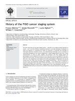
Initially the continuous variable age was plotted against
Sept9 outcome to look for trends of association, and to
determine a cut-point for dichotomization of the age variable (Fig. 1). The plot revealed a tendency towards an increased false positive rate for NEDs with ages above >65
years, particularly for the high specificity 2/3 algorithm
(Fig. 1, right panel). After dichotomizing age at 65 years
and testing for association to Sept9, the group with age >65
was significantly associated with increased false positive
rates for both the 1/3 and 2/3 algorithms (p = 0.015 and
p = 0.05 respectively). The analysis also revealed that
age >65 was associated with an increased false negative rate
Ørntoft et al. BMC Cancer (2015) 15:819
Page 4 of 10
Table 1 Demographic distribution of the nested cohort from
the Endoscopy II prospective sample collection
Table 2 Summary of Epi proColon test performance in a nested
case-control cohort from the Endoscopy II population
Variable
Sex
Female
Total
CRC
Adenoma NED
1/3 algorithm
299
128
21
150
Diagnosis
Subjects (n)
Positive (n)
Negative (n)
Fraction (95 % CI)
151
65 (51)
11 (52)
75 (50)
CRC
128
93
35
0.73 (0.64–0.80)
Male
148
63 (49)
10 (48)
75 (50)
stage I
35
13
22
0.37 (0.21–0.55)
Age
≤65
183
74 (58)
11 (52)
98 (65)
stage II
35
32
3
0.91 (0.76–0.98)
>65
116
54 (42)
10 (48)
52 (35)
stage III
30
23
7
0.77 (0.58–0.90)
Rectal bleeding
No
166
54 (42)
10 (48)
102 (68)
stage IV
28
25
3
0.89 (0.72–0.98)
Yes
133
74 (58)
11 (52)
48 (32)
Non-CRC
171
30
141
0.18 (0.12–0.24)
Anemia
No
250
102 (80)
19 (90)
129 (86)
Adenoma
21
3
18
0.14 (0.03–0.63)
Yes
49
26 (20)
2 (10)
21 (14)
NED
150
27
123
0.18 (0.12–0.25)
Weightloss
No
211
81 (63)
17 (81)
113 (75)
2/3 algorithm
Yes
88
47 (37)
4 (19)
37 (25)
CRC
128
75
53
0.59 (0.50–0.67)
Altered Defaecation
No
126
58 (45)
12 (57)
56 (37)
stage I
35
6
29
0.17 (0.07–0.34)
Yes
173
70 (55)
9 (43)
94 (63)
stage II
35
26
9
0.74 (0.57–0.88)
Abdominal Pain
No
172
72 (56)
17 (81)
83 (55)
stage III
30
19
11
0.63 (0.44–0.80)
Yes
127
56 (44)
4 (19)
67 (45)
stage IV
28
24
4
0.86 (0.67–0.96)
Abdominal mass
No
283
119 (93)
0
143 (95)
Non-CRC
171
7
164
0.04 (0.02–0.08)
Yes
16
9 (7)
0
7 (5)
Adenoma
21
0
21
0.00 (0.00–0.16)
Distention
No
241
103 (80)
21 (100)
117 (78)
NED
150
7
143
0.05 (0.02–0.09)
Yes
58
25 (20)
0 (0)
33 (22)
Hypertension
No
183
81 (63)
10 (48)
92 (61)
Yes
116
47 (37)
11 (52)
58 (39)
NED No Evidence of Disease
Fraction: Positive fraction detected
Difference in 1/3 vs 2/3 algorithm: Fischer’s Exact Test, p< 0.001
Increasing proportion of true positive results with increasing tumor stage:
Wilcoxon rank sum test for trend, z<0.001
Diabetes
No
280
117 (91)
16 (76)
147 (98)
Yes
19
11 (9)
5 (24)
3 (2)
No
243
108 (84)
19 (90)
116 (77)
Yes
56
20 (16)
2 (10)
34 (23)
Respiratory disease
No
267
117 (91)
21 (100)
129 (86)
Yes
32
11 (9)
0 (0)
21 (14)
Arthritis
No
281
120 (94)
20 (95)
141 (94)
Yes
18
8 (6)
1 (5)
9 (6)
Smoke#
No
123
47 (37)
12 (57)
64 (43)
81 (63)
9 (43)
86 (57)
Alcohol##
Normal
245
103 (80)
15 (71)
127 (85)
Abuse
54
25 (20)
6 (29)
23 (15)
BMI###
<18,5
10
4 (3)
1 (5)
5 (3)
18,5 to 25
162
70 (55)
10 (48)
82 (55)
25 to 30
94
39 (31)
7 (33)
48 (32)
Arteriosclerosis
Yes
Plasma volume¤
>30
33
15 (12)
3 (14)
15 (10)
< 3.5 ml
160
70 (55)
6 (29)
84 (56)
3.5 ml
139
58 (45)
15 (71)
66 (44)
Data are n (%)
# Former smokers and current smokers pooled
## Abuse: Women > 7 units per week, Men >14 units per week
### Underweight < 18,5, Normal 18,5–25, Overweight 25–30, Heavy
overweight >30 ¤ median 3.0 ml, range (2–3.4 ml)
NED No Evidence of Disease
for the 2/3 algorithm (p = 0.007, Tables 3 and 4). However,
this significance was probably driven by an unintended imbalance in tumor stage distribution in the two age groups,
with the >65 age group having significantly fewer stage III
and IV tumors (Additional file 2: Table S2).
Of the other variables only Arthritis and Arteriosclerosis consistently affected Sept9 outcome. Arthritis was
associated with an increased false negative rate (Table 3),
which was significant for the 1/3 algorithm (p = 0.005)
and borderline for the 2/3 algorithm (p = 0.07). Arteriosclerosis was associated with an increased false positive
rate (Table 4). This association was significant for the 2/
3 algorithm (p = 0.007) and borderline for the 1/3 algorithm (p = 0.07).
Female gender was associated with an increased false
negative rate for the more sensitive 1/3 algorithm but
not the more specific 2/3 algorithm.
As a low sample input means less cfDNA available for
analysis, it was tested whether a low plasma volume had
any effect on Sept9 performance. No effect on sensitivity
was observed (Fishers exact test p = 0.69 vs p = 0.59 for
the two algorithms). On the opposite, a low plasma volume surprisingly seemed to produce more false positive
Ørntoft et al. BMC Cancer (2015) 15:819
Page 5 of 10
Fig. 1 Age plotted against Sept9 outcome. FP: False positive, TN: True negative, FN: False negative, TP: True positive. N: Number of subjects in each
category. Age: All ages younger or equal to the age interval mentioned. -1/3 and 2/3 refers to the PCR-algorithms used
controls (Table 4). However, this significance was probably driven by age, as more subjects aged >65 had lower
plasma volumes (Additional file 3: Table S3).
Sept9 as predictor of CRC
Logistic regression models were built to evaluate: i) how
well a positive Sept9 test predicts CRC, ii) which, if any,
of the available exposure variables might modify Sept9’s
ability to predict CRC, and iii) the strength of the association between the Sept9 test and the diagnosis of CRC,
when taking these variables into account.
First, it was evaluated which of the available variables
(including symptoms) were associated with CRC. In univariate models, only Sept9 (OR 8.25, 95 % CI 4.83–
14.09, p < 0.001), rectal bleeding (OR 2.82 95 % CI
1.76–4.5, p < 0.001) and Diabetes (OR 5.89, 95 % CI
1.68–20.68, p = 0.006) showed a significant association
(Additional file 4: Table S4).
To identify variables associated with Sept9 outcome,
univariate logistic regression models were built with Sept9
as outcome variable and all other variables consecutively
as explaining variable. Diabetes was significant with an
OR of 5.2 (95 % CI 1.4–19.1), and likewise was age when
adjusted for tumor stage (OR 2.06, 95 % CI 1.1–3.8), but
none of the other variables (Additional file 5: Table S5).
Next, we checked if any variables modified the outcome of
the Sept9 test. Age >65 and Arthritis was found to be significant modifiers with an OR of 2.46 (95 % CI 1.14–5.30)
and 0.03 (95 % CI 0.00–0.22), respectively. Consequently,
we allowed for effect modification from these factors in
the final multivariate regression model (Additional file 6:
Table S6).
All variables found to be associated with Sept9 by
either Fishers exact test or regression (age, Arthritis,
Arteriosclerosis, and Diabetes) were included in a
final multivariate regression model with CRC as outcome. Interestingly, the adjusted OR associated with a
positive Sept9 test increased from 8.25 to 29.46 (95 %
CI 12.58–69.02, p < 0.001) for the 1/3 algorithm
Table 5. Similar results were obtained for the 2/3 algorithm (Additional file 7: Table S7).
Discussion
Clinical performance
It is well examined that the Sept9 test can be used to
identify occult CRC. More than 15.000 subjects have
been tested and the reported sensitivity ranges from 36.6
to 95.6 % [14]. The test has primarily been applied to
cases and controls selected from separate populations
i.e. cases were typically patients with symptomatic CRC
Ørntoft et al. BMC Cancer (2015) 15:819
Page 6 of 10
Table 3 Sept9 positivity of individuals with CRC
1/3 algorithm
Sex
Age
Hypertension
Diabetes
Arteriosclerosis
Respiratory disease
Arthritis
Smoke#
Alcohol##
BMI
Tumor site
MSI¤
Plasma volume
2/3 algorithm
P
N
% (95 % CI)
p*
P
N
% (95 % CI)
p*
93
35
73
NR
75
53
59
NR
Female
42
23
65 (52–76)
0.05
34
31
52 (40–65)
0.16
Male
51
12
81 (69–90)
41
22
65 ( 52–77)
51
23
69 (59–81)
24
30
44 (31–59)
49
32
60 (49–71)
26
21
55 (40–70)
67
50
57 (48–66)
8
3
73 (39–94)
65
43
60 (50–69)
10
10
50 (27–73)
71
46
61 (51–70)
4
7
36 (11–69)
73
47
61 (52–70)
2
6
25 (3–65)
29
18
62 (46–75)
46
35
57 (45–68)
64
39
62 (52–72)
11
14
44 (24–65)
Variable
≤65
57
17
77 (66–86)
>65
36
18
67 (53–79)
No
60
21
74 (63–83)
Yes
33
14
70 (55–83)
No
84
33
72 (63–80)
Yes
9
2
82 (48–98)
No
80
28
74 (65–82)
Yes
13
7
65 (41–85)
No
87
30
74 (65–82)
Yes
6
5
55 (23–83)
No
91
29
76 (61–83)
Yes
2
6
25 (3–65)
No
38
9
81 (67–91)
Yes
55
26
68 (57–78)
Normal use
78
25
76 (66–84)
Abuse
15
10
60 (39–79)
<18,5
3
1
75 (19–99)
3
1
75 (19–99)
18,5 to 25
52
18
74 (62–84)
41
29
59 (46–70)
25 to 30
27
12
69 (52–83)
22
17
56 (40–72)
>30
11
4
73 (45–92)
9
6
60 (32–84)
17
13
57 (37–75)
34
20
63 (49–76)
24
20
55 (39–70)
28
22
56 (41–70)
6
5
55 (23–83)
43
27
61 (49–73)
32
26
55 (42–68)
Left
23
7
77 (58–90)
Right
37
17
69 (54–80)
Rectum
33
11
75 (60–87)
Stable
35
15
70 (55–82)
Unstable
8
3
73 (39–94)
< 3.5 ml
52
18
74 (62–84)
3.5 ml
41
17
71 (57–82)
0.23
0.68
0.73
0.42
0.17
0.005
0.08
0.14
0.97
0.67
1.00
0.69
0.007
0.58
0.36
0.46
0.20
0.07
0.36
0.12
0.95
0.70
1.00
0.59
*p-value, two-sided Fisher’s exact test, p<0.05 considered statistically significant
NR Not relevant, P Positive, N Negative, % Positive fraction detected
#Former smokers and smokers pooled
##Abuse: Women > 7 units per week, Men >14 units per week
¤MSI determined by Immunohistochemistry, data not available on all cases
found at colonoscopy, while controls were often screening subjects or symptomatic individuals found to be
tumor negative after colonoscopy. This makes statistical
comparison uncertain, and furthermore does not fulfil
the REMARK criteria [15]. The present study, with cases
and controls selected from the same cohort of symptomatic subjects, who all had their blood samples drawn and
processed according to the same standard operating procedure, fulfills the requirement of the REMARK criteria.
Moreover the included subjects have gender and comorbidity distributions similar to what can be expected in a
screening population. This might be the reason that the
overall results are more similar to that of recent screening based cohorts than of earlier case–control studies
[10, 12, 13]. The present study indicated that the Sept9
assay had low sensitivity in detecting early stage tumors
(adenomas and stage I carcinomas). However, that may
potentially be explained by the limited plasma volume
used for analysis (≤3.5 ml). It has been reported that the
number of ctDNA (cell-free tumor DNA) genome equivalents per 5 milliliter blood often is less than ten for
patients with stage I carcinomas [16]. Accordingly, to
Ørntoft et al. BMC Cancer (2015) 15:819
Page 7 of 10
Table 4 Sept9 positivity of individuals with NED
1/3 algorithm
Sex
Age
Hypertension
Diabetes
Arteriosclerosis
Respiratory disease
Arthritis
Smoke#
Alcohol##
BMI
Plasma volume
2/3 algorithm
P
N
% (95 % CI)
p*
P
N
% (95 % CI)
p*
27
123
18
NR
27
123
18
NR
Female
11
64
15 (8–25)
0.4
2
73
3 (0–9)
0.44
Male
16
59
21 (13–32)
5
70
7 (2–15)
≤65
12
86
12 (6–20)
2
96
2 (0–7)
>65
15
37
29 (17–43)
5
47
10 (3–21)
No
18
74
20 (12–29)
4
88
4 (1–11)
Yes
9
49
16 (7–27)
3
55
5 (1–14)
No
25
122
17 (11–24)
6
141
4 (2–9)
Yes
2
1
67 (9–99)
1
2
33 (0–91)
2
114
2 (0–6)
5
29
15 (5–31)
5
124
4 (1–8)
2
19
10 (1–30)
7
134
5 (2–10)
0
9
0 (0–34)
2
62
3 (0–11)
5
81
6 (2–13)
6
121
5 (2–10)
1
22
4 (0–22)
Variable
No
17
99
15 (9–22)
Yes
10
24
29 (12–39)
No
24
105
19 (12–26)
Yes
3
18
14 (3–36)
No
23
118
16 (11–23)
Yes
4
5
44 (14–79)
No
10
54
16 (8–27)
Yes
17
69
20 (12–30)
0.015
0.66
0.08
0.07
0.77
0.06
0.33
Normal use
24
103
19 (12–27)
Abuse
3
20
13 (3–34)
0.77
<18,5
0
5
0 (0–52)
0
5
0 (0–52)
18,5 to 25
15
67
18 (11–28)
5
77
6 (2–14)
25 to 30
9
39
19 (9–33)
1
47
2 (0–11)
>30
3
12
20 (4–48)
1
14
7 (0–32)
< 3.5 ml
20
64
24 (15–34)
6
78
7 (3–15)
3.5 ml
7
59
11 (4–21)
1
65
2 (0–8)
0.92
0.05
0.05
1.00
0.13
0.007
0.25
1.00
0.36
1.00
0.61
0.14
*p-value, two-sided Fisher’s exact test, p<0.05 considered statistically significant
NR Not relevant, P Positive, N Negative, % Positive fraction, NED No Evidence of Disease
#Former smokers and current smokers pooled
##Abuse: Women > 7 units per week, Men >14 units per week
Table 5 Predictors of Colorectal Cancer, 1/3 algorithm
p-value*
Predictor
OR (95 % CI)
Sept9 -crude estimate
8.25 (4.83–14.09)
0.000
Correction for other variables
Multivariate OR (95 % CI)
p-value*
Sept9 -adjusted OR
29.46 (12.58–69.02)
0.000
Age >65
2.80 ( 1.23–6.36)
0.014
Age as effect–modificator of Sept9
0.24 (0.07–0.80)
0.020
Demographic characteristics
Co-morbidities
Diabetes
2.26 (0.51–10.05)
0.283
Arterioschlerosis
0.39 (0.18–0.86)
0.019
Arthritis
4.88 (1.30–18.42)
0.019
Arthritis as effect-modificator of Sept9
0 .02 (0.000.22)
0.001
*p<0.05 considered statistically significant
Ørntoft et al. BMC Cancer (2015) 15:819
increase the sensitivity towards early stage tumors it will
probably be necessary to increase the plasma volume. In
addition to plasma volume other factors may potentially
also influence adenoma sensitivity. A recent report indicated that the methylation of the Sept9 locus is a late
event in the transformation of adenomas to carcinomas
[17]; even if adenomas release ctDNA, it may not be
methylated and hence may not be detected by the Sept9
assay. One way to mitigate this particular problem could
be to use multiple markers, including markers targeting
adenomas, rather than Sept9 alone. Along these lines we
hypothesize that to reach optimal sensitivity and specificity of both adenomas and early stage carcinomas an increased plasma volume and a multiplex test, targeting
several colorectal neoplasia specific methylation markers,
is needed.
Factors with impact on assay performance
The influence of demographic parameters on the Sept9
test has previously only been sparsely examined. In line
with other reports we showed that gender and tumor
localization did not affect assay sensitivity [10, 12, 13,
18, 19]. Previously, deregulation of Sept9 expression has
been reported to be associated with genomic instability
by at least two mechanisms associated to chromosomal
instability (CIN), namely by mitotic spindle defects and/
or incomplete cell division [20]. Therefore we investigated whether Sept9 methylation was better at predicting CIN than microsatellite unstable (MSI) tumors?
Surprisingly, we could not identify differences between
the positivity rates for MSI and microsatellite stable
(MSS) cancers (Fishers exact test, p = 1.00, Table 3).
The only factor besides tumor stage recurrently reported to influence Sept9 performance is age. Higher
age has been described to be associated with both decreasing sensitivity and specificity [12, 13]. Our findings
are in support of this observation, as we also showed the
assay to have reduced specificity for the oldest test subjects (age > 65). The decreasing specificity with older
age might be partly explained by the known correlation
between chronological age and increased genome-wide
DNA methylation changes [21], but could also be due to
a higher prevalence of chronic diseases in elderly compared to younger subjects. Several studies associate
various chronic diseases with DNA methylation changes
[22, 23]. This might lead to a higher risk of coincident
and non-CRC related methylation of the Sept9 locus in
elderly subjects. Though a positive Sept9 test should not
be regarded as confirmative evidence for CRC, and should
always be confirmed by a colonoscopy, a decreased specificity with age >65 challenges a test aimed at subjects at
age 50–75 years, and lead to larger down-stream costs. To
address this problem an age-differentiated use of the two
Sept9 scoring algorithms could be considered: if the 1/3
Page 8 of 10
algorithm is applied to subjects ≤ 65 and the 2/3 algorithm
is applied to subjects >65 years the combined sensitivity
and specificity is 64 % and 89 % compared to 0.73 % and
0.82 % for the 1/3 algorithm alone (Additional file 8:
Table S8).
In contrast to earlier studies, several co-morbidities influenced the Sept9 test in the present study [10, 13, 24].
Particularly, subjects with Arthritis were difficult to
score correctly for the Sept9 test. This has not been reported earlier. Nevertheless, in 2008 the assay was tested
in a cohort of 315 control subjects without CRC, but
with different comorbidities [24]. By going through the
reported data we observed, consistent with the present
study, that 20 % of NED subjects with Rheumatoid arthritis were false positive and similarly that patients with
Lupus also had a high false positive rate (14.2 %). Since
1966 it has been known that autoimmune inflammatory
diseases such as Lupus, Polyarthritis, or Rheumatoid
arthritis are associated with significant elevated levels of
cfDNA [25–27]. We therefore speculate that the decreased assay sensitivity observed in subjects with Arthritis could be due to increased circulating levels of
arthritis-associated cfDNA, making it difficult to detect
the few copies of methylated ctDNA from the CRC. Further, a recently published study of DNA methylome
changes in Rheumatoid arthritis indicates that DNA
hypermethylation is a part of the disease etiology and
that the methylation alterations continue to evolve as
the disease progresses to chronic Rheumatoid arthritis.
We consider that this dynamic pattern may lead to
cancer-independent methylation of Sept9, and hence a
higher false positivity rate among subjects with NED and
arthritis [28].
For subjects with NED, Diabetes and Arteriosclerosis showed borderline significant association to false
positive Sept9 results. Both diseases generate generalized inflammation in the body, and hence potentially
increased levels of cfDNA and methylome changes,
however additional studies are needed to establish
this confidently.
Strengths and limitations of our study
A major strength of this study is that it is based on a
well-characterized nested case–control design, which
minimizes the risk of the selection bias that was seen in
several of the early Sept9 studies. The available lifestyle
factors were self-reported by the patients, with the uncertainty this may cause, whereas BMI, follow-up and information about chronic diseases were collected from
the medical records. In order to eliminate potential confounding effects from age and gender we matched cases
and controls on these parameters. The male and female
cases were further matched on tumor site and stage.
Collectively this fulfills the requirements for statistical
Ørntoft et al. BMC Cancer (2015) 15:819
comparison of case and controls. One obvious limitation
of the study is the size of the cohort, which counted only
299 subjects. Accordingly, near-significant differences
between cases and controls (Type II error) may still reflect potentially interesting observations. Another limitation is that the Sept9 test is validated for 3.5 mL of
plasma and only 139 subjects fulfilled this requirement.
Though no significant difference was observed as a result of the lower plasma volume, this could influence especially the overall assay sensitivity. Finally, we allowed
for a wide age range in our cohort with 45 subjects <50
years of age. Therefore the age of the cohort differs
slightly from that of a screening cohort, where all subjects
are >50. The wide age range may enhance the differences
in assay performance due to age when subjects >65 are
compared to subjects ≤65.
Conclusions
In conclusion, the present nested case–control study indicates that the Sept9 assay has an overall sensitivity of 73 %
and a specificity of 82 % (1/3 algorithm). While these
numbers appear promising, the sensitivity for adenomas
and stage I tumors was limited. Naturally, the utility of the
assay for CRC population screening will require improved
sensitivity for detection of these early stage tumors. We
consider that increasing the plasma volume will be essential to achieve the needed improvement, but this must be
tested in future studies. In addition, we showed that high
age and comorbidities like Arthritis, Arteriosclerosis, and
Diabetes affected assay performance negatively. Taken together this might partly explain why the performance of
the Sept9 assay in recent screening based studies varies
from the performance estimates of previous retrospective
case–control studies. In addition, the findings indicate that
age and comorbidities alter both the DNA methylome and
the levels of circulating DNA in an individual. This implies
that all future blood-based assays, targeting a few ctDNA
copies in a large pool of cfDNA, especially methylationsensitive assays, may be affected.
Additional files
Additional file 1: Table S1. Association between variables in cohort.
*Two-sided Fisher’s exact test, all numbers are p-values. p < 0.05
considered statistically significant. # Former smokers and current smokers
pooled vs non-smokers. ## Abuse: Women > 7 units per week, Men >14
units per week. ### Underweight < 18,5, Normal 18,5-25, Overweight
25–30, Heavy overweight >30. (DOC 35 kb)
Additional file 2: Table S2. Individuals with CRC stratified by
tumorstage and age. p-value <0.001 Fishers exact test. (DOC 30 kb)
Additional file 3: Table S3. Positivity of subjects with NED stratified by
plasma volume and age. * Two-sided Fisher’s exact test, p < 0.05 considered
statistically significant Fraction: Positive fraction detected. NR: Not Relevant.
NED: No Evidence of Disease. (DOC 35 kb)
Additional file 4: Table S4. Predictors of CRC in univariate regression,
1/3 algorithm. ¤ p-values for Sept9 2/3 algorithm similar (data not
Page 9 of 10
shown). * p-value < 0.05 is considered statistically significant. # Former
smokers and current smokers pooled vs non-smokers. ## Abuse: Women
> 7 units per week, Men >14 units per week. ### Underweight < 18,5,
Normal 18,5–25, Overweight 25–30, Heavy overweight >30. (DOC 39 kb)
Additional file 5: Table S5. Factors associated with a positive Sept9
outcome. ¤ p-values for Sept9 2/3 algorithm similar (data not shown).
* p-value < 0.05 is considered statistically significant. # Former smokers
and current smokers pooled vs non-smokers. ## Abuse: Women > 7 units
per week, Men >14 units per week. ### Underweight < 18,5, Normal
18,5–25, Overweight 25–30, Heavy overweight >30. (DOC 33 kb)
Additional file 6: Table S6. Effect modificators of Sept9 positivity in
CRC. ¤ p-values for Sept9 2/3 algorithm similar (data not shown).
* p-value < 0.05 is considered statistically significant. # Former smokers
and current smokers pooled vs non-smokers. ## Abuse: Women > 7 units
per week, Men >14 units per week. ### Underweight < 18,5, Normal
18,5–25, Overweight 25–30, Heavy overweight >30. (DOC 34 kb)
Additional file 7: Table S7. Predictors of Colorectal Cancer, 2/3
algorithm. * p < 0.05 considered statistically significant. (DOC 37 kb)
Additional file 8: Table S8. Sensitivity and specificity for Sept9 after
age adjusted combinations of positivity algorithms. (DOC 31 kb)
Abbreviations
CRC: Colorectal cancer; DNA: Deoxyribo-nucleic-acid; PCR: Polymerase chain
reaction; CI: Confidence interval; OR: Odds ratio; FOBT/FIT: Fecal occult blood
test/ Fecal immunochemical test; RNA: Ribo-nucleic-acid; HNPCC: Hereditary
nonpolyposis colorectal cancer/Lynch syndrome; FAP: Familial adenomatous
polyposis; TNM: Classification system for malignant tumors; DCCG: Danish
colorectal cancer group; AMI: Acute myocardial infarction; NED: No evidence
of disease; UICC: Union for international cancer control; EDTA: Ethylenediamine-tetraacetic-acid; cfDNA: Circulating cell-free DNA; ACTB: Actin beta,
protein coding gene used as internal control in this setting; Ct: Cycle
threshold in PCR reactions; REMARK: REporting recommendations for tumour
MARKer prognostic studies, article; ctDNA: Circulating cell-free tumor DNA;
CIN: Chromosomal instability; MSI: Microsatellite unstable; MSS: Microsatellite
stable.
Competing interest
The authors declare that they have no competing interests.
Authors’ contributions
MWO, HJN, TFO and CLA contributed to the study design. HJN collected and
managed the Endoscopy II cohort. MWO and CLA performed statistical
analyses. MWO drafted the manuscript. TFO provided constructive critical
input. HJN, TFO and CLA revised the manuscript. All authors approved of the
final manuscript.
Authors’ informations
HJN participated on behalf of the Danish Study Group on Early Detection of
Colorectal Cancer:
Lars N. Jørgensen, MD, DMSc, Department of Surgical Gastroenterology,
Bispebjerg Hospital, Copenhagen,
Mogens R. Madsen, MD, Department of Surgery, Herning Hospital, Herning,
Jesper Vilandt, MD, Department of Surgery, Hillerød Hospital, Hillerød,
Thore Hillig, MSc, Ph.D., Department of Clinical Biochemistry, Hillerød
Hospital, Hillerød,
Michael Klærke, MD, Department of Surgery, Horsens Hospital, Horsens,
Jens Andersen, MD, Department of Surgical Gastroenterology, Hvidovre
Hospital, Hvidovre
Knud T. Nielsen, MD, Department of Surgery, Randers Hospital, Randers,
Søren Laurberg, MD, DMSc, Department of Surgical Gastroenterology, Aarhus
Hospital THG, Aarhus
Acknowledgements
We would like to thank all the subjects who agreed to participate in the
Endoscopy II cohort. We would also like to thank research nurses, technicians
and secretaries at the collaborating hospitals and laboratories for their skillful
work and major engagement in the entire project. Finally we wish to thank
Epigenomics GmbH Berlin, for skillfully processing the plasma samples and
running the Sept9 assay (EpiproColon).
Ørntoft et al. BMC Cancer (2015) 15:819
Author details
1
Department of Molecular Medicine, MOMA, Aarhus University Hospital,
Skejby DK-8200Aarhus N, Denmark. 2Department of Surgical
Gastroenterology 360, Hvidovre Hospital, University of Copenhagen, DK-2650
Hvidovre, Denmark.
Received: 14 July 2015 Accepted: 16 October 2015
References
1. GLOBOCAN 2012 v1.0, Cancer Incidence and Mortality Worldwide: IARC
CancerBase No. 11 [database on the Internet]. International Agency for
Research on Cancer. 2012. Available from: . Accessed:
31/05/2015
2. Bjerregaard NC, Tottrup A, Sorensen HT, Laurberg S. Diagnostic value of
self-reported symptoms in Danish outpatients referred with symptoms
consistent with colorectal cancer. Colorectal Dis. 2007;9(5):443–51.
doi:10.1111/j.1463-1318.2006.01170.x.
3. Mandel JS, Bond JH, Church TR, Snover DC, Bradley GM, Schuman LM, et al.
Reducing mortality from colorectal cancer by screening for fecal occult
blood. Minnesota Colon Cancer Control Study. N Engl J Med.
1993;328(19):1365–71. doi:10.1056/NEJM199305133281901.
4. Pande R, Froggatt P, Baragwanath P, Harmston C. Survival outcome of
patients with screening versus symptomatically detected colorectal cancers.
Colorectal Dis. 2013;15(1):74–9. doi:10.1111/j.1463-1318.2012.03120.x.
5. Heichman KA. Blood-based testing for colorectal cancer screening. Mol
Diagn Ther. 2014;18(2):127–35. doi:10.1007/s40291-013-0074-z.
6. Nielsen HJ, Jakobsen KV, Christensen IJ, Brunner N, Danish Study Group on
Early Detection of Colorectal C. Screening for colorectal cancer: possible
improvements by risk assessment evaluation? Scand J Gastroenterol.
2011;46(11):1283–94. doi:10.3109/00365521.2011.610002.
7. Regueiro CR, Committee AGAFT. AGA Future Trends Committee report:
Colorectal cancer: a qualitative review of emerging screening and
diagnostic technologies. Gastroenterology. 2005;129(3):1083–103.
doi:10.1053/j.gastro.2005.06.012.
8. Ladabaum U, Allen J, Wandell M, Ramsey S. Colorectal cancer screening
with blood-based biomarkers: cost-effectiveness of methylated septin 9
DNA versus current strategies. Cancer Epidemiol Biomarkers Prev.
2013;22(9):1567–76. doi:10.1158/1055-9965.EPI-13-0204.
9. deVos T, Tetzner R, Model F, Weiss G, Schuster M, Distler J, et al. Circulating
methylated SEPT9 DNA in plasma is a biomarker for colorectal cancer. Clin
Chem. 2009;55(7):1337–46. doi:10.1373/clinchem.2008.115808.
10. Johnson DA, Barclay RL, Mergener K, Weiss G, Konig T, Beck J, et al. Plasma
Septin9 versus fecal immunochemical testing for colorectal cancer
screening: a prospective multicenter study. PLoS One. 2014;9(6), e98238.
doi:10.1371/journal.pone.0098238.
11. Lofton-Day C, Model F, Devos T, Tetzner R, Distler J, Schuster M, et al. DNA
methylation biomarkers for blood-based colorectal cancer screening. Clin
Chem. 2008;54(2):414–23. doi:10.1373/clinchem.2007.095992.
12. Church TR, Wandell M, Lofton-Day C, Mongin SJ, Burger M, Payne SR,
et al. Prospective evaluation of methylated SEPT9 in plasma for
detection of asymptomatic colorectal cancer. Gut. 2014;63(2):317–25.
doi:10.1136/gutjnl-2012-304149.
13. Potter NT, Hurban P, White MN, Whitlock KD, Lofton-Day CE, Tetzner R, et al.
Validation of a real-time PCR-based qualitative assay for the detection of
methylated SEPT9 DNA in human plasma. Clin Chem. 2014;60(9):1183–91.
doi:10.1373/clinchem.2013.221044.
14. Li Y, Song L, Gong Y, He B. Detection of colorectal cancer by DNA
methylation biomarker SEPT9: past, present and future. Biomark Med.
2014;8(5):755–69. doi:10.2217/bmm.14.8.
15. McShane LM, Altman DG, Sauerbrei W, Taube SE, Gion M, Clark GM, et al.
REporting recommendations for tumour MARKer prognostic studies
(REMARK). Br J Cancer. 2005;93(4):387–91. doi:10.1038/sj.bjc.6602678.
16. Bettegowda C, Sausen M, Leary RJ, Kinde I, Wang Y, Agrawal N, et al.
Detection of circulating tumor DNA in early- and late-stage human
malignancies. Sci Transl Med. 2014;6((224):224ra24. doi:10.1126/
scitranslmed.3007094.
17. Wasserkort R, Kalmar A, Valcz G, Spisak S, Krispin M, Toth K, et al. Aberrant
septin 9 DNA methylation in colorectal cancer is restricted to a single CpG
island. BMC Cancer. 2013;13:398. doi:10.1186/1471-2407-13-398.
Page 10 of 10
18. Ahlquist DA, Taylor WR, Mahoney DW, Zou H, Domanico M, Thibodeau SN,
et al. The stool DNA test is more accurate than the plasma septin 9 test in
detecting colorectal neoplasia. Clin Gastroenterol Hepatol. 2012;10(3):272–7.
doi:10.1016/j.cgh.2011.10.008. e1.
19. Toth K, Wasserkort R, Sipos F, Kalmar A, Wichmann B, Leiszter K, et al.
Detection of methylated septin 9 in tissue and plasma of colorectal patients
with neoplasia and the relationship to the amount of circulating cell-free
DNA. PLoS One. 2014;9(12), e115415. doi:10.1371/journal.pone.0115415.
20. Peterson EA, Stanbery L, Li C, Kocak H, Makarova O, Petty EM. SEPT9_i1 and
genomic instability: mechanistic insights and relevance to tumorigenesis.
Genes Chromosomes Cancer. 2011;50(11):940–9. doi:10.1002/gcc.20916.
21. Fraga MF, Ballestar E, Paz MF, Ropero S, Setien F, Ballestar ML, et al.
Epigenetic differences arise during the lifetime of monozygotic twins. Proc
Natl Acad Sci U S A. 2005;102(30):10604–9. doi:10.1073/pnas.0500398102.
22. Handy DE, Castro R, Loscalzo J. Epigenetic modifications: basic mechanisms
and role in cardiovascular disease. Circulation. 2011;123(19):2145–56.
doi:10.1161/CIRCULATIONAHA.110.956839.
23. Keating ST, El-Osta A. Epigenetics and metabolism. Circ Res.
2015;116(4):715–36. doi:10.1161/CIRCRESAHA.116.303936.
24. Grutzmann R, Molnar B, Pilarsky C, Habermann JK, Schlag PM, Saeger HD,
et al. Sensitive detection of colorectal cancer in peripheral blood by septin
9 DNA methylation assay. PLoS One. 2008;3(11), e3759. doi:10.1371/
journal.pone.0003759.
25. Bartoloni E, Ludovini V, Alunno A, Pistola L, Bistoni O, Crino L, et al.
Increased levels of circulating DNA in patients with systemic autoimmune
diseases: A possible marker of disease activity in Sjogren’s syndrome. Lupus.
2011;20(9):928–35. doi:10.1177/0961203311399606.
26. Tan EM, Schur PH, Carr RI, Kunkel HG. Deoxybonucleic acid (DNA) and
antibodies to DNA in the serum of patients with systemic lupus
erythematosus. J Clin Invest. 1966;45(11):1732–40. doi:10.1172/JCI105479.
27. Zhong XY, von Muhlenen I, Li Y, Kang A, Gupta AK, Tyndall A, et al.
Increased concentrations of antibody-bound circulatory cell-free DNA in
rheumatoid arthritis. Clin Chem. 2007;53(9):1609–14. doi:10.1373/
clinchem.2006.084509.
28. Ai R, Whitaker JW, Boyle DL, Tak PP, Gerlag DM, Wang W et al. DNA
methylome signature in early rheumatoid arthritis synoviocytes compared
with longstanding rheumatoid arthritis synoviocytes. Arthritis Rheum. 2015.
doi:10.1002/art.39123
Submit your next manuscript to BioMed Central
and take full advantage of:
• Convenient online submission
• Thorough peer review
• No space constraints or color figure charges
• Immediate publication on acceptance
• Inclusion in PubMed, CAS, Scopus and Google Scholar
• Research which is freely available for redistribution
Submit your manuscript at
www.biomedcentral.com/submit