Factors that influence persistence or recurrence of high-grade squamous intraepithelial lesion with positive margins after the loop electrosurgical excision procedure: A retrospective study
Bạn đang xem bản rút gọn của tài liệu. Xem và tải ngay bản đầy đủ của tài liệu tại đây (528.91 KB, 10 trang )
Zhu et al. BMC Cancer (2015) 15:744
DOI 10.1186/s12885-015-1748-1
RESEARCH ARTICLE
Open Access
Factors that influence persistence or
recurrence of high-grade squamous
intraepithelial lesion with positive margins
after the loop electrosurgical excision
procedure: a retrospective study
Menghan Zhu1†, Yuan He1, Jan PA Baak1,2, Xianrong Zhou3, Yuqing Qu3, Long Sui2,4, Weiwei Feng1,2*†
and Qing Wang2,4*
Abstract
Background: In 5–20 % of patients with cervical high-grade squamous intraepithelial lesion (HSIL), a positive
margin after the loop electrosurgical excision procedure (LEEP) is associated with persistence/recurrence, but the
prognostic value of other clinico-pathological factors is less clear.
Methods: Among 4336 patients with HSIL who underwent an initial LEEP, 275 (6 %) had HSIL-positive margins, 37
of whom were lost to follow-up. We evaluated the remaining 238 patients. Persistence/recurrence was defined as
histopathological HSIL during follow-up.
Results: The age of the patients ranged from 21 to 69 years (median: 40). The median follow-up period was
25 months (range: 6–43). Of the 238 patients, 211 (88.7 %) patients remained free of persistence/recurrence,
while 27 (11.3 %) experienced persistence/recurrence. According to a univariate analysis, age (P = 0.03) and
maximum specimen diameter (P = 0.043) were associated with persistence/recurrence, but number/location
of involved margin sections and the pathology of the endocervical curettage were not (P > 0.10). The relative
risk of the subjects (greater than or equal to 35 years ages) was 4.6 times of the subject less than 35 years, the
difference was statistically significant (14 % vs. 3 %, P < 0.05). A multivariate analysis indicated that an age of
35 years or older was the only independent risk factor (OR 4.97, 95 % CI 1.14–21.62, P = 0.03).
Conclusion: In patients with HSIL and HSIL-involved margins after an initial LEEP, age is a strong independent
predictor of persistence/recurrence. Follow-up with screening cytology and/or biopsy may be considered in
younger patients, whereas a secondary LEEP/hysterectomy may be considered in older patients.
Keywords: HSIL, LEEP, Positive margins, Recurrence, Persistence, Follow-up
* Correspondence: ;
†
Equal contributors
1
Department of Gynecology, Obstetrics and Gynecology Hospital, Fudan
University, Shen Yang Road 128, Shanghai 200090, China
2
Shanghai Key Laboratory of Female Reproductive Endocrine-Related
Disease, Fudan University, Shanghai, China
Full list of author information is available at the end of the article
© 2015 Zhu et al. Open Access This article is distributed under the terms of the Creative Commons Attribution 4.0
International License ( which permits unrestricted use, distribution, and
reproduction in any medium, provided you give appropriate credit to the original author(s) and the source, provide a link to
the Creative Commons license, and indicate if changes were made. The Creative Commons Public Domain Dedication waiver
( applies to the data made available in this article, unless otherwise stated.
Zhu et al. BMC Cancer (2015) 15:744
Background
Worldwide, cervical cancer is the most common malignant tumor of the female reproductive system. Cervical
intraepithelial neoplasia (CIN) 2/3, which is also called
high-grade squamous intraepithelial lesion (HSIL) according to the Bethesda system, is a well-defined precursor
lesion of cervical invasive squamous cell carcinoma and is
much more frequent than its invasive counterpart. Cervical cancer can be prevented by early detection and
proper treatment of HSIL. However, there is a trend toward conservative treatment, particularly in young women
and/or in those who desire to preserve fertility. As one
type of cervical conization surgery, the loop electrosurgical excision procedure (LEEP) has been widely applied
with ideal therapeutic effects. However, 2–48 % of patients
with HSIL who are treated with LEEP have been reported
to have persistent/recurrent disease after an initial LEEP
for HSIL [1–7].
Many studies have been conducted that have investigated the predictors of persistent/recurrent HSIL. Age,
histological grade of the conization specimen, number of
involved margin sections, location of involved margin
sections, preoperative human papilloma virus (HPV)
load, postoperative HPV status, pathology of the endocervical curettage (ECC), and human immunodeficiency
virus (HIV) infection are among the possible predictors
of persistent/recurrent HSIL [6–13]. A positive margin
after LEEP (defined as a histopathological finding of CIN
along the specimen margin regardless of the CIN grade)
is a well-defined predictor of persistent/recurrent disease
[4, 11, 12, 14]. Some investigations have suggested that
secondary conization (including cold knife conization
and LEEP) or hysterectomy should be applied in patients
who have positive margins, while other studies have
demonstrated that this population can be followed-up
without the need for secondary surgery.
As the spontaneous regression rate of HSIL is much
lower than that of LSIL [15], it is reasonable to assume
that patients with HSIL margins are more likely to have
persistence/recurrence than patients with LSIL margins;
therefore, a “wait-and-see” strategy would carry a high risk
for persistence/recurrence in patients with HSIL margins
in the initial cervical cone specimen. In contrast, if this hypothesis cannot be validated, secondary surgery for these
patients may result in overtreatment to a certain extent. A
previous study demonstrated that HSIL can regress [16],
which definitely challenged this hypothesis. However, as
data on the persistence/recurrence rate in patients with
HSIL, LSIL margins or HSIL margins are not available,
the optimal treatment for patients with HSIL with positive
margins remains controversial.
Therefore, we analyzed the data of patients with HSIL
and HSIL margins to distinguish the factors that influence
persistent/recurrent disease.
Page 2 of 10
Methods
This study was approved by the ethics committee of
Obstetrics and Gynecology Hospital of Fudan University. We reviewed the medical records of all 6443 patients who had undergone LEEP at the Obstetrics and
Gynecology Hospital of Fudan University from October
2010 to September 2013. All of these 6443 patients
underwent LEEP due to abnormal cervical biopsy revealed as LSIL or higher.
Of these patients, 4336 were diagnosed with HSIL, 895
were diagnosed with LSIL, 903 were diagnosed with
chronic cervicitis, 308 were diagnosed with invasive cervical cancer, and 1 was diagnosed with neuroendocrine
carcinoma. Among the 4336 patients with HSIL, 275
(6.34 %) had HSIL-involved surgical margins and the
others were margin-free or LSIL-involved margins. In
275 patients, 37 were excluded due to loss of follow-up
or rejection of follow-up. Finally, a total of 238 patients
were enrolled in this study.
The data in the study were collected from hospital’s
archived database and for the purpose of research. This
study didn’t involve in the diagnosis and treatment of
the patients. Informed consent was waived by the the
ethics committee of Obstetrics and Gynecology Hospital
of Fudan University.
Information that was collected included patient characteristics, details regarding the initial LEEP specimens
(e.g., depth, thickness and maximum diameter), histology
of the first LEEP specimen and ECC specimen, number
and location of involved margin sections, cervical cytology results, high-risk HPV results, biopsy and ECC
findings during follow-up, and histology from the second
LEEP and hysterectomy after the initial LEEP.
The initial LEEP
The initial LEEP was performed in an outpatient setting
by attending physicians in department of Cervical Disease of Obstetrics and Gynecology hospital through a
standard procedure with a loop electrode attached to an
electrosurgical unit. The unit was operated in a blended
mode that consisted of 70 W for cutting and 30 W for
coagulation. In all of the LEEPs performed, specimens
were obtained from the transformation zone and the
endocervical canal for histopathological evaluation.
The standardized procedure was as the follows: on the
day of operation, every patient underwent a repeat colposcopy prior to LEEP to indentify cervical lesions and
transformation zone. 5 % acetic acid and Lugol’s iodine
solution were used in sequence to distinguish cervical lesions and active transformation zone. The excision
started at 2 o’clock position of the cervix. A rotary cut
was performed with an electrode moving clockwise to
remove the entire active transformation zone. The presence of lesions were far from the edge of the specimen
Zhu et al. BMC Cancer (2015) 15:744
(≥3 mm). The depth of excision depended on the transformation zone: 1) approximate 1 cm if the squamocolumnar junction (SCJ) was observed; 2) if the SCJ
could not be observed, the addition LEEP in the canal
was performed (top hat LEEP) that the depth of the specimen was approximate 2.5 cm. Cauterization was applied on the cervical crater and bleeders for hemostasis.
The duration of the operation was about 5 min.
The LEEP specimens were marked length-wise with
ink and were radially sectioned after the endocervical
portion had been marked for orientation. Before fixation,
each LEEP specimen was measured to determine its
depth, thickness and maximum diameter. The margins
of the LEEP specimens were subdivided into the following categories: the upper margin, lower margin, bilateral
margin, outer margin, inner margin and stromal margin.
Sequential treatment
According to ASCCP’s guidelines: “If CIN2, 3 is identified at the margins of an excisional procedure or postprocedure ECC, cytology and ECC at 4–6 month is
preferred, but repeat excision is acceptable and hysterectomy is acceptable if re-excision is not feasible”. So
we performed either a strict follow-up or a secondary
LEEP or hysterectomy according to patients’ conditions
and intentions.
1. Forty-three patients underwent a ThinPrep®
cytologic test (TCT) with an HPV test at the first
follow-up, which was 3–4 months after the initial
LEEP. The subsequent follow-ups occurred every
6–12 months. If the TCT showed abnormal cytology,
including atypical squamous cells of undetermined
significance (ASC-US), LSIL and HSIL, a biopsy was
performed. If the TCT was normal and if two
consecutive HPV tests were positive, a biopsy was
also performed. If HSIL was confirmed by biopsy, a
secondary LEEP or hysterectomy was performed.
This group was named TCT group.
2. One hundred and six patients underwent a TCT, an
HPV test at the first follow-up, and a colposcopically
directed biopsy simultenously. If the cytology was
abnormal and the biopsy showed LSIL or a less
pathological lesion, the TCT was repeated and a
biopsy was performed 3–6 months later. If the biopsy
showed HSIL, a second LEEP or hysterectomy was
performed. If both the TCT and biopsy were normal,
the patients were followed every 6–12 months thereafter. This group was named TCT and biopsy group.
3. Twenty-two patients received a secondary LEEP at a
median of 48 days (range 12–120 days) after the
initial one (LEEP group). After the second LEEP, the
patients were regularly followed-up with a TCT and
an HPV test.
Page 3 of 10
4. Sixty-seven patients received hysterectomy at a
median of 33 days (range 8–250 days) after the
initial LEEP (Hysterectomy group) as they had the
following reasons : 1) They feared disease
persistence/recurrence unless the uterus was
removed and they had no reproductive need; 2)
Secondary LEEP was technically impossible; 3) They
experienced difficulties with a regular follow-up. After
the hysterectomy, the patients were followed-up with
the TCT and HPV test to detect vaginal lesions.
During the follow-up period, the cervical cytology
sample, which simultaneously contained both an ectocervical and an endocervical sample, was obtained with
a disposable ThinPrep® brush (Hologic, Marlborough,
MA, USA). The sample was then fixed in ThinPrep®
PreservCyt® Solution (Hologic, Marlborough, MA, USA).
Cervical samples for the HPV test were collected with the
Digene cervical sampler kit (Digene, Gaithersburg, MD,
USA). The sample was then stored in a tube that contained
Digene Specimen Transport Medium (Digene, Gaithersburg,
MD, USA). An HPV test was performed with the HC2 system. The chemiluminescent reaction was analyzed by a
luminometer and compared with the relative light units of
clinical samples and the positive control containing
1.0 pg/mL. With regard to a relative light unit, a positive
control ratio of 1 or more was considered a positive result.
All samples were analyzed for the presence of the HR-HPV
types (16, 18, 32, 34, 36, 39, 45, 51, 52, 56, 58, 59 and 68).
Colposcopy was performed with 5 % acetic acid and
Lugol’s iodine solution to distinguish cervical lesions
during colposcopically directed biopsies. If no obvious
abnormality was found after staining,biopsies were taken
from 3, 6, 9 and 12 o’clock positions of the cervix. Along
with biopsy, the ECC specimens were obtained for histopathological evaluation using an endocervical curette.
A subsequent LEEP was performed with the same approach as the initial LEEP. Laparoscopic hysterectomy
was performed by experienced chief physicians.
All specimens were evaluated at the Department of
Pathology, Obstetrics and Gynecology Hospital of Fudan
University. A consensus was reached for all specimens
after an independent review of the original diagnoses by
two experienced gynecological pathologists.
Definition of persistence/recurrence
Persistence/recurrence was defined as histopathological
HSIL, which was diagnosed from a biopsy or a subsequent surgical (including hysterectomy and LEEP) specimen at any time after the initial LEEP was performed.
HSIL (CIN2/3) was diagnosed based on Pathology and
Genetic of Tumours of the Breast and Female Genital
Organs, World Organization Classification of Tumours
(2003) [17].
Zhu et al. BMC Cancer (2015) 15:744
Page 4 of 10
Statistics
Analysis of the data was performed with IBM© SPSS® 20
software for Windows (SPSS Inc., Chicago, IL, USA).
Fisher’s exact tests, the t-test, one-way analysis of variance (ANOVA), and Mann-Whitney tests were utilized
to identify factors that were related to the presence of
persistent/recurrent disease in the univariate analysis.
Fisher’s exact test was used to determine the age group
that was associated with a significantly increased risk of
disease persistence/recurrence. Results were considered
statistically significant if a P < 0.05 was obtained. A
multivariate logistic regression was used to determine
the independent value of the factors found to be significant in the univariate analysis.
Results
Patient characteristics
All patients had HSIL-involved margins, and 96.6 % (230/
238) of the primary LEEP HSIL lesions were positive for
high-risk HPV. Based on the first follow-up or treatment,
the patients were divided into four groups. The median
follow-up period was 25 months (range: 6–43 months) for
the entire population. For each group, the follow-up times
were as follows, and no significant differences were observed between the four groups: ① TCT group: 80–540
days (median: 240); ② TCT and biopsy group: 37–967
days (median: 280); ③ LEEP group: time from initial to
second LEEP: 12–120 days (median: 48), after second
LEEP: 90–974 days, (median: 188) and entire follow-up:
12–1040 days (median: 180); and ④ Hysterectomy group:
time from initial LEEP to hysterectomy: 7–250 days
(median: 33), after hysterectomy: 30–900 days (median:
171), entire follow-up: 30–914 days (median: 227).
The patient characteristics are shown in Table 1. The
age of the patients ranged from 21 to 69 (median: 40)
years. The mean age of the patients in the hysterectomy
group was higher than that of patients in the other three
groups (47.9 ± 8.7 vs. 37.2 ± 8.0). Other factors, including
the diameter, depth and thickness of the LEEP specimen
and the number of involved margins, were not different
between the four groups. However, in the hysterectomy
group, more cases had ECC HSIL compared with the
other groups.
Rate of persistence/recurrence of cervical HSIL
Among the 4336 patients with HSIL, 275 (6.34 %) had
HSIL-involved surgical margins, and 37 were excluded
due to loss of follow-up or rejection of follow-up . Of
the remaining 238 patients with HSIL and HSIL-positive
margins, 211 (88.7 %) remained free of persistent/recurrent disease, while 27 (11.3 %) had persistent/recurrent
disease. In the TCT group, 4 patients demonstrated cytology ≥ ASC-US, and 2 of these patients were excluded
due to negative biopsy results. HSIL was confirmed in
two (2/43, 5 %) patients by biopsy; one patient underwent a second LEEP, and the other patient received a
hysterectomy. In the TCT and biopsy group, 11 patients
demonstrated TCT cytology ≥ ASC-US. The biopsy
results showed 3 patients with cervicitis, 4 with LSIL
and 4 with HSIL (4/106, 4 %). Of the 4 patients with
HSIL, 3 underwent a second LEEP and 1 received a
hysterectomy. In the LEEP group, 7 of the 22 (32 %)
Table 1 Characteristics of four treatment groups
TCT
TCT + Biopsy
2nd LEEP
Hysterectomy
Age (range)
24–57
21–60
23–47
28–69
Mean
35.6 ± 8.7
37.9 ± 8.0
37.4 ± 5.8
47.9 ± 8.7
Median size of LEEP
35
38
38
46
Diameter (cm,mean)
1.92 ± 0.33
1.96 ± 0.42
2.13 ± 0.73
1.82 ± 0.41
Median (range)
1.8 (1–2.5)
1.8 (1.2–3.5)
2.15 (1–3)
1.8 (1–3)
Thickness (cm, mean)
0.89 ± 0.22
0.93 ± 0.21
0.91 ± 0.45
0.84 ± 0.20
Median (range)
0.8 (0.3–1.8)
0.9 (0.6–1.7)
0.85 (0.3–1.8)
0.8 (0.3–1.5)
Depth (cm, mean)
1.44 ± 0.29
1.38 ± 0.29
1.54 ± 0.38
1.39 ± 0.33
Median
1.5 (1–2.2)
1.2 (0.8–2)
1.55 (1–2)
1.2 (0.8–2)
Single
42
96
17
57
Multiple
1
10
5
10
p
<0.001a
0.0856a
0.1124a
0.4128a
Number of involved margins
0.0512b
Endocervical curettage histopathology
≤ LSIL
37
101
18
50
HSIL
6
5
4
17
Data analysis
a
One way ANOVA
b
Fisher exact test
<0.001b
Zhu et al. BMC Cancer (2015) 15:744
Page 5 of 10
patients were found to have persistence of HSIL according to the second LEEP specimen. Afterward, recurrence
was not detected in any of these patients. In the hysterectomy group, 14 of the 67 (21 %) patients were found
to have HSIL persistence according to the hysterectomy
specimen. The persistent/recurrent cases were all confirmed within 8–127 days, except 2 cases in the TCT +
biopsy group (226 and 522 days).
Factors that influence the persistence/recurrence of HSIL
We tested whether a correlation existed between persistence/recurrence and various factors. No difference was
noted in the depth and thickness of the LEEP specimens,
the number of involved margins, the location of the involved margin section, and the pathology of the ECC
(Table 2) between patients with and without persistent/
recurrent HSIL. However, the mean age was significantly
higher in patients with persistence/recurrence (46.44 ±
11.79 years vs. 39.42 ± 8.87 years, P < 0.001). Furthermore, the mean maximum diameter of the LEEP specimens was smaller in patients with persistence/
recurrence compared with patients without recurrence
(1.75 ± 0.55 cm vs. 1.93 ± 0.40 cm, P = 0.043).
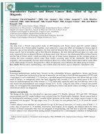
We further observed the recurrent rate in four age
groups. Figure 1 showed the rate of patients younger
younger than 35 years was the lowest than that of other
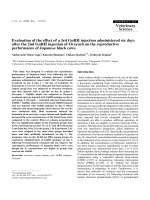
groups. In addition, the distributions of age were
different in two groups (recurrent vs. non-recurrent),
the median, 25 % percentile and 75 % percentile of age
in the recurrent group were greater than the nonrecurrent group respectively (Fig. 2). In order to clarify
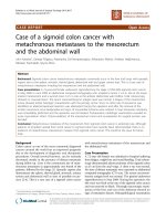
the relationship between age and recurrence, these subjects were categorized into two age groups to differentiate the recurrence rate. The relative risk of the subjects
(greater than or equal to 35 years ages) was 4.6 times of
the subject less than 35 years, the difference was statistically significant (14 % vs. 3 %, P < 0.05) (Fig. 3).
A multivariate logistic regression analysis indicated
that an age of 35 years or older was the only independent risk factor (OR 4.97, 95 % CI 1.14–21.62, P = 0.03).
Discussion
As both a diagnostic and therapeutic procedure, LEEP
provides a conservative approach to treat HSIL, particularly for women who are young or who desire to preserve their fertility. However, cervical lesions persist or
recur in a certain portion of patients after LEEP. Positive
margins have been identified as a predictive factor of
disease persistence/recurrence [4, 11, 12, 14]. As few
studies have specifically analyzed the risk of recurrent/
persistent HSIL and its related factors in patients with
HSIL who have HSIL-involved margins, we specifically
investigated the outcome and its related factors in those
patients to determine the reasonable treatment options
Table 2 The influence factors for persistence/recurrence
Persistence/recurrence of HSIL
(n = 27)
No persistence/recurrence
(n = 211)
P value
46.44 ± 11.79 (28 ~ 69)
39.42 ± 8.87 (21 ~ 67)
Two-tailed 0.000,
single-tailed 0.000a
<35
2 (3 %)
60 (97 %)
0.03c
≥35
25 (14 %)
151 (86 %)
maximum diameter
(cm, range)
1.75 ± 0.55 (1.00 ~ 3.00)
1.93 ± 0.40 (1.00 ~ 3.50)
0.043b
Thickness
(cm, range)
0.90 ± 0.22 (0.30 ~ 1.80)
0.82 ± 0.23 (0.30 ~ 1.20)
0.120b
Depth (cm, range)
1.40 ± 0.33 (0.80 ~ 2.00)
1.41 ± 0.31 (0.80 ~ 2.20)
0.917b
Single
21 (9.9 %)
191 (90.1 %)
0.092c
Multiple
6 (23.1 %)
20 (76.9 %)
Upper
7 (14.9 %)
40 (85.1 %)
Lower
5 (7.0 %)
66 (93.0 %)
Lateral
7 (10.6 %)
59 (89.4 %)
Outer
2 (13.3 %)
13 (86.7 %)
Inner
0 (0)
4 (100 %)
Stromal
0 (0)
9 (100 %)
≤LSIL
24 (11.7 %)
182 (88.3 %)
≥HSIL
3 (9.4 %)
29 (90.6 %)
Age (years)
Age
The features of LEEP specimen
Number of involved margin section
Location of involved margin section
Pathology of ECC
t test
b
Mann-Whitney test
c
Fisher exact test
a
0.681b
1.000c
Zhu et al. BMC Cancer (2015) 15:744
Page 6 of 10
100
90
80
70
%
60
Recurrence
No Recurrence
50
40
30
20
10
0
<35
40-45
35-39
Age (Years)
>45
Fig. 1 Recurrent rate of patients in four age groups. The rate of patients younger than 35 years was the lowest than that of other groups
for this population. We found that the rate of persistent/
recurrent HSIL was 11.3 % in patients with HSIL with
HSIL-involved resection margins at the initial LEEP, as
well as that age is a strong independent predictor.
In patients with HSIL, the rate of positive margins after
LEEP ranges from 5.7 to 19 % in the literature [18-23]. In
our study, the rate of HSIL margins in patients with HSIL
after LEEP was consistent with that in previously published reports (6.34 %). However, in some studies, positive
margins are defined as the histological diagnosis of CIN
along the LEEP specimen margin, regardless of the CIN
grade.
The range of the rates of persistence/recurrence in
published references varies greatly, which may be attributed to the different inclusion criteria, definitions of persistence/recurrence and follow-up times. We compared
our study with other publications that contained this
information as well as predictive factors for persistence/
recurrence (Table 3). Among 6 other studies, cases of
CIN 2/3 with or without positive margins were included
in 5 studies [7, 11, 14, 24-26], whereas cases of CIN1,2
and 3 were included in 1 study [26]. In these studies, the
authors also mentioned the rate of persistence/recurrence
in patients with positive margins, which varied from 14.2
70
60
Age
50
40
30
20
No
Yes
Recurrence HSIL
Fig. 2 The distributions of age in two groups (recurrent vs. non-recurrent). The distributions of age were different in two groups (recurrent vs.
non-recurrent), the median, 25 % percentile and 75 % percentile of age in the recurrent group were greater than the non-recurrent group respectively
Zhu et al. BMC Cancer (2015) 15:744
Page 7 of 10
100
90
80
70
%
60
No Recurrence
Recurrence
50
40
P=0.03
30
20
10
0
<35
>34
Age (Years)
Recurrence
No Recurrence
Total
<35 yrs
>34 yrs
2 (3%)
25 (14%)
60 (97%) 151 (86%)
62 (100%) 176 (100%)
Total
27
221
238
Fig. 3 The recurrent rate in two age groups (<35 vs. >34 years). The relative risk of the subjects (greater than or equal to 35 years ages) was 4.6
times of the subject less than 35 years (14 % vs. 3 %, P < 0.05)
to 60 %. However, the numbers of patients with positive
margins were small (range: 5–71). Furthermore, the definition of a “positive margin” in 4 studies was the presence
of CIN (regardless of the grade) [7, 11, 25, 26]. In addition,
the definition of recurrence in 4 studies was pathologic
findings of CIN3 while in 2 studies was biopsy confirmed
evidence of CIN of any grade (1,2,3) [7, 11]. In our study,
we found that the persistence/recurrence rate in patients
with HSIL with HSIL margins was 11.3 %, which was
lower than the rates in the other 6 studies. The possible
reason may be our inclusion criteria were critical. Therefore, it remains to be discussed whether subsequent surgeries are necessary for patients who have HSIL-involved
margins.
In addition, the authors analyzed the predictive factors
in all cases of CIN and found positive margins (3/6 studies), HPV positivity during follow-up (2/6 studies), and
tumor characteristics (e.g., depth, height or size) (2/6
studies) were related to recurrence. However, we analyzed the predictive factors in a particular group of patients, namely, those with HSIL and HSIL-involved
margins (238 patients), and found that age and diameter
of the tumor (size) were associated with persistence/recurrence, while age ≥35 years was the only independent
predictive factor.
Increasing age has been identified as a possible presurgical predictor of persistence/recurrence in some studies [8, 24, 27]. Our study indicated that in patients with
HSIL who have HSIL margins, an age of 35 years or older
was an independent risk factor for persistent/recurrent
disease. It is still unclear why older women may be more
likely to experience persistence/recurrence. One possible
reason may be altered immunity or positive selection over
time toward viruses with a higher oncogenic risk [24]. In
clinical practice, patients in this age group often desire
fertility or uterus preservation; thus, conservative management is frequently considered during follow-up. Given the
relatively high risk of persistence/recurrence, patients who
are older than 35 years require close follow-up.
The size of the lesion is also a factor that has been reported to predict recurrence because it correlates with
the completeness of the excision/ablation [28]. The maximum diameter of the LEEP specimen is a reflection of
the lesion size. In our study, the maximum diameter was
shown to be statistically significant in a univariate analysis. Patients who experienced persistence/recurrence
had LEEP specimens with smaller maximum diameters
than patients who were free of disease. However, maximum tumor diameter was not an independent risk factor in the multivariate logistic regression. The causes of
this difference may be the variation of cervix sizes.
Some studies indicated that extensive involvement of
the endocervical cone margin [9] and involvement of
multiple margins after LEEP for CIN 2/3 are strong predictors of residual disease. In our study, ECC ≥ HSIL
and, multiple involved margin sections were not associated with persistence/recurrence according to a univariate analysis. However, it was notable that the rate of
persistence/recurrence was higher in patients with multiple involved margin sections compared with patients
who had a single involved margin (23.08 % vs. 9.91 %, P =
0.092). Thus,the the significance of involvement of multiple margin sections needs to be investigated in large
sample size study.
High-risk HPV (HR-HPV) testing is also useful to
predict recurrence. Alonso et al. demonstrated that HRHPV load (N1000 RLU) prior to LEEP was significantly
associated with a higher risk of recurrence, and the most
Zhu et al. BMC Cancer (2015) 15:744
Table 3 Rates of persistence/recurrence in patients with positive margin published in literature
No. of cases with positive
margin and its definition
Definition of Persistence/
Recurrence
Predict factor of Persistence/Recurrencea
11.3 % (27/238)
Age and diameter of specimen . in univariate
analysis. Age ≥35 was an independent factor
Year
Total
case
No.
Case
inclusion
criteria
Menghan Z
Current
study
238
HSIL with 25 months
HSIL
margin
238 (the presence of HSIL in Histolopathological
the margin)
confirmed HSIL
Verguts J [24]
2006
72
CIN 2 or
worse
24 months
14 (the presence of CIN
Sun X [25]
2009
207
CIN 3
not
mentioned
10 (the presence of CIN 1 or pathologic findings of
worse)b
CIN3
50 % (5/10)b
Cytological grade, depth of conization, parity
and multi-quadrants of CIN 3 in punch biopsy
were related to increased risk for positive margin a
Leguevaque P [7]
2010
352
CIN 2,3
73 months
71 (definition not
mentioned)
25.3 % (18/71)
a positive HR-HPV test at 6 months postoperatively,
positive endocervical margins,positive
pre-treatment HPV typinga
Baloglu A [11]
2010
42
CIN 2,3
8.6 months
5 (the presence of CIN 1,2,3) pathology finding of CIN 1 60 % (3/5)
or CIN 2,3
surgical margin positivitya
Kliemann L M [14] 2012
97
CIN 2,3
160 days
39 (the presence of
CIN 2,3)
Serati M [26]
282
CIN 1,2,3
26.7 months 21 [the presence of CIN3
was close to (≤1 mm) or
involved the margin]
2012
Follow-up
period
(mean)
Rate of Persistence
/Recurrence in cases
with positive margin
Author
presence of > CIN2, biopsy 14.2 % (2/14)
confirmed
Pap smear or biopsy
confirmed evidence of
CIN of any grade (1,2,3)
Persistence of HR-HPV DNAa
pathology findings of CIN
2,3
46.3 % (18/39)
positive margin, cone height, tumor sizea
colposcopically directed
biopsy proved CIN 3
38.1 %(8/21)
the surgical margin statusa
a
The predictive factors were analyzed based on the whole case sample, not the particular subgroup of cases with positive margins
Authors observed residual CIN in 67 of 207 patients who received hysterectomy, but not including those who received other follow-ups
b
Page 8 of 10
Zhu et al. BMC Cancer (2015) 15:744
important predictor of recurrence was a positive HRHPV test at 6 months post-surgery [4]. Another study
demonstrated that a test for HPV DNA conducted
12 months post-therapy was the best predictor of recurrent or residual disease [29]. Ovestad et al. found that
CIN 2/3 lesions with non-HPV16 HR-HPV infection
may spontaneously regress [30]. Since 2006, the guidelines of the American Society for Colposcopy and Cervical Pathology (ASCCP) have included HPV testing for
the management of atypical glandular cytology and for
the follow-up after treatment for cervical intraepithelial
neoplasia. In our study, 96 % of patients were HR-HPVpositive before the initial LEEP. As we did not perform
HR-HPV testing before the second LEEP and before
hysterectomy in these two groups, we were unable to
compare HPV load before and after the initial LEEP
treatment.
The treatment options for patients with HSIL and
positive margins depend on different factors. The
ASCCP updated its consensus guidelines in 2013, as follows: “If CIN2, 3 is identified at the margins of an excisional procedure or post-procedure ECC, cytology and
ECC at 4–6 month is preferred, but repeat excision is
acceptable and hysterectomy is acceptable if re-excision
is not feasible”. The results presented herein may assist
in the personalization of the choice of procedure for patients who have HSIL-involved margins after LEEP. It
should be considered that the majority of patients with
HSIL-involved margins remain disease-free throughout
the follow-up period. Therefore, disease management
should be individualized based on desired fertility, age,
patient preference and other factors. If patients choose
to undergo additional treatment, the risk of complications must be weighed against the desire to eradicate potential residual disease.
In our study, the rates of HSIL persistence/recurrence in patients who had a subsequent LEEP and hysterectomy were 31.82 % (7/22) and 20.90 % (14/67),
respectively, while that in patients who were selected
for close follow-up (TCT or TCT combined with
colposcopic-guided biopsy) was 4.02 % (6/149). The
mean age of the patients who underwent a subsequent
hysterectomy was 47.87 ± 8.71 years, which was significantly higher than the mean age of patients in the
other three groups. Significantly more patients who were
older than 35 years experienced persistence/recurrence
compared with patients who were younger than 35 years
(14 % vs. 3 %). Thus, we considered that the presence of
HSIL margins is still an important factor associated with
recurrence but that this does not always warrant surgical
retreatment. A closer, strict surveillance after an initial
LEEP is acceptable for younger patients. For patients who
are older than 35 years, a subsequent LEEP or a carefully
decided hysterectomy is reasonable.
Page 9 of 10
Conclusion
In patients with HSIL and HSIL-involved margins after
an initial LEEP, age is a strong independent predictor of
persistence/recurrence. Follow-up with screening cytology
and/or biopsy may be considered in younger patients,
whereas a secondary LEEP/hysterectomy may be considered in older patients.
One weakness of our study is its retrospective nature
and lack of HPV tests before patients received a direct
second LEEP and hysterectomy, which, therefore, limits
its application. In addition, the follow-up period was
relatively short. The strengths of our study are the large
size of the study population and the extensive investigation of preoperative variables. We are currently in the
process of developing a prospective study to confirm or
refute our results.
Abbreviations
HSIL: High-grade squamous intraepithelial lesion; LEEP: Loop electrosurgical
excision procedure; LSIL: Low-grade squamous intraepithelial lesion;
CIN: Cervical intraepithelial neoplasia; ECC: Endocervical curettage;
HPV: Human papilloma virus; OR: Odd ratio; CI: Confidence interval;
ASCCP: American Society for Colposcopy and Cervical Pathology.
Competing interests
The authors declare that they have no competing interests.
Authors’ contributions
MZ collected the data, did the data analysis and wrote the manuscript.YH is
a statistician and analyzed the data. JB reviewed the data and revised the
manuscript. XZ and YQ reviewed the pathology reports. LS did part of the
surgeries as well as the quality control of the surgical procedure and revised
the manuscript. WF designed the study, analyzed the data and wrote the
manuscript. QW did the surgeries and designed the study. All authors have
read and approved the final manuscript.
Acknowledgements
This study was supported by a research funding from the National Natural
Science Foundation of China (Grant No. 81272877) to Dr. Long Sui.
Author details
1
Department of Gynecology, Obstetrics and Gynecology Hospital, Fudan
University, Shen Yang Road 128, Shanghai 200090, China. 2Shanghai Key
Laboratory of Female Reproductive Endocrine-Related Disease, Fudan
University, Shanghai, China. 3Department of Pathology, Obstetrics and
Gynecology Hospital, Fudan University, Shanghai, China. 4Department of
Cervical Diseases, Obstetrics and Gynecology Hospital, Fudan University,
Shanghai, China.
Received: 24 May 2015 Accepted: 9 October 2015
References
1. Mahadevan N, Horwell DH. Histological incomplete excision of CIN after
large loop excision of the transformation zone (LLETZ) merits careful followup, not retreatment. Br J Obstet Gynaecol. 1993;100:794–5.
2. Debarge VH, Collinet P, Vinatier D, Ego A, Dewilde A, Boman F, et al. Value
of human papillomavirus testing after conization by loop electrosurgical
excision for high-grade squamous intraepithelial lesions. Gynecol Oncol.
2003;90:587–92.
3. Tillmanns TD, Falkner CA, Engle DB, Wan JY, Mannel RS, Walker JL, et al.
Preoperative predictors of positive margins after loop electrosurgical
excisional procedure-Cone. Gynecol Oncol. 2006;100:379–84.
4. Alonso I, Torne A, Puig-Tintore LM, Esteve R, Quinto L, Campo E, et al.
Pre- and post-conization high-risk HPV testing predicts residual/recurrent
disease in patients treated for CIN 2–3. Gynecol Oncol. 2006;103:631–6.
Zhu et al. BMC Cancer (2015) 15:744
5.
6.
7.
8.
9.
10.
11.
12.
13.
14.
15.
16.
17.
18.
19.
20.
21.
22.
23.
24.
25.
26.
27.
Milojkovic M. Residual and recurrent lesions after conization for cervical
intraepithelial neoplasia grade 3. Int J Gynaecol Obstet. 2002;76:49–53.
Ghaem-Maghami S, Sagi S, Majeed G, Soutter WP. Incomplete excision of
cervical intralepithelial neoplasia and risk of treatment failure: a metaanalysis. Lancet Oncol. 2007;8:985–93.
Leguevaque P, Motton S, Decharme A, Soule-Tholy M, Escourrou G, Hoff J.
Predictors of recurrence in high-grade cervical lesions and a plan of
management. Eur J Surg Oncol. 2010;36:1073–9.
Lindeque BG. Management of cervical premalignant lesions. Best Pract Res
Clin Obstet Gynaecol. 2005;19:545–63.
Kietpeerakool C, Khunamornpong S, Srisomboon J, Siriaunkgul S, Suprasert
P. Cervical intraepithelial neoplasia II-III with endocervical cone margin
involvement after cervical loop conization: Is there any predictor for residual
disease? J Obstet Gynaecol Res. 2007;33:660–4.
Park JY, Kim DY, Kim JH, Kim YM, Kim YT, Nam JH. Human Papillomavirus
Test After Conization in Predicting Residual Disease in Subsequent
Hysterectomy Specimens. Obstet Gynecol. 2009;114:87–92.
Baloglu A, Uysal D, Bezircioglu I, Bicer M, Inci A. Residual and recurrent
disease rates following LEEP treatment in high-grade cervical intraepithelial
lesions. Arch Gynecol Obstet. 2010;282:69–73.
Malapati R, Chaparala S, Cejtin HE. Factors Influencing Persistence or
Recurrence of Cervical Intraepithelial Neoplasia After Loop Electrosurgical
Excision Procedure. J Low Genit Tract Dis. 2011;15:177–9.
Brockmeyer AD, Wright JD, Gao F, Powell MA. Persistent and recurrent
cervical dysplasia after loop etectrosurgical excision procedure. Am J Obstet
Gynecol. 2005;192:1379–81.
Kliemann LM, Silva M, Reinheimer M, Rivoire WA, Capp E, Dos Reis R.
Minimal cold knife conization height for high-grade cervical squamous
intraepithelial lesion treatment. Eur J Obstet Gynecol Reprod Biol.
2012;165:342–6.
Ostor AG. Natural history of cervical intraepithelial neoplasia: a critical
review. Int J Gynecol Pathol. 1993;12:186–92.
Munk AC, Kruse AJ, van Diermen B, Janssen EA, Skaland I, Gudlaugsson E,
et al. Cervical intraepithelial neoplasia grade 3 lesions can regress. APMIS.
2007;115:1409–14.
Wells M, Östör AG, Crum CP, et al. Tumours of the Uterine Cervix, Epithelial
tumours. In: Tavassoli FA , Devilee P, editors. Pathology and Genetics of
Tumours of the Breast and Female Genital Organs. Lyon: IARC; 2003. p.260.
Husseinzadeh N, Shbaro I, Wesseler T. Predictive value of cone margins and
post-coneendocervical curettage with residual disease in subsequent
hysterectomy. Gynecol Oncol. 1989;33:198–200.
Townsend DE. Cervical cone margins as a predictor for residual dysplasia in
post-cone hysterectomy specimens. Obstet Gynecol. 1994;84:898.
MohamedNoor K, Quinn MA, Tan J. Outcomes after cervical cold knife
conization with complete and incomplete excision of abnormal epithelium:
A review of 699 cases. Gynecol Oncol. 1997;67:34–8.
Kalogirou D, Antoniou G, Karakitsos P, Botsis D, Kalogirou O, Giannikos L.
Predictive factors used to justify hysterectomy after loop conization:
Increasing age and severity of disease. Eur J Gynaecol Oncol. 1997;18:113–6.
Livasy CA, Maygarden SJ, Rajaratnam CT, Novotny DB. Predictors of
recurrent dysplasia after a cervical loop electrocautery excision procedure
for CIN-3: A study of margin, endocervical gland, and quadrant
involvement. Mod Pathol. 1999;12(3):233–8.
Park JY, Lee SM, Yoo CW, Kang S, Park SY, Seo SS. Risk factors predicting
residual disease in subsequent hysterectomy following conization for
cervical intraepithelial neoplasia (CIN) III and microinvasive cervical cancer.
Gynecol Oncol. 2007;107:39–44.
Verguts J, Bronselaer B, Donders G, Arbyn M, Van Eldere J, Drijkoningen M,
et al. Prediction of recurrence after treatment for high-grade cervical
intraepithelial neoplasia: the role of human papillomavirus testing and age
at conisation. BJOG. 2006;113:1303–7.
Sun XG, Ma SQ, Zhang JX, Wu M. Predictors and clinical significance of the
positive cone margin in cervical intraepithelial neoplasia III patients. Chin
Med J (Engl). 2009;122:367–72.
Serati M, Siesto G, Carollo S, Formenti G, Riva C, Cromi A, et al. Risk factors
for cervical intraepithelial neoplasia recurrence after conization: a 10-year
study. Eur J Obstet Gynecol Reprod Biol. 2012;165:86–90.
Sarian L, Derchain S, Pitta DD, Morais SS, Rabelo-Santos SH. Factors
associated with HPV persistence after treatment for high-grade cervical
intra-epithelial neoplasia with large loop excision of the transformation
zone (LLETZ). J Clin Virol. 2004;31:270–4.
Page 10 of 10
28. Arbyn M, Simoens C, Goffin F, Noehr B, Bruinsma F. Treatment of cervical
cancer precursors: influence of age, completeness of excision and cone
depth on therapeutic failure, and on adverse obstetric outcomes. BJOG.
2011;118:1274–5.
29. Arbyn M, Ronco G, Anttila A, Meijer CJ, Poljak M, Ogilvie G, et al. Evidence
regarding human papillomavirus testing in secondary prevention of cervical
cancer. Vaccine. 2012;30 Suppl 5:F88–99.
30. Ovestad IT, Gudlaugsson E, Skaland I, Malpica A, Munk AC, Janssen EA, et al.
The impact of epithelial biomarkers, local immune response and human
papillomavirus genotype in the regression of cervical intraepithelial
neoplasia grades 2–3. J Clin Pathol. 2011;64:303–7.
Submit your next manuscript to BioMed Central
and take full advantage of:
• Convenient online submission
• Thorough peer review
• No space constraints or color figure charges
• Immediate publication on acceptance
• Inclusion in PubMed, CAS, Scopus and Google Scholar
• Research which is freely available for redistribution
Submit your manuscript at
www.biomedcentral.com/submit