The existence of Th22, pure Th17 and Th1 cells in CIN and cervical cancer along with their frequency variation in different stages of cervical cancer
Bạn đang xem bản rút gọn của tài liệu. Xem và tải ngay bản đầy đủ của tài liệu tại đây (1.66 MB, 11 trang )
Zhang et al. BMC Cancer (2015) 15:717
DOI 10.1186/s12885-015-1767-y
RESEARCH ARTICLE
Open Access
The existence of Th22, pure Th17 and Th1
cells in CIN and Cervical Cancer along with
their frequency variation in different stages
of cervical cancer
Wenjing Zhang1,2†, Xinli Tian1,3,4†, Fidia Mumtahana1, Jun Jiao1,3, Teng Zhang1,3, Kimiko Della Croce5, Daoxin Ma3,
Beihua Kong1 and Baoxia Cui1*
Abstract
Background: Recently, it is found that T-helper (Th) 22 cells are involved in different types of autoimmune and tumor
diseases. But, till now, no study has been carried out to understand the involvement of these cells in cervical cancer (CC).
Methods: Flow cytometry was used to determine the expression of interferon gamma (IFN-γ), Interleukin-22 (IL-22), IL-17
in the peripheral blood of healthy controls (HC), CIN and cervical cancer patients. From peripheral blood mononuclear
cells (PBMCs), mRNA expression levels of Aryl hydrocarbon receptor (AHR), RAR-related orphan receptor C (RORC), TNF-α
and IL-6 were respectively determined. Using the method of ELISA, plasma concentrations of IL-22, IL-17 and TNF-α were
examined.
Results: Th22 and Th17 cells were elevated in CC and CIN patients. Th1 cells and the plasma concentrations of IL-22 in
CC patients were significantly increased compared with HC. In CC patients, an increased prevalence of Th22 cells was
associated with lymph node metastases. There was a positive correlation between Th22 and Th17 cells, but an
approximately negative correlation between Th22 and Th1 cells in CC patients. The mRNA expression of RORC,
TNF-α and IL-6 was significantly high in CC patients.
Conclusions: Our results indicate that there is a higher circulatory frequency of Th22, Th17 and Th1 cells in CC which
may conjointly participate in the pathogenesis and growth of CC.
Keywords: Cervical cancer, Th17, Th22, Th1, IL-22
Background
Cervical cancer (CC) is one of the leading gynecological
cancers in developing countries. The main etiology behind
this occurrence is the persistent infection of high-risk human papillomavirus (HPV) [1–3]. Even if the incidence of
HPV is high, with the help of cell mediated immunity, it
can be cleared spontaneously [4–6]. A very few cases may
develop into advanced CC from precancerous lesions
which may indicate a substantial role of immune regulation
* Correspondence:
†
Equal contributors
1
Department of Obstetrics and Gynecology, Qilu Hospital, Shandong
University, Jinan 250012, P.R. China
Full list of author information is available at the end of the article
in the controlling of HPV associated lesions and cancer
progression [7].
We know that T helper (Th) cells, one subgroup of
lymphocytes, have an essential role in the immune system. Recently it was demonstrated that Th cells such as
Th1, Th2, Th17, Treg cells, participate in the pathogenesis and progression of different solid tumors [7–10]. A
newly discovered T cell subset - Th22 cells, which were
detected in autoimmune and inflammatory diseases,
have the ability to secrete IL-22 and TNF-α, but do not
express IL-4 (Th2 marker), IL-17 (Th17 marker) or
IFN-γ (Th1 marker). In the human body (in the presence of IL-6 or/and TNF-α) the naive CD4+cells differentiate into Th22 cells with the aid of plasmacytoid
dendritic cells, AHR and RORC [11, 12]. It is known
© 2015 Zhang et al. Open Access This article is distributed under the terms of the Creative Commons Attribution 4.0
International License ( which permits unrestricted use, distribution, and
reproduction in any medium, provided you give appropriate credit to the original author(s) and the source, provide a link to
the Creative Commons license, and indicate if changes were made. The Creative Commons Public Domain Dedication waiver
( applies to the data made available in this article, unless otherwise stated.
Zhang et al. BMC Cancer (2015) 15:717
that Th22 is a distinct subset with novel characteristics
compared to other Th cells (Th17, Th2 and Th1 cells).
It is demonstrated that Th22 cells play an important
role in the pathogenesis of inflammatory diseases and
autoimmunity diseases such as psoriasis, Graves’ disease
and rheumatoid arthritis. [13–15]. However, the nature
of Th22 cells are not properly umderstood in human
cancer. Recently some studies concluded that Th22 cells
contribute to the the progression of hepatocellular and
gastric carcinoma which indicates that Th22 cells may
be involved in the development of tumors. [16–18].
IL-22 which belongs to the IL-10 cytokine family is
mainly an effector cytokine of Th22 cells. IL-22 maintains its function by binding to a heterodimeric transmembrane receptor complex consisting of IL-10R2 and
IL-22R1, and activates Janus kinase (signal transducers
and activators of transcription signaling pathways which
acts with a dual role in inflammatory and autoimmune
diseases [19–21]). It is seen that IL-22 leads to tumor
proliferation, apoptosis suppression and metastasis promotion by activation of STAT3 in colon cancer [22]. Reversely, IL-22 exerts a protective role in mucosal wound
healing acceleration by inducing STAT3-dependent expression in ulcerative colitis [23, 24].
To the best of our knowledge, no previous study has
shown data that considers Th22 cells and their association
with Th17 or Th1 in cervical cancer. To examine the possible status of these cells in the pathophysiology of CC, we
measured the frequency of peripheral Th22, Th17, Th1,
mRNA expression levels of RORC, AHR, IL-6, TNF-α in
PBMCs along with plasma concentrations of IL-22, IL-17
and TNF-α in PB of CC, CIN patients and HC for assessing their relevance.
Page 2 of 11
Table 1 Clinical characteristics of CC patients
Characteristics
Category
N = 61(%)
IA
10 (16)
IB
37 (61)
IIA
9 (15)
IIB
5 (8)
SCC
54(88)
FIGO stage
Histology type
ADC
4(7)
Unknown
3(5)
Well
9(15)
Moderate
11(18)
Poor
33(54)
Unknown
8(13)
Positive
11(18)
Negative
48(79)
Unknown
2(3)
Tumor differentiation
Lymph node metastases
Tumor size(cm)
<4
43(70)
≥4
18(30)
Yes
12(20)
Vasoinvasion
No
44(72)
Unknown
5(8)
Abbreviation: FIGO, International Federation of Gynecologists and
Obstetricians; SCC,
squamous cell carcinoma; ADC, adenocarcinoma.
Methods
Patients and controls
Six-one pathologically confirmed CC patients (age 24 –
60 years, median 48 years) and 38 CIN patients (age 27–61
years, median 42 years) were enrolled in this study. All the
patients of CIN group have biopsy results of CINIII. Before
the study none of the patients had received anticancer
treatment.
Thirty-two healthy women with normal results of pap
smear (TCT) and HPV (HC2) tests served as controls
(age 22–47 years, median 27 years).They are from our
Gynecologic Clinic and Regular Physical Examination
Center.
The participants with simultaneous active or chronic infection, autoimmune disease, diabetes, or a history of
other malignant tumors or connective tissue diseases were
excluded. The characteristics of the patients are given in
Table 1. The clinical stage of CC patients was based on
FIGO 2009 criteria. Informed written consent was obtained from each participant. Medical Ethical Committee
of Qilu Hospital, Shandong University, China provided the
ethical approval for the study.
Flow Cytometric Analysis
Intracellular cytokines were evaluated by flow cytometry to identify the cytokine-producing cells. Briefly,
heparinized peripheral whole blood (200 μl) with an
equal volume of Roswell Park Memorial Institute
(RPMI) 1640 medium (Sigma Chemical, St Louis, MO,
USA) was incubated for 4 h at 37 °C and 5 % CO2 in the
presence of 25 ng/ml of phorbolmyristate acetate (PMA),
1 μg/ml of ionomycin and 1.7 μg/ml of Monensin (all from
Alexis Biochemicals, San Diego, CA, USA). After incubation, the cells were stained with anti-CD4-PE-Cy5 monoclonal antibodies at room temperature (RT) in the dark for
15 minutes to delimitate CD4+ T cells. Then, to fix the
cells, 100 μl Reagent A (FIX &PERM Kit, MultiSciences
Biotech Co., Ltd.) was added to each sample at RT in the
dark for 15 min. After washing the cells with 3 ml PBS,
Zhang et al. BMC Cancer (2015) 15:717
100 μl of Reagent B (FIX &PERM Kit, MultiSciences Biotech Co., Ltd.) and the recommended dose of anti-IL17APE and anti-IL22-APC and anti-IFNγ-FITC monoclonal
antibody were added to each sample after fixation and
permeabilization. Samples were then incubated at RT in
the dark for 15 min to stain IL-17, IL-22 and IFN-γ. Isotype controls were used to correct compensation and confirm antibody specificity. After washing the cells with 3 ml
PBS, we added 300 μl PBS to re-suspend the cells for cytometric analysis. Stained cells were analyzed by flow cytometric analysis using a FACS cytometer equipped with
Cell Quest software (BD Bioscience Pharmingen). All
antibodies mentioned above were from eBioscience
(San Diego, CA, USA). Th17, Th22 and Th1 cells are
defined as CD4+IFNγˉIL17+IL22ˉ, CD4+IFNγˉIL17ˉIL22+ and CD4+IFNγ+ cells respectively.
Quantitative real-time PCR analysis
Trizol (Invitrogen, America) was used for isolation of Total
RNA from PBMCs. For reverse transcription reaction the
Prime Script RT reagent kit (Perfect Real Time; Takara)
was used according to the instruction of the manufacturer.
Reverse transcription reaction was done at 37 °C for 15 minutes, followed by 85 °C for 5 seconds. Real-time PCR was
done by Applied Biosystems 7500 Real-Time PCR System
(Applied Biosystems, Foster City, CA, USA). The primers
are shown as follows: AHR forward: CAA ATC CTT CCA
AGC GGC ATA; reverse: CGC TGA GCC TAA GAA
CTG AAA G; RORC forward: TTT TCC GAG GAT GAG
ATT GC; reverse: CTT TCC ACA TGC TGG CTA CA;
TNF-α forward: CGA GTG ACA AGC CTG TAG C,
reverse: GGT GTG GGT GAG GAG CAC AT; GAPDH
forward: GCT CTC TGC TCC TCC TGT TC, reverse:
GTT GAC TCC GAC CTT CAC CT; IL-6 forward: TTC
TCC ACA AGC GCC TTC GGT CCA, reverse: AGG
GCT GAG ATG CCG TCG AGG ATG TA. All experiments were conducted in triplicate. For calculation of
amplification efficiency of the PCR products Applied
Biosystems System software was used. The results were
signified relative to the number of GAPDH transcripts
used as a reference control.
IL-22, IL-17 and TNF-α Enzyme-linked Immunosorbent
Assay (ELISA)
Heparin-anticoagulant vacutainer tubes were used for
collection of PB. For cytokines determination plasma was
attained from all the subjects by centrifugation and stored
at a temperature of −80 °C. A quantitative sandwich enzyme immunoassay technique was used for plasma level
determination of IL-22, IL-17 and TNF-α according to
the manufacturer’s instructions (eBioscience, San Diego,
CA, USA).
Page 3 of 11
Statistical analysis
Mean ± SD or median (range) were used for expression
of values. Distribution of the data was obtained from
Kolmogorov-Smirnov test (K-S test). ANOVA and
Newman-Kuels multiple comparison tests were used for
the assessment of normal distribution data. KruskalWallis test (H test) and Nemenyi tests were used for
unusual data. Assessment of Correlation analysis was
obtained from Pearson correlation. A p value less than
0.5 was considered statistically significant. All tests were
performed by SPSS 17.0 software.
Results
Elevated Th22 and Th17 cells in PB of CIN and CC
patients
The percentage of Th22 cells (CD4+IFNγˉIL17ˉIL22+ T
cells, pure Th22 cells) and Th17 cells (CD4+IFNγˉIL17+
IL22ˉ T cells, pure Th17 cells) of CIN (Th22: 1.27 ±
0.56 %, p = 0.001; Th17: 3.10 ± 1.40 %, p < 0.001) and CC
patients (Th22: 1.75 % ± 0.704 %, p < 0.001; Th17: 3.35
± 1.34, p < 0.001) significantly increased compared with
HC (Th22: 0.77 % ± 0.36 %; Th17: 1.78 ± 0.80 % ). Besides, significant difference was also found in Th22 cells
between CIN and CC patients (p < 0.001), but none in
Th17 cells (Fig. 2a and b).
Elevated Th1 cells in PB of CC patients
Significantly elevated frequencies of Th1 cells were
found in CC (7.95 % ± 3.95 %) compared with HC
(4.98 % ± 2.92 %, p < 0.001) and CIN patients (6.23 % ±
2.52 %, p = 0.015). However, no significant difference
was found between HC and CIN patients (p > 0.05)
(Fig. 2c).
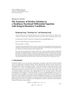
A typical dot plot of the percentage of Th1 cells, Th22
cells and Th17 cells in representative patients and HC is
shown in Fig. 1.
Comparisons of Th17/Th22 ratio
Regarding the ratio of Th17/Th22, we detected a significant decrease in CC patients (2.12 ± 1.02, p = 0.007)
compared with CIN patients (3.09 ± 2.60) (Fig. 2d).
Correlation Analysis among Th22, Th17 and Th1 Cells in
CC and CIN patients
A positive correlation was discovered among Th22 cells
and Th17 cells in CC patients (r = 0.546, p < 0.0001,
Pearson correlation analysis), but none in CIN patients
(r = 0.163, p = 0.328). In CC patients, an approximately
negative correlation was seen among Th22 and Th1 cells
(r = − 0.235, p = 0.068, Pearson correlation analysis), but
none in CIN patients (r = − 0.144, p = 0.388) (Fig. 3).
Zhang et al. BMC Cancer (2015) 15:717
Page 4 of 11
Fig. 1 Circulating percentages of Th17, Th22 and Th1 cells in representative HC, CIN and CC patients. a. Lymphocytes were gated in R1 by flow
cytometry. b, c, d The percentages of circulating Th1(CD4+ IFNγ+ T cells) cells in HC and CIN and CC patients. CD4+IFNγˉ T cells were gated in R2.
e, f, g The proportions of pure Th17 (CD4+IFNγˉIL17+IL22ˉ T cells) and pure Th22 cells (CD4+IFNγ−IL17ˉIL22+ T cells) in representative controls, CIN
and CC patients
Zhang et al. BMC Cancer (2015) 15:717
Page 5 of 11
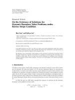
Fig. 2 Results of circulating Th subsets in HC, CIN and CC patients. a The percentages of circulating Th22 (CD4+IFNγ−IL17ˉIL22+ T cells) cells. Significantly
higher percentage of Th22 cells was present in CC patients (1.75 ± 0.704 %) in comparison with CIN patients (1.27 ± 0.56 %, p < 0.001) and HC
(0.77 ± 0.36 %, p < 0.001); again increased percentage of Th22 cells noticed in CIN patients than HC (p = 0.001). b The percentages of circulating pure
Th17 (CD4+IFNγˉIL17+IL22ˉ T cells) cells. There was a significantly high percentage of pure Th17 cells in CIN patients (3.10 ± 1.40 %, p < 0.001) or CC
patients (3.35 ± 1.34 %, p < 0.001) than HC (1.78 ± 0.80 %). c The percentages of circulating Th1 (CD4+IFNγ+ T cells) cells. Significantly elevated frequencies
of Th1 cells were found in CC (7.95 % ± 3.95 %) compared with HC (4.98 % ± 2.92 %, p < 0.001) and CIN patients (6.23 % ± 2.52 %, p < 0.001). However, no
significant difference was found between HC and CIN patients. d Correlation of Th17/Th22 ratio in HC, CIN and CC patients. Significant difference was
found between CIN (3.09 ± 2.60) and CC patients (2.12 ± 1.02, p = 0.007). Bars symbolized SD. * p < 0.05, ** p < 0.01, *** p < 0.001. NS no significance
mRNA expression levers of AHR, RORC, TNF-α and IL-6 in
CC, CIN patients and controls
There was an increased trend of AHR in CC patients
(0.274 ± 0.160) and CIN patients (0.299 ± 0.16) compared
with HC (0.257 ± 0.103), though both values of P were
more than 0.05 (Fig. 4a).
In comparison, CC patients (0.305 ± 0.188, p = 0.002)
or CIN patients (0.256 ± 0.188, p = 0.036) exhibited increased level of the RORC mRNA expression than normal
controls (0.128 ± 0.099) but the CIN patients and CC patients had no important difference in between (p > 0.05)
(Fig. 4b). In addition, CC patients (r = 0.60, p < 0.01,
Pearson correlation) and CIN patients (r = 0.521, p = 0.015,
Pearson correlation) had a positive correlation between
RORC and Th17 cells. Furthermore, CC patients (r = 0.612,
p < 0.01, Pearson correlation) and CIN patients (r = 0.509,
p = 0.018, Pearson correlation) showed a positive correlation between RORC and Th22 cells (Fig. 5).
The CC patients (median, 0.369; range, 0.016 – 1.59)
showed TNF-α mRNA expression significantly high in
comparison with HC (0.264 ± 0.28, p = 0.043) and CIN
patients (median, 0.193; range, 0.009 – 4.27, p = 0.015)
but CIN patients and HC did not show any significant
high level of this expression (Fig. 4c).
The HC (median, 0.029; range, 0.002 – 0.139) had lower
IL-6 mRNA expression in PBMCs than the CC patients
(median, 0.101; range, 0.006 – 0.763, p = 0.001) and CIN
patients (median, 0.085; range, 0.003 – 1.74, p = 0.019) but
CIN patients and CC patients had no significant difference
in between (p > 0.05) (Fig. 4).
Correlation on the frequencies of Th17 and Th22 cells
with clinical characters in CC patients
CC patients with lymph node metastasis exhibited profoundly increased frequency of Th22 cells (2.20 ± 0.85 %,
n = 11) compared to CC patients without lymph node
metastases (1.68 ± 0.64 %, p = 0.026, n = 48) (Fig. 6). No
significant diversity was detected among Th22, Th17 and
Th1 cells frequency and other prognostic factors including
clinical stage, tumor size and vasoinvasion in CC patients
(p > 0.05).
Zhang et al. BMC Cancer (2015) 15:717
Page 6 of 11
Fig. 3 Correlations between Th subsets in CIN and CC patients. a The correlation between the levels of Th17 and Th22 cells in patients with CIN
(r = 0.163, p = 0.328); b The correlation between the levels of Th22 and Th1 cells in patients with CIN (r = − 0.144, p = 0.388); c There was a positive
correlation between Th22 cells and Th17 cells in CC patients (r = 0.546, p < 0.0001) d There was an approximately negative correlation between Th22
cells and Th1 cells in CC patients (r = − 0.235, p = 0.068).
Increased IL-22 concentrations in plasma of CC patients
The CIN or CC patients and HC all showed plasma IL-22,
IL17 and TNF-α. Significantly higher levels of IL-22 were
revealed in CC patients (median 37.46; range 24.84 –
120.06 pg/ml, n = 31, p = 0.039) than those in HC (median 26.8; range 11.3-42.7 pg/ml, n = 19) (Fig. 7a). No
remarkable diversities were found among CIN patients
(CIN: median 31.17; range 20.93 - 82.68 pg/ml, n = 22,
p > 0.05) and CC patients or CIN patients and HC.
However, concentration of plasma IL-17 and TNF-α
were found similar in HC, CIN and CC patients (p > 0.05)
(Fig. 7b and c).
Discussion
Persistent infection with HPV is the main cause of CC and
CIN [25, 26]. That CIN and CC arise more frequently in
immunosuppressive women indicates that elimination of
HPV is related to immunity function. In the evolution of
these diseases, local or systemic immune mechanisms abnormalities may be involved [27, 28]. A vast and dynamic
crosstalk among immune cells, along with cytokines turmoil has been regarded as a crucial element of cancer
pathophysiology [29]. In our current study, we focused on
immune cells, mainly three subtypes of T helper cellsTh1, Th17 and Th22 cells and their probable role in CC
and CIN.
Interferon (IFN)-γ causes activation of immune cells in
the tumor microenvironment. It is known that Th1 cells,
the main source of IFN-γ, have a powerful anti-tumor
function. To enhance the function of antigen presenting
cells, tumor antigen specific CD4+Th1 cells can travel to
the tumor site and secrete inflammatory cytokines and
modulate the microenvironment [30]. It was observed that
for cancer inhibition and better outcomes, Th1 adaptive
Zhang et al. BMC Cancer (2015) 15:717
Page 7 of 11
Fig. 4 The mRNA expression of AHR, RORC, TNF-α and IL-6 in CIN and CC patients and HC. a AHR mRNA expression level between CIN patients, CC
patients and HC was comparable (p > 0.05); b A remarkably high expression of the RORC mRNA was seen in CC patients (0.305 ± 0.188, p = 0.002) or
CIN patients (0.256 ± 0.188, p = 0.036) compared to HC; c A significantly high expression of TNF-α was observed in CC patients (median, 0.369; range,
0.016 - 1.59) compared to CIN patients (median, 0.193; range, 0.009 - 4.27, p = 0.015) or HC (0.264 ± 0.28, p = 0.043); d The expression of IL-6 is significantly
increased in CC patients (median, 0.101; range, 0.006 - 0.763, p = 0.001) or CIN patients (median, 0.085; range, 0.003 - 1.74, p = 0.019) when compared with
HC (median, 0.029; range, 0.002 - 0.139). Bars symbolize SD. * p < 0.05, ** p < 0.01 NS no significance
immunity is essential [31]. In our study, we demonstrated
a significant elevated frequency of Th1 cells in CC patients, compared to CIN patients and HC, which is consistent with other previous studies of the involvement of
Th1 cells in tumors.
It was noticed that another two inflammatory cell subgroups, Th17 and Th22 cells are involved in viral infection
and mucosal immunity [32, 33, 34]. In our previous study,
we saw that there was a significant increase of Th17 cells
(CD4+IL17+ cells) in CIN and CC patients [8, 11]. In order
to exclude multiple positive cells Th17 cells are defined as
CD4+IFNγ−IL17+IL22− cells, which also were called as
“pure Th17 cells”. Th22 cells are now defined as CD4+
IL17ˉIL22+IFNγ− cells, which is an independent subset of
T helper cells from Th1 and Th17 cells [35–37]. In the
current study, we evaluate the frequencies of pure Th17
and Th22 cells to confirm the probable role of these two
famous types of T helper cells in PB of CIN and CC by
flow cytometry. As expected, increased frequencies of
Th17 and Th22 cells were found in both CIN and CC
compared to HC. Moreover, the increased change of Th22
cells in CC was much higher than that of CIN. It suggested that as cervical precancerous lesion occurs, Th22
cells might gradually elevate from CIN to CC. However,
no significant difference of Th17 cells was found between
CIN and CC. But the data indeed shows that there are frequencies of Th17 and Th22 cells changed in the tumorigenesis of both CIN and CC which indicate these two
types of cells may paticipate in tumor immunity.
IL-22 is known to have a relationship with virusinfection reactions and whose receptor is confined to nonhematopoietic cells (mainly epithelial cells). Previously it
was considered that IL-22 is a cytokine of Th17 cells. Now
it is considered as the characteristic product of Th22 cells.
Our study also revealed elevated levels of plasma IL-22 in
CC patients. Additionally, expression of a series of molecules, which are responsible for cellular differentiation
and survival was triggered by IL-22 [38, 39]. In our
study, a raised level of plasma IL-22 was found, which
indicated that Th22 cells, the main T helper cells which
product IL-22, may be involved in the process of CC.
However, plasma IL-17 did not show a significant
change. This might be due to the fact that concentration
of IL-17 was too low to present the change, as it showed
low levels in both of CC and HC. In addition, there was
a positive correlation between the frequencies of Th17
and Th22 cells in CC patients, suggesting that differentiation of Th22 cells may be linked to Th17 cells or even
Zhang et al. BMC Cancer (2015) 15:717
Page 8 of 11
Fig. 5 Correlations between RORC and Th subsets in CIN and CC patients. a, b RORC had the positive correlation with Th17 cells and Th22 cells
in CIN patients (Th17 cells, r = 0.521, p = 0.015, n = 21; Th22 cells, r = 0.509, p = 0.018, n = 21); c, d RORC had the positive correlation with Th17 cells and
Th22 cells in CC patients (Th17 cells, r = 0.600, p < 0.01, n = 31; Th22 cells, r = 0.612, p < 0.01, n = 31)
Fig. 6 The Th22, Th17 or Th1 cells frequency in positive or negative
lymph node metastases. Increased frequency (p = 0.026) of Th22 was
observed in CC patients with lymph node metastases (2.20 ± 0.85 %,
n = 11) comparing to CC patients without lymph node metastases
(1.68 ± 0.64 %, n = 48). *p < 0.05, NS no significance
Th22 cells might partly derive from Th17 cells. This
derivation may partly explain the type of IL22+Th17
cells. However, no correlation was found in CIN III or
HC. One reason for this is that the frequencies level of
Th17 and Th22 cells are very low, hence the difference
between detected results and real conditions multiplied
and distorted the statistic results. Another reason is
that, in a normal situation, Th17 and Th22 cells are derived from a different origin and induced by different
stimuli. However, when cancer appears, inflammatory
cells show a partly inter-related differentiation, which
also causes elevated frequency of IL22+Th17 cells during the process.
It was seen that RORC is the key transcription factor
directing Th17 lineage and modulates the polarization of
Th22 cells [12, 40]. In our study we noticed a notably elevated expression of RORC in CIN and CC patients.
Zhang et al. BMC Cancer (2015) 15:717
Page 9 of 11
Fig. 7 Results of plasma cytokines in CIN, CC patients and HC. a A significantly elevated expression of IL-22 was seen among CC patients (median 37.46;
range 24.84 - 120.06 pg/ml, p = 0.039) and HC (median 26.8;range 11.3-42.7 pg/ml). b No significant difference was found on concentration of IL-17 in
control, CIN and CC patients. c No significant elevation was found on concentration of TNF-α in control, CIN and CC patients. *p < 0.05 NS
no significance
Also, the expression of RORC is positively correlated with
both Th22 and Th17 cells. It is assumed that in CIN and
CC patients the differentiation of Th22 and Th17 cells is
mainly regulated by RORC. We previously found that IL6, which promoted differentiation of Th22 cells, is highly
expressed in CIN and CC patients [11, 12, 19]. Elevated
IL-6 mRNA expression was found in CIN and CC patients
compared to HC. The data showed that, in CC and CIN
patients, immune environment may be more suitable for
polarization of Th22 cells.
However, no significance was found in AHR expression. Although AHR is the most important transcription
factor of Th22 cells, AHR pathway is not unique. It is
demonstrated that TGF-β could inhibit IL-22 secreting
of Th17 cells by AHR-independent pathways. In our
study of CIN and CC, no significant change was found.
The explanation for increase of Th22 cells may not be
caused by AHR (transcription level), but others pathways, such like stimulation and transformation.
Referring to clinic factors, in CC patients, lymph node
metastases were found to correlate with aggregation of
Th22 cells. Again, a positive association between Th22
cells and Th17 cells was also observed. Consequently, it
is imaginable that co-increased levels of Th22 and Th17
cells along with pro-inflammatory cytokines may play a
synergistic role in the progression of CC. Nevertheless,
there was an approximately negative correlation between
Th1 cells and Th22 cells in CC patients. This argues that
the beneficial Th1 cells gradually declined while more
Th22 was produced toward disease progression. However, the interaction among these three different cells demands further investigation.
Conclusion
It is seen that patients with CC possess a high frequency
of circulating Th22 cells, Th17 cells and Th1 cells. The
higher prevalence of Th22 cells was found in patients with
advanced CC, arguing an important role for this T-cell
subtype in the growth and acceleration of CC. For a better
understanding of this development (i.e., regulation and
function of these cells in CC) more extensive experiments
are needed which may lead to the evolution of promising
therapeutic strategy for CC patients.
Abbreviations
AHR: Aryl hydrocarbon receptor; CC: Cervical cancer; CIN: Cervical
intraepithelial neoplasia; HC: Healthy control; HPV: Human papilloma virus;
Zhang et al. BMC Cancer (2015) 15:717
PB: Peripheral blood; PBMCs: Peripheral blood mononuclear cells;
PCR: polymerase chain reaction; RORC: RAR-related orphan receptor C;
SD: Standard deviation; STAT: signal transducer and activator of transcription;
Th: T helper cell; TNF: Tumor necrosis factor; Treg: The regulatory T cells.
Competing interests
The authors declare no conflicts of interest.
Authors’ contribution
Conceived and designed the experiments: BXC, WJZ and XLT. Collected
samples: WJZ, XLT and JJ. Performed the experiments: WJZ and XLT.
Analyzed the data: XLT, JJ and TZ; Contributed reagents/materials/analysis
tools: DXM and BHK. Wrote the paper: WJZ and XLT. Edited the paper: FM
and KDC. All authors read and approved the final manuscript.
Acknowledgements
This study was supported by the National Natural Science Foundation of
China (Nos. 81172486, 81470319 and 81072122).
Author details
1
Department of Obstetrics and Gynecology, Qilu Hospital, Shandong
University, Jinan 250012, P.R. China. 2Key Laboratory of Gynecologic
Oncology, Qilu Hospital, Shandong University, Jinan 250012, P.R. China.
3
Hematology Oncology Center, Qilu Hospital, Shandong University, Jinan
250012, P.R. China. 4Department of Obstetrics and Gynecology, Weifang
Maternal and Child Health Hospital, Weifang 261011, P.R. China. 5Department
of Molecular & Cellular Biology, University of Arizona, Tucson, AZ, USA.
Received: 23 December 2014 Accepted: 10 October 2015
References
1. Parkin DM, Bray F, Ferlay J, Pisani P. Global cancer statistics 2002. CA Cancer
J Clin. 2005;55:74–108.
2. Das BC, Sharma JK, Gopalakrishna V, Luthra UK. Analysis by polymerase
chain reaction of the physical state of human papillomavirus type16 DNA in
cervical preneoplastic and neoplastic lesions. J Gen Virol. 1992;73:2327–36.
3. Bosh FX, Lorincz A, Munoz N, Meijer CJ, Shah KV. The causal relation
between human papillomavirus and cervical cancer. J Clin Pathol.
2002;55:244–65.
4. Sheu BC, Chang WC, Lin HH, Chow SN, Huang SC. Immune concept of
human papillomaviruses and related antigens in local cancer milieu of
human cervical neoplasia. J Obstet Gynaecol Res. 2007;33:103–13.
5. Hebner CM, Laimins LA. Humanpapilloma viruses: basic mechanisms of
pathogenesis and oncogenicity. Rev Med Virol. 2006;16:83–97.
6. Nguyen HH, Broker TR, Chow LT, Alvarez RD, Vu HL, Andrasi J, et al. Immune
responses to human papillomavirus in genital tract of women with cervical
cancer. Gynecol Oncol. 2005;96:452–61.
7. Cheng WF, Lee CN, Su YN, Chang MC, Hsiao WC, Chen CA, et al. Induction
of human papillomavirus type 16-specific immunologic responses in a
normal and an human papillomavirus-infected populations. Immunology.
2005;115:136–49.
8. Zhang Y, Ma D, Zhang Y, Tian Y, Wang X, Qiao Y, et al. The imbalance of
Th17/Treg in patients with uterine cervical cancer. Clin Chim Acta.
2011;412:894–900.
9. Gu-Trantien C, Loi S, Garaud S, Equeter C, Libin M, Wind AD. CD4+ follicular
helper T cell infiltration predicts breast cancer survival. J Clin Invest.
2013;123:2873–92.
10. Goedeqebuure PS, Eberlein TJ. The role of CD4+ tumor-infiltrating
lymphocytes in human solid tumors. Immunol Res. 1995;14:119–31.
11. Hou F, Li Z, Ma D, Zhang W, Zhang Y, Zhang T, et al. Distribution of Th17
cells and Foxp3-expressing T cells in tumor-infiltrating lymphocytes in
patients with uterine cervical cancer. Clin Chim Acta. 2012;413:1848–54.
12. Duhen T, Geiger R, Jarrossay D, Lanzavecchia A, Sallusto F. Production of
interleukin 22 but not interleukin 17 by a subset of human skin-homing
memory T cells. Nat Immunol. 2009;10:857–63.
13. Trifari S, Kaplan CD, Tran EH, Crellin NK, Spits H. Identification of a human
helper T cell population that has abundant production of interleukin 22
and is distinct from T(H)-17, T(H)1 and T(H)2 cells. Nat Immunol.
2009;10:864–71.
Page 10 of 11
14. Kagami S, Rizzo HL, Lee JJ, Koguchi Y, Blauvelt A. Circulating Th17, Th22,
and Th1 cells are increased in psoriasis. J Invest Dermatol. 2010;130:1373–83.
15. Peng D, Xu B, Wang Y, Guo H, Jiang Y. A high frequency of circulating th22
and th17 cells in patients with new onset graves' disease. PLoS One.
2013;8:e68446.
16. Zhang L, Li YG, Li YH, Qi L, Liu XG, Yuan CZ, et al. Increased frequencies of
Th22 cells as well as Th17 cells in the peripheral blood of patients with
ankylosing spondylitis and rheumatoid arthritis. PLoS One. 2012;7:e31000.
17. Qin S, Ma S, Huang X, Lu D, Zhou Y, Jiang H. Th22 cells are associated with
hepatocellular carcinoma development and progression. Chin J Cancer Res.
2014;26:135–41.
18. Liu T, Peng L, Yu P, Zhao Y, Shi Y, Mao X, et al. Increased Circulating Th22 cells
and Th17 cells are Associated with Tumor Progression and Patients Survival in
Human Gastric Cancer. J Clin Immunol. 2012;32:1332–9.
19. Zhuang Y, Peng LS, Zhao YL, Shi Y, Mao XH, Guo G, et al. Increased
intratumoral IL-22-producing CD4+ T cells and Th22 cells correlate with
gastric cancer progression and predict poor patient survival. Cancer
Immunol Immunother. 2012;61:1965–75.
20. Wolk K, Witte E, Witte K, Warszawska K, Sabat R. Biology of interleukin-22.
Semin Immunopathol. 2010;32:17–31.
21. Ouyang W, Rutz S, Crellin NK, Valdez PA, Hymowitz SG. Regulation and
functions of the IL-10 family of cytokines in inflammation and disease. Annu
Rev Immunol. 2011;29:71–109.
22. Sonnenberq GF, Fouser LA, Artis D. Border patrol: regulation of immunity,
inflammation and tissue homeostasis at barrier surfaces of IL-22. Nat
Immunol. 2011;12:383–90.
23. Jiang R, Wang H, Deng L, Hou J, Shi R, Yao M, et al. IL-22 is related to
development of human colon cancer by activation of STAT3. BMC Cancer.
2013;13:59.
24. Pickert G, Neufert C, Leppkes M. STAT3 links IL-22 signaling in intestinal
epithelia cells of mucosal wound healing. J Exp Med. 2009;206:1465–72.
25. Wilczynski SP, Bergen S, Walker J, Liao SY, Pearlman LF. Human
papillomaviruses and cervical cancer: analysis of histopathologic features
associated with different viral types. Hum Pathol. 1998;19:697–704.
26. Bosch FX, Manos MM, Muñoz N, Sherman M, Jansen AM, Peto J, et al.
Prevalence of human papillomavirus in cervical cancer: a worldwide
perspective. International biological study on cervical cancer (IBSCC) Study
Group. J Natl Cancer Inst. 1995;87:796–802.
27. Kosmaczewska A, Bocko D, Ciszak L, Wlodarska-Polinska I, Kornafel J,
Szteblich A, et al. Desregulated expression of both costimulatory CD28 and
inhibitory CTLA4 molecules in PB T cells of advanced cervical cancer
patients suggests systemic immunosuppression related to disease
progression. Pathol Oncol Res. 2012;18:479–89.
28. Huang Y, Zhang J, Cui ZM, Zhao J, Zheng Y. Expression of the CXCL12/CXCR4
and CXCL16/CXCR6 axes in cervical intraepithelial neoplasia and cervical
cancer. Chin J Cancer. 2013;32:289–96.
29. Grivennikov SI, Greten FR, Karin M. Immunity, Inflammation and Cancer. Cell.
2010;140:883–99.
30. Cohen PA, Peng L, Plautz GE, Kim JA, Weng DE, Shu S. CD4+ T cells in
adoptive immunotherapy and the indirect mechanism of tumor rejection.
Crit Rev Immunol. 2000;20:17–56.
31. Galon J, Costes A, Sanchez-Cabo F, Kirilovsky A, Mlecnik B, Lagorce-Pagès C,
et al. Type, density, and location of immune cells within human colorectal
tumors predict clinical outcome. Science. 2006;313:1960–4.
32. Levillayer F, Mas M, Levi-Acobas F, Brahic M, Bureau JF. Interleukin 22 is a
candidate gene for Tmevp3, a locus controlling Theiler's virus-induced
neurological diseases. Genetics. 2007;176:1835–44.
33. Ryan-Payseur B, Ali Z, Huang D, Chen CY, Yan L, Wang RC, et al. Virus infection
stages and distinct Th1 or Th17/Th22 T-cell responses in malaria/SHIV coinfection
correlate with different outcomes of disease. J Infect Dis. 2011;204:1450–62.
34. Eyerich S, Eyerich K, Pennino D, Carbone T, Nasorri F, Pallotta S, et al. Th22
cells represent a distinct human T cell subset involved in epidermal
immunity and remodeling. J Clin Invest. 2009;119:3573–85.
35. Zheng Y, Danilenko DM, Valdez P, Kasman I, Eastham-Anderson J, Wu J,
et al. Interleukin-22, a T(H)17 cytokine, mediates IL-23-induced dermal
inflammation and acanthosis. Nature. 2007;445:648–51.
36. Chung Y, Yang X, Chang SH, Ma L, Tian Q, Dong C. Expression and
regulation of IL-22 in the IL-17-producing CD4+ T lymphocytes. Cell Res.
2006;16:902–7.
37. Liang SC, Tan XY, Luxenberg DP, Karim R, Dunussi-Joannopoulos K, Collins
M, et al. Interleukin (IL)-22 and IL-17 are coexpressed by Th17 cells and
Zhang et al. BMC Cancer (2015) 15:717
Page 11 of 11
cooperatively enhance expression of antimicrobial peptides. J Exp Med.
2006;203:2271–9.
38. Zenewicz LA, Flavell RA. Recent advances in IL-22 biology. Int Immunol.
2011;23:159–63.
39. Sonnenberg GF, Fouser LA, Artis D. Functional biology of the IL-22-IL-22R
pathway in regulating immunity and inflammation at barrier surfaces. Adv
Immunol. 2010;107:1–29.
40. Ivanov II, McKenzie BS, Zhou L, Tadokoro CE, Lepelley A, Lafaille JJ, et al. The
orphan nuclear receptor RORgammat directs the differentiation program of
proinflammatory IL-17+ T helper cells. Cell. 2006;126:1121–33.
Submit your next manuscript to BioMed Central
and take full advantage of:
• Convenient online submission
• Thorough peer review
• No space constraints or color figure charges
• Immediate publication on acceptance
• Inclusion in PubMed, CAS, Scopus and Google Scholar
• Research which is freely available for redistribution
Submit your manuscript at
www.biomedcentral.com/submit