Lymphocyte activation by IL28B protein in mice
Bạn đang xem bản rút gọn của tài liệu. Xem và tải ngay bản đầy đủ của tài liệu tại đây (328.59 KB, 5 trang )
Int.J.Curr.Microbiol.App.Sci (2017) 6(5): 1129-1133
International Journal of Current Microbiology and Applied Sciences
ISSN: 2319-7706 Volume 6 Number 5 (2017) pp. 1129-1133
Journal homepage:
Original Research Article
/>
Lymphocyte Activation by IL28B Protein in Mice
S. Barathiraja*, N. Vijay, Brijesh Kumar, Lijo john, V. Umapathi,
M. Sedhumadhavan and G.R. Reddy
ICAR-Indian Veterinary research Institute, Hebbal, Bengaluru-560024, India
*Corresponding author
ABSTRACT
Keywords
Interleukin 28B
(IL28B), mice,
Spleenocytes,
Lymphocytes,
Fluorescent Activated
Cell Sorting (FACS).
Article Info
Accepted:
12 April 2017
Available Online:
10 May 2017
Interleukin 28B (IL28B) which is also called as Interferon lambda3
(IFNλ3) has been analyzed for its immunomodulatory activity in mice.
Bovine IL28B protein was administered to mice intramuscularly and then
the spleenocytes were harvested from mice on 7 th day post injection. The
collected spleenocytes were analyzed for the activation of B and T
lymphocytes using Fluorescent Activated Cell Sorting (FACS) analysis.
Helper T cell activation using CD3-FITC, CD4-PE/Cy7 and CD69-PE;
cytotoxic T cell activation using CD3-FITC, CD8-PE/Cy7 and CD69-PE
and B cell activation using CD19-PE and CD22-FITC was studied. The
results show that the IL28B protein activates both B and T lymphocytes.
These immunomodulatory activities will have an added value in addition to
its antiviral activity to elicit better immune response.
Introduction
Interferon lambdas (IFNλs) are the most
recently discovered IFNs which have
important immunomodulatory activity in
addition to its antiviral activity. Type III IFNs
include IFNλ1 (IL29), IFNλ2 (IL28A), IFNλ3
(IL28B) and the very recently described
IFNλ4. Like the type I IFN, binding of the
IFNλ receptor results in the activation of
JAK/ STAT signaling and further expression
of interferon-stimulated genes (ISGs) which
induces antiviral state. However, unlike type I
IFN receptors, which is expressed on virtually
all cell types, IFNλ receptor is only expressed
on specific tissues such as epithelia (Odendall
and Kagan, 2015). So, In contrast to the type I
and type II IFNs, type III IFNs is apparently
acting in specific areas of the body to activate
resident immune cells and induces a local
immunity (Lasfar et al., 2016).
Zheng et al., (2013) reported that IL28B can
influence dendritic cells (DCs) by which it
can regulate the function of T cells and it can
also directly affect T cells through inhibition
of the T helper 2 cell (Th2) responses.
Dolganiuc et al., (2013) identified that IFNλ
enables generation of DC populations with
regulatory
capacity,
which
facilitates
expansion of regulatory T cells.
Role of IFNs in innate and adaptive immunity
that complement their antiviral functions is
1129
Int.J.Curr.Microbiol.App.Sci (2017) 6(5): 1129-1133
yet to be well characterized. In this present
study, the immunomodulatory activity of
bovine IL28B was studied by Fluorescent
Activated Cell Sorting (FACS) using specific
CD markers.
Materials and Methods
Mice
Mice used in our study were approved by
Institutional Animal Ethics Committee,
Bangalore, India. Male mice (Balb/c) of about
6 weeks old were obtained from experimental
animal facility in Indian Institute of Science,
Bangalore. They were provided with ad
libitum food and water. All mice were kept
under observation seven days before
treatment. Mice were observed daily during
the treatment and there was no adverse events
occurred.
IL28B protein
Bovine IL28B protein expressed and purified
from Pichia pastoris was used for the study.
IL28B protein coated with Poly (lactic-coglycolic acid) (PLG) microsphere which was
prepared by double
emulsion-solvent
evaporation technique (Feczko et al., 2011)
was used for the study.
Experimental conditions
Mice were grouped in to two (group I and II)
each containing three animals. Group I
(control group) was given 1XPBS and group
II
were
given
10µg
of
PLG
micosphere/IFNλ3 protein on day 0. The
route of inoculation of IL28B protein was
intra-muscular on left leg. The animals were
sacrificed on day7 and preceded for analysis.
pentobarbital sodium anesthesia and the
abdomen was opened, spleen was located and
separated from rest of the viscera. The
collected spleen was washed 3 times in sterile
1X Phosphate Buffered Saline (PBS).
Spleenocytes were harvested by ballooning
method using 1XPBS and filtered using a
70µM cell strainer. Spleenocytes were
collected by centrifugation at 1500 rpm for 5
min at 25ºC. The cell pellet was made free of
red blood cells (RBC) by treating with RBC
lysis buffer and washed once with 1X cell
staining buffer. The spleenocytes were
resuspended in 5ml of staining buffer and
preceded with the staining procedure.
FACS analysis
CD molecules conjugated with different
fluorescent tags used for FACS analysis: B
cell activation markers CD19-PE and CD22FITC, cytotoxic T cell markers CD3-FITC,
CD8-PE/Cy7 and CD69-PE and helper T cell
markers CD3-FITC, CD4-PE/Cy7 and CD69PE were used for the study. Spleenocytes
about one million cells per 100µl of cell
staining solution was used for the study.
Spleenocytes with antibodies in each reaction
tubes were incubated at 4ºC for 20 min. After
incubation about 400 µl of cell staining buffer
was added and cells were pelleted at 4000
rpm for 6 min. Cell pellet was washed using
500 µl of 1XPBS to remove the unbound
antibodies. Cells were again pelleted at 4000
rpm for 6 min and resuspended in 500µl of
cell suspension buffer and analysed by FACS.
Before analysis of B and T cell activation,
colour compensation was done using prestained beads.
Results and Discussion
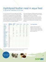
B cell activation
Spleenocyte preparation
Mice were sacrificed by exsanguination
through cutting cervical artery under
Spleenocytes harvested from mice were
analyzed for activation of B lymphocytes
using the markers CD19-PE and CD22-FITC
1130
Int.J.Curr.Microbiol.App.Sci (2017) 6(5): 1129-1133
where CD19 is B cell marker and CD22 is B
cell activation marker in mice. FACS analysis
had shown the activated B cell population of
20.3% in protein treatment group compared to
6.7% in control group which is statistically
significant (Figure 1A and B).
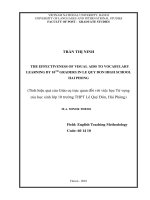
activation marker in mice. There was an about
14.1% increase in cytotoxic T cell activation
in IL28B protein treatment group compared to
control group (Figure 2).
Cytotoxic T cell activation
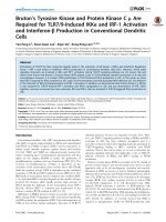
Spleenocytes isolated from mice were
analyzed for activation of helper T
lymphocytes using CD3-FITC, CD4-PE/Cy7
and CD69-PE, where CD69 is T cell
activation
marker
in
mice.
Spleenocytes were analyzed for activation of
cytotoxic T cells in mice spleen by FACS
using the molecules CD3-FITC, CD8-PE/Cy7
and CD69-PE, where CD69 is T cell
Helper T cell activation
Fig.1 B lymphocyte activation in mice spleen. (A): Control group. (B): IL28B protein group
showed 13.6% more increase in activated B lymphocytes compared to control
Fig.2 Cytotoxic T cell activation in mice. (A): Control group. (B) IL28B protein group showing
14.1% more increase in cytotoxic T cells compared to control
1131
Int.J.Curr.Microbiol.App.Sci (2017) 6(5): 1129-1133
Fig.3 T-helper cell activation. (A): control group. (B): IL28B protein group showing 6.3% more
increase in activated B lymphocytes compared to control
There was increase in activation of helper T
cell population in mice spleenocytes with
protein treatment. IL28B protein group has
shown 8% increase when compared to 1.1%
in the control group (Figure 3).
Type III Interferons are being tested for its
antiviral activity in many disease conditions.
Egli et al., (2014) reported that IFNλ3 is a key
regulator of B and T cell vaccine responses
against
Influenza.
As
they
have
Immunomodulatory activities in addition to
their antiviral activity, the study were done by
analyzing the B and T lymphocyte activation
in mice spleenocytes. Analysis of cells
surface molecules of B and T lymphocytes by
FACS analysis will provide an indication of
immunodulatory activity of IL28B protein.
Mice were administered IL28B protein and
spleenocytes after one week were analyzed
for the presence of activated B and T
lymphocytes by FACS analysis using specific
CD markers. The present study shows that
there was increase in activated B lymphocytes
(about 13.6%) in mice spleenocytes in
comparison to control groups. Lasfar et al.,
(2011) reported that IFNλ1 activates B
lymphocytes and modulates of Th2 cytokines.
There was no specific report available on
IL28B protein.
IL28B found to activate both cytotoxic and
helper T cells in mice spleenocytes but there
was also a significant activation of cytotoxic
T cells in comparison to Helper T cells.
Cytotoxic T cell activation was 14% high and
helper T cells activation was 6% high in
comparison to control animals. Mennechet
and Uze (2006) have shown that the treatment
of monocyte-derived DCs with IL-29,
member of type III IFNs led to induction of
the proliferation of regulatory T cells.
Lymphocyte activation by IL28B protein will
provide an added advantage to host immune
system to overcome the viral diseases. As
there is no demonstrated data available on
lymphocyte activation by bovine IL28B
protein, these findings will improve it and
also help the host to clear viral infection
effectively.
Acknowledgement
The authors acknowledge the Director, IVRI
(Indian Veterinary Research Institute)
Izatnagar and Joint Director, IVRI, Bangalore
1132
Int.J.Curr.Microbiol.App.Sci (2017) 6(5): 1129-1133
for providing facilities. The first author also
acknowledges ICAR (Indian Council of
Agricultural Research) India for the ICARSenior Research Fellowship.
References
Dolganiuc, A., Kodys, K., Marshall, C., Saha,
B., Zhang, S., Bala, S. and Szabo, G.
2012. Type III Interferons, IL-28 and
IL-29, Are Increased in Chronic HCV
Infection and Induce Myeloid Dendritic
Cell- Mediated FoxP3+ Regulatory T
Cells. PLOS ONE, 7(10): e44915.1-10.
Egli, A., Santer, D.M., Shea, D.O., Tyrrell,
D.L. and Houghton, M. 2014. The
impact of the interferon-lambda family
on the innate and adaptive immune
response to viral infections. Emerg.
Micro. and Infec., 3: 1-12.
Feczko, T., Toth, J., Dosa, G. and Gyenis, J.
2011.
Optimization
of
protein
encapsulation in PLGA nanoparticles.
Chem. Engi. Processing: Process
Intensification., 50(8): 757-765.
Lasfar, A., Abushahba, W., Balan, M. and
Cohen-Solal, K.A. 2011. Interferon
Lambda: A New Sword in Cancer
Immunotherapy.
Clin.
Develop.
Immunol., 1-11.
Lasfar, A., Zloza, A., de laTorre, A. and
Cohen-Solal, K.A. 2016. IFN-λ: A
New Inducer of Local Immunity against
Cancer and Infections. Frontiers in
Immunol., 7: 1-7.
Mennechet, F.J. and Uze, G. 2006. Interferonlambdatreated
dendritic
cells
specifically induce proliferation of
FOXP3-expressing suppressor T cells.
Blood, 107: 4417–4423
Odendall, C. and Kagan, J.C. 2015. The
unique regulation and functions of type
III interferons in antiviral immunity.
Curr. Opinion in Virol., 12: 47–52.
Zheng, Y., Hui Li, Yu, J., Zhao, H., Wang,
S.E., Ren, X. 2013. Interferon λs:
Special Immunomodulatory Agents and
Potential Therapeutic Targets. J Innate
Immun., 5: 209–218.
How to cite this article:
Barathiraja, S., N. Vijay, Brijesh Kumar, Lijo john, V. Umapthi, M. Sedhumadhavan and
Reddy, G.R.. 2017. Lymphocyte Activation by IL28B Protein in Mice.
Int.J.Curr.Microbiol.App.Sci. 6(5): 1129-1133. doi: />
1133