Serological and epidemiological picture of dengue during the year 2014: An exclusive study of Kutch district, Gujarat, India
Bạn đang xem bản rút gọn của tài liệu. Xem và tải ngay bản đầy đủ của tài liệu tại đây (254.64 KB, 7 trang )
Int.J.Curr.Microbiol.App.Sci (2017) 6(5): 2100-2106
International Journal of Current Microbiology and Applied Sciences
ISSN: 2319-7706 Volume 6 Number 5 (2017) pp. 2100-2106
Journal homepage:
Original Research Article
/>
Serological and Epidemiological Picture of Dengue during the Year 2014: An
Exclusive Study of Kutch District, Gujarat, India
Jigar Kiritkumar Gusani*, Hitesh Jayprakash Assudani, Krupali Kothari and A.N. Ghosh
Department of Microbiology, Gujarat Adani Institute of Medical Sciences, Bhuj, India
*Corresponding author
ABSTRACT
Keywords
Dengue,
Kutch,
ELISA,
Serological markers,
Epidemiology.
Article Info
Accepted:
19 April 2017
Available Online:
10 May 2017
The present study was carried out to find out data on Dengue specific
serological markers like NS1 antigen, IgM antibody and epidemiological
scenario of Dengue cases in Kutch region during the year 2014. Total 765
blood samples from across the Kutch region were tested by ELISA for NS1
antigen and/or IgM antibody as per the protocols and personal,
demographic and clinical details of each patient was recorded. Analysis of
this data revealed that age group 21-30 years had maximum dengue cases.
Male cases (429) were more than females (336). Even though major chunk
of Kutch population leaves in rural areas; overall dengue cases were high in
urban areas. Out of 765 cases 331 tested positive for dengue. Amongst all
positive cases 181(54.68%) were tested positive for NS1 antigen which
helped in early diagnosis of dengue. Rise of dengue cases started after the
month of August and falls down by the end of December 2014 which
coincides with post monsoon season.
Introduction
Dengue is a flavivirus transmitted by Aedes
aegypti mosquito. Dengue virus infection has
emerged as a notable public health problem in
recent decades in term of the mortality and
morbidity associated with it (Ukey et al.,
2010; WHO, 1997). Dengue is a major
problem in many parts of India and large
outbreaks are reported from various parts of
India and abroad (George et al., 1975; Kaur et
al., 1997). The clinical picture of dengue
mimics many other viral illnesses, which
makes laboratory confirmation very crucial
for the patient management. The benefits of
early diagnosis of dengue fever go beyond
treatment and encase epidemiological efforts.
For diagnosis of dengue cases many tests such
as virus isolation, RNA, Antigen and antibody
detection methods are available (Chakravarti
et al., 2006). However virus isolation, RNA
detection by PCR, requires well trained staff
and dedicated set up which is not feasible in
most of the health centers in India. In most of
the cases detection of IgM antibody is used
for diagnosis of dengue infection. However
time required for appearance of IgM antibody
is approximately 4-6 days (WHO, 1997).
Dengue non-structural 1 antigen (NS1) is
highly conserved glycoprotein produced in
2100
Int.J.Curr.Microbiol.App.Sci (2017) 6(5): 2100-2106
both membrane associated and secretary
forms is used as a novel biomarker for early
diagnosis of dengue infection (Subedi et al.,
2014). NS1 antigen detected by ELISA is
present in high concentrations in sera of
dengue virus infected patients during early
clinical phase of disease (Kumarasamy et al.,
2007).
details which were recorded in same forms.
The date of onset of fever and the date of
blood sample collection were also recorded in
the data entry form. Onset of fever was taken
as Day 0 and accordingly sample age was
defined as the gap between the date of onset
of fever and the date of collection of blood
sample.
Kutch is the largest district of India which has
a very special & diverse ecological system.
Literature search suggests that there is
scarcity of specific & exclusive data on
prevalence of Dengue in Kutch region
(Madhulika Mistry et al., 2015).
Serum was separated and then according to
the gap between onset of fever and sample
collection type of ELISA test was decided. If
the gap was 0 to 4 days then NS1 Ag ELISA
was performed and if it was more than 6 days
then IgM capture ELISA was performed. For
the samples having 5 to 6 days of fever both
IgM & NS1 Ag ELISA were performed. The
Dengue IgM capture ELISA kits were
supplied from NIV, Pune under NVBDCP
and for NS1 Ag detection Platelia dengue
NS1 Ag (Bio-Rad) ELISA kits were used. If
any of these test came positive by ELISA it
was considered as confirmed case and
notified to the concerned government
authorities.
The present study was carried out to find out
data on Dengue specific serological markers
like NS 1 antigen (Ag), IgM antibody and
epidemiological scenario of Dengue cases in
Kutch region during the year 2014.
Materials and Methods
This was a prospective & observational study
carried out from January 2014 to December
2014 at Department of Microbiology, Gujarat
Adani Institute of Medical Sciences, G K
General Hospital, Bhuj.
As a designated sentinel surveillance center
by National Vector Borne Disease Control
Program (NVBDCP), department is receiving
serum samples from Primary & Community
Health centers as well as affiliated private
hospitals from all over Kutch for testing and
confirmation of suspected Dengue cases.
Patients with acute onset of illness, high grade
fever,
severe
headache,
backache,
musculoskeletal pain or retro-bulbar pain with
or without rashes were considered as
clinically suspected case of dengue virus
infection (Durani et al., 2014).
There was standard laboratory requisition
form with Personal, Demographic and clinical
Results and Discussion
Total 765 blood samples from across the
Kutch region were received at our Institute for
serological diagnosis of Dengue fever during
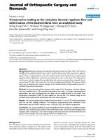
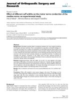
the year 2014. Following Figure 1 shows age
group wise distribution of clinically suspected
and positive dengue cases.
Age group of 21-30 years reported highest
number of dengue cases followed by age
group of 31-40 years. Figure 2 shows that
overall males are more affected then females.
Positive cases are also more in males.
From figure 3 it can be observed that
403(52.67%) cases were from urban areas &
362(47.32%) were belonging to rural areas.
Positive cases were more from urban areas
(46.65%) as compared to rural (39.5%).
2101
Int.J.Curr.Microbiol.App.Sci (2017) 6(5): 2100-2106
Table 1 displays Dengue case presented to the
hospital after onset of fever and positive cases
amongst them. Majority of cases appeared
between 3 to 5 days of fever (325 cases)
followed by >5 days of fever (274 cases).
Highest positivity was observed amongst the
cases that came to hospital within 2 days of
fever.
Table 2 shows Number positive cases
detected by different ELISA based tests. More
than half (54.68%) cases were detected by NS
1 antigen test. While almost 10% of cases
were having both the tests, NS 1 Antigen &
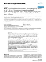
IgM antibody, positive. Figure 4 depicts
month wise trend of Dengue cases for the
year 2014. Peak rise of cases starts after the
month of August and falls down by the end of
December 2014. Another interesting finding
is there are small peaks of rise in dengue
cases noted between the months of April to
July.
Out of 765 suspected cases, highest cases
were seen in age group of 21 – 30 years
(34.64%) followed by 11 – 20 years age
group (22.22%). Also the positive cases
amongst both these group showed similar
pattern. These show predilection of dengue
infection toward young adults and
adolescents. These findings are in accordance
to other studies by (Durani et al., 2014 &
Pandey et al., 2012).
Gender distribution of cases revealed that 429
(56.08%) cases were males and 336(43.92%)
were females. Amongst the positive cases
202(61.03%) were males and 129(38.97%)
were females. Overall males were more
affected which can be due to various factors
like their frequent outdoor activities in this
region and less reporting of female cases etc.
These findings are similar to other studies by
Durani et al., 2014; Piyush tripathi et al.,
2008; Nidhi pandey et al., 2012.
Table 1 Dengue cases distribution as per days of fever
Number of days between
onset of fever and sample
collection
0 – 2 days
3 -5 days
> 5 days
Total numbers of sample
collected / Suspected Cases
(n= 765)
166 (21.7%)
325 (42.8%)
274 (35.8%)
Dengue Positive (n=331)
81 (48.8%)
137 (42.15%)
113 (41.24%)
Table 2 Dengue positive cases by ELISA tests
Test (ELISA Based)
NS 1 Antigen
NS 1 Antigen + IgM Antibody
IgM Antibody
Numbers of Dengue
positive Cases (n= 331)
181
32
118
2102
Percentage
54.68 %
9.67 %
35.65 %
Int.J.Curr.Microbiol.App.Sci (2017) 6(5): 2100-2106
2103
Int.J.Curr.Microbiol.App.Sci (2017) 6(5): 2100-2106
Demographic distribution of Dengue cases in
Kutch in 2014 showed that cases from urban
areas (403, 52.67%) were slightly higher than
the rural (362, 46.65%) areas. Although
previous studies from Gujarat & India showed
that prevalence of dengue cases were much
higher in urban areas; difference here in the
present study is much less Madhulika Mistry et
al., 2015; Durani et al., 2014; Patankar et al.,
2014; Dutta et al., 2012; Khan et al., 2014. One
reason for this can be natural demography of
Kutch region where majority of population lives
in rural areas as per Census 2011 data
(Government of India, 2011 Census).
In this study testing of samples were done as
per the age of sample and it was divided into 3
categories based on gap between onset of fever
2104
Int.J.Curr.Microbiol.App.Sci (2017) 6(5): 2100-2106
and sample collection. As NS1 antigen and IgM
antibody levels follow a pattern after the onset
of fever; according that the type of test was
decided (CDC laboratory guidance for
laboratory testing). Maximum number of
samples were from 3-5 days of fever (325,
42.48%) followed by >5 days of fever (274,
35.81%). However almost half (81, 48.8%)
patients from total 166 patients who came
within 2 days of fever were tested positive for
Dengue. This could be due to high sensitivity
and specificity of Dengue NS 1 antigen ELISA
test which was the sole test selected for patients
presents early in acute stage of Dengue
(Kulkarni et al., 2011; Peeling et al., 2010).
It is concluded that this study has just opened
the first door for analysis of Dengue in Kutch
region. It indicates that young adults, both from
rural and urban region, are affected more by
Dengue. The high number of positive cases
during typical post monsoon season indicates
failure in vector control steps and alerts
clinicians and epidemiologists to be more
vigilant for more such upcoming outbreaks. It
also emphasizes role of early detection which
can be more accurately done by tests like NS 1
antigen and thus helps in implementation of
better clinical and preventive measures for
entire population at risk.
Acknowledgment
Amongst the positive cases (331) detected in
this study, more than half (181, 54.68%) were
alone detected by NS 1 antigen test followed by
Ig M antibody positive cases (118, 35.65%). In
rest of 32 positive cases both NS 1 antigen and
Ig M antibody were detected. Various studies
have indicated that NS 1 antigen helps in early
detection and confirmation of diagnosis
(Kulkarni et al., 2011; Datta et al., 2010; Krunal
Mehta et al., 2016). When month wise trend
was depicted on graph it reinforced the fact that
surge in Dengue vector and so the rise in
dengue cases occur in Monsoon and post
monsoon season (WHO Dengue fever 2nd
Edition 1997, Madhulika Mistry et al., 2015;
Durani et al., 2014; Piyush tripathi et al., 2008).
Rain starts late in Kutch around end of June and
lasts till September. In the year 2014 rise in
dengue cases occurred from July; peak reached
in October and then started declining. Apart
from this; there was unusual small peak of cases
during the months of April & May which are
hot & dry months. This requires further
epidemiological investigation for search of
factors which increased dengue transmission in
Non seasonal months. Also there is need of long
term studies with analysis of dengue serotypes
to know more about the epidemiological,
demographic and biological variations of
dengue cases in Kutch region. It will
tremendously help in taking vector control
measures and formulating preventive strategies
for Dengue in this region.
We sincerely thank the entire staff of the
Department of Microbiology, GAIMS, Bhuj for
their support.
References
Chakravarti A., Kumaria R., Batra V., Verma V.
2006. Improved detection of dengue virus
serotypes from serum samples; evaluation
of single tube multiplex RTPCR with cell
culture. Dengue Bull., 30:133 40.
Datta, S., Wattal, C. 2010. Dengue NS1 antigen
detection: A useful tool in early diagnosis
of dengue virus infection. Indian J. Med.
Microbiol., 28(2): 107–10.
Durani, K., Dund, J., Shingala, H., Sinha, M.
2014. Epidemilogical trend analysis of
dengue virus infection in western part of
Gujarat. Indian J. Res., 3(6): 146–8.
Dussart, P., Labeau, B., Lagathu, G., Louis, P.,
Nunes, M.R.T., Rodrigues, S.G., et al.
2006.
Evaluation
of
an
enzyme
immunoassay for detection of dengue virus
NS1Antigen in human serum. Clin. Vaccine
Immunol., 13: 1185 89.
Dutta, P., Khan, S.A., Borah, J., Mahanta, J. 2012.
Demographic and clinical features of
patients with dengue in Northeastern region
of India: a retrospective cross-sectional
study during 2009–2011. J. Virol.
Microbiol., article ID 786298. doi:
10.5171/2012.786298.
2105
Int.J.Curr.Microbiol.App.Sci (2017) 6(5): 2100-2106
George, S., Soman, R.S. 1975. Studies on dengue
in Bangalore city: Isolation of virus from
man and mosquitoes. Indian J. Med. Res.,
63: 396-401.
Kaur, H., Prabhakar, H., Mathew, P., Marshalla,
R., Arya, M. 1997. Dengue haemorrhagic
fever outbreak in October- november 1996
in Ludhiana, Punjab, India. Indian J. Med.
Res., 106: 1-3.
Khan, S.A., Dutta, P., Topno, R., Soni, M.,
Mahanta, J. 2014. Dengue outbreak in a
hilly state of Arunachal Pradesh in
northeast India. Scientific World J., 584093.
doi: 10.1155/2014/584093.
Krunal, D., Mehta, et al. 2016. Study of
Correlation between Platelet Count and
Serological Markers of Dengue Infection
with Importance of NS1 Antigen in
Western Region of India. National J.
Laboratory Med., 5 (2): 55.
Kulkarni, R.D., Patil, S.S., Ajantha, G.S.,
Upadhya, A.K., Kalabhavi, A.S., Subhada,
R.M. et al. 2011. Association of platelet
count and serological markers of dengue
infection – importance of NS1 antigen.
Indian J. Med. Microbiol., 29(4): 359-62.
Kumarasamy, V., Wahab, A.H.A., Chua, S.K.,
Hassan, Z., et al. 2007. Evaluation of a
commercial dengue NS1 antigen- capture
ELISA for laboratory diagnosis of acute
dengue virus infection. J. Virol. Methods,
140: 75-79.
Madhulika Mistry, Yogesh Goswami, Rajesh, K.,
Chudasama,
Dhara Thakkar. 2015.
Epidemiological
and
demographic
characteristics of dengue disease at a
tertiary care centre in Saurashtra region
during the year 2013. J. Vector Borne Dis.,
52: p. 299–303
Nidhi Pandey, Rachna Nagar, Shikha Gupta,
Omprakash, Danish Khan, Desh Deepak
Singh,Gitika Mishra, Shantanu Prakash,
K.P. Singh, Mastan, Singh & Amita Jain.
2012. Trend of dengue virus infection at
Lucknow, north India (2008- 2010): a
hospital based study, Indian J. Med. Res.,
136, p 862-867.
Pandey, N., Nagar, R., Gupta, S., Omprakash,
Khan, D., Singh, D.D., Mishra, G., Prakash,
S., Singh, K.P., Singh, M., Jain, A. 2012.
Trend of dengue virus infection at
Lucknow, north India (2008- 2010): A
hospital based study. Indian J. Med. Res.,
136: 862-7.
Patankar, M.C., Patel, B.V., Gandhi, V.P., Shah,
P.D., Vegad, M.M. 2014. Seroprevalence of
dengue in Gujarat, Western India: a study at
a tertiary hospital in north India. Int. J.
Med. Sci. Public Health, 3(1): 16–8.
Peeling, R.W., Artsob, H., Pelegrino, J.L., Buchy,
P., Cardosa, M.J., Devi, S., et al. 2010.
Evaluation of diagnostic tests: Dengue. Nat.
Rev. Microbiol., 8: S30-37.
Piyush Tripathi, Rashmi Kumar, Sanjeev Tripathi,
J.J. Tambe and Vimala Venkatesh. 2008.
Descriptive Epidemiology of Dengue
Transmission in Uttar Pradesh. Indian
Pediatrics, 45: 315-31.
Subedi, D. and Taylor-Robinson, A.W. 2014.
Laboratory diagnosis of dengue infection:
current techniques and future strategies.
Open J. Clin. Diag., 4: 63-70.
Ukey, P.M., Bondade, S.A., Paunipagar, P.V.,
Power, R.M., Akulwar, S.L. 2010. Study of
seroprevalence of dengue fever in central
India. Indian J. Community Med., 35(4):
517-9.
World Health Organization. 1997. Dengue
haemorrhagic fever: Diagnosis, treatment,
prevention and control. 2nd
edition.
Geneva, Switzerland.
How to cite this article:
Jigar Kiritkumar Gusani and Ghosh, A.N. 2017. Serological and Epidemiological Picture of Dengue
During the Year 2014: An Exclusive Study of Kutch District, Gujarat, India.
Int.J.Curr.Microbiol.App.Sci. 6(5): 2100-2106. doi: />
2106