Phenotypic characterization and antimicrobial susceptibility of blood borne pathogens in a Tertiary Care Center, Ujjain (M.P), India
Bạn đang xem bản rút gọn của tài liệu. Xem và tải ngay bản đầy đủ của tài liệu tại đây (324.63 KB, 8 trang )
Int.J.Curr.Microbiol.App.Sci (2017) 6(5): 2271-2278
International Journal of Current Microbiology and Applied Sciences
ISSN: 2319-7706 Volume 6 Number 5 (2017) pp. 2271-2278
Journal homepage:
Original Research Article
/>
Phenotypic Characterization and Antimicrobial Susceptibility of Blood Borne
Pathogens in a Tertiary Care Center, Ujjain (M.P), India
Komal Singh, Ramesh Agrawal*, Yogyata Marothi and Harshada Shah
Department of Microbiology, R.D. Gardi Medical College, Ujjain, M.P. India
*Corresponding author
ABSTRACT
Keywords
BacT/alert, Blood
stream infection,
Blood samples,
Bacterial isolate,
Septicaemia,
Antimicrobial
susceptibility.
Article Info
Accepted:
25 April 2017
Available Online:
10 May 2017
Blood stream infections can lead to life threatening sepsis and require immediate
antimicrobial treatment. Blood culture is an essential tool for the investigation of clinically
suspected sepsis. The present study has been conducted to describe the profile of bacterial
isolates from blood cultures and their antibiotic resistance. This is prospective study of 273
blood cultures, collected from clinically suspected cases of bacteraemia studied over a
period of five months in a tertiary care hospital in Ujjain, M.P. The isolates were identified
by standard biochemical tests and antimicrobial resistance patterns were determined by
CLSI guidelines. Blood cultures were positive in 70 (25.6%) patients by BacT alert
system. Gram positive organism accounted for 51.4% cases; most common being
Staphylococcus aureus (47.3%) followed by Enterococci (4.3%), Of the Gram negative
isolates, Klebsiella spp (14.3%) was the most common followed by Pseudomonas spp.
(8.6%). Candida albicans was isolated in 2.8% cases. Gram positive isolates showed high
resistance to penicillin (81.8%) and least resistance to linezolid (18.2%). Gram negative
isolates were found high resistance to amoxy-clav (90%) and least resistance to Imipenem
(20%). This study provides information on antibiotic resistance of blood isolates. It may
be a useful guide for physicians initiating empiric therapy and will help in formulation of
antibiotic therapy strategy.
Introduction
Blood stream infection (BSI) is the major
cause of morbidity and mortality globally.
Blood stream infections range from selflimiting infections to life threatening sepsis,
which requires rapid and appropriate
antimicrobial treatment. Hence periodic
monitoring of blood culture isolates and
determination of susceptibility to antibiotics
are necessary to improve the empirical
therapy (Chitralekha et al., 2015). Many
bacterial pathogens have developed resistance
to most of the antibiotics; it has become a
serious health problem with many economic
and social inferences all over the worlds (Jo
Ann, 2009). Many bacteria have been
reported which cause bacteraemia with
variation in distribution from place to place
(Gohel et al., 2014). Rapid identification and
antimicrobial susceptibility testing of the
causative agents of bloodstream infections are
the most important tasks of the clinical
microbiology laboratory, that provide
essential information to
clinicians for
selecting appropriate antimicrobial therapy
for patients with bloodstream infections
(Lupetti et al., 2009). Automated blood
culture system is non invasive continuous
monitoring technology that reduces the time
2271
Int.J.Curr.Microbiol.App.Sci (2017) 6(5): 2271-2278
needed to detect positive blood cultures as
well as decreases the specimen handling (Kim
et al., 2010). Published guidelines
recommend that the interval between the
collection of blood and the entry of the bottles
into an automated blood culture system
should not be longer than 2 or 4 h; also
manufacturer instructions indicate that
inoculated vials should be transported to the
laboratory as quickly as possible (Clinical
Laboratory Standards Institute, 2007; Public
Health England, 2014). The changing
epidemiology and susceptibility patterns of
microorganisms emphasize the necessity of
constant surveillance of blood stream
infections (Muhammad et al., 2013). The
present study was done to analyze various
organisms causing bacteremia and their
antibiotic resistance pattern. This study wide
enable using appropriate antibiotic, may
decrease the hospital stay and cost of
treatment and reduce Mortality.
The main objectives of this study includes,
Isolation and identification of pathogens from
blood
samples
by
automated
3D
BacT/ALERT blood culture system and their
antibiotic resistance patterns in a tertiary care
center, Ujjain M.P, India.
Materials and Methods
This study was carried out at the Department
of Microbiology, R.D. Gardi Medical
College, Ujjain, Central India. A total of 273
blood samples from clinically suspected cases
of septicemia were studied during a 5 months
period from Oct 2016 to Feb 2017. Blood
samples were collected by using strict aseptic
precautions and inoculated immediately into
BacT/ALERT FA plus and PF plus aerobic
blood culture bottles. After collection these
bottles were immediately incubated in
BacT/ALERT 3D (bioMerieoux) – a fully
automated blood culture system for detection
of growth in blood culture. The negative
results were followed up to 7days and final
report was issued. The BacT/ALERT
automatically gives a signal alert. The
positive bottles were then subculture on
blood, MacConkey and chocolate agar. These
plates were incubated aerobically at 37 C and
examined
after
18-24
hours.
Final
identification
was
done
by colony
characteristics, Gram's staining, motility
testing (hanging drop preparation) and routine
biochemical test (Catalase, coagulase, indole,
methyl red, citrate, urease, Triple sugar iron,
PPA, and oxidase testing). Fungal isolate was
identified by Gram’s staining showing gram
positive budding yeast cells and germ tube
testing. Antimicrobial susceptibility testing of
bacterial isolates was done by the KirbyBauer disc diffusion method using Muller
Hinton agar media as per CLSI guidelines.
Results and Discussion
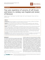
During the study period, 273 blood cultures
were analyzed of which 70 (25.6%) were
positive and 203 (74.4%) were culture
negative (Figure 1). Amongst total samples
147 (53.8%) were male and 126 (46.2%) were
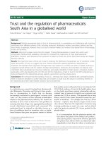
female patients. The age range varies from 1
month to 87 years. The incidence of blood
culture samples was the highest (26.4%) in 110 years age group followed by 0-1 yrs
(21.3%). Detail of age groups distribution was
given in (Table 1 and Figure 2). Blood
samples were collected from different wards
and ICUs in the hospital. Maximum number
(37.7%) of blood samples were obtained from
pediatrics wards. Details are given in (Table
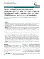
2). Out of 70 blood culture positive isolates,
36 (51.4%) were Gram positive organisms, 32
(45.7%) were Gram negatives organisms,
while 2 (2.8%) isolates were fungi (Candida
spp.). We have also isolate 3 gram positive
bacilli (bacillus) and 1 coagulase negative
staphylococcus (CoNS) were probably skin
contaminant excluded from the positive
isolates. Detail description of isolates was
2272
Int.J.Curr.Microbiol.App.Sci (2017) 6(5): 2271-2278
shown in (Table 3 and Figure 3). Staph aureus
(47.2%) was the predominant organism in all
bacterial isolates followed by Klebsiella spp
(14.3%). Antibiotic resistance pattern of the
gram positive organism are shown in (Table
4). Resistance ranges from 18.2% to 81%.
Staph aureus exhibit most resistance to
penicillin (81.8%) followed by cefoxitin
(MRSA) 63.3% and least resistance to
linezolid (18.2%). Among gram negative
organisms Klebsiella was the predominant
isolate which exhibit most resistance to amoxclav (90%) followed by Amikacin (80%) and
least resistance to imipenem (20%).
Pseudomonas exhibit most resistance to
Cefepime (66.6%) followed by Ceftazidime
(50%) and least resistance to imipenem
andAmikacin (16.6%). Detail description of
antibiotic resistance pattern of gram negative
organisms is shown in (Table 5).
The findings are matching these respected by
sepsis is one of the leading causes of death,
and rapid identification of blood stream
infection is mandatory to perform adequate
antibiotic therapy. In the present study a total
of 273 blood culture samples were collected
and analyzed, of which 70 (25.6%) were
positive by BacT/Alert system. which is quite
similar to Sahoo et al., (2016) and Alam et
al., (2011) but quite lower Kavitha et al.,
(2010) and Maimoona et al.,(2014). Majority
of the patients (26.4%) were in the 1-10 years
age group. Maximum number (37.7%) of
blood culture came from pediatrics ward. The
incidence of Gram-positive organism was
(51.4%) while (45.7%) were Gram-negative
organisms which was quite similar to Kalpesh
et al., (2014) and China et al., (2013), but in
other studies like Maimoona et al., and
Ayobola et al., (2011) Gram-negative
organisms have taken over Gram-positive
organisms in hospital settings. This indicates
that infections by Gram-positive organisms
constitute a significant threat to bacteremia
and septicemia in our hospital setup and the
spectrum of organisms is subject to
geographical alterations. Among Gram
positive organism staphylococcus aureus was
the commonest (47.2%) isolate followed by
Enterococcus (4.3%) which was quite similar
to study by Kalpesh et al., (2014) and
Anbumani et al., (2008).
While Klebsiella spp was the most
predominant (14.3%) isolate among gram
negative organism which was in accordance
with Panday et al., (2017).
Table.1 Age and gender wise distribution of blood culture of sepsis patients
Age group
1month - 1year
>1-10 year
11-20 year
21- 30 year
31- 40 year
41-50 year
51- 60 year
>61 year
Total
Total
58
72
40
23
18
25
16
21
273
2273
Male
28
43
21
14
11
13
9
8
147
Female
30
29
19
9
7
12
7
13
126
Int.J.Curr.Microbiol.App.Sci (2017) 6(5): 2271-2278
Table.2 Blood samples collected from different wards and ICUs
Word
NICU
MICU
PICU
SICU
P/W
MEDICINE
SURGERY
OTHER ICUs
Number of cases
10
19
60
12
103
29
7
24
ORTHO
9
Percentage (%)
3.7
6.9
21.9
4.4
37.7
10.7
2.5
8.9
3.3
Table.3 Organisms isolated from positive blood samples
Organism
Staphylococcus
Enterococcus
Pseudomonas spp.
E. coli
Klebsiella spp.
Citrobacter spp.
Enterobacter
Candida
Non-fermenter GNB
Total
Number of positive cases Percentage %
33
47.2
3
4.3
6
8.5
4
5.7
10
14.3
3
4.3
4
5.7
2
2.8
5
7.2
70
100%
Table.4 Antibiotic resistant pattern of gram positive bacteria
Staph. aureus (n=33)
Antibiotic drugs
Penicillin
Cefoxitin
Cotrimoxazole
Erythromycin
Clindamycin
Tetracycline
Vancomycin
Linezolid
Ampicillin
High level
Gentamycin
Enterococcus (n=3)
No of resistance
isolates
27
21
17
19
14
7
12
6
-
Percentage
(%)
81.8
63.6
51.5
57.6
42.4
21.2
36.4
18.2
-
No of resistance
isolates
2
1
0
2
Percentage
(%)
66.6
33.3
0
66.6
-
-
1
33.3
2274
Int.J.Curr.Microbiol.App.Sci (2017) 6(5): 2271-2278
Table.5 Antibiotic resistant pattern of gram negative bacteria
Antibiotic drugs
Amikacin
Aztronem
Piperacillin
Cefepime
Ceftazidime
Ciprofloxacin
Imipenem
Amoxy-clav
Piperacillin
tazobactum
E.coli (n=4)
Resistance No
1
3
3
2
3
1
4
1
(%)
25
75
75
50
75
25
100
25
Pseudomonas (n=6)
Resistance No
(%)
1
16.6
2
33.3
2
33.3
4
66.6
3
50
2
33.3
1
16.6
2
33.3
Klebsiella (n=10)
Resistance No (%)
8
80
3
30
7
70
6
60
4
40
2
20
9
90
5
50
Fig.1 blood culture results (n=273) by BacT alert system
total, 273
positive blood
sample, 70
negative
blood sample,
203
Fig.2 Age and gender wise distribution of blood culture cases
2275
Int.J.Curr.Microbiol.App.Sci (2017) 6(5): 2271-2278
Fig.3 Organisms isolated from automated blood culture
Higher prevalence of antimicrobial resistance
was noted in this study, especially in gram
negative organism. This might be due to
indiscriminate use of antibiotics in hospital.
Most of the gram negative organisms were
multidrug resistance with a very high
resistance to beta-lactam antibiotics and least
resistance to Imipenem.
The overall
resistance of gram positive organism was
from 0 to 81.8%, and for gram negative
organism from 0 to 100% these results are in
concordance with Araya et al., (2015) which
was 0–83% and 0–100% for gram positive
and negative, respectively. Staphylococcus
aureus showed high resistance to cefoxitin
(MRSA) which is quite similar to Garg et al.,
(2007). E. coli and Klebsiella was showed
high resistance to amoxy-clav which was
accordance to Veena mangunath et al.,
(2015), and least resistance to imipenem
accordance to Aziz japoni et al., (2008).
In the current study fungal septicaemia caused
by Candida albicans was observed 2.8%
which was quite similar with the Panday et
al., (2017) they have observed 3% Candida
albicans in their study. The rise in antibiotic
resistance in blood isolates emphasizes the
importance of sound hospital infection
control, rational prescribing policies, and the
need for awareness to use antimicrobial drugs.
It may be concluded from the study that early
diagnosis and appropriate treatment of BSIs
should be based on the current knowledge of
bacterial profile and antibiotic resistance
pattern, which should be provided by
microbiology laboratory from time to time.
We observed that Staphylococcus aureus and
organisms belonging to Enterobacteriaceae
family are the leading causes of septicemia.
Increasing incidence of drug resistant
organisms like MRSA and multidrug
resistance gram negative bacilli raises serious
concerns and mandates strict antibiotic policy
to prevent emergence and spread of antibiotic
resistance. We hope that these results could
support microbiologists, clinicians and
hospital managers in the identification and
implementation of strategic targeted actions to
coordinate infection control interventions and
antimicrobial policies in order to decrease the
rate the emergence of resistance and minimize
mortality of septicaemic patients.
2276
Int.J.Curr.Microbiol.App.Sci (2017) 6(5): 2271-2278
References
Alam, M.S., Pillai, P.K., Kapur, P., Pillai,
K.K. 2011. Resistant patterns of
bacteria isolated from bloodstream
infections at a university hospital in
Delhi. J. Pharm. Bioallied Sci., 3:
525-30.
Anbumani, N., et al. 2008. “Distribution and
antimicrobial susceptibility of bacteria
isolated from blood cultures of
hospitalized patients in a tertiary care
hospital, Indian J. Practicing Doctor,
vol. 5, no. 2, pp. 1–7.
Araya Gebreyesus Wasihun, Letemichael
Negash Wlekidan, Senay Aregawi
Gebremariam, Tsehaye
Asmelash
Dejene,
Abadi
Luel
Welderufael, Tadesse
Dejenie
Haile and Saravanan
Muthupandian.
2015. Bacteriological profile and
antimicrobial susceptibility patterns of
blood culture isolates among febrile
patients in Mekelle Hospital, Northern
Ethiopia,
Springerplus,
4:
314.
Published
online
2015
Jul
3. doi: 10.1186/s40064-015-1056-x
Ayobola, E.D., et al. 2011. “Study of
prevalence
and
antimicrobial
susceptibility of blood culture bacterial
isolates,” Malaysian J. Microbiol., vol.
7, no.2, pp.78–82.
Aziz Japoni, Shohreh Farshad, Abdolvahab
Alborzi, Mehdi Kalani, Nouradin
Rafaatpour, Barat Oboodi and Bahman
Pourabbas, 2008. Epidemiology and
Antibacterial Susceptibility Patterns of
Bloodstream Infections, 2001-2004: An
Experience with BACTEC 9240 in
Southern Iran. Pakistan J. Biol. Sci., 11:
422-427.
China, D., and V. Gupta. 2013.
“Bacteriological
profile
and
antimicrobial susceptibility pattern of
blood isolates from a tertiary care
hospital in North India,” Int. J.
Pharmaceutical Res. Biosci., vol. 2, no.
2, pp. 24–35.
Chitralekha
Saikumar,
Bindu,
Kiran
Madhusudhan, Praveena, R., Illamani,
V. 2015. Aerobic Microbial Profile and
Antibiotic Susceptibility of Blood
Isolates in a Tertiary Care Center, Int. J.
Pharm. Sci. Rev. Res., 34(2), Article
No. 23, Pages: 135-137.
Clinical Laboratory Standards Institute. 2007.
Principles and procedures for blood
cultures; approved guideline CLSI
document M47-A. CLSI, Wayne, PA.
Debananda Sahoo, Lalatendu Mohanty, S.S.
Panda,
S.N.,
Mishra.
2016.
Bacteriological Analysis of Blood
Culture Isolates in Patients with Sepsis
in A Tertiary Care Hospital of Eastern
India, IJCMR, Volume 3 | Issue 12 |
December 2016 | ICV (2015): 77.83 |
ISSN (Online): 2393-915X; (Print):
2454-7379.
Garg, A., et al. 2007. “Bacteriological profile
and antimicrobial resistance of blood
culture isolates from a university
hospital,” J. Indian Academy of Clin.
Med., vol. 8, no. 2, pp. 139–143.
Gohel, K., Jojera, A., Soni, S., Gang, S.,
Sabnis,
R.,
Desai,
M.
2014.
Bacteriological profile and drug
resistance patterns of blood culture
isolates in a tertiary care nephrourology
teaching institute. Biomed. Res. Int.,
153747. doi: 10.1155/2014/153747
Jo Ann, D. 2009. Antibiotic resistance: the
ongoing challenge for effective drug
therapy. JAAPA, 22(3): 18–22A.
Kalpesh Gohel, et al. 2014. Bacteriological
Profile and Drug Resistance Patterns of
Blood Culture Isolates in a Tertiary
Care Nephrourology Teaching Institute,
Hindawi
Publishing
Corporation
BioMed Res. Int., Article ID 153747, 5
pages
/>
2277
Int.J.Curr.Microbiol.App.Sci (2017) 6(5): 2271-2278
Kavitha, P., et al. 2010. “Bacteriological
profile and antibiogram of blood culture
isolates in a pediatric care unit,” J.
Laboratory Physicians, vol. 2, pp. 85–
88.
Kim, K.E. and J.Y. Han, 2010. Evaluation of
the clinical performance of an
automated pro calcitonin assay for the
quantitative detection of bloodstream
infection. Korean. J. La. Med., 30: 153159. DOI:10.3343/kjlm.2010.30.2.153
Lupetti, et al. 2009. Rapid identification and
antimicrobial susceptibility testing of
Gram-positive cocci in blood cultures
by direct inoculation into the BD
Phoenix system, Clin. Microbiol.
Infect., Volume 16, Issue 7.
Maimoona Mustafa and Syed Laeeq Ahmed.
2014. Bacteriological profile and
antibiotics susceptibility pattern in
neonatal septicemia in view of
emerging drug resistance, J. Med. Allied
Sci., 4(1): 02-08 www. jmas. in Print
ISSN:
22311696
Online
ISSN:
2231170X.
Manjusha Pandey, Devendra Niranjan and
R.C., Pande. 2017. Bacteriological
Profile and Antimicrobial Resistance of
Blood Culture Isolates from a 350
bedded Hospital Lucknow, India. Int. J.
Curr. Microbiol. App. Sci., 6(1): 184193.
doi:
/>601.023
Mathew, A., et al. 2006. “Performance
standards
for
antimicrobial
susceptibility testing,” Clinical and
Laboratory Standards Institute, vol. 26,
supplement 16.
Muhammad Fayez, Irfan Ali Mirza, Aamer
Ikram, Aamir Hussain, Tahir Ghafoor
and Umer Shujat. 2013. Pathogens
causing blood stream infections in
immunocompromised
patients,
J.
College of Physicians and Surgeons
Pakistan, 13, Vol. 23 (12): 848-851
Public Health England. 2014. UK standards
for
microbiology
investigations.
Investigation of blood cultures (for
organisms other than Mycobacterium
Species). Bacteriol., 8: 1-51
Veena Manjunath, Chillargi, C., Prathiba, S.,
Amarnath, S.K., Hegde, S.M. 2015.
Microbial and antimicrobial resistance
profile of bloodstream infections: A
hospital-based
study,
Medica
Innovatica, Volume 4- Issue 2.
How to cite this article:
Komal Singh, Ramesh Agrawal, Yogyata Marothi, Harshada Shah. 2017. Phenotypic
Characterization and Antimicrobial Susceptibility of Blood Borne Pathogens in a Tertiary Care
Center, Ujjain (M.P). Int.J.Curr.Microbiol.App.Sci. 6(5): 2271-2278.
doi: />
2278