Utility of rapid antigen detection for diagnosis of rota viral infection in children
Bạn đang xem bản rút gọn của tài liệu. Xem và tải ngay bản đầy đủ của tài liệu tại đây (237.93 KB, 5 trang )
Int.J.Curr.Microbiol.App.Sci (2017) 6(5): 1124-1128
International Journal of Current Microbiology and Applied Sciences
ISSN: 2319-7706 Volume 6 Number 5 (2017) pp. 1124-1128
Journal homepage:
Original Research Article
/>
Utility of Rapid Antigen Detection for Diagnosis of
Rota Viral Infection in Children <2 Yrs
Shivani Singh*, K. Tukaram Prabhu, Avinash Laghawe, Navinchandra Kaore and Arti Jain
People's College of Medical Sciences, Bypass road, Bhanpur, Bhopal 462037, India
*Corresponding author
ABSTRACT
Keywords
Rotavirus,
Diarrhoea,
Rapid immunochromatography,
Gastroenteritis,
Infants.
Article Info
Accepted:
12 April 2017
Available Online:
10 May 2017
Rotavirus is a major cause of severe gastroenteritis in young children between 6
months to 2 years of age This study was done with the aim to determine the utility
of rapid antigen detection kit for diagnosis of rota viral infection in suspected
cases of diarrhoea in children ≤ 2yrs. We found that out of the 32 samples, 06
tested postive for the rotaviral antigen by rapid immunochromatography method.
66.7% of the cases were in 13 – 24 months age group. Detection of Rotavirus
infection is necessary in determining the clinical severity as well as finding the
prevalence and incidence of this infection in the community. The rapid test kit
used is easy to use and inexpensive. This can detect the cases of Rotaviral
diarrhoea and be helpful in diagnosis as well as in epidemiological stuides. It can
also prevent the unnecessary use of antibiotics.The rapid antigen detection kit for
diagnosis of rota viral infection in suspected cases of diarrhoea in children ≤ 2yrs
can be a useful tool.
Introduction
Rota virus is a member of the family
Reoviridae, the only RNA virus family that
has a double stranded RNA. When observed
under electron microscope Rotaviruses have a
distinct wheel like appearance. Hence they
have been named rota which in Latin means
wheel. The virus has a genome of 11
segments of double-stranded RNA, of
molecular weight 2 x 10⁵ to 2.2 x 10⁶ Daltons.
This RNA is present in the core which is
covered with a triple layered capsid (Shobha
Broor, 2003). Seven groups (A-G) of
Rotaviruses have been described and only
groups A, B and C infect humans. Group A
has multiple strains and causes most of the
childhood diseases.
Worldwide Rotaviruses are a leading cause of
acute gastroenteritis in infants and young
children worldwide, infecting nearly all
children by the age of 5, often more than
once.
Each
year
rotavirus
causes
approximately 111 million episodes of
gastroenteritis, 25 million outpatient visits,
and 2 million hospitalizations in children
under age 5 worldwide. The incubation period
of rotavirus diarrhoea varies from 1-7 days
(Parashar et al., 2003; Parashar et al., 2006).
In infants and young children, there is an
abrupt onset of severe vomiting and diarrhea
with vomiting usually preceding diarrhoea.
Stools are usually loose and watery, mucus
1124
Int.J.Curr.Microbiol.App.Sci (2017) 6(5): 1124-1128
may be present but blood is very rare. Mild to
moderate dehydration is seen in 80 per cent of
cases and severe loss of fluids and electrolytes
may be fatal if untreated. Mild fever is seen in
a large majority of cases. The illness usually
lasts 3-8 days, but virus shedding continues
for about 10 days to 1 month. In
immunodeficient children, rotavirus can
persist for months. Older children and adults
are infected but they generally suffer from
subclinical infections and virus is infrequently
detected in their stool samples (Steele, 1999).
Rotavirus diarrhoea may show a seasonal
variation with a high incidence of the disease
in winter months at low relative humidity in
north India. Treatment of acute rotavirus
infection is nonspecific and involves
management of symptoms and, most
importantly, maintenance of hydration
(Bhautik Modi, 2013).
Transmission of Rotavirus occurs through the
feco–oral route (Deepali Masurkar, 2013).
Rotavirus is continuously shed in large
numbers during the course of disease and
stool specimens collected from the first to
fourth days of illness are optimal for rotavirus
detection (Fischer and Gentsch, 2004). They
can be easily identified on electron
microscopy of stool samples which is one of
the most specific tests for diagnosis. Direct
electron microscopy examination of stools for
rotavirus has a high sensitivity. However it
requires expensive equipment and trained
personnel, hence cannot be used in field
studies. Other methods like immunoelectro
osmophoresis and modified complement
fixation test were developed, but they lacked
sensitivity.
The recent advent of antigen detection
methods based on immunological techniques
using monoclonal and polyclonal antibodies
has gained attention of researchers. Direct
detection of viral antigen by rapid one step
immunochromatography technique is an
inexpensive, easy to handle sensitive test with
no need of invasive procedures and special
instrumentation (Sushmita Roy et al., 2008).
It is estimated that 1 in every 250 children
born in India dies from rotavirus by the age of
5 yr. India accounts for 17 per cent of the
world’s estimated rotavirus associated deaths.
A number of studies have been conducted on
the prevalence of childhood rotavirus
diarrhoea in various parts of the country in
which rotavirus was detected in 5 - 71 % of
the hospitalized children less than 5 years of
age with acute gastroenteritis.
The clinical manifestations of rotavirus
diarrhoea alone are not very distinctive to
permit exact diagnosis hence testing of
samples in the laboratory is the best way to
confirm the diagnosis. Most cases of
diarrhoea are treated with antibiotics,
irrespective of the causative agent.
However, if infection due to rotavirus can be
diagnosed early, the misuse and unnecessary
usage of antibiotics can be avoided.
Etiological diagnosis may not be essential in
the treatment of individual patients, but the
knowledge of the relative importance and
seasonal prevalence of different pathogens in
different regions is essential for proper
management of outbreaks and for the
planning and implementation of control
measures.
In this study, stool samples from cases of
diarrhoea patients’ ≤ 2 years of age were
tested to detect the Rotavirus antigen in the
stool specimen by immuno-chromatography
test with an aim to know the cause of
diarrhoea. If it is of viral origin, unnecessary
administration of antibiotics can be prevented
and thus help in right and proper management
of the patient.
1125
Int.J.Curr.Microbiol.App.Sci (2017) 6(5): 1124-1128
Materials and Methods
This cross-sectional prospective study was
conducted in a tertiary care hospital of Bhopal
and samples were collected from patients
attending the pediatric IPD and OPD for a
period of Two months – July 15th to
September 15th, 2016.
Cases were defined as children whose main
complaint was acute diarrhea, characterized
by occurrence of three or more loose, liquid
or watery stools with or without mucous in a
24 hours period. Other symptoms like fever,
vomiting etc were also recorded.
SD Bioline kit, Lot no. 14BD0034 Expiry
date: 2018/06/29 was used for the test. This
kit uses rabbit polyclonal anti-rota virus
antibodies which enables identification of
Group A Rotavirus antigens.
Stool sample from 32 patients ≤ 2yrs of age
with diarrhoea were collected in a clean wide
mouth screw capped bottle. The samples were
transported immediately to Microbiology
Laboratory.
They were then processed as per instructions
of the manufacturer. Briefly, a portion of
faeces (about 50g) from a stool sample was
taken and the swab provided was placed into
the sample collection tube and swirled at least
10 times. The swab was squeezed against the
wall of the tube and discarded. The dropping
cap was placed on the sample collection tube.
3-4 drops of the prepared sample was dropped
into the immuno-chromatography device, the
results were read after 10-20 minutes and
appearance of test line was taken to be
positive.
Results and Discussion
A total of 32 samples were collected from
patients ≤ 2yrs of age with diarrhea. Out of
these 12 patients were from the Out-patient
department and 20 were admitted to the
Paediatric ward. There were 18 male and 14
female patients with distribution as shown in
table 1.
Out of the 32 samples, 6 (18.75%) were
positive for rota viral antigen. Out of the 6
positive cases, 1 (16.67%) was a patient from
OPD while 5(83.33%) were from the ward.
Age wise distribution of the cases were as
shown in table 2.
Among the 06 positive cases 03 (50 %) were
males and 03 (50 %) were females and one
male among them was an OPD patient.
The average duration of the diarrhoea was 7
days and the average frequency was 6 times a
day in our study. Fever was present in all the
patients with rota viral diarrhoea with
vomitting in 83.3% (5) of the patients. None
of them displayed any signs of dehydration.
Acute gastroenteritis remains a leading cause
of post-neonatal under-five mortality in India
contributing about 13% of under-five
mortality. Rotavirus is the most important
cause for severe gastroenteritis in this age
group. Studies in the last decade estimate the
annual mortality due to rotavirus in India to
be between 90,000 and 153,000 (Jacob John
et al., 2014).
Of India’s more than 2.3 million annual
deaths among children, about 334 000 are
attributable to diarrhoeal diseases. Rotavirus
is the leading cause of severe diarrhoea in
children in developed and developing
countries. Almost all children have been
infected by the time they reach five years of
age. In developing countries rotavirus is
responsible for approximately half a million
deaths per year (Bhautik Modi, 2013). The
immune-chromatography test (ICT) used for
detection of Rota virus in stool samples give
1126
Int.J.Curr.Microbiol.App.Sci (2017) 6(5): 1124-1128
rapid
results.
According
to
Salwa
Badrelsabbha Ibrahim et al., (2015) the ICT is
quick, inexpensive, easy to perform and
requires very little equipment. Jayoung Kim
et al., (2014) have reported that rapid tests
show no interference, no cross reactivity, high
reproducibility and acceptable agreement
rates with other detection technologies like
ELISA, ELFA and PCR.
In our study the 18.75% of the cases tested
were positive for rotaviral antigen by rapid
immune-chromatography test. This is in
concordance with studies by Razaq Hadi
Eissa et al., (2014) and Jayoung Kim et al.,
(2014) However other studies have found
higher percentages like Sushmita et al.,
(2012)- 52.5%. Hussein (2013) found that
distribution of Rotavirus among infants with
diarrhea was 50.5% (52/103). About the
distribution of these viruses among age
groups, the results show that the most affected
age group was 1- 4 months (51.5%) followed
by less than 1 month group which consist
34%.
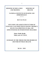
Table.1 Distribution of the patients – gender wise and OPD/IPD
OPD (n=12)
IPD (n=20)
Total
Male (n=18)
6
12
18
Female (n=14)
6
8
14
Table.2 Age wise distribution of the cases
Age
0-12 months
13-24 months
Positive (n=6)
2 (33.3%)
4 (66.7%)
Among the 6 patients, 01 patient was from
OPD while rests 05 were from paediatric
ward, suggesting the seriousness of the
disease.
In our study, 4 (66.7%) of the positive cases
were in the age group of 13-24 months.
Similar result has been reported by Salwa
Badrelsabbha Ibrahim et al., (2015) with
patients in 6-12 months range having highest
rate of rotavirus infection - 54%. As far as
gender distribution was concerned, there was
no significant difference in the distribution of
patients or in the number of cases, suggesting
that the patients of both genders are equally
affected by the disease. In contrast to study
by Wg Cdr John et al., (2014) whose study
showed 33.2% had fever and 43.6% had
vomiting along with diarrhea and study by
Negative (n=26)
10 (38.5%)
16 (61.5%)
Hussein et al., (2013) had 78.6% fever and
68% vomiting symptoms, in our study all the
patients presented with fever while 05
patients among the 06 positive cases had
vomiting. One patient from OPD did not have
vomiting. All the 06 patients were treated
symptomatically and all of them recovered
without administration of antibiotics.
In a study from Punjab, rotavirus infection
has been observed throughout the year with
maximum occurrence in November and
another peak in the hot and dry months of
May (Ram, 1990). The maximum incidence
in Pune occurred in winter and the minimum
in the rainy season (Kelkar, 1997). This study
was conducted during the period of July to
September, which can probably explain the
low number of positives. Also the number of
1127
Int.J.Curr.Microbiol.App.Sci (2017) 6(5): 1124-1128
samples tested was 32 and a larger number of
samples will have to be tested to make the
result statistically significant. This was the
limitation of our study.
Acknowledgement
The authors thank Indian Council of Medical
Research (ICMR) for their support for this
Short Term Studentship (STS) – 2016
research project.
References
Bhautik Modi. 2013. Rotavirus diarrhoea current scenario and preventive strategies
Guest editorial. Natl. J. Med. Res., 3(2):
104-105.
Deepali,
M.,
Masurkar.
2013.
Latex
Agglutination Test: A tool for rapid
diagnosis of Rotavirus from HIV seropositive and sero-negative patients with
diarrhea, Biol. Med., 5: 34-39.
Fischer, T.K. and Gentsch, J.R. 2004. Rotavirus
typing methods and algorithms. Rev. Med.
Virol., 14: 7182.
Hussein, O.M., Al-Dahmoshi, et al. 2013.
Rapid
identification
of
rotavirus,
adenovirus
and
Norovirus
using
immunochromatography test among
Infantile diarrhea, Iraq I.J.S.N., 4(4): 598602.
Jacob John, et al. 2011. Rotavirus
gastroenteritis in India, 2011–2013:
Revised estimates of disease burden and
potential impact of vaccines. Vaccine,
32S: A5–A9.
Jayoung Kim, et al. 2014. Evaluation of an
Immunochromatographic Assay for the
Rapid and Simultaneous Detection of
Rotavirus and Adenovirus in Stool
Samples. Ann. Lab. Med., 34: 216-222.
How to cite this article:
Kelkar, S.D. 1990. Prevalence of human group
A rotavirus serotypes in Pune, India
(1990-1993). Indian J. Med. Res., 106:
508-12.
Parashar, U.D., Givson, C.J., Bresee, J.S.,
Glass, R.I. 2006. Rotavirus and severe
childhood diarrhea. Emerg. Infect. Dis.,
12: 304-306.
Parashar, U.D., Hummelman, E.G., Bresee, J.S.,
et al. 2003. Global illness and deaths
caused by rotavirus disease in children.
Emerg. Infect. Dis., 9: 565-572.
Ram, S., Khurana, S., Kusana, S.B., Sharma, S.,
Vadehra, D.V., Broor, S. 1990.
Bioecological factors and rotavirus
diarrhoea. Indian J. Med. Res., 91: 16770.
Razaq Hadi Eissa, et al. 2014. Rapid Diagnosis
of
Rota-Adenoviruses
for
Acute
Gastroenteritis in hospitalized Children
under 4 Years Old, Baghdad. Int. J. Curr.
Microbiol. App. Sci., 3(1): 453-458.
Salwa Badrelsabbha Ibrahim, et al. 2015.
Detection of Rotavirus in children with
acute
gastroenteritis
in
Zagazig
University Hospitals in Egypt. Electronic
Physician, 7: 1227-1233.
Shobha Broor, Dhrubaa Ghosh, Purva Mathur.
2003. Molecular epidemiology of
rotaviruses in India. Indian J. Med. Res.,
118: 59-67.
Steele, J.C. 1999. Rotavirus. Clin. Lab. Med.,
19(3): 691703
Sushmita Roy, et al. 2012. Rapid detection of
Rotavirus antigen in stool sample of acute
diarrheic children. Bangladesh J. Med.
Microbiol., 06(01): 11-13.
Wg Cdr, B.M., John, et al. 2014. Prevalence of
rotavirus infection in children below two
years presenting with diarrhea. Med. J.
Armed Forces India, DOI:
http:
//dx.doi.org/10.1016/j.mjafi.2014.02.008
Shivani Singh, K. Tukaram Prabhu, Avinash Laghawe, Navinchandra Kaore, Arti Jain. 2017.
Utility of Rapid Antigen Detection for Diagnosis of Rota Viral Infection in Children <2 Yrs.
Int.J.Curr.Microbiol.App.Sci. 6(5): 1124-1128. doi: />
1128