Long non-coding RNA SNHG3 promotes breast cancer cell proliferation and metastasis by binding to microRNA-154-3p and activating the notch signaling pathway
Bạn đang xem bản rút gọn của tài liệu. Xem và tải ngay bản đầy đủ của tài liệu tại đây (5.55 MB, 13 trang )
Jiang et al. BMC Cancer
(2020) 20:838
/>
RESEARCH ARTICLE
Open Access
Long non-coding RNA SNHG3 promotes
breast cancer cell proliferation and
metastasis by binding to microRNA-154-3p
and activating the notch signaling pathway
Hongnan Jiang1, Xiaojun Li2, Wei Wang1 and Honglin Dong3*
Abstract
Background: Breast cancer (BC) is a malignant tumor that occurs in the epithelial tissue of the breast gland. Long
non-coding RNA (lncRNA) small nucleolar RNA host gene 3 (SNHG3) has been found to promote BC cell proliferation
and invasion by regulating the microRNA (miR)-101/zinc-finger enhancer binding axis in BC. Herein, the objective of
the present study is to evaluate the effect of lncRNA SNHG3 on BC cell proliferation and metastasis with the Notch
signaling pathway.
Methods: Differentially expressed lncRNA in BC tissues and normal breast tissues was analyzed. SNHG3 si-RNA-1 and
SNHG3 si-RNA-2 were constructed to detect the mechanism of SNHG3 interference in BC cell proliferation, viability,
migration and invasion. Then, dual-luciferase reporter gene assay was utilized to verify the binding relation between
SNHG3 and miR-154-3p as well as miR-154-3p and Notch2. Moreover, xenograft transplantation was applied to confirm
the in vitro experiments.
Results: Highly expressed SNHG3 was observed in BC tissues. The growth of BC cells in vivo and in vitro was evidently
repressed after silencing SNHG3. BC cell invasion and migration were inhibited by silencing SNHG3 in vitro. SNHG3
could act as a competing endogenous RNA of miR-154-3p and upregulate the Notch signaling pathway to promote
BC cell development. Activation of the Notch signaling pathway can partly reverse the inhibition of cell activity
induced by silencing SNHG3.
Conclusion: Our study demonstrated that interfered lncRNA SNHG3 promoted BC cell proliferation and metastasis by
activating the Notch signaling pathway. This investigation may offer new insight for BC treatment.
Keywords: Breast cancer, Long non-coding RNA SNHG3, microRNA-154-3p, Notch signaling pathway, Competing
endogenous RNA
* Correspondence:
3
Department of Vascular Surgery, The Second Hospital of Shanxi Medical
University, No. 382, Wuyi Road, Taiyuan 030001, Shanxi, PR China
Full list of author information is available at the end of the article
© The Author(s). 2020 Open Access This article is licensed under a Creative Commons Attribution 4.0 International License,
which permits use, sharing, adaptation, distribution and reproduction in any medium or format, as long as you give
appropriate credit to the original author(s) and the source, provide a link to the Creative Commons licence, and indicate if
changes were made. The images or other third party material in this article are included in the article's Creative Commons
licence, unless indicated otherwise in a credit line to the material. If material is not included in the article's Creative Commons
licence and your intended use is not permitted by statutory regulation or exceeds the permitted use, you will need to obtain
permission directly from the copyright holder. To view a copy of this licence, visit />The Creative Commons Public Domain Dedication waiver ( applies to the
data made available in this article, unless otherwise stated in a credit line to the data.
Jiang et al. BMC Cancer
(2020) 20:838
Background
Breast cancer (BC) is a malignant tumor that occurs in
the epithelial tissue of the breast gland and is the most
prevalent cancer among the female globally [1]. BC
can be triggered by factors like age, menarche history,
reproductive patterns, physical activity, breast characteristics and body habitus [2]. Increasing data indicate
that incidence and mortality rates in developed countries are declining but growing in developing countries
[2]. At present, women give little attention to clinical
inspection and examination of BC, thus it is often diagnosed in advanced stage [1]. Surgery, molecular
treatment, radiation therapy and chemotherapy are
considered as approaches for BC treatment [3]. However, it remains challenging to ascertain an individual
basis who would benefit from these treatments while
who would be possible to encounter toxicities [4]. In
this context, novel therapeutic strategies for BC are in
urgent need. Towards this, we undertook a long noncoding RNA (lncRNA)-based approach to understand
the underlying mechanism in BC development, in
order to develop novel intervention strategies.
LncRNAs are important in disease occurrence and development, and its associations with these diseases contribute to insightful perspectives about the pathogenesis,
diagnosis and treatments of diseases [5]. A recent study
has suggested that lncRNA regulates gene at transcriptional, post-transcriptional and epigenetic levels to get
involved in tumor progression, including BC [6]. Upregulated lncRNA small nucleolar RNA host gene 3
(SNHG3) serves as an oncogene in BC cells [7]. LncRNA
SNHG3 serves as a competing endogenous RNA
(ceRNA), encouraging the growth of colorextal cancer
[8]. Dysregulated miR is observed in many malignancies
indicating a tumor suppressive or oncogenic role [9]. It
has been reported that miR-154 is a therapeutic target in
BC treatment by serving as a tumor inhibitor [10]. Additionally, another study has demonstrated that miR154-3p is found to be remarkably deregulated in ductal
carcinoma in situ, the most common type of noninvasive BC [11]. Notch2 has been found to play an important role in promoting BC cell dormancy and
mobilization [12]. Additionally, the Notch signaling
pathway is a fundamental mechanism operating in
multicellular organisms as well as in most cells, playing
a significant role in promoting cell development and
differentiation [13, 14]. Notch signaling pathway regulates key target genes’ transcriptional activity and acts
as a therapeutic target in treating several cancers, including BC [15]. From all above, it is reasonable to
hypothesize that there may be interactions among
lncRNA SNHG3, miR-154-3p and Notch2 in BC cell
proliferation and metastasis. Thus, we conducted a
series of experiments to verify the hypothesis.
Page 2 of 13
Methods
Clinical samples
Women with BC were consecutively recruited at the
Second Hospital of Shanxi Medical University from
January 2015 to January 2018. Before being enrolled in
the study, they received routine chest X-ray, mammography and abdominal ultrasonography, but did not receive
chemotherapy or radiotherapy. Criteria for exclusion from
the study were as follows: inflammatory breast cancer,
metastasis, pre-existing treatment or recurrence of the disease, the presence of diseases such as liver disease, arthritis, or other cancers. All patients received radical
mastectomy or modified radical mastectomy. Sixty patients diagnosed with BC were recruited in the carcinoma
group and sixty patients with benign breast lesions were
recruited in the control group. Furthermore, 3 breast cancer and benign breast lesions specimens were collected to
perform transcriptome analysis.
Reverse transcription-quantitative polymerase chain
reaction (RT-qPCR)
Trizol (Invitrogen, Carlsbad, CA, USA) was employed to
extract total RNA. PrimeScript RT kit (Takara, Bio Inc.,
Shiga, Japan) was applied to conduct reverse transcription PCR. Quantitative PCR was performed by AceQ
qPCR SYBR Green Master Mix kit (Vazyme Biotech Co.
Ltd., Nanjing, China) on a LightCycler 480 (Roche, Basel,
Switzerland). The primers were synthesized via TransGen Biotech (Shanghai, China). Their sequences are
listed in Table 1. All the experiments were performed
three times.
Cell lines selection
Human BC cell lines MCF-7, MDA-MB-231, HCC1937,
BT474, SKBr-3 and breast epithelial cell line MCF10A
were purchased from the Experimental Cell Center,
Chinese Academy of Sciences (Beijing, China). Subsequently, cells were cultivated in Roswell Park Memorial
Institute 1640 medium consisting of 10% fetal bovine
serum in a 37 °C incubator with 5% CO2 for 48 h and
subcultured.
Small interfere RNA (siRNA)
SNHG3 siRNA-1 and SNHG3 siRNA-2 were synthesized
via GenePharma Biotech (Shanghai, China) and transfected
using HilyMax kit (Dijindo Laboratories, Kumamoto,
Kyushu, Japan) with a firm compliance to its instructions.
Afterwards, SNHG3 level was verified with RT-qPCR 48 h
later.
Cell proliferation and viability assays
ZCell proliferation ability was measured as per the requirements of 5-ethynyl-2′-deoxyuridine (EdU) staining
[16] and colony formation assay [17]. Cell viability was
Jiang et al. BMC Cancer
(2020) 20:838
Page 3 of 13
Table 1 Primers sequence
Sequence
Forward
Reverse
SNHG3
TTCAAGCGATTCTCGTGCC
AAGATTGTCAAACCCTCCCTGT
LNC00680
TTCGGTCTCTTCGACGACG
TGCGAACTCTTGGTGTAGGTC
AC017048.4
CAGCAGAGGAAGACCATGTG
GGCGTTTGGAGTGGTAGAAA
miR181A2HG
TTGCTGGCGTCTCGGTTAAT
GCCACCACACTGCCATATCT
AC007461.2
AACGCTT CACGAATTTGCGT
CTCGCTTCGGCAGCACA
LNC00277
CACGGGGGGCATTTGGAGATTTT
GCCACCACACTGCCATATCT
GATA3-AS1
CGAGTCGGGTTCTGATCCAC
GGATGCTGCTTTCCACCCAT
AC017048.3
AGGGGCCTTCCAGATTAAGG
CGAGTCGGGTTCTGATCCAC
miR-154-3p
GTGGTACTTGAAGATAGGTT
TTGGTACTGAAAAATAGGTC
Notch1
GTCAACGCCGTAGATGACC
TTGTTAGCCCCGTTCTTCAG
Notch2
TCCACTTCATACTCACAGTTGA
TGGTTCAGAGAA AACATACA
Notch3
GGGAA AAAGGCAATAGGC
GGAGGGAGAAGCCAAGTC
GAPDH
GAAGAGAGAGACCCTCACGCTG
ACTGTGAGGAGGGGAGATTCAGT
Note: SNHG3 Small nucleolar RNA host gene 3, LNC Long non-coding, miR microRNA; GAPDH Glyceraldehyde-3-phosphate dehydrogenase
detected in the light of the instructions of 3-(4, 5dimethylthiazol-2-yl)-2, 5-diphenyltetrazolium bromide
(MTT) kit [18].
Cell invasion and migration assays
Cell invasion and migration ability was performed by
Transwell assay based on previously described [19].
Western blot analysis
Cells were washed twice by pre-cooling phosphate buffered saline (PBS) and lysed for 30 min at 4 °C before
centrifuged at 15,000×g for 15 min at 4 °C to remove cell
debris. Then, the separated proteins were transferred
onto the polyvinylidene fluoride (PVDF) membranes
after using 10% sodium dodecyl sulfate polyacrylamide
gel electrophoresis. To ensure all the samples were
transferred, the PVDF membranes were stained with
ponceau staining solution. Then, the membranes were
incubated in sealing solution for 2 h at room
temperature. Next, the membranes reacted with antiNotch1 (1/500, ab8925, Abcam, Cambridge, MA, USA),
anti-Notch2 (1/200, ab8926, Abcam) and anti-Notch3
(1 μg/mL, ab23426, Abcam) for 2 h. And then, the membranes were fully washed twice in PBS and twice in trisbuffered saline tween (TBST). Afterwards, the membranes were cultivated with secondary antibody goat
anti-mice (1:1000, ab7068, Abcam) labeled by horseradish peroxidase (HRP) for 1 h, washed in TBST again and
finally visualized with Super Signal West Pico kit. βactin was applied as the internal reference.
hybridization was stayed overnight at 65 °C. The sections
were washed with sodium chloride-sodium citrate buffer
with the original concentration. Then the slides were
treated in 5% sealing solution for 30 min at room
temperature, and each section was cultivated in sealing
buffer overnight at 4 °C with the anti digoxigenin
(NEF832001EA, Perkin-Elmer, Waltham, Massachusetts,
USA) labeled by 100 μL HRP at the ratio of 1: 500. After
3 times of tris buffered saline (TBS) washes (10 min/
time), trichostatin (TSA) staining solution was prepared
in accordance with instructions of Perkin-Elmer TSA
Plus kit (NEL753001KT, Perkin-Elmer). After that, the
sections were incubated in TBS containing 4′, 6diamidino-2-phenylindole (DAPI), washed and air-dried,
and finally fixed in aqueous fluorescent mounting reagent. The pictures were captured using a Leica SP8
laser scanning confocal microscope (Leica, Solms,
Germany).
RNA pull-down assay
A total of 100 μg RNA was extracted. Then, 500 μg
streptavidin beads were combined with miR-154 labeled
with 200 pmol biotin, and incubated with the extracted
RNA for 1 h. Next, the elution buffer was added to collect the pull-down RNA complex. The mRNA levels of
lncRNA SNHG3 and Notch2 were quantitatively analyzed by RT-qPCR. The specific operations strictly
followed the instructions of Magnetic RNA-Protein PullDown kit (GENEWIZ, Beijing, China).
Dual luciferase reporter gene assay
Fluorescence in situ hybridization (FISH) assay
MCF-7 and HCC1937 cells were hybridized with
lncRNA SNHG3 probe (Exiqon, Vedbaek, Denmark).
The probe mixture was denatured at 85 °C and the
Cells were transfected with 2 μg pMiR-report vectorSNHG3/Notch2 3′UTR (GenePharma, Shanghai, China)
and miRNA-154-3p using Lipofectamine 2000. Transfected cells were lysed at 48 h and then luciferase activities
Jiang et al. BMC Cancer
(2020) 20:838
Fig. 1 (See legend on next page.)
Page 4 of 13
Jiang et al. BMC Cancer
(2020) 20:838
Page 5 of 13
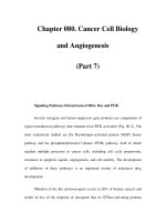
(See figure on previous page.)
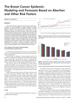
Fig. 1 SNHG3 was upregulated in BC. a. Volcano map of lncRNAs between BC and benign breast lesions specimens by transcriptome analysis.
The blue dots indicated high lncRNA expression; the red dots indicated low lncRNA expression and the black dots showed the lncRNAs with an
expression of |log2FC| < 2. Log2FC was logarithm of fold-change with base 2 and the fold-change was cancer over normal. The Y axis
represented an adjusted FDR, and the X axis represented the log2FC value. Aberrantly expressed lncRNAs were identified by DESeq R. Altogether,
137 highly expressed and 139 low expressed lncRNAs were identified; b. Different expressions of the top 8 lncRNAs between BC and benign
breast lesions specimens by RT-qPCR; c. SNHG3 expression in normal tissue and primary tumor assessed by UALCAN; d. SNHG3 level among BC
cell lines and human mammary epithelial cells detected using RT-qPCR. Three independent experiments were performed. Data are expressed as
mean ± standard deviation; one-way ANOVA and Tukey’s multiple comparisons test was used, *p < 0.05, **p < 0.01
comparisons test was used for the pairwise comparison
after ANOVA analysis. An adjusted p-value < 0.05 was
regarded as a statistically significant result.
were detected using Dual-luciferase Reporter Assay System. All the experiments were performed three times.
Xenografts transplantation
Results
Twelve specific pathogen-free BALB/c nude mice (4–6
week-old, 20 ± 2 g) [Beijing Vital River Laboratory Animal Technology Co., Ltd., Beijing, China, SCXK (Beijing)
2015–0001] were numbered with body weight as a parameter and randomly assigned into two groups (n = 6).
The stably transfected 4 × 10⁶ MCF-7 cells by si-SNHG3
or Scramble siRNA were dispersed by 2 mL saline and
injected subcutaneously into the right axilla of mice.
Tumor volume was measured every 5 days and every 3
days after the 20th day. Mice were suffocated to death
by CO2 35 days later. The tumors were taken out and
weighed for immunohistochemistry, with every step following the guidance in a literature report [20]. Primary
antibodies used in the immunohistochemistry were antiNotch1 (1/200, ab8925, Abcam), anti-Notch2 (1/200,
ab8926, Abcam) and anti-Notch3 (5 μg/mL, ab23426,
Abcam), as well as the secondary antibody (1:1000,
ab150117, Abcam) labeled by HRP.
LncRNA SNHG3 was highly expressed in BC patients
Firstly, the expression difference of lncRNA between BC
tissues and normal breast tissues were detected by transcriptome sequencing. A total of 478 lncRNAs were obtained, 276 of which were differentially expressed, 137 of
which were highly expressed, and 139 of which were
poorly expressed in cancer tissues (Fig. 1a). Eight lncRNAs
with the most significant differential expression were selected: SNHG3, LNC00680, AC017048.4, MIR181A2HG,
AC007461.2, LNC00277, GATA3-AS1 and AC017048.3
(Table 2), and their levels were verified in 60 pairs of BC
tissues and normal breast tissues. Result of RT-qPCR was
consistent with that of transcriptome sequencing (p <
0.05) (Fig. 1b). Chen J. et al. have indicated in a literature
report that lncRNA SNHG promoted osteosarcoma via
sponging miR-196a-5p [21]. Liu L. et al. have suggested
that lncRNA SNHG3 existed as an oncogene in lung
adenocarcinoma, and upregulation of lncRNA SNHG3
promoted lung adenocarcinoma cell growth [22]. It has
also been found that the malignancy of glioma was encouraged by SNHG3 via silent kruppel-like factor3 and
p21 [23]. Taherian-Esfahani Z. et al. have found that
lncRNA SNHG family played an important role in occurrence and hallmark of BC. SNHG1 expression was related
to clinical staging; SNHG5 was related to malignance
Statistical analysis
All the experiments were performed in triplicate. The
measurement data were expressed as mean ± standard
deviation. Statistical analysis was performed with GraphPad Prism 8 software (GraphPad, San Diego, CA, USA).
The p-values were calculated using the one-way or twoway analysis of variance (ANOVA). Tukey’s multiple
Table 2 Characteristics of the top 10 lncRNAs
Ensemble
Gene
Dysregulation
Fold Change
P Value
ENSG00000242125
SNHG3
Up
14.57442588
1.15E-68
ENSG00000215190
LNC00680
Up
3.616287083
3.75E-27
ENSG00000224577
AC017048.4
Up
7.63389477
5.67E-49
ENSG00000224020
miR-181A2HG
Up
10.62338023
1.55E-55
ENSG00000226101
AC007461.2
Up
4.326357933
5.34E-30
ENSG00000212766
LNC00277
Up
4.302321175
9.37E-47
ENSG00000197308
GATA3-AS1
Up
5.294820578
2.35E-41
ENSG00000163364
AC017048.3
Up
14.0691325
4.78E-24
Note: SNHG3 Small nucleolar RNA host gene 3, LNC Long non-coding, miR microRNA
Jiang et al. BMC Cancer
(2020) 20:838
Fig. 2 (See legend on next page.)
Page 6 of 13
Jiang et al. BMC Cancer
(2020) 20:838
Page 7 of 13
(See figure on previous page.)
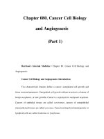
Fig. 2 SNHG3 silencing effectively inhibited BC cells proliferation, invasion and migration. Two siRNAs targeted SNHG3 and scramble siRNA were
transfected into MCF-7 and HCC1937 cells. a. RT-qPCR was performed to validate siRNA transfection. MCF-7 and HCC1937 cell biological
behaviors were detected with EdU staining (b); BC cell proliferation detected by MTT proliferation assay (c) and colony formation assays (d); E.
MCF-7 and HCC1937 cells migrating from upper Transwell chambers into lower ones, without Matrigel (× 200); f. MCF-7 and HCC1937 cells
invading from Matrigel-coated upper Transwell chambers into lower ones (× 200); g. Western blot analysis was carried out to determine Ecadherin and N-cadherin protein levels (representative images were shown, full-length gels are presented in Supplementary Figure 1). Three
independent experiments were performed. Data are expressed as mean ± standard deviation; one-way ANOVA and Sidak’s multiple comparisons
test was used to determine statistical significance, or two-way ANOVA and Tukey’s multiple comparisons test was used, *p < 0.05, **p < 0.01
while SNHG3 expressed higher in estrogen receptor/progesterone receptor (ER/PR) compared with ER/PR positive
BC [24]. However, there was less study about SNHG3 in
BC. According to UALCAN ( />index.html), an online bioinformatics analysis site [25], we
found that lncRNA SNHG3 expression in BC patients was
evidently higher than that in healthy people (p < 0.05)
(Fig. 1c). Besides, SNHG3 had a higher expression in BC
cell lines than that in MCF10A cells (p < 0.05) (Fig. 1d).
Interfered lncRNA SNHG3 repressed BC cell proliferation,
invasion and migration
To further prove the effect of SNHG3 on BC cells,
siRNA were used to construct MCF-7 and HCC1937
cells with stable knockdown of SNHG3. Firstly, after
siRNA interference was verified by RT-qPCR, the expressions of SNHG3 in MCF-7 and HCC1937 cells
showed an evident decline and siRNA-2 had a more
powerful intervention capacity (p < 0.05) (Fig. 2a). Next,
EdU staining, colony formation assay and MTT assay
were performed to measure BC cell viability and proliferation. As the results shown, BC cell viability and proliferation significantly decreased after intervening SNHG3 (p <
0.05) (Fig. 2b-d). Invasion and migration of BC cells decreased obviously as showed by Transwell assay (p < 0.05)
(Fig. 2e/f). The expressions of epithelial-mesenchymal transition (EMT)-related proteins E-cadherin (1:50, ab1416,
Abcam) and N-cadherin (1:100, ab18203, Abcam) in BC
cell were further tested by Western blot analysis. The result
revealed that after the interference of SNHG3, the expression of E-cadherin increased remarkably while the expression of N-cadherin decreased (p < 0.05) (Fig. 2g).
SNHG3 strengthened Notch2 viability by competitively
combination with miR-154-3p
Firstly, lncATLAS database ( [26]
was used to predict that the subcellular fractions of
lncRNA SNHG3 were mainly localized in cytoplasm
(Fig. 3a). Afterwards, FISH assay verified that lncRNA
SNHG3 was mainly localized in the cytoplasm of MCF-7
and HCC1937 cells. The probes of lncRNA SNHG3 in
MCF-7 and HCC1937 cells were stained into red, and
the nucleus was stained into blue by DAPI (Fig. 3b).
Then, the total RNA of MCF-7 and SNHG3 cells was
extracted by separating cytoplasm and nucleus to detect
lncRNA SNHG3 expression in cytoplasm and nucleus
respectively. As showed in Fig. 3c, SNHG3 mainly appeared in cytoplasm (p < 0.05), suggesting that SNHG3
affected the development of BC through the mechanism
of CeRNA. Thereafter, a large number of miRs were predicted to be possibly combined with SNHG3 by Starbase
( [27], and we focused on
miR-154-3p, which was regarded as a tumor suppressor
in bladder cancer by targeting ATG7 according to Junfeng Wang et al. [28]. According to Hui Hu et al., BC
cell proliferation and migration were inhibited when
miR-154 targeted E2F5 transcription factors [29].
Kalpan-Meier plotter ( />php? P = Service) [30] website was emplyed to predict
the relationship between miR-154 and prognosis of BC
patients, and it was found that patients with low expression of miR-154 had worse prognosis (Fig. 3d). In
addition, dual luciferase reporter gene assay was conducted to verify the binding relation between miR-1543p and SNHG3; the result of RNA pull-down experiment also revealed that there was a binding complex
between SNHG3 and miR-154-3p; specifically, SNHG3
could be detected in the bio-miR-154 group, (p < 0.05)
(Fig. 3e/f). Then, RT-qPCR was applied to verify the
miR-154-3p expression in MCF-7 and HCC1937 cells
after intervening SNHG3 expression. As showed in
Fig. 3g, miR-154-3p expression was evidently increased
after the intervention of SNHG3 (p < 0.05). Later, we further considered the downstream mechanism of miR154-3p and predicted the target gene of miR-154 on
Starbase website. And we focused on Notch2 by consulting the literature. Anuradha Sehrawat et al. have found
that activating Notch discouraged BC cell apoptosis at
initial stage [31]. The dual luciferase reporter gene assay
confirmed the binding relation between miR-154 and
Notch2, and RNA pull-down assay verified that miR-154
and Notch2 colud form binding complex (p < 0.05)
(Fig. 3e/f). After that, RT-qPCR and Western blot analysis were employed to detect Notch2 expression in
MCF-7 and HCC1937 cells after intervening SNHG3 expression. The expression of Notch2 was obviously decreased after intervention of SNHG3 (p < 0.05) (Fig. 3h).
From the above results, it was concluded that SNHG3
Jiang et al. BMC Cancer
(2020) 20:838
Fig. 3 (See legend on next page.)
Page 8 of 13
Jiang et al. BMC Cancer
(2020) 20:838
Page 9 of 13
(See figure on previous page.)
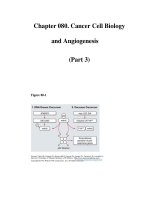
Fig. 3 SNHG3 competitively bound to miR-154-3p and regulated Notch2. a. Subcellular localization of SNHG3 in the LncATLAS database; b. FISH
experiments with probes targeting SNHG3 were performed to validate the subcellular localization of SNHG3 in MCF-7 and HCC1937 were stained
with probes targeting SNHG3 (red stain), and the nuclei were stained with 4′,6-diamidino-2-phenylindole (blue stain). The merged image showed
SNHG3 was cytoplasm-sublocalized in MCF-7 and HCC1937; c. Nuclear and cytoplasmic expression of SNHG3 in MCF-7 and HCC1937 cells
determined by RT-qPCR; d. Kalpan-Meier plotter predicted breast cancer prognosis via miR-154-3p expression level; e. Luciferase reporter plasmid
containing SNHG3-WT or SNHG3-Mut was transfected into 293 T cells together with miR-154-3p in parallel with an miR-NC plasmid vector;
luciferase reporter plasmid containing SNHG3-WT or SNHG3-Mut was transfected into 293 T cells together with miR-154-3p in parallel with an
miR-NC plasmid vector; luciferase reporter plasmid containing NOTCH2-WT or NOTCH2-Mut was transfected into 293 T cells together with miR154-3p in parallel with an miR-NC plasmid vector; f. the binding relationship between miR-154-3p, SNHG3 and Notch2 was verified by RNA pulldown assay; g. RT-qPCR was performed to determine the levels of miR-154-3p and Notch2 mRNA in MCF-7 and HCC1937 cells.; h. Western blot
assay was performed to determine Notch2 protein level in MCF-7 and HCC1937 cells (representative images were shown, full-length gels are
presented in Supplementary Figure 2); J. RT-qPCR and western blot analysis were performed to determine Notch2 level in MCF-7 and HCC1937
cells. Three independent experiments were performed. Data are expressed as mean ± standard deviation; one-way ANOVA and Tukey’s Multiple
comparison test were used to determine statistical significance, *p < 0.05
enhanced Notch2 activity by competitively binding to
miR-154-3p, thus promoting BC cell proliferation and
metastasis.
Activation of the notch signaling pathway partly reversed
the inhibition of cell activity induced by intervening SNHG3
Jagged 1, a specific activator of the Notch signaling pathway,
was added into MCF-7 cells after intervening SNHG3 expression. The results of RT-qPCR and Western blot analysis
showed that mRNA and protein levels of Notch1, Notch2
and Notch3 improved apparently (p < 0.05) (Fig. 4a/b), accompanied by the improvement of cell activity, proliferation,
invasion and migration (p < 0.05) (Fig. 4c-h).
SNHG3 intervention inhibits the growth of BC cell
xenograft tumor in vivo
The growth and weight of transplanted tumors were
measured to evaluate the effect of SNHG3 on MCF-7
cells in vivo. It was showed that inhibited SNHG3 suppressed the growth of tumor (p < 0.05) (Fig. 5a/b). The
result of immunohistochemistry revealed that after the
inhibition of SNHG3 expression, Notch1-, Notch2- and
Notch3-positive cells in MCF-7 xenograft tumor increased (p < 0.05) (Fig. 5c).
Discussion
As the most common malignant cancer and main cause
of mortality in women, BC showed a high survival rate,
but reducing BC incidence and mortality remains a priority for the public [32]. Besides, lncRNAs are deregulated in a variety of cancers and regulate cancer-related
pathways, indicating that they play vital roles in cancer
prognosis [33]. A prior study has demonstrated that
lncRNA MIAT promotes BC progression and functions
as ceRNA to regulate DUSP7 expression by sponging
miR-155-5p [34]. In this study, we assumed that there
may be roles of lncRNA SNHG3 in BC cell proliferation
and metastasis via the Notch signaling pathway.
Consequently, our data showed that SNHG3 competitively bound to miR-154-3p and activated the Notch signaling pathway to promote BC cell proliferation and
metastasis.
Firstly, the results of transcriptome sequencing showed
that SNHG3 was expressed higher in BC cells than that in
normal breast cells. Consistently, another study reported
that SNHG3 expression was remarkably higher in ovarian
cancer tissues than in adjacent normal tissues, and upregulating SNHG3 expression linked with poor prognosis
and enhanced malignant progression of ovarian cancer
[35]. LncRNA SNHG3 was proved to be upregulated in
BC cells [7]. Functional assays by Liang Liu et al. have suggested that upregulated SNHG3 led to growth of cell proliferation, cell cycle progress and decrease of cell
apoptosis, indicating that SNHG3 served as an oncogene
in lung cancer by controlling tRNA processing, transcription, apoptosis, cell adhesion and signal transduction [22].
Additionally, this current study also suggested that BC cell
proliferation, invasion and migration evidently decreased
with inhibited SNHG3. Lan Hong and his colleagues
found that ovarian cancer cell proliferation and invasion
were inhibited after SNHG3 knockdown [35]. Similarly,
SNHG1 promoted miR-448 expression, suppressed regulatory T cell differentiation, and eventually impeded the
immune escape of BC [36]. Meanwhile, a recent article
has indicated that overexpressed SNHG3 encouraged
osteosarcoma (OS) cell invasion and migration, lessening
the survival rate of OS patients [37]. That’s to say, a higher
survival rate could be achieved by the inhibition of
SNHG3. Therefore, poor expression of SNHG3 might act
as a possible therapeutic target for BC. What’s more, functional assays in our study found that the E-cadherin level
was expressly enhanced and N-cadherin level was noticeably declined after interfering SNHG3. As a tumor
suppressor, E-cadherin played an important role in encouraging BC cell progression and metastasis [38]. Ncadherin expression promoted BC cell mobility, invasion
Jiang et al. BMC Cancer
(2020) 20:838
Page 10 of 13
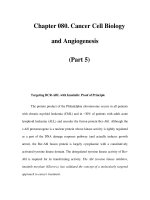
Fig. 4 Notch signaling pathway activation reversed BC cells proliferation and viability by SNHG3 silencing. MCF-7 stably expressed si-SNHG3–2
was treated with Notch signaling pathway specific activator, Jagged 1. RT-qPCR and Western blot analysis were performed to determine Notch1,
Notch2 and Notch3 mRNA (a) and protein (b) levels after Jagged 1 treatment (representative images were shown, full-length gels are presented
in Supplementary Figure 3); MCF-7 cells were performed with MTT proliferation assay (c) and EdU staining (d) and colony formation assays (e) to
determine Notch signaling pathway activation effectiveness; f. MCF-7 cells migrating from upper Transwell chambers into lower ones, without
Matrigel (× 200); g. MCF-7 cells invading from Matrigel-coated upper Transwell chambers into lower ones (× 200); h. Western blot analysis was
carried out to determine E-cadherin and N-cadherin protein level (representative images were shown, full-length gels are presented in
Supplementary Figure 4). Three independent experiments were performed. Data are expressed as mean ± standard deviation; one-way ANOVA
and Sidak’s multiple comparisons test was used to determine statistical significance, or two-way ANOVA and Tukey’s multiple comparisons test
was used, *p < 0.05, **p < 0.01
and migration [39]. So, interfered SNHG3 could repress
BC cell biological behaviors.
Additionally, dual-luciferase reporter gene assay found
a link between SNHG3 and miR-154-3p. Then, we focused on miR-154-3p. Recently, it has been found that
in BC cells where lncRNA SNHG5 was negatively correlated with miR-154-5p, increase of SNHG5 suppressed
miR-154-5p and upregulated proliferation cell nuclear
antigen, promoting BC cell biological processes [40]. Another study has unearthed that SNHG1 served as a
sponge in weakening miR-154-5p, which could regulate
BC cell proliferation and apoptosis [41]. Besides, in our
study, the binding relation between miR-154-3p and
Notch2 was also found in a dual-luciferase reporter gene
assay. Highly expressed Notch2 was found to improve
survival rate in many BC patients and was important in
Notch signaling pathway activation [42, 43]. Mattia
Capulli et al. have demonstrated that BC cell proliferation was repressed by endosteal niche cells in a Notch2related way [12]. However, this study was the first to
Jiang et al. BMC Cancer
(2020) 20:838
Fig. 5 (See legend on next page.)
Page 11 of 13
Jiang et al. BMC Cancer
(2020) 20:838
Page 12 of 13
(See figure on previous page.)
Fig. 5 SNHG3 intervention can inhibit the growth of BC cell xenograft tumor in vivo. MCF-7 cells stably SNHG3-siRNA and scramble siRNA were
inoculated subcutaneously into BALB/c nude mice at a dose of 5 × 10⁶ per mouse (n = 6 in each group). Tumor growth was measured
continuously every 5 days, and 20 days later, tumor growth was monitored every 3 days. At 35 days post-implantation, the mice were euthanized
by carbon dioxide asphyxiation. a. Tumor size; b. Tumor weight and representative view of xenografts. Tumor sections were obtained and stained
with anti-Notch1, anti-Notch2 and anti-Notch3 antibodies; c. Representative views of Notch1, Notch2 and Notch3-positive tumor cells and
quantification of immunohistochemistry. Data are expressed as mean ± standard deviation. One-way ANOVA and Sidak’s multiple comparisons
test was used to determine statistical significance, or two-way ANOVA and Tukey’s multiple comparisons test was used, *p < 0.05, **p < 0.01
explore the molecular mechanism between SNHG3,
miR-154 and Notch2 pathway. It was found that SNHG3
could act as a ceRNA of miR-154-3p and upregulate the
Notch signaling pathway to promote BC cell proliferation and metastasis. Moreover, RT-qPCR and Western
blot analysis found that activating the Notch signaling
pathway encouraged BC cell viability, proliferation, invasion and migration. Previous research suggested that aberrant Notch signaling pathway played a significant role
in implicating BC cell progression [44], which was in
agreement with our results. Interestingly, effect of
Notch2 on BC cell proapoptotic and anti-migratory
response has been revealed to be inhibited when it was
activated by zerumbone [31]. Furthermore, Notch signaling pathway has been found to play a significant role in
breast epithelial cell differentiation and participated in
BC growth by Notch receptors and ligands [45].
Conclusion
In summary, our study supported that lncRNA SNHG3
promoted BC cell proliferation and metastasis by competitively binding to miR-154-3p and activating the
Notch signaling pathway. Now, molecule-targeted treatment of tumors has been widely accepted. The results in
this study may provide novel insights for the molecular
therapy of BC. In the future, we will further explore the
mechanism of other targets of lncRNA SNHG3, and explore the role of Notch 1 and Notch 3 in breast cancer.
We will carry out relevant researches, for example the
rescue experiments in which inhibition of miR-154 negates growth inhibitory effects caused by SNHG3 knockdown under the permission of experimental conditions
and funds. Although our findings provide therapeutic
implication in BC treatment, the experiment results and
effective application into clinical practice need further
validation.
Supplementary information
Supplementary information accompanies this paper at />1186/s12885-020-07275-5.
Additional file 1.
Abbreviations
ANOVA: Analysis of variance; BC: Breast cancer; ceRNA: Competing
endogenous RNA; DAPI: 4′,6-diamidino-2-phenylindole; EdU: 5-ethynyl2′-deoxyuridine; EMT: Epithelial-mesenchymal transition; ER/PR: Estrogen
receptor/progesterone receptor; FISH: Fluorescence in situ
hybridization; HRP: Horseradish peroxidase; lncRNA: Long non-coding
RNA; miR: MicroRNA; MTT: 3-(4, 5-dimethylthiazol-2-yl)-2, 5diphenyltetrazolium bromide; OC: Ovarian cancer; OS: Osteosarcoma;
PBS: Phosphate buffered saline; PVDF: Polyvinylidene fluoride; RTqPCR: Reverse transcription quantitative polymerase chain reaction;
siRNA: Small interfere RNA; SNHG3: Small nucleolar RNA host gene 3;
TBS: Tris buffered saline; TBST: Tris-buffered saline tween;
TSA: Trichostatin
Acknowledgements
Not applicable.
Authors’ contributions
HNJ is the guarantor of integrity of the entire study and contributed
to the study concepts; XJL and WW contributed to the study design
and experiment; HLD took charge of the acquisition and analysis of
data; HNJ contributed to the manuscript preparation and manuscript
editing. All authors read and approved the final manuscript.
Funding
Not applicable.
Availability of data and materials
The datasets used and/or analysed during the current study available from
the corresponding author on reasonable request.
Ethics approval and consent to participate
This study was approved and supervised by the ethics committee of Second
Hospital of Shanxi Medical University. All subjects or their families were
informed and had signed the informed consent. Animal experiments were
performed in compliance with the recommendations in the Guide for the Care
and Use of Laboratory Animals of the National Institutes of Health. The protocol
was approved by the Animal Ethics Committee of the Clinical Ethical
Committee of the Second Hospital of Shanxi Medical University.
Consent for publication
Not applicable.
Competing interests
All authors declare that there is no conflict of interests in this study.
Author details
Department of Breast Surgery, The Second Hospital of Shanxi Medical
University, Taiyuan 030001, Shanxi, PR China. 2Department of Rdaiology, The
Second Hospital of Shanxi Medical University, Taiyuan 030001, Shanxi, PR
China. 3Department of Vascular Surgery, The Second Hospital of Shanxi
Medical University, No. 382, Wuyi Road, Taiyuan 030001, Shanxi, PR China.
1
Received: 19 November 2019 Accepted: 9 August 2020
Additional file 2.
Additional file 3.
Additional file 4.
References
1. Akram M, Iqbal M, Daniyal M, Khan AU. Awareness and current knowledge
of breast cancer. Biol Res. 2017;50(1):33.
Jiang et al. BMC Cancer
2.
3.
4.
5.
6.
7.
8.
9.
10.
11.
12.
13.
14.
15.
16.
17.
18.
19.
20.
21.
22.
23.
24.
25.
(2020) 20:838
Winters S, Martin C, Murphy D, Shokar NK. Breast Cancer epidemiology,
prevention, and screening. Prog Mol Biol Transl Sci. 2017;151:1–32.
Peart O. Breast intervention and breast cancer treatment options. Radiol
Technol. 2015;86(5):535M–58M quiz 559-562.
Braden AM, Stankowski RV, Engel JM, Onitilo AA. Breast cancer biomarkers:
risk assessment, diagnosis, prognosis, prediction of treatment efficacy and
toxicity, and recurrence. Curr Pharm Des. 2014;20(30):4879–98.
Guo ZH, You ZH, Wang YB, Yi HC, Chen ZH. A Learning-Based Method for
LncRNA-Disease Association Identification Combing Similarity Information
and Rotation Forest. iScience. 2019;19:786–95.
Liu Y, Sharma S, Watabe K. Roles of lncRNA in breast cancer. Front Biosci
(Schol Ed). 2015;7:94–108.
Liang C, Hu Z, Zhou Z, Zhang H. SNHG3 promotes proliferation and
invasion by regulating the miR-101/ZEB1 axis in breast cancer. RSC Adv.
2018;8(27):15229–40.
Huang W, Tian Y, Dong S, Cha Y, Li J, Guo X, Yuan X. The long non-coding
RNA SNHG3 functions as a competing endogenous RNA to promote
malignant development of colorectal cancer. Oncol Rep. 2017;38(3):1402–10.
Khan S, Ayub H, Khan T, Wahid F. MicroRNA biogenesis, gene silencing
mechanisms and role in breast, ovarian and prostate cancer. Biochimie.
2019;167:12–24.
Xu H, Fei D, Zong S, Fan Z. MicroRNA-154 inhibits growth and invasion of
breast cancer cells through targeting E2F5. Am J Transl Res. 2016;8(6):2620–30.
Li S, Meng H, Zhou F, Zhai L, Zhang L, Gu F, Fan Y, Lang R, Fu L, Gu L, et al.
MicroRNA-132 is frequently down-regulated in ductal carcinoma in situ
(DCIS) of breast and acts as a tumor suppressor by inhibiting cell
proliferation. Pathol Res Pract. 2013;209(3):179–83.
Capulli M, Hristova D, Valbret Z, Carys K, Arjan R, Maurizi A, Masedu F,
Cappariello A, Rucci N, Teti A. Notch2 pathway mediates breast cancer
cellular dormancy and mobilisation in bone and contributes to
haematopoietic stem cell mimicry. Br J Cancer. 2019;121(2):157–71.
Braune EB, Lendahl U. Notch -- a goldilocks signaling pathway in disease
and cancer therapy. Discov Med. 2016;21(115):189–96.
Li L, Tang P, Li S, Qin X, Yang H, Wu C, Liu Y. Notch signaling pathway
networks in cancer metastasis: a new target for cancer therapy. Med Oncol.
2017;34(10):180.
Krishna BM, Jana S, Singhal J, Horne D, Awasthi S, Salgia R, Singhal SS.
Notch signaling in breast cancer: from pathway analysis to therapy. Cancer
Lett. 2019;461:123–31.
Fortunato GM, Da Ros F, Bisconti S, De Acutis A, Biagini F, Lapomarda A,
Magliaro C, De Maria C, Montemurro F, Bizzotto D, et al. Electrospun
structures made of a hydrolyzed keratin-based biomaterial for development
of in vitro tissue models. Front Bioeng Biotechnol. 2019;7:174.
Lu M, Wang T, He M, Cheng W, Yan T, Huang Z, Zhang L, Zhang H, Zhu W,
Zhu Y, et al. Tumor suppressor role of miR-3622b-5p in ERBB2-positive
cancer. Oncotarget. 2017;8(14):23008–19.
Cui F, Hu J, Ning S, Tan J, Tang H. Overexpression of MCM10 promotes cell
proliferation and predicts poor prognosis in prostate cancer. Prostate. 2018;
78(16):1299–310.
Li J, Guo Y, Duan L, Hu X, Zhang X, Hu J, Huang L, He R, Hu Z, Luo W, et al.
AKR1B10 promotes breast cancer cell migration and invasion via activation
of ERK signaling. Oncotarget. 2017;8(20):33694–703.
Campbell PS, Mavingire N, Khan S, Rowland LK, Wooten JV, Opoku-Agyeman
A, Guevara A, Soto U, Cavalli F, Loaiza-Perez AI, et al. AhR ligand aminoflavone
suppresses alpha6-integrin-Src-Akt signaling to attenuate tamoxifen resistance
in breast cancer cells. J Cell Physiol. 2018;234(1):108–21.
Chen J, Wu Z, Zhang Y. LncRNA SNHG3 promotes cell growth by sponging
miR-196a-5p and indicates the poor survival in osteosarcoma. Int J
Immunopathol Pharmacol. 2019;33:2058738418820743.
Liu L, Ni J, He X. Upregulation of the long noncoding RNA SNHG3
promotes lung adenocarcinoma proliferation. Dis Markers. 2018;2018:
5736716.
Fei F, He Y, He S, He Z, Wang Y, Wu G, Li M. LncRNA SNHG3 enhances the
malignant progress of glioma through silencing KLF2 and p21. Biosci Rep.
2018;38(5):BSR20180420.
Taherian-Esfahani Z, Taheri M, Dashti S, Kholghi-Oskooei V, Geranpayeh L,
Ghafouri-Fard S. Assessment of the expression pattern of mTOR-associated
lncRNAs and their genomic variants in the patients with breast cancer. J
Cell Physiol. 2019;234(12):22044–56.
Chandrashekar DS, Bashel B, Balasubramanya SAH, Creighton CJ, PonceRodriguez I, Chakravarthi B, Varambally S. UALCAN: a portal for facilitating
Page 13 of 13
26.
27.
28.
29.
30.
31.
32.
33.
34.
35.
36.
37.
38.
39.
40.
41.
42.
43.
44.
45.
tumor subgroup gene expression and survival analyses. Neoplasia. 2017;
19(8):649–58.
Mas-Ponte D, Carlevaro-Fita J, Palumbo E, Hermoso Pulido T, Guigo R,
Johnson R. LncATLAS database for subcellular localization of long
noncoding RNAs. RNA. 2017;23(7):1080–7.
Li JH, Liu S, Zhou H, Qu LH, Yang JH. starBase v2.0: decoding miRNA-ceRNA,
miRNA-ncRNA and protein-RNA interaction networks from large-scale CLIPSeq data. Nucleic Acids Res. 2014;42(Database issue):D92–7.
Zhang J, Mao S, Wang L, Zhang W, Zhang Z, Guo Y, Wu Y, Yi F, Yao X.
MicroRNA154 functions as a tumor suppressor in bladder cancer by directly
targeting ATG7. Oncol Rep. 2019;41(2):819–28.
Zhang JY, Gong YL, Li CJ, Qi Q, Zhang QM, Yu DM. Circulating MiRNA
biomarkers serve as a fingerprint for diabetic atherosclerosis. Am J Transl
Res. 2016;8(6):2650–8.
Nagy A, Lanczky A, Menyhart O, Gyorffy B. Author correction: validation of
miRNA prognostic power in hepatocellular carcinoma using expression data
of independent datasets. Sci Rep. 2018;8(1):11515.
Sehrawat A, Sakao K, Singh SV. Notch2 activation is protective against
anticancer effects of zerumbone in human breast cancer cells. Breast Cancer
Res Treat. 2014;146(3):543–55.
Miller JW, Smith JL, Ryerson AB, Tucker TC, Allemani C. Disparities in breast
cancer survival in the United States (2001-2009): Findings from the
CONCORD-2 study. Cancer. 2017;123(Suppl 24):5100–18.
Soudyab M, Iranpour M, Ghafouri-Fard S. The role of long non-coding RNAs
in breast Cancer. Arch Iran Med. 2016;19(7):508–17.
Luan T, Zhang X, Wang S, Song Y, Zhou S, Lin J, An W, Yuan W, Yang Y, Cai
H, et al. Long non-coding RNA MIAT promotes breast cancer progression
and functions as ceRNA to regulate DUSP7 expression by sponging miR155-5p. Oncotarget. 2017;8(44):76153–64.
Hong L, Chen W, Wu D, Wang Y. Upregulation of SNHG3 expression
associated with poor prognosis and enhances malignant progression of
ovarian cancer. Cancer Biomark. 2018;22(3):367–74.
Pei X, Wang X, Li H. LncRNA SNHG1 regulates the differentiation of Treg
cells and affects the immune escape of breast cancer via regulating miR448/IDO. Int J Biol Macromol. 2018;118(Pt A):24–30.
Zheng S, Jiang F, Ge D, Tang J, Chen H, Yang J, Yao Y, Yan J, Qiu J, Yin Z,
et al. LncRNA SNHG3/miRNA-151a-3p/RAB22A axis regulates invasion and
migration of osteosarcoma. Biomed Pharmacother. 2019;112:108695.
Hugo HJ, Gunasinghe N, Hollier BG, Tanaka T, Blick T, Toh A, Hill P, Gilles C,
Waltham M, Thompson EW. Epithelial requirement for in vitro proliferation
and xenograft growth and metastasis of MDA-MB-468 human breast cancer
cells: oncogenic rather than tumor-suppressive role of E-cadherin. Breast
Cancer Res. 2017;19(1):86.
Derycke LD, Bracke ME. N-cadherin in the spotlight of cell-cell adhesion,
differentiation, embryogenesis, invasion and signalling. Int J Dev Biol. 2004;
48(5–6):463–76.
Chi JR, Yu ZH, Liu BW, Zhang D, Ge J, Yu Y, Cao XC. SNHG5 promotes breast
Cancer proliferation by sponging the miR-154-5p/PCNA Axis. Mol Ther
Nucleic Acids. 2019;17:138–49.
Xu M, Chen X, Lin K, Zeng K, Liu X, Pan B, Xu X, Xu T, Hu X, Sun L, et al. The
long noncoding RNA SNHG1 regulates colorectal cancer cell growth
through interactions with EZH2 and miR-154-5p. Mol Cancer. 2018;17(1):141.
Xu J, Song F, Jin T, Qin J, Wu J, Wang M, Wang Y, Liu J. Prognostic values of
notch receptors in breast cancer. Tumour Biol. 2016;37(2):1871–7.
Zhou S, Yu L, Xiong M, Dai G. LncRNA SNHG12 promotes tumorigenesis
and metastasis in osteosarcoma by upregulating Notch2 by sponging miR195-5p. Biochem Biophys Res Commun. 2018;495(2):1822–32.
Lamy M, Ferreira A, Dias JS, Braga S, Silva G, Barbas A. Notch-out for breast
cancer therapies. New Biotechnol. 2017;39(Pt B):215–21.
Kontomanolis EN, Kalagasidou S, Pouliliou S, Anthoulaki X, Georgiou N,
Papamanolis V, Fasoulakis ZN. The notch pathway in breast Cancer
progression. ScientificWorldJournal. 2018;2018:2415489.
Publisher’s Note
Springer Nature remains neutral with regard to jurisdictional claims in
published maps and institutional affiliations.