Salvage chemoradiation therapy for recurrence after radical surgery or palliative surgery in esophageal cancer patients: A prospective, multicenter clinical trial protocol
Bạn đang xem bản rút gọn của tài liệu. Xem và tải ngay bản đầy đủ của tài liệu tại đây (3 MB, 8 trang )
Chang et al. BMC Cancer
(2020) 20:877
/>
STUDY PROTOCOL
Open Access
Salvage chemoradiation therapy for
recurrence after radical surgery or palliative
surgery in esophageal cancer patients: a
prospective, multicenter clinical trial
protocol
Xiao Chang1†, Lei Deng1†, Wenjie Ni1, Chen Li1, Weiming Han1, Lin-rui Gao1, Shijia Wang1, Zongmei Zhou1,
Dongfu Chen1, Qinfu Feng1, Jun Liang1, Nan Bi1, Jima Lv1, Shugeng Gao2, Yousheng Mao2, Qi Xue2 and
Zefen Xiao1*
Abstract
Background: Currently, adjuvant therapy is not recommended for patients with thoracic esophageal squamous cell
cancer (TESCC) after radical surgery, and a proportion of these patients go on to develop locoregional recurrence
(LRR) within 2 years. Besides, there is no evidence for salvage chemoradiation therapy (CRT) in patients with residual
tumor after esophagectomy (R1/R2 resection). In addition, factors like different failure patterns and relationship with
normal organs influence the decision for salvage strategy. Here, we aimed to design a modularized salvage CRT
strategy for patients without a chance of salvage surgery according to different failure patterns (including R1/R2
resection), and further evaluated its efficacy and safety.
Methods: Our study was designed as a one arm, multicenter, prospective clinical trial. All enrolled patients were
stratified in a stepwise manner based on the nature of surgery (R0 or R1/2), recurrent lesion diameter, involved
regions, and time-to-recurrence, and were further assigned to undergo either elective nodal irradiation or involved
field irradiation. Then, radiation technique and dose prescription were modified according to the distance from the
recurrent lesion to the thoracic stomach or intestine. Ultimately, four treatment plans were established.
Discussion: This prospective study provided high-level evidence for clinical salvage management in patients with
TESCC who developed LRR after radical surgery or those who underwent R1/R2 resection.
Trial registration: Prospectively Registered. ClinicalTrials.gov NCT03731442, Registered November 6, 2018.
Keywords: Esophageal neoplasm, Locoregional recurrence, R1/R2 resection, Chemoradiation therapy, Palliative
management
* Correspondence:
†
Xiao Chang and Lei Deng are first authors responsible for paper writing and
patients enrollment
1
Department of Radiation Oncology, National Cancer Center/National Clinical
Research Center for Cancer/Cancer Hospital, Chinese Academy of Medical
Sciences and Peking Union Medical College, No. 17 South Panjiayuan lane,
Chaoyang District, Beijing 100021, China
Full list of author information is available at the end of the article
© The Author(s). 2020 Open Access This article is licensed under a Creative Commons Attribution 4.0 International License,
which permits use, sharing, adaptation, distribution and reproduction in any medium or format, as long as you give
appropriate credit to the original author(s) and the source, provide a link to the Creative Commons licence, and indicate if
changes were made. The images or other third party material in this article are included in the article's Creative Commons
licence, unless indicated otherwise in a credit line to the material. If material is not included in the article's Creative Commons
licence and your intended use is not permitted by statutory regulation or exceeds the permitted use, you will need to obtain
permission directly from the copyright holder. To view a copy of this licence, visit />The Creative Commons Public Domain Dedication waiver ( applies to the
data made available in this article, unless otherwise stated in a credit line to the data.
Chang et al. BMC Cancer
(2020) 20:877
Background
According to the 2019 National Comprehensive Cancer
Network (NCCN) guidelines for esophageal cancer [1],
adjuvant treatment is not recommended for patients
with thoracic esophageal squamous cell cancer (TESCC)
who received radical surgery as their first treatment, regardless of the T and N status. However, the recurrence
rate is as high as 23.8–58%, and the median time to recurrence is about 10.5 months [2–6]. Even in Japan
where three-field lymphadenectomy is the preferred
treatment option, 24–46% patients go on to experience
recurrence after R0 resection, which is the main cause of
surgical treatment failure [7–10]. Besides, in patients
with residual tumor (R1/R2), salvage chemoradiation
therapy (CRT) is recommended as the main component
of palliative management for locoregional recurrence
(LRR) disease. However, data of large samples or highlevel evidence are still lacking.
Our previous retrospective analysis [11] indicated that
most patients developed lymph node recurrence in the
supraclavicular (25.8%) and upper mediastinal (44.4%)
regions, and those who underwent salvage CRT had significantly better survival than those that underwent
radiotherapy alone, chemotherapy, or best supportive
care. Similar results were found in other studies [12–15].
Overall survival (OS) directly depended on failure patterns and corresponding treatment strategies, so prospective clinical trials were necessary for screening of
specific patients to attain survival benefit from the optimal salvage strategy.
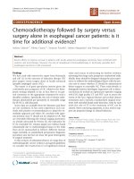
This study was aimed to design a modularized salvage
CRT strategy for patients unsuited for salvage surgery
Fig. 1 Flow chart of the trial
Page 2 of 8
based on different failure patterns (including R1/R2 resection) and further evaluate its efficacy and safety.
Methods/design
Study design and objectives
The current study was designed as a one-arm, multicenter, prospective clinical trial. The enrolled patients were
stratified in a stepwise manner based on the nature of
surgery (R0 or R1/2), recurrent lesion diameter, involved
regions, and time-to-recurrence, and were further
assigned to undergo either elective nodal irradiation
(ENI) or involved field irradiation (IFI). Then, radiation
technique and dose prescription were modified according to the distance from the recurrent lesion to thoracic
stomach or intestine. Ultimately, four treatment plans
were established. A flow chart of the study overview is
shown in Fig. 1.
The primary end point is the 1-, 2-, and 3-year OS.
The secondary end points include the 1-, 2-, and 3-year
rates of progression-free survival (PFS), completion
rates, out-field recurrence, and toxicity profiles.
The study began on November 2018, and patients will
continue to be included until November 2022.
Patient population
Patients enrolled thus far mainly comprise untreated patients after LRR or palliative surgery. The inclusion criteria include: (1) R1/R2 resection, (2) LRR after radical
surgery, (3) out-field LRR after adjuvant chemoradiation
or radiotherapy, (4) LRR after adjuvant chemotherapy,
(5) no prior therapy after LRR, (6) age 16–70 years, (7)
good general condition (i.e., Karnofsky Performance
Chang et al. BMC Cancer
(2020) 20:877
Status [KPS] ≥70)], (8) normal complete blood count
(CBC), especially white blood cell count > 4.0*10^9/L,
(9) satisfactory liver and kidney functions.
The exclusion criteria include: (1) prior malignancies
within 5 years, (2) pregnant status or lactation, (3) history of drug allergy, (4) refused informed consent (5)
non-regional lymph node (except for metastasis to
supraclavicular or celiac lymph nodes) or distant metastasis (including metastasis to organs including bone,
lung or liver etc.) (6) severe cardiovascular diseases, infections, active ulcerations, diabetes mellitus with unstable blood sugar, and mental disorders.
Recurrence
Tumor residue includes positive pathological margins of
the specimens (R1) and incomplete tumor resection during the operation (R2). LRR is defined as recurrence at
sites of the anastomosis, tumor bed, mediastinal lymph
nodes, or para-gastric lymph nodes (including nodes adjacent to the cardia or along the course of the left gastric
artery). Recurrence in the deep cervical, supraclavicular,
or celiac regions are also defined as regional relapse. Distant metastasis was defined as metastasis in the liver,
lung, bone, and pleura; subcutaneous metastasis; and
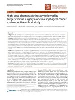
Fig. 2 Illustration depicting reclassified regions
Page 3 of 8
other nonregional lymph node metastasis such as axillary and inguinal lymph nodes. If a second recurrence
was detected within 4 weeks after the first occurrence, it
was considered synchronous. Once suspicious recurrent
lesions are identified on imaging, biopsy is attempted.
The diagnostic standard for imaging should meet the
criteria of significant enlargement or increase in the
number of existing lymph nodes, or the appearance of
the new lymph nodes compared with previous examinations. Otherwise, positron emission tomographycomputed tomography (PET-CT) clearly diagnoses recurrence through metabolic activity and imaging
features.
To comprehensively describe the design of target volume, the 8th American Joint Committee on Cancer
(AJCC) regional lymph node stations [16] were reclassified into four regions (Fig. 2). Region I includes the area
above the sternal notch, including the supraclavicular
space and No. 1 lymphatic drainage region; region II includes the mediastinal No. 2, 4, and 8 U lymphatic drainage regions; region III includes mediastinal No. 7, 8 M/
Lo, and 9 lymphatic drainage regions; and region IV
includes the abdominal No. 15–20 lymphatic drainage
regions. Close region recurrence was defined as
Chang et al. BMC Cancer
(2020) 20:877
recurrences within the sites of (1) regions I and II, (2)
regions II and III, (3) regions III and IV, or (4) regions I
and III. Distant regional metastasis was defined as recurrences at the both sites of regions I and IV or region II
and IV.
Radiotherapy
The planning CT was recommended to be fused with
planning magnetic resonance imaging (MRI) or PETCT, if available, to further improve the contouring accuracy. The gross tumor volume (GTV-T) or metastatic
regional nodes (GTV-N) is defined as the residual
tumor, tumor-bed recurrence, or metastatic lymph node.
The planning gross tumor volume (PGTV) is created by
expanding GTV-T or GTV-N with a uniform 0.5-cm
margin. As for delineation of clinical target volume
(CTV), both IFI and ENI were adopted.
In the ENI group, the principle to design prophylactic
target volume of high-risk lymphatic drainage regions basically comprised GTV-T/GTV-N plus a 3.0–5.0-cm craniocaudal and 0.6-cm horizontal margin. For recurrence
in regions I or II, CTV comprised the region with the
upper boundary at the upper margin of the T1 vertebral
body or 1.0–1.5-cm superior to GTV-N and lower boundary in the 2.0–3.0-cm inferior to the carina, including the
supraclavicular space and No. 1, 2, 4, 7, and 8 U stations.
For recurrence in region III, the CTV comprised the
Page 4 of 8
region with upper boundary at the level of the clavicular
head and lower boundary in the margin 2.0-cm inferior to
the carina or 1.0–1.5-cm inferior to GTV-N, including
No. 2, 4, 7, and 8 U/M stations. For recurrence in region
IV, CTV comprised the region with upper boundary in
the 1.0–1.5-cm superior to GTV-N and lower boundary
in the celiac axis or 1.5-cm inferior to GTV-N, including
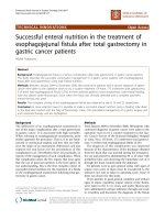
No. 15–20 stations. The technique of intensity-modulated
radiation therapy (IMRT) with simultaneously integrated
boost (SIB) or sequential boost was modified according to
the safety of the thoracic stomach or intestine. Figure 3a
shows the SIB-IMRT being applied to a recurrent lesion
far from the thoracic stomach with a prescription dose of
PTV 50.4 Gy/1.8 Gy/28 f and PGTV 59.92–62.16 Gy/
2.14–2.22 Gy/28 f. Figure 3b shows the IMRT with sequential boost applied to a recurrent lesion close to the
thoracic stomach with a prescription dose of PTV 50.4
Gy/1.8 Gy/28 f and a sequential boost to PGTV 10–12
Gy/1.8–2 Gy/5–7 f.
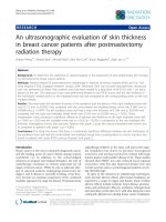
In the IFI group, CTV only consisted of GTV-T/GTVN plus a 0.6–0.8-cm horizontal margin and 1.0–1.5-cm
craniocaudal margin. No prophylactic irradiation was
delivered to any lymph node drainage regions. For lesions located away from the thoracic stomach (Fig. 4a),
the prescribed dose was 60 Gy/2 Gy/�������������������������������������������������������������������������������������������������������������������������������������������������������������������������������������������������������������������������������������������������������������������������������������������������������������������������������������������������������������������������������������������������������������������������������������������������������������������������������������������������������������������������������������������������������������������������������������������������������������������������������������������������������������������������������������������������������������������������������������������������������������������������������������������������������������������������������������������������������������������������������������������������������������������������������������������������������������������������������������������������������������������������������������������������������������������������������������������������������������������������������������������������������������������������������������������������������������������������������������������������������������������������������������������������������������������������������������������������������������������������������������������������������������������������������������������������������������������������������������������������������������������������������������������������������������������������������������������������������������������������������������������������������������������������������������������������������������������������������������������������������������������������������������������������������������������������������������������������������������������������������������������������������������������������������������������������������������������������������������������������������������������������������������������������������������������������������������������������������������������������������������������������������������������������������������������������������������������������������������������������������������������������������������������������������������������������������������������������������������������������������������������������������������������������������������������������������������������������������������������������������������������������������������������������������������������������������������������������������������������������������������������������������������������������������������������������������������������������������������������������������������������������������������������������������������������������������������������������������������������������������������������������������������������������������������������������������������������������������������������������������������������������������������������������������������������������������������������������������������������������������������������������������������������������������������������������������������������������������������������������������������������������������������������������������������������������������������������������������������������������������������������������������������������������������������������������������������������������������������������������������������������������������������������������������������������������������������������������������������������������������������������������������������������������������������������������������������������������������������������������������������������������������������������������������������������������������������������������������������������������������������������������������������������������������������������������������������������������������������������������������������������������������������������������������������������������������������������������������������������������������������������������������������������������������������������������������������������������������������������������������������������������������������������������������������������������������������������������������������������������������������������������������������������������������������������������������������������������������������������������������������������������������������������������������������������������������������������������������������������������������������������������������������������������������������������������������������������������������������������������������������������������������������������������������������������������������������������������������������������������������������������������������������������������������������������������������������������������������������������������������������������������������������������������������������������������������������������������������������������������������������������������������������������������������������������������������������������������������������������������������������������������������������������������������������������������������������������������������������������������������������������������������������������������������������������������������������������������������������������������������������������������������������������������������������������������������������������������������������������������������������������������������������������������������������������������������������������������������������������������������������������������������������������������������������������������������������������������������������������������������������������������������������������������������������������������������������������������������������������������������������������������������������������������������������������������������������������������������������������������������������������������������������������������������������������������������������������������������������������������������������������������������������������������������������������������������������������������������������������������������������������������������������������������������������������������������������������������������������������������������������������������������������������������������������������������������������������������������������������������������������������������������������������������������������������������������������������������������������������������������������������������������������������������������������������������������������������������������������������������������������������������������������������������������������������������������������������������������������������������������������������������������������������������������������������������������������������������������������������������������������������������������������������������������������������������������������������������������������������������������������������������������������������������������������������������������������������������������������������������������������������������������������������������������������������������������������������������������������������������������������������������������������������������������������������������������������������������������������������������������������������������������������������������������������������������������������������������������������������������������������������������������������������������������������������������������������������������������������������������������������������������������������������������������������������������������������������������������������������������������������������������������������������������������������������������������������������������������������������������������������������������������������������������������������������������������������������������������������������������������������������������������������������������������������������������������������������������������������������������������������������������������������������������������������������������������������������������������������������������������������������������������� Our prospective phase I/II trial [25]
supported the safety and efficacy of the dose patterns
adopted in this trial (95% PGTV/PTV 59.92 Gy/ 50.40
Gy/28 f, EQD2 = 60.62 Gy). In addition, for patients who
are intolerant to SIB-IMRT, concurrent chemoradiotherapy with a sequential boost of about 10 Gy was adopted.
Welsh et al. [24] reported that 50% patients experienced
local failure and 90% LRR cases were within GTV after
definitive CRT with a prescription dose of 50.4 Gy. This
result indicated that the local control rate was unsatisfactory and therapeutic intensification should be carried
out for the primary tumor. Therefore, in order to keep
the toxicity level stable, we speculated whether it was
possible to improve the local control rate and prolong
survival by appropriately increasing the radiotherapy
dose.
Although CRT was preferred, the role of chemotherapy in palliative management remains controversial.
Nemoto et al. [17] reported that combined chemotherapy was correlated with a better 2-year local control rate,
but failed to show better survival. However, previously
noted trial RTOG 8501 [26, 27] showed that the 5-year
OS of definitive radiotherapy with or without chemotherapy was 26 and 0% (P < 0.001), respectively. Our
Chang et al. BMC Cancer
(2020) 20:877
findings appear consistent with other studies [11–15]
and have indicated that CRT correlates with better survival than radiotherapy alone and is well tolerated in patients who developed LRR. Further, it was also unclear
whether patients should receive consolidation chemotherapy. A propensity score-matched analysis [28]
showed that consolidation chemotherapy did not further
prolong PFS and OS following definitive CRT, and no
prospective randomized clinical trials supported the
addition of consolidation chemotherapy following salvage CRT. However, there was still high risk of LRR with
synchronous distant metastases [3, 5, 7–10], so consolidation chemotherapy was only recommended to patients
who has a good general status and responded well to the
primary treatment.
However, concerning the target volumes of CRT for
esophageal cancer, there is no global consensus regarding whether ENI or IFI should be performed
[29–34]. In this trial, target volumes were determined
by the goal of treatment. For LRR patients with potential curable possibility, prophylactic irradiation to
high-risk lymph node regions should be considered
because of the following reasons: (1) The median time
to recurrence is short, and most studies reported 50%
patients develop recurrence within 7–12 months. The
median time to recurrence in our hospital was even
shorter (7 months), and we rechecked cases to find
that that a major proportion of patients with LRR
were identified by clinical examinations and close
follow-up without any symptoms such as dysphagia,
obstruction, or pain. (2) The lymphatic metastasis of
esophageal cancer occurred early, and lymph node
dissection is known to be difficult given the complex
anatomy of the upper mediastinum. (3) The recurrence rate in multiple lymphatic regions was high. Ni
et al. [11] reported that > 50% patients had recurrence
in multiple regions of the upper mediastinum. For patients with widespread recurrence or giant tumor
bulk, IFI was mainly applied to relieve symptoms,
achieve high completion rate, and thereby prolong
survival.
Abbreviations
TESCC: Thoracic esophageal squamous cell cancer; LRR: Locoregional
recurrence; CRT: Chemoradiation therapy; ENI: Elective nodal irradiation;
IFI: Involved field irradiation; NCCN: National comprehensive cancer network;
OS: Overall survival; SIB: Simultaneously integrated boost; IMRT: Intensitymodulated radiation therapy; KPS: Karnofsky performance status;
CBC: Complete blood count; PET-CT: Positron emission tomographycomputed tomography; MRI: Magnetic resonance imaging; AJCC: American
joint committee on cancer; GTV-T: Gross tumor volume; GTV-N: Metastatic
regional nodes; PGTV: Planning gross tumor volume; CTV: Clinical target
volume; PTV: Planning target volume; OAR: Organ at risk; PEG-rhGCSF: Polyethylene glycol recombinant human granulocyte colony-stimulating
factor; RTOG: Radiation therapy oncology group; CTCAE: Common
terminology criteria of adverse events; CRF: Case report form; SAE: Serious
adverse events; RECIST: Response evaluation criteria in solid tumors
Page 7 of 8
Acknowledgements
We thank all the patients who participated in this trial, all participating
branch-centers and investigators who devote their time and passion in the
implementation of this study.
Trial status
The study protocol was approved by the institutional review board in
October 2018. Recruitment started in November 2018 and is currently
ongoing.
Authors’ contributions
ZFX made substantial contributions to the conception and design of the
study, revised the article critically for important intellectual content, and
approved the final version to be published; XC drafted the manuscript; LD
participated in designing study; XC and LD participated in conducting the
study and equally contributed to the paper; WJN, CL, WMH, LRG, SJW made
substantial contribution to the delivery of this study and collected data; ZMZ,
DFC, QFF, JL, NB, JML, SGG, YSM and QX are currently involved in study
implementation. All authors read and approved the final manuscript.
Funding
This work was supported by the Capital Fund for Health Improvement and
Research [grant number 2016–2-4021]. The manuscript has been peer
reviewed by the funding body.
The funding source has no role in study design, data collection, analysis,
interpretation, the writing of the manuscript, or the decision to submit the
current study.
Availability of data and materials
Not applicable – data collection is still ongoing.
Ethics approval and consent to participate
The study protocol has been approved by the ethics committee of the
Chinese Academy of Medical Sciences (18–175/1753). Written informed
consent will be obtained from all participants.
Consent for publication
Not applicable.
Competing interests
The authors declare that they have no competing interests.
Author details
1
Department of Radiation Oncology, National Cancer Center/National Clinical
Research Center for Cancer/Cancer Hospital, Chinese Academy of Medical
Sciences and Peking Union Medical College, No. 17 South Panjiayuan lane,
Chaoyang District, Beijing 100021, China. 2Department of Thoracic Surgery,
National Cancer Center/National Clinical Research Center for Cancer/Cancer
Hospital, Chinese Academy of Medical Sciences and Peking Union Medical
College, Beijing, China.
Received: 18 June 2020 Accepted: 18 August 2020
References
1. National Comprehensive Cancer Network: NCCN clinical practice guidelines
in oncology: esophageal and Esophagogastric junction cancers, Version 2,
2018.
2. Hsu PK, Wang BY, Huang CS, Wu YC, Hsu WH. Prognostic factors for postrecurrence survival in esophageal squamous cell carcinoma patients with
recurrence after resection. J Gastrointest Surg. 2011;15(4):558–65 http://
www.ncbi.nlm.nih.gov/pubmed/21327531.
3. Miyata H, Yamasaki M, Kurokawa Y, et al. Survival factors in patients with
recurrence after curative resection of esophageal squamous cell carcinomas.
Ann Surg Oncol. 2011;18(12):3353–61.
4. Lu J, Tao H, Song D, Chen C. 549-555 recurrence risk model for esophageal
cancer after radical surgery. Chin J Cancer Res. 2013;25(5):549–55.
5. Oppedijk V, Van Der Gaast A, Van Lanschot JJB, et al. Patterns of recurrence
after surgery alone versus preoperative chemoradiotherapy and surgery in
the CROSS trials. J Clin Oncol. 2014;32(5):385–91.
Chang et al. BMC Cancer
6.
7.
8.
9.
10.
11.
12.
13.
14.
15.
16.
17.
18.
19.
20.
21.
22.
23.
24.
25.
26.
(2020) 20:877
Liu Q, Cai XW, Wu B, Zhu ZF, Chen HQ, Fu XL. Patterns of failure after
radical surgery among patients with thoracic esophageal squamous cell
carcinoma: implications for the clinical target volume design of
postoperative radiotherapy. PLoS One. 2014;9(5):e97225.
Bhansali MS, Fujita H, Kakegawa T, et al. Pattern of recurrence after
extended radical esophagectomy with three- field lymph node dissection
for squamous cell carcinoma in the thoracic esophagus. World J Surg. 1997;
21(3):275–81.
Nakagawa S, Kanda T, Kosugi SI, Ohashi M, Suzuki T, Hatakeyama K.
Recurrence pattern of squamous cell carcinoma of the thoracic esophagus
after extended radical esophagectomy with three-field lymphadenectomy. J
Am Coll Surg. 2004;198(2):205–11.
Baba M, Aikou T, Yoshinaka H, et al. Long-term results of subtotal
esophagectomy with three-field lymphadenectomy for carcinoma of the
thoracic esophagus. Ann Surg. 1994;219(3):310–6.
Kyriazanos ID, Tachibana M, Shibakita M, et al. Pattern of recurrence after
extended esophagectomy for squamous cell carcinoma of the esophagus.
Hepatogastroenterology. 2003;50(49):115–20.
Ni W, Yang J, Deng W, et al. Patterns of recurrence after surgery and
efficacy of salvage therapy after recurrence in patients with thoracic
esophageal squamous cell carcinoma. BMC Cancer. 2020;20(1):144.
Raoul JL, Le Prise E, Meunier B, et al. Combined radiochemotherapy for
postoperative recurrence of oesophageal cancer. Gut. 1995;37(2):174–6.
Su X-D, Zhang D-K, Zhang X, Lin P, Long H, Rong T-H. Prognostic factors in
patients with recurrence after complete resection of esophageal squamous
cell carcinoma. J Thorac Dis. 2014;6(7):949–57.
Lu J-C, Kong C, Tao H. Radiotherapy with or without concurrent
chemotherapy for lymph node recurrence after radical surgery of thoracic
esophageal squamous cell carcinoma. Int J Radiat Oncol Biol Phys. 2010;
78(3):710–4.
Zhang W-W, Zhu Y-J, Yang H, et al. Concurrent radiotherapy and weekly
chemotherapy of 5-fluorouracil and platinum agents for postoperative
locoregional recurrence of oesophageal squamous cell carcinoma. Sci Rep.
2015;5:8071.
Rice TW, Ishwaran H, Ferguson MK, Blackstone EH, Goldstraw P. Cancer of
the esophagus and Esophagogastric junction: an eighth edition staging
primer. J Thorac Oncol. 2017;12(1):36–42. />10.016.
Nemoto K, Ariga H, Kakuto Y, et al. Radiation therapy for loco-regionally
recurrent esophageal cancer after surgery. Radiother Oncol. 2001;61(2):165–8.
Zhang J, Peng F, Li N, et al. Salvage concurrent radio-chemotherapy for
post-operative local recurrence of squamous-cell esophageal cancer. Radiat
Oncol. 2012;7:93.
Shioyama Y, Nakamura K, Ohga S, et al. Radiation therapy for recurrent
esophageal cancer after surgery: clinical results and prognostic factors. Jpn J
Clin Oncol. 2007;37(12):918–23.
Zhang W, Liu X, Xiao Z, et al. Postoperative intensity-modulated
radiotherapy improved survival in lymph node-positive or stage III thoracic
esophageal squamous cell carcinoma. Oncol Res Treat. 2015;38(3):97–102.
/>Sher DJ, Koshy M, Liptay MJ, Fidler MJ. Influence of conformal radiotherapy
technique on survival after chemoradiotherapy for patients with stage III
non-small cell lung cancer in the National Cancer Data Base. Cancer. 2014;
120(13):2060–8.
Lai S-Z, Li W-F, Chen L, et al. How does intensity-modulated radiotherapy
versus conventional two-dimensional radiotherapy influence the treatment
results in nasopharyngeal carcinoma patients? Int J Radiat Oncol Biol Phys.
2011;80(3):661–8.
Welsh JW, Riley B, Palmer MB, et al. Intensity Modulated Proton Therapy
Allows Dose Escalation and Normal-Tissue Sparing in Locally Advanced
Distal Esophageal Tumors. Int J Radiat Oncol. 2010;78(3, Supplement):S808
/>Zhang W-Z, Chen J-Z, Li D-R, et al. Simultaneous modulated accelerated
radiation therapy for esophageal cancer: a feasibility study. World J
Gastroenterol. 2014;20(38):13973–80.
Li C, Deng W, Wang X, et al. A Phase 1/2 Radiation Dose Escalation Trial
Using SIB-IMRT Technique With Concurrent Chemotherapy in Unresectable
Esophageal Carcinoma. Int J Radiat Oncol. 2017;99(2, Supplement):E166
/>Herskovic A, Martz K, al-Sarraf M, et al. Combined chemotherapy and
radiotherapy compared with radiotherapy alone in patients with Cancer of
Page 8 of 8
27.
28.
29.
30.
31.
32.
33.
34.
the esophagus. N Engl J Med. 1992;326(24):1593–8 />doi/full/10.1056/NEJM199206113262403%5Cnxy.
library.vcu.edu/doi/full/10.1056/NEJM199206113262403%5Cnhttp://www.
nejm.org.proxy.library.vcu.edu/doi/pdf/10.1056/NEJM199206113262403.
Cooper JS, Guo MD, Herskovic A, et al. Chemoradiotherapy of locally
advanced esophageal cancer: long-term follow-up of a prospective
randomized trial (RTOG 85-01). Radiation Therapy Oncology Group. JAMA.
1999;281(17):1623–7.
Chen Y, Guo L, Cheng X, et al. With or without consolidation chemotherapy
using cisplatin/5-FU after concurrent chemoradiotherapy in stage II-III
squamous cell carcinoma of the esophagus: a propensity score-matched
analysis. Radiother Oncol. 2018;129(1):154–60.
Yamashita H, Takenaka R, Omori M, et al. Involved-field radiotherapy (IFRT)
versus elective nodal irradiation (ENI) in combination with concurrent
chemotherapy for 239 esophageal cancers: a single institutional
retrospective study. Radiat Oncol. 2015;10(1):1–10. />s13014-015-0482-9.
Onozawa M, Nihei K, Ishikura S, et al. Elective nodal irradiation (ENI) in
definitive chemoradiotherapy (CRT) for squamous cell carcinoma of the
thoracic esophagus. Radiother Oncol. 2009;92(2):266–9. />1016/j.radonc.2008.09.025.
Li M, Zhang Y, Zhu H, et al. Feasibility of elective nodal irradiation (ENI) and
involved field irradiation (IFI) in radiotherapy for the elderly patients (aged
≥ 70 years) with esophageal squamous cell Cancer: a retrospective analysis
from a single institute. PLoS One. 2015;10(12):e0143007. />1371/journal.pone.0143007.
Ji K, Zhao L, Yang C, Meng M, Wang P. Three-dimensional conformal
radiation for esophageal squamous cell carcinoma with involved-field
irradiation may deliver considerable doses of incidental nodal irradiation.
Radiat Oncol. 2012;7(1):1–8.
Huang W, Huang Y, Sun J, et al. Atlas of the thoracic lymph nodal
delineation and recommendations for lymph nodal CTV of esophageal
squamous cell cancer in radiation therapy from China. Radiother Oncol.
2015;116(1):100–6. />Yamashita H, Okuma K, Wakui R, Kobayashi-Shibata S, Ohtomo K, Nakagawa
K. Details of recurrence sites after elective nodal irradiation (ENI) using 3Dconformal radiotherapy (3D-CRT) combined with chemotherapy for thoracic
esophageal squamous cell carcinoma - a retrospective analysis. Radiother
Oncol. 2011;98(2):255–60. />
Publisher’s Note
Springer Nature remains neutral with regard to jurisdictional claims in
published maps and institutional affiliations.