Homeobox proteins are potential biomarkers and therapeutic targets in gastric cancer: A systematic review and meta-analysis
Bạn đang xem bản rút gọn của tài liệu. Xem và tải ngay bản đầy đủ của tài liệu tại đây (3.9 MB, 17 trang )
Jin et al. BMC Cancer
(2020) 20:866
/>
RESEARCH ARTICLE
Open Access
Homeobox proteins are potential
biomarkers and therapeutic targets in
gastric cancer: a systematic review and
meta-analysis
Xiao Jin, Lu Dai, Yilan Ma, Jiayan Wang, Haihao Yan, Ye Jin, Xiaojuan Zhu and Zheng Liu*
Abstract
Background: An increasing number of studies have described the aberrant expression of homeobox (HOX)
proteins in gastric cancer (GC), which is critically associated with the prognosis and clinicopathological
characteristics of GC. This study was conducted to investigate the clinical value and action mechanisms of HOX
proteins in GC.
Methods: A comprehensive search of PubMed, Embase, Web of Science and Cochrane Library was performed in
accordance with the Preferred Reporting Items for Systematic Reviews and Meta-Analysis (PRISMA) statement. The
pooled hazard ratio (HR) with its 95% confidence interval (95% CI) and the pooled odds ratio (OR) with its 95% CI
were used to assess the effect of HOX protein expression on the prognosis and clinicopathological features of GC,
respectively.
Results: Nineteen studies containing 3775 patients were selected for this study. Heterogeneity among HRs of
overall survival (OS) was markedly high (I2 = 90.5%, p = 0.000). According to the subgroup analysis, increased
expression of HOX protein in the downregulated subgroup was associated with a good prognosis for patients with
GC (pooled HR: 0.46, 95% CI: 0.36–0.59, I2 = 3.1%, p = 0.377), while overexpression of HOX protein in the
upregulated subgroup was correlated with a reduced OS (pooled HR: 2.59, 95% CI: 1.79–3.74, I2 = 73.5%, p = 0.000).
The aberrant expression of HOX protein was crucially related to the TNM stage, depth of tumour invasion, tumour
size, lymph node metastasis, distant metastasis, vascular invasion, histological differentiation and Lauren
classification in patients with GC. In addition, the molecular mechanisms by which HOX proteins regulate
tumorigenesis and development of GC were also explored.
Conclusions: HOX proteins play vital roles in GC progression, which might serve as prognostic markers and
therapeutic targets for GC.
Keywords: Homeobox proteins, Gastric cancer, Prognosis, Clinicopathological characteristics, Meta-analysis
* Correspondence:
Institute of Digestive Endoscopy and Medical Centre for Digestive Disease,
The Second Affiliated Hospital, Nanjing Medical University, Nanjing, Jiangsu
Province 210011, People’s Republic of China
© The Author(s). 2020 Open Access This article is licensed under a Creative Commons Attribution 4.0 International License,
which permits use, sharing, adaptation, distribution and reproduction in any medium or format, as long as you give
appropriate credit to the original author(s) and the source, provide a link to the Creative Commons licence, and indicate if
changes were made. The images or other third party material in this article are included in the article's Creative Commons
licence, unless indicated otherwise in a credit line to the material. If material is not included in the article's Creative Commons
licence and your intended use is not permitted by statutory regulation or exceeds the permitted use, you will need to obtain
permission directly from the copyright holder. To view a copy of this licence, visit />The Creative Commons Public Domain Dedication waiver ( applies to the
data made available in this article, unless otherwise stated in a credit line to the data.
Jin et al. BMC Cancer
(2020) 20:866
Background
Gastric cancer (GC) is one of the most common cancers.
Although the incidence of GC is decreasing, it remains
the sixth most common malignancy and accounts for
8.2% of global cancer-related deaths, according to the
most recent global cancer statistics reported in 2018 [1].
GC is highly heterogeneous, and patients with GC are
usually diagnosed in an advanced stage. Despite significant developments in surgical techniques and adjuvant
therapy, the overall prognosis of patients with GC is still
poor. Therefore, it is urgent to identify molecular
markers to predict the prognosis and evaluate the therapeutic effect of GC.
Homeobox (HOX) genes encode a highly conserved
family of 39 transcription factors that are divided into
four clusters (HOXA, HOXB, HOXC, and HOXD).
HOX proteins play crucial roles in the early development of embryos and organs, including vertebrae, external genitalia, gastrointestinal tract and central
nervous system [2]. Because embryogenesis and
tumorigenesis share similar characteristics such as
growth and differentiation, the deregulation of HOX
protein has been observed in abnormal development
and malignancy [3]. HOX proteins function as oncogenes or tumour suppressors, depending on the context [4]. An increasing number of HOX proteins have
been investigated in various tumours, including acute
myeloid leukaemia [5], breast cancer [6], lung cancer
[7], and digestive tract neoplasms [8–10]. Currently,
the implications of HOX proteins in tumorigenesis
and development of GC have been reported in many
studies. Nevertheless, the roles of HOX proteins in
GC remain controversial.
Therefore, considering the conflicting conclusions of
current researches, we conducted this systematic review
and meta-analysis to explore the prognostic and clinicopathological value of HOX proteins in GC and summarized the molecular mechanisms by which HOX
proteins regulate tumorigenesis and development of GC.
Methods
This study was conducted according to the PRISMA
guidelines [11].
Page 2 of 17
or “homeo box gene” or “homeo box genes” or “homeotic genes” or “homeo box sequence” or “homeo box sequences” or “sequences, homeo box” or “homeo boxes”
or “sequence, homeo box” or “homeobox sequence” or
“homeobox sequences” or “sequence, homeobox” or “sequences, homeobox” or “homeoboxes” or “homeo box”
or “homeobox” or “hoxa1” or “hoxa5” or “hoxa9” or
“hoxa10” or “hoxa11” or “hoxa13” or “hoxb5” or “hoxb7”
or “hoxb8” or “hoxb9” or “hoxb13” or “hoxc6” or
“hoxc9” or “hoxc10” or “hoxd4” or “hoxd9” or “hoxd10”
or “hoxd13” or “stomach neoplasms” or “neoplasm,
stomach” or “stomach neoplasm” or “neoplasms, stomach” or “gastric neoplasms” or “gastric neoplasm” or
“neoplasm, gastric” or “neoplasms, gastric” or “cancer of
stomach” or “stomach cancers” or “gastric cancer” or
“cancer, gastric” or “cancers, gastric” or “gastric cancers”
or “stomach cancer” or “cancer, stomach” or “cancers,
stomach” or “cancer of the stomach” or “gastric cancer,
familial diffuse”. The references of eligible studies in this
field were also searched manually. Two investigators (XJ
and LD) reviewed the titles and abstracts of studies and
retrieved studies that met the inclusion criteria for fulltext evaluation.
Selection criteria
All authors jointly deliberated and set the selection
criteria. The following inclusion criteria were established: (1) GC was pathologically and histologically
confirmed; (2) HOX protein expression was detected
using immunohistochemical (IHC) staining in GC tissues and paired noncancerous mucosae; (3) studies
evaluated the correlation between HOX protein expression and the outcome of GC; and (4) studies supplied sufficient information for calculating the HR
with its 95% CI for survival and the OR with its 95%
CI for clinicopathological parameters. The following
exclusion criteria were used: (1) overlapping or duplicate data; (2) reviews, letters, case reports, conference
abstracts, retracted articles, editorials, and full texts
not published in English; (3) studies of cancer cells or
animal models, or irrelevant studies; and (4) studies
without adequate information for calculating HRs and
95% CIs.
Search strategies
Two researchers (XJ and LD) independently performed a
comprehensive literature search of PubMed, Embase,
Web of Science and Cochrane Library through March 6,
2020. The following MeSH terms and text words were
used in combination: “genes, homeobox” or “gene,
homeobox” or “homeobox gene” or “homeobox genes”
or “genes, homeotic” or “gene, homeotic” or “homeotic
gene” or “hox genes” or “gene, hox” or “genes, hox” or
“hox gene” or “genes, homeo box” or “gene, homeo box”
Quality assessment
Two researchers (YM and JW) assessed the quality of
studies using the Newcastle-Ottawa Quality Assessment Scale (NOS) [12]. The NOS consists of three
aspects of evaluation: selection, comparability and
outcomes between the case group and the control
group. Studies that scored ≥6 points were considered
high quality. Any disagreement was resolved by
discussion.
Jin et al. BMC Cancer
(2020) 20:866
Data extraction
Two investigators (XJ and LD) independently reviewed
all included studies and extracted the available data. The
following information was collected: (1) publication details, including the first author’s name, publication year
and origin of the studied population; (2) characteristics
of the studied population, including HRs with 95% CIs
and clinicopathological features. In the studies that reported HRs from both univariate and multivariate
models, we extracted the HR from the latter model that
had been adjusted for potential confounders. If the HR
was not reported, it was extrapolated using Engauge
Digitizer v.11.1 software, Tierney’s spreadsheet [13], and
(3) a cut-off value.
Statistical analysis
HR and its 95% CI from each study were used to calculate the pooled HR and its 95% CI. The heterogeneity of
the combined HR was determined using Cochran’s Q
test and Higgins’ I-squared statistics. A p value< 0.05
was considered significant. We used the random effects
model if heterogeneity was observed (I2 ≥ 50%). The
fixed effects model was used in the absence of
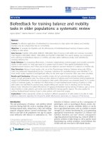
Fig. 1 Flow diagram of this systematic review and meta-analysis
Page 3 of 17
heterogeneity (I2<50%) [14]. A subgroup analysis was
conducted based on the expression level of HOX proteins in patients with GC. The sensitivity analysis was
conducted to evaluate the stability of the results, after
excluding each study. Publication bias was assessed
using the Begg’s funnel plots and Egger’s tests, and a p
value< 0.05 was considered significant. Statistical analyses were performed using Stata statistical software
(version 15.0).
Results
Literature search
Our search strategy preliminarily identified 329 potential
records. One hundred seventy-three articles remained
after the removal of duplicated studies. Forty-eight of
these studies were removed after perusing the titles and
abstracts. Then, reviews, editorials, letters, conference
abstracts, retracted articles, full texts not published in
English, and studies of cancer cells or animal models
were excluded. Subsequently, 18 studies lacking insufficient data were rejected. Finally, 19 studies including
3775 patients with GC were included in this analysis.
The selection process is shown in Fig. 1.
Jin et al. BMC Cancer
(2020) 20:866
Study characteristics
All included studies were conducted in China, Japan
and Korea and were published between 2012 and
2019. These studies involve the following HOX proteins: HOXB9 [15], HOXD10 [16], HOXA5 [17, 18],
HOXA10 [19–21], HOXA13 [22, 23], HOXC6 [24],
HOXB7 [25, 26], HOXA1 [27], HOXA9 [28], HOXC9
[29], HOXC10 [30], HOXD4 [31], HOXA11 [32] and
HOXD9 [33]. These studies explored the prognostic
value of HOX protein expression for determining OS
or disease-free survival (DFS) and the correlation between the expression of HOX proteins and clinicopathological characteristics of patients with GC. HOX
expression at the protein level was detected using immunohistochemical staining. All included studies divided HOX protein expression into high (positive)
and low (negative) groups, but the cut-off value was
slightly different among these studies. A detailed description of the characteristics of the included studies
is provided in Table 1.
Correlation of HOX protein expression with the prognosis
This meta-analysis included a total of 19 articles containing 14 HOX proteins. HOXB9, HOXD10 and
HOXA5 were expressed at low levels in GC and acted
as tumour suppressors. In contrast, HOXA13,
HOXC6, HOXB7, HOXA1, HOXC9, HOXC10,
HOXD4, HOXA11 and HOXD9 were expressed at
high levels and functioned as tumour promotors in
patients with GC. In addition, HOXA10 expression
was increased in GC, but its role in predicting the
prognosis of GC was unclear. In a pooled analysis including all studies with data on the prognostic effects
of HOX proteins in GC, considerable heterogeneity
among pooled HRs for OS was observed. A subgroup
analysis stratified by the expression level was performed, and the results revealed different trends between the downregulated subgroup and the
upregulated subgroup. High expression of HOX proteins in the downregulated subgroup was associated
with a good prognosis for patients with GC (pooled
HR: 0.46, 95% CI: 0.36–0.59, I2 = 3.1%, p = 0.377),
while the overexpression of HOX proteins in the upregulated subgroup was correlated with a poor OS
(pooled HR: 2.59, 95% CI: 1.79–3.74, I2 = 73.5%, p =
0.000) (Fig. 2a). The explanation for the high level of
heterogeneity of the upregulated subgroup might be
that HOXA10 had different prognostic values in the
existing studies. The result of the analysis of the upregulated subgroup after excluding HOXA10 suggested that overexpressed HOX proteins significantly
indicated a poor prognosis (pooled HR = 3.03, 95% CI:
2.45–3.74, I2 = 16.5%, p = 0.283) (Fig. 3). DFS was reported in 6 studies involving 5 HOX proteins.
Page 4 of 17
HOXA5 expression was associated with an increased
DFS in patients with GC (pooled HR = 0.46, 95% CI:
0.23–0.91). In contrast, HOXA13, HOXA10, HOXB7
and HOXA1 expression was associated with a decreased DFS (pooled HR = 3.77, 95% CI: 2.61–5.45)
(Fig. 2b).
Correlation of HOX protein expression with
clinicopathological characteristics
Seventeen studies with 2899 patients were included to
detect the relationship between HOX protein expression and tumour stage. As shown in Fig. 4a, increased
expression of HOXB9 and HOXD10 was significantly
correlated with an earlier TNM stage (HOXB9: OR =
0.22, 95% CI: 0.12–0.41, HOXD10: OR = 0.21, 95% CI:
0.14–0.31), while increased expression of HOXA13,
HOXB7, HOXA1, HOXA9, HOXC9, HOXC10,
HOXA11 and HOXD9 was notably associated with an
advanced TNM stage (I2 = 92.6%, p = 0.000). Due to
the high level of heterogeneity, we performed a subgroup analysis based on the expression levels of HOX
proteins. The heterogeneity of the upregulated group
was decreased but still at a high level (I2 = 75.8%, p =
0.000) (Fig. 4b). A subsequent analysis showed that
the studies of HOXA10 contributed a considerable
amount of heterogeneity (data not shown). In
addition, the difference of scoring systems for assessing expression levels of HOX proteins in the included studies was also one of the main sources of
heterogeneity. The pooled analysis of the relationship
between HOX proteins and the depth of tumour invasion showed that HOXD10 indicated a low T category (HOXD10: OR = 0.20, 95% CI: 0.09–0.41), while
HOXA13, HOXC6, HOXB7 and HOXA1 were related
to a high T category (HOXA13 (2013): OR = 4.18,
95% CI: 1.75–10.01; HOXA13 (2018): OR = 1.90, 95%
CI: 1.08–3.35; HOXC6: OR = 3.55, 95% CI: 1.11–
11.31; HOXB7 (2015): OR = 3.44, 95% CI: 1.32–8.95;
HOXB7 (2017): OR = 10.14, 95% CI: 4.36–23.58; and
HOXA1: OR = 2.03, 95% CI: 1.18–3.48) (Fig. 5a). We
pooled 11 studies including 2087 patients and found
that HOXD10, HOXA5 and HOXC10 were associated
with a decreased tumour size (HOXD10: OR = 0.37,
95% CI: 0.25–0.54; HOXA5 (2018): OR = 0.20, 95%
CI: 0.07–0.55; HOXA5 (2019): OR = 0.23, 95% CI:
0.08–0.67; and HOXC10: OR = 0.38, 95% CI: 0.15–
0.98), while the overexpression of HOXA10, HOXB7
and HOXD4 was associated with an increased tumour
size (HOXA10 (2015): OR = 2.39, 95% CI: 1.40–4.09;
HOXB7 (2017): OR = 2.60, 95% CI: 1.61–4.20; and
HOXD4: OR = 2.71, 95% CI: 1.28–5.74) (Fig. 5b).
Similarly, the heterogeneity was significantly reduced
by conducting a subgroup analysis according to expression levels of HOX proteins (Fig. 5c). Sixteen
2018
2018
2019
2019
2019
Yao et al. [30]
Liu et al. [31]
Wang et al. [32]
Zhu et al. [33]
China
China
China
China
China
China
China
China
China
China
China
China
Korea
China
Japan
China
China
China
China
Country
HOXD9
HOXA11
HOXD4
HOXC10
HOXC9
HOXA13
HOXA9
HOXB7
HOXA1
HOXB7
HOXA10
HOXC6
HOXA10
HOXA13
HOXA10
HOXA5
HOXA5
HOXD10
HOXB9
HOX protein
Upregulated
Upregulated
Upregulated
Upregulated
Upregulated
Upregulated
Upregulated
Upregulated
Upregulated
Upregulated
Upregulated
Upregulated
Upregulated
Upregulated
Upregulated
Downregulated
Downregulated
Downregulated
Downregulated
Expression
90 (55/35)
114 (58/56)
127 (68/59)
73 (38/35)
95 (68/27)
264 (186/78)
128 (88/40)
330 (195/135)
264 (144/120)
96 (66/30)
264 (159/105)
161 (76/85)
57 (29/28)
132 (103/29)
749 (221/528)
124 (60/64)
81 (29/52)
436 (242/194)
190 (86/104)
Sample size
(high/low)
IS:3
IOD/Area = 0.31
IS:7
IS:4
IS:4
IS:3
IS:4
IS:4
IS:3
IS:2
IS:2
IS:2
compare to the staining smooth muscle
IS:3
percentage of stained cancer cells = 10%
median value
IS:4
IS:4
IS:4
Cut-off value
OS
OS
OS
OS
OS
OS, DFS
OS
OS
OS, DFS
OS, DFS
OS, DFS
OS
OS
OS, DFS
OS
OS, DFS
OS
OS
OS
Survival
U
U, M
U, M
U, M
U, M
U, M
U, M
U
U
U, M
U, M
U
U
U, M
U, M
U
U, M
U
U, M
Survival analysis
OS Overall survival, DFS Disease free survival, U Univariate analysis, M Multivariate analysis, IS Immunostaining score, IOD Integrated option density, NOS Newcastle-Ottawa Scale
2018
Peng et al. [29]
2015
Han et al. [21]
Han et al. [23]
2013
Zhang et al. [24]
2017
2013
Lim et al. [20]
2017
2013
Han et al. [22]
Ma et al. [28]
2012
Sentani et al. [19]
He et al. [26]
2019
Wu et al. [18]
2015
2018
Peng et al. [17]
2016
2015
Wang et al. [16]
Yuan et al. [27]
2013
Sha et al. [15]
Tu et al. [25]
Year
First author
Table 1 Characteristics of the included studies
Kaplan-Meier curves
Text
Text
Text
Text
Text
Text
Kaplan-Meier curves
Text
Text
Text
Kaplan-Meier curves
Kaplan-Meier curves
Text
Text
Kaplan-Meier curves
Text
Kaplan-Meier curves
Text
HR availability
6
6
7
7
7
6
8
8
6
8
6
8
7
7
7
7
8
7
7
NOS score
Jin et al. BMC Cancer
(2020) 20:866
Page 5 of 17
Jin et al. BMC Cancer
(2020) 20:866
Page 6 of 17
Fig. 2 Subgroup analysis of OS (a) or DFS (b) by HOX protein expression in GC
studies with 3509 patients reported that HOXB9 and
HOXD10 were unfavourable factors for lymph node metastasis in patients with GC (HOXB9: OR = 0.35, 95% CI:
0.19–0.63 and HOXD10: OR = 0.24, 95% CI: 0.16–0.37),
and overexpression of HOXA13, HOXA1, HOXA9,
HOXC10, HOXD4 and HOXD9 was correlated with the
presence of lymph node metastasis (HOXA13 (2013):
OR = 2.38, 95% CI: 1.02–5.54; HOXA13 (2018): OR =
2.38, 95% CI: 1.39–4.09; HOXA1: OR = 2.45, 95% CI:
1.49–4.04; HOXA9: OR = 2.68, 95% CI: 1.23–5.83;
HOXC10: OR = 6.18, 95% CI: 2.22–17.18; HOXD4: OR =
5.53, 95% CI: 2.55–12.02; and HOXD9: OR = 23.11, 95%
CI: 6.04–88.49) (Fig. 6a). The results of the pooled analysis
revealed that HOXD10 was not conducive to the distant
metastasis of GC (HOXD10: OR = 0.34, 95% CI: 0.19–
0.60), but that HOXC10 and HOXA11 promoted distant
metastasis of GC (HOXC10: OR = 5.55, 95% CI: 1.42–
21.61 and HOXA11: OR = 19.02, 95% CI: 1.07–337.91)
(Fig. 6b). In addition, the upregulation of HOXB7 promoted vascular invasion in patients with GC (HOXB7
(2017): OR = 5.12, 95% CI: 3.18–8.23) (Fig. 6c). Moreover,
HOXB9, HOXD10, HOXA5 and HOXC9 were factors
contributing to good or moderate histological differentiation (HOXB9: OR = 0.17, 95% CI: 0.09–0.33, HOXD10:
Jin et al. BMC Cancer
(2020) 20:866
Page 7 of 17
Fig. 3 Subgroup analysis of OS by HOX protein expression in GC (excluded HOXA10)
OR = 0.66, 95% CI: 0.44–0.99, HOXA5 (2018): OR = 0.26,
95% CI: 0.10–0.68; and HOXC9: OR = 0.28, 95% CI: 0.11–
0.71), and overexpression of HOXA13, HOXA1, HOXA9
and HOXD9 was related to poorly differentiated status of
GC (HOXA13 (2013): OR = 2.41, 95% CI: 1.02–5.67;
HOXA13 (2018): OR = 1.84, 95% CI: 1.06–3.18; HOXA1:
OR = 2.37, 95% CI: 1.41–4.00; HOXA9: OR = 4.98, 95%
CI: 2.12 11.70; and HOXD9: OR = 14.63, 95% CI: 4.81–
44.43) (Fig. 7a). Additionally, HOXD10 and HOXB7 was
correlated with the intestinal phenotype of GC (HOXD10:
OR = 5.02, 95% CI: 3.34–7.57 and HOXB7 (2017): OR =
6.27, 95% CI: 3.81–10.31) (Fig. 7b). None of the HOX proteins included in the pooled analysis exhibited significant
associations with age (Fig. 8a), sex (Fig. 8b) or tumour
Fig. 4 Forest plots of the pooled analysis for the association between HOX protein expression and TNM stage (a), TNM stage subgroup
analysis (b)
Jin et al. BMC Cancer
(2020) 20:866
Page 8 of 17
Fig. 5 Forest plots of the pooled analysis for the association between HOX protein expression and T categories (a), tumour size (b), tumour size subgroup
analysis (c)
location (Fig. 8c). Additionally, the relationships between
HOXA5, HOXA10, HOXA13 and HOXB7 expression and
clinicopathological characteristics were all explored in several studies. As shown in Fig. 9, HOXA5 expression predicted a smaller tumour size (OR = 0.22, 95% CI: 0.10–0.45)
(Fig. 9a), but there is no correlation between HOXA10 expression and clinicopathological features (Fig. 9b). The
overexpression of both HOXA13 (Fig. 9c) and HOXB7
(Fig. 9d) was significantly associated with an advanced
tumour stage (HOXA13: OR = 2.31, 95% CI: 1.44–3.71 and
HOXB7: OR = 3.48, 95% CI: 2.28–5.32) and a high T category (HOXA13: OR = 2.62, 95% CI: 1.23–5.60 and
HOXB7: OR = 6.05, 95% CI: 2.08–17.57), and HOXA13
was also related to lymph node metastasis (OR = 2.38, 95%
CI: 1.51–3.75) and poor differentiation status (OR = 1.99,
95% CI: 1.25–3.15).
Sensitivity analysis
A sensitivity analysis was performed to verify the robustness of our results. As shown in Fig. 10, the pooled HR
was not significantly altered when each study was removed, which confirmed the reliability of overall results
for the OS of patients with GC.
Jin et al. BMC Cancer
(2020) 20:866
Page 9 of 17
Fig. 6 Forest plots of the pooled analysis for the association between HOX protein expression and lymph node metastasis (a), distant metastasis
(b), vascular invasion (c)
Publication bias
Begg’s test and Egger’s test were performed to evaluate
publication bias. The results did not reveal substantial
publication bias (Fig. 11: Begg’s test: p = 0.576, Egger’s
test: p = 0.166).
Mechanisms by which HOX proteins regulate GC
In Table 2 and supplementary Fig. 1, we summarize
the molecular mechanisms by which HOX proteins
included in this study modulate carcinogenesis and
development of GC [15–57]. HOXB9 inhibits GC
progression via AKT and NF-κB pathways [34].
HOXD10 suppresses the migration and invasion of
GC cells through insulin-like growth factor binding
protein-3 (IGFBP3) and RhoC-AKT pathway [36, 39].
HOXA5 suppresses GC progression by inhibiting the
G1/S transition during the cell cycle [17]. HOXA13
promotes GC development via TGF-β, ERK1/2,
MDM2-p53- MRP1 pathways, and Wnt/β-catenin signalling [23, 44–46]. HOXC6 enhances invasive and
metastatic abilities of GC cells by upregulating the expression of MMP9 via activating ERK pathway [48].
Jin et al. BMC Cancer
(2020) 20:866
Page 10 of 17
Fig. 7 Forest plots of the pooled analysis for the association between HOX protein expression and histologic differentiation (a), Lauren
classification (b)
HOXA1 increases the proliferation of GC cells by upregulating cyclin D1 expression [27]. HOXB7 mediates GC cell
malignancy by activating AKT/MAPK signalling, Src-FAK
pathway, PIK3R3/AKT pathway, and epithelial mesenchymal transition (EMT) [26, 49, 50]. The miR-182/HOXA9
axis is implicated in RUNX3-mediated GC development
[51]. In addition, HOXC9 contributes to GC progression
by inducing EMT, MMP2 expression, and stem cell-like
properties [29]. HOXC10 activates ATM/NF-kB pathway
and MAPK signalling, functioning as an oncogene in GC
[30, 54]. HOXD4 increases the proliferation and invasion
of GC cells by upregulating c-Myc and cyclin D1 [31].
HOXD9 activates RUFY3, increasing the proliferation, migration and invasion of GC cells [33]. However, the effects
of HOXA10 and HOXA11 on tumorigenesis and development of GC are controversial.
Discussion
GC is a main cause of cancer-related mortality. Currently, radical gastrectomy combined with adjuvant
chemotherapy is recognized as the most effective treatment for GC. Nevertheless, many patients with GC are
usually diagnosed in an advanced stage, missing the opportunity for radical surgical resection. Based on the
current situation, it is important to identify factors
which is helpful to improving prediction accuracy and
promoting curative effect of GC. Most of the HOX
genes are generally activated and expressed during
Jin et al. BMC Cancer
(2020) 20:866
Page 11 of 17
Fig. 8 Forest plots of the pooled analysis for the association between HOX protein expression and age (a), sex (b), tumour location (c)
embryogenesis, and many of these proteins are aberrantly expressed during tumorigenesis. According to
the literatures, HOX proteins are related to the prognosis and clinicopathological features of GC, but the
results are controversial. We conducted this study to
further clarify the effects of HOX proteins on the
prognosis and clinicopathological characteristics of
GC and describe the molecular mechanisms by which
HOX proteins regulate tumorigenesis and development of GC.
The present systematic review and meta-analysis enrolled 19 eligible studies containing 3775 patients. In
the pooled analysis of the effects of HOX proteins on
the GC prognosis, HOXB9, HOXD10 and HOXA5
were correlated with a good prognosis in patients
with GC, while HOXA13, HOXC6, HOXB7, HOXA1,
HOXC9, HOXC10, HOXD4, HOXA1 and HOXD9
were related to a poor prognosis. However, Kato
et al. identified positive HOXB9 expression in GC as
a marker of a poor prognosis. Unfortunately, the
study by Kato was not included in this meta-analysis
due to the lack of an analysis of HOXB9 expression
in paired noncancerous mucosae [58].
The relationship between HOX proteins and clinicopathological features of GC were also analysed in
this study. The results revealed correlations between
Jin et al. BMC Cancer
(2020) 20:866
Page 12 of 17
Fig. 9 Forest plots of the pooled analysis for the association between HOX protein expression and clinicopathological characteristics: HOXA5 (a),
HOXA10 (b), HOXA13 (c), HOXB7 (d)
the expression of HOX proteins and TNM stage, T
category, tumour size, lymph node metastasis, distant
metastasis, vascular invasion, histological differentiation, and Lauren classification in GC. Based on the
results of the meta-analysis described above, we speculated that HOX proteins might predict the prognosis
of patients with GC, which was also confirmed in
each included original study. Therefore, we inferred
that combined detection of the expression of various
HOX proteins might provide a novel perspective for
predicting the prognosis of patients with GC.
Currently, some clinicopathological parameters such
as age, sex, tumour stage, depth of invasion, lymph
node metastasis, distant metastasis, and resection
margins, have been proven to be prognostic indicators
of GC [59, 60]. At the same time, several molecules
are under investigation as predictors of survival, such
as gene mutations, DNA methylation, RNAs, and
proteins [61]. Regrettably, many studies have only explored the individual relationship between clinicopathological characteristics or molecular markers and the
prognosis of patients with GC, although a few studies
have established prognostic models [62]. Bria et al.
Jin et al. BMC Cancer
(2020) 20:866
Page 13 of 17
Fig. 10 Sensitivity analysis of the included studies on OS
combined clinicopathological parameters (sex, age,
Lauren classification, stage, margins, grade, site, size,
and resected nodes) with molecular markers (HER2,
FHIT, and APC) to construct a risk stratification of
GC, establishing a scientific model to determine its
prognosis. In addition, the authors conducted a large
prospective validation with a larger sample size to
eliminate all sources of bias in the retrospective study
[63]. GC is highly heterogeneous, and even similar
clinicopathological features result in different outcomes, suggesting that a more reasonable classification system is needed for predicting the prognosis
and therapeutic effect of GC. A novel classification
system with four molecular subtypes was developed
by The Cancer Genome Atlas (TCGA) [64]. Besides,
Sohn et al. developed a scoring system (TCGA risk
score) based on TCGA to predict prognosis and adjuvant chemotherapy outcomes in patients with GC,
which was validated as an independent prognostic
factor for GC in multivariate Cox regression analyses
[65]. Analogously, Lin et al. established a novel prognosis scoring system based on TCGA and Gene Expression Omnibus to predict the prognosis of GC,
which comprised signatures of tumour protein-coding
genes (P), tumour noncoding genes (N) and immune/
stroma cells in the tumour microenvironment (M)
(PMC score). Furthermore, the combination of PNM
scores with American Joint Committeeon Cancer
(AJCC) staging significantly increased its predictive
value [66]. In addition, Tahara et al. investigated the
prognosis and clinicopathological characteristics of
GC by combining genetic and epigenetic abnormalities. The CpG island methylator phenotype (CIMP)
and TP53 hot spot mutation status (R175, G245,
R248, R273, and R282) were sufficient to predict the
prognosis and clinicopathological features of GC.
Among these features, patients with the CIMP−TP53
hot spot+ subtype presented the worst overall survival
[67]. Moreover, Ooi et al. selected three oncogenic
pathways (NF-κB, Wnt/β-catenin, and proliferation/
stem cells) by analysing a GC pathway heatmap and
combined them to predict its prognosis, which was
validated in vitro [68].
The development of GC is determined by both genes
and environmental factors, which has been confirmed in
mouse models. Microbial infections, particularly Helicobacter pylori (H. pylori) and Epstein-Barr virus, are important environmental factors and have been confirmed
as prognostic factors for GC [69, 70]. Although H. pylori
infection is the strongest risk factor for GC, very few H.
pylori-infected populations develop GC. This outcome is
attributed to the duration of infection, strain type and
host genetic signatures [71]. The crucial effects of genetic factors on GC development have been revealed
using progress in genetic technology, including the construction of genetically engineered mice via recombinant
DNA technology to achieve molecular overexpression or
deficiency, as well as gene mutations, clarifying the
pathogenesis of GC and the interactions between various
factors. For example, INS-GAS transgenic mice on the
FVB genetic background that overexpress gastrin develop intramucosal carcinomas with submucosal and
Jin et al. BMC Cancer
(2020) 20:866
Page 14 of 17
Fig. 11 Tests for publication bias of OS: Begg’s test (a), Egger’s test (b)
Table 2 Action mechanisms of HOX proteins in gastric cancer
HOX proteins
Expression
Upstream
Downstream
Pathways
Reference
HOXB9
Downregulated
NA
NA
↓cells proliferation, migration and invasion; ↑MET;
AKT and NF-κB pathway
[15, 34, 35]
HOXD10
Downregulated
miR-10b, miR-92b-3p
IGFBP3
↓cells proliferation, migration and invasion; AKT
pathway; RhoC pathway
[16, 36–39]
HOXA5
Downregulated
miR-196a
NA
↓cells G1-S transition, proliferation and colony
formation; ↓angiogenesis
[17, 18]
HOXA10
Upregulated
NA
miR-196b-5p, BCL2
↑cells viability, proliferation, colony information,
migration and invasion ↓apoptosis; ↑tumor
metastasis; JAK1/STAT3 signaling;
HOXA10/miR-196b-5p axis;
↓cells growth, motility and invasive activity;
[19–21, 40–42]
HOXA13
Upregulated
lncRNA HOTTIP
DHRS2, cadherin17
↑cells proliferation, migration and invasion;
↑EMT; TGF-β pathway, ERK1/2 pathway,
Wnt/β-catenin pathway, MDM2-p53-MRP1
pathway; chemotherapy resistance to 5-FU
[22, 23, 43–46]
HOXC6
Upregulated
lncRNA HOTAIR
NA
↑cells proliferation, colony formation, migration
and invasion; ERK signaling;
[24, 47, 48]
HOXB7
Upregulated
NA
NA
↑cells G1-S transition, proliferation, migration
and invasion; ↑EMT; ↓apoptosis; AKT/MAPK
pathway; Src-FAK pathway; PIK3R3/ AKT pathway
[25, 26, 49, 50]
HOXA1
Upregulated
NA
NA
↑cells proliferation, invasion and migration;
↑cyclin D1
[27]
HOXA9
Upregulated
miR-182
NA
↑cells proliferation, migration and invasion;
↑tumor progression
[28, 51]
HOXC9
Upregulated
miR-26a
NA
↑EMT and stem cell-like phenotypic
acquisition; ↑tumor metastasis
[29]
HOXC10
Upregulated
miR-136
CST1
↑cells migration and invasion; ↑tumor growth and
peritoneal metastasis; ATM/NF-kB pathway;
MAPK signaling
[30, 52–55]
HOXD4
Upregulated
NA
NA
↑cells proliferation, migration and invasion; ↑c-Myc
and cyclinD1
[31]
HOXA11
Controversial
STAT3
STAT3
Wnt pathway
[32, 56, 57]
HOXD9
Upregulated
NA
RUFY3
↑cells proliferation, invasion and migration;
↑tumorigenesis and metastasis
[33]
↓: inhibit; ↑: promote; NA Not available, AKT Protein kinase B, ATM Ataxia telangiectasia mutated, BCL2 B cell lymphoma-2, CST1 Cystatin 1, DHRS2 Dehydrogenase/
reductase 2, ERK Extracellular regulated protein kinases, FAK Focal adhesion kinase, IGFBP3 Insulin-like growth factor binding protein-3, JAK1 Janus kinase 1, MAPK
Mitogen-activated protein kinase, MDM2 Murine double minute 2, MET Mesenchymal epithelial transition, MRP1 Multidrug resistance-associated protein 1, PIK3R3
Phosphoinositide-3-kinase, regulatory subunit 3, RhoC Ras superfamily of GTP-binding protein, Src Steroid receptor coactivator, RUFY3 RUN and FYVE domain
containing 3, 5-FU 5-fluorouracil
Jin et al. BMC Cancer
(2020) 20:866
intravascular invasion in less than 1 year when infected
by Helicobacter felis (H. felis) or H. pylori, with males
showing a higher prevalence than females, indicating sex
differences in GC tumorigenesis [72, 73]. However, INSGAS mice on a C57BL/6 background infected with H.
felis do not progress to GC [74]. Surprisingly, gastrin
knockout mice (GAS−/− mice) are also confirmed to be
susceptible to GC and exhibit antral GC, in contrast to
INS-GAS mice, which develop corpus cancers [75].
Moreover, GAS−/− mice are more susceptible to antral
cancer induced by MNU, a gastric carcinogen used in
mouse models, compared to WT mice on the same genetic background [76].
Taken together, these studies reveal important roles
of genetic signatures in the development of GC, and
the external factor such as infection is also indispensable. Thus, the establishment of a comprehensive and
detailed scoring system containing the most basic
clinicopathological parameters, molecular markers,
gene expression profiles, microbial infections, etc.,
might be more accurate in predicting the prognosis of
patients with GC than a single factor. Our manuscript
analysing the effects of HOX proteins in GC development aimed to predict the prognosis and provide
therapeutic targets for GC. The results of this metaanalysis recommend the inclusion of HOX proteins in
the model predicting the prognosis of GC.
Several limitations of this systematic review and
meta-analysis should be noted. First, several HRs and
their 95% CIs for OS were extracted from the survival
curves, which might affect the reliability of the results. Second, the sample size of each study was not
large enough, which might affect the accuracy of the
results. Third, IHC methodologies including the primary antibody used, antibody dilutions, and the scoring system applied, differed, which might partially
contribute to the heterogeneity. Finally, all patients
included in our study were Asians, which might restrict the applicability of our results to other races.
Conclusions
This systematic review and meta-analysis firstly generalized and evaluated the significance of HOX proteins in
modulating the prognosis and clinicopathological characteristics of GC. We also summarized the molecular
mechanisms by which HOX proteins regulate tumorigenesis and development of GC. Based on these findings,
HOX proteins might serve as biomarkers and therapeutic targets for GC. Considering the limitations of this
study, further large-scale prospective and high-quality
studies are required to confirm the potential values of
HOX proteins in GC.
Page 15 of 17
Supplementary information
Supplementary information accompanies this paper at />1186/s12885-020-07346-7.
Additional file 1: Figure S1. Molecular mechanisms how HOX proteins
regulate tumorigenesis and development of GC.↑: promote; ⊥: inhibit;
AKT: protein kinase B; ATM: ataxia telangiectasia mutated; BCL2: B cell
lymphoma-2; CDH17: cadherin 17; CST1: cystatin SN; DHRS2: dehydrogenase/reductase 2; EGF: epidermal growth factor; ERK: extracellular regulated
protein kinases; FAK: focal adhesion kinase; IGFBP3: insulin-like growth
factor binding protein-3; JAK1: janus kinase 1; MAPK: mitogen-activated
protein kinase; MDM2: murine double minute 2; MET: mesenchymal epithelial transition; MMP2: matrix metalloproteinase 2; MMP9: matrix metalloproteinase 9; MMP14: matrix metalloproteinase 14; MRP1: multidrug
resistance-associated protein 1; NF-κB: nuclear factor-kappa B; NKD1:
naked cuticle homolog 1; PIK3R3: phosphoinositide-3-kinase, regulatory
subunit 3; RhoC: ras superfamily of GTP-binding protein; RUFY3: RUN and
FYVE domain containing 3; RUNX3: runt-related transcription factor 3; Src:
steroid receptor coactivator; STAT3: signal transducers and activators of
transcription 3; TFF1: trefoil factor 1; TGF-β: transforming growth factor-β;
TNF-α: tumour necrosis factor-α; uPA: urokinase-type plasminogen activator; uPAR: urokinase-type plasminogen activator receptor.
Abbreviations
AJCC: American Joint Committeeon Cancer; AKT: Protein kinase B;
ATM: Ataxia telangiectasia mutated; CI: Confidence interval; CIMP: CpG island
methylator phenotype; DFS: Disease-free survival; EMT: Epithelial
mesenchymal transition; ERK: Extracellular regulated protein kinases;
FAK: Focal adhesion kinase; GC: Gastric cancer; H. felis: Helicobacter felis;
HOX: Homeobox; HR: Hazard ratio; IGFBP3: Insulin-like growth factor binding
protein-3; IHC: Immunohistochemical; MAPK: Mitogen-activated protein
kinase; MDM2: Murine double minute 2; MRP1: Multidrug resistanceassociated protein 1; NF-κB: Nuclear factor-kappa B; NOS: Newcastle-Ottawa
Quality Assessment Scale; OR: Odds ratio; OS: Overall survival;
PIK3R3: Phosphoinositide-3-kinase, regulatory subunit 3; RhoC: Ras
superfamily of GTP-binding protein; RUFY3: RUN and FYVE domain
containing 3; TGF-β: Transforming growth factor-β
Acknowledgements
We would like to thank all researchers for their contributions to this study.
Authors’ contributions
XJ designed the research, searched the literatures, extracted and analysed
the data, and wrote the manuscript. LD searched the literatures, extracted
and analysed the data. YM and JW conducted literatures quality assessment.
XJ, LD, YM, JW, HY, YJ, XZ and ZL established selection criteria. All authors
read, reviewed and approved the final manuscript.
Funding
Not applicable.
Availability of data and materials
All data generated or analysed in this study are included in this published
article.
Ethics approval and consent to participate
Not applicable.
Consent for publication
Not applicable.
Competing interests
The authors declare no conflict of interests.
Jin et al. BMC Cancer
(2020) 20:866
Received: 22 April 2020 Accepted: 26 August 2020
References
1. Bray F, Ferlay J, Soerjomataram I, Siegel RL, Torre LA, Jemal A. Global cancer
statistics 2018: GLOBOCAN estimates of incidence and mortality worldwide
for 36 cancers in 185 countries. CA Cancer J Clin. 2018;68(6):394–424.
2. Mark M, Rijli FM, Chambon P. Homeobox genes in embryogenesis and
pathogenesis. Pediatr Res. 1997;42(4):421–9.
3. Samuel S, Naora H. Homeobox gene expression in cancer: insights from
developmental regulation and deregulation. Eur J Cancer (Oxford, England :
1990). 2005;41(16):2428–37.
4. Shah N, Sukumar S. The Hox genes and their roles in oncogenesis. Nat Rev
Cancer. 2010;10(5):361–71.
5. Nagy A, Osz A, Budczies J, Krizsan S, Szombath G, Demeter J, et al. Elevated
HOX gene expression in acute myeloid leukemia is associated with NPM1
mutations and poor survival. J Adv Res. 2019;20:105–16.
6. de Bessa Garcia SA, Araujo M, Pereira T, Mouta J, Freitas R. HOX genes
function in breast Cancer development. Biochimica et biophysica acta
Reviews on cancer. 1873;2020(2):188358.
7. Li L, Zhang X, Liu Q, Yin H, Diao Y, Zhang Z, et al. Emerging role of HOX
genes and their related long noncoding RNAs in lung cancer. Crit Rev
Oncol Hematol. 2019;139:1–6.
8. Joo MK, Park JJ, Chun HJ. Impact of homeobox genes in gastrointestinal
cancer. World J Gastroenterol. 2016;22(37):8247–56.
9. Kuo TL, Cheng KH, Chen LT, Hung WC. Deciphering the potential role of
Hox genes in pancreatic cancer. Cancers (Basel). 2019;11(5):734.
10. Quagliata L, Quintavalle C, Lanzafame M, Matter MS, Novello C, di Tommaso
L, et al. High expression of HOXA13 correlates with poorly differentiated
hepatocellular carcinomas and modulates sorafenib response in in vitro
models. Lab Invest. 2018;98(1):95–105.
11. Moher D, Liberati A, Tetzlaff J, Altman DG. Preferred reporting items for
systematic reviews and meta-analyses: the PRISMA statement. BMJ (Clinical
research ed). 2009;339:b2535.
12. Lo CK, Mertz D, Loeb M. Newcastle-Ottawa scale: comparing reviewers’ to
authors’ assessments. BMC Med Res Methodol. 2014;14:45.
13. Tierney JF, Stewart LA, Ghersi D, Burdett S, Sydes MR. Practical methods for
incorporating summary time-to-event data into meta-analysis. Trials. 2007;8:
16.
14. DerSimonian R, Laird N. Meta-analysis in clinical trials revisited.
Contemporary Clin Trials. 2015;45(Pt A):139–45.
15. Sha S, Gu Y, Xu B, Hu H, Yang Y, Kong X, et al. Decreased expression of
HOXB9 is related to poor overall survival in patients with gastric carcinoma.
Digestive Liver Dis. 2013;45(5):422–9.
16. Wang YY, Li L, Ye ZY, Zhao ZS, Yan ZL. MicroRNA-10b promotes migration
and invasion through Hoxd10 in human gastric cancer. World J Surg Oncol.
2015;13:259.
17. Peng X, Zha L, Chen A, Wang Z. HOXA5 is a tumor suppressor gene that is
decreased in gastric cancer. Oncol Rep. 2018;40(3):1317–29.
18. Wu Y, Zhou T, Tang Q, Xiao J. HOXA5 inhibits tumor growth of gastric
cancer under the regulation of microRNA-196a. Gene. 2019;681:62–8.
19. Sentani K, Oue N, Naito Y, Sakamoto N, Anami K, Oo HZ, et al. Upregulation
of HOXA10 in gastric cancer with the intestinal mucin phenotype: reduction
during tumor progression and favorable prognosis. Carcinogenesis. 2012;
33(5):1081–8.
20. Lim JY, Yoon SO, Seol SY, Hong SW, Kim JW, Choi SH, et al. Overexpression
of miR-196b and HOXA10 characterize a poor-prognosis gastric cancer
subtype. World J Gastroenterol. 2013;19(41):7078–88.
21. Han Y, Lu S, Wen YG, Yu FD, Zhu XW, Qiu GQ, et al. Overexpression of
HOXA10 promotes gastric cancer cells proliferation and HOXA10(+)/CD44(+)
is potential prognostic biomarker for gastric cancer. Eur J Cell Biol. 2015;
94(12):642–52.
22. Han Y, Tu WW, Wen YG, Li DP, Qiu GQ, Tang HM, et al. Identification and
validation that up-expression of HOXA13 is a novel independent prognostic
marker of a worse outcome in gastric cancer based on
immunohistochemistry. Med Oncol (Northwood, London, England). 2013;
30(2):564.
23. Han Y, Song C, Wang J, Tang H, Peng Z, Lu S. HOXA13 contributes to
gastric carcinogenesis through DHRS2 interacting with MDM2 and confers
5-FU resistance by a p53-dependent pathway. Mol Carcinog. 2018;57(6):
722–34.
Page 16 of 17
24. Zhang Q, Jin XS, Yang ZY, Wei M, Liu BY, Gu QL. Upregulated Hoxc6
expression is associated with poor survival in gastric cancer patients.
Neoplasma. 2013;60(4):439–45.
25. Tu W, Zhu X, Han Y, Wen Y, Qiu G, Zhou C. Overexpression of HOXB7 is
associated with a poor prognosis in patients with gastric cancer. Oncol Lett.
2015;10(5):2967–73.
26. He X, Liu Z, Xia Y, Xu J, Lv G, Wang L, et al. HOXB7 overexpression
promotes cell proliferation and correlates with poor prognosis in gastric
cancer patients by inducing expression of both AKT and MARKs.
Oncotarget. 2017;8(1):1247–61.
27. Yuan C, Zhu X, Han Y, Song C, Liu C, Lu S, et al. Elevated HOXA1 expression
correlates with accelerated tumor cell proliferation and poor prognosis in
gastric cancer partly via cyclin D1. J Exp Clin Cancer Res. 2016;35:15.
28. Ma YY, Zhang Y, Mou XZ, Liu ZC, Ru GQ, Li E. High level of homeobox A9
and PBX homeobox 3 expression in gastric cancer correlates with poor
prognosis. Oncol Lett. 2017;14(5):5883–9.
29. Peng X, Kang Q, Wan R, Wang Z. miR-26a/HOXC9 Dysregulation promotes
metastasis and stem cell-like phenotype of gastric Cancer. Cell Physiol
Biochem. 2018;49(4):1659–76.
30. Yao S, He L, Zhang Y, Ye L, Lai Y, Huang L, et al. HOXC10 promotes gastric
cancer cell invasion and migration via regulation of the NF-κB pathway.
Biochem Biophys Res Commun. 2018;501(3):628–35.
31. Liu H, Tian H, Zhao J, Jia Y. High HOXD4 protein expression in gastric
adenocarcinoma tissues indicates unfavorable clinical outcomes. Saudi J
Gastroenterol. 2019;25(1):46–54.
32. Wang C, Shi M, Ji J, Cai Q, Jiang J, Zhang H, et al. A self-enforcing HOXA11/
Stat3 feedback loop promotes stemness properties and peritoneal
metastasis in gastric cancer cells. Theranostics. 2019;9(25):7628–47.
33. Zhu H, Dai W, Li J, Xiang L, Wu X, Tang W, et al. HOXD9 promotes the
growth, invasion and metastasis of gastric cancer cells by transcriptional
activation of RUFY3. J Exp Clin Cancer Res. 2019;38(1):412.
34. Zhang L, Wu Q, He C, Liang D, Yi Q, Shi J, et al. HOXB9 inhibits proliferation
in gastric carcinoma cells via suppression of phosphorylated-Akt and NF-κBdependent snail expression. Digestive Liver Dis. 2019;51(1):157–65.
35. Chang Q, Zhang L, He C, Zhang B, Zhang J, Liu B, et al. HOXB9 induction of
mesenchymal-to-epithelial transition in gastric carcinoma is negatively
regulated by its hexapeptide motif. Oncotarget. 2015;6(40):42838–53.
36. Liu Z, Zhu J, Cao H, Ren H, Fang X. miR-10b promotes cell invasion through
RhoC-AKT signaling pathway by targeting HOXD10 in gastric cancer. Int J
Oncol. 2012;40(5):1553–60.
37. Li C, Huo B, Wang Y, Cheng C. Downregulation of microRNA-92b-3p
suppresses proliferation, migration, and invasion of gastric cancer SGC-7901
cells by targeting Homeobox D10. J Cell Biochem. 2019;120(10):17405–12.
38. Wang L, Chen S, Xue M, Zhong J, Wang X, Gan L, et al. Homeobox D10
gene, a candidate tumor suppressor, is downregulated through promoter
hypermethylation and associated with gastric carcinogenesis. Molecular
Med (Cambridge, Mass). 2012;18(1):389–400.
39. Xue M, Fang Y, Sun G, Zhuo W, Zhong J, Qian C, et al. IGFBP3, a
transcriptional target of homeobox D10, is correlated with the prognosis of
gastric cancer. PLoS One. 2013;8(12):e81423.
40. Shao L, Chen Z, Peng D, Soutto M, Zhu S, Bates A, et al. Methylation of the
HOXA10 promoter directs miR-196b-5p-dependent cell proliferation and
invasion of gastric Cancer cells. Mol Cancer Res. 2018;16(4):696–706.
41. Chen W, Wu G, Zhu Y, Zhang W, Zhang H, Zhou Y, et al. HOXA10
deteriorates gastric cancer through activating JAK1/STAT3 signaling
pathway. Cancer Manag Res. 2019;11:6625–35.
42. Song C, Han Y, Luo H, Qin Z, Chen Z, Liu Y, et al. HOXA10 induces BCL2
expression, inhibits apoptosis, and promotes cell proliferation in gastric
cancer. Cancer Med. 2019;8(12):5651–61.
43. Chang S, Liu J, Guo S, He S, Qiu G, Lu J, et al. HOTTIP and HOXA13 are
oncogenes associated with gastric cancer progression. Oncol Rep. 2016;
35(6):3577–85.
44. He YX, Song XH, Zhao ZY, Zhao H. HOXA13 upregulation in gastric cancer is
associated with enhanced cancer cell invasion and epithelial-tomesenchymal transition. Eur Rev Med Pharmacol Sci. 2017;21(2):258–65.
45. Qin Z, Chen Z, Weng J, Li S, Rong Z, Zhou C. Elevated HOXA13 expression
promotes the proliferation and metastasis of gastric cancer partly via
activating Erk1/2. Onco Targets Ther. 2019;12:1803–13.
46. Qu LP, Zhong YM, Zheng Z, Zhao RX. CDH17 is a downstream effector of
HOXA13 in modulating the Wnt/β-catenin signaling pathway in gastric
cancer. Eur Rev Med Pharmacol Sci. 2017;21(6):1234–41.
Jin et al. BMC Cancer
(2020) 20:866
47. Lin J, He J, He X, Wang L, Xue M, Zhuo W, et al. HoxC6 functions as an
oncogene and isoform HoxC6-2 may play the primary role in gastric
carcinogenesis. Dig Dis Sci. 2020.
48. Chen SW, Zhang Q, Xu ZF, Wang HP, Shi Y, Xu F, et al. HOXC6 promotes
gastric cancer cell invasion by upregulating the expression of MMP9. Mol
Med Rep. 2016;14(4):3261–8.
49. Wu J, Long Z, Cai H, Yu S, Liu X. Homeobox B7 accelerates the cancer
progression of gastric carcinoma cells by promoting epithelial-mesenchymal
transition (EMT) and activating Src-FAK pathway. Onco Targets Ther. 2019;
12:3743–51.
50. Cai JQ, Xu XW, Mou YP, Chen K, Pan Y, Wu D. Upregulation of HOXB7
promotes the tumorigenesis and progression of gastric cancer and
correlates with clinical characteristics. Tumour Biol. 2016;37(2):1641–50.
51. Yu J, Tian X, Chang J, Liu P, Zhang R. RUNX3 inhibits the proliferation and
metastasis of gastric cancer through regulating miR-182/HOXA9.
Biomedicine & pharmacotherapy = Biomedecine & pharmacotherapie. 2017;
96:782–91.
52. Kim J, Bae DH, Kim JH, Song KS, Kim YS, Kim SY. HOXC10 overexpression
promotes cell proliferation and migration in gastric cancer. Oncol Rep. 2019;
42(1):202–12.
53. Zheng J, Ge P, Liu X, Wei J, Wu G, Li X. MiR-136 inhibits gastric cancerspecific peritoneal metastasis by targeting HOXC10. Tumour Biol. 2017;39(6):
1010428317706207.
54. Guo C, Hou J, Ao S, Deng X, Lyu G. HOXC10 up-regulation promotes gastric
cancer cell proliferation and metastasis through MAPK pathway. Chinese J
Cancer Res = Chung-kuo yen cheng yen chiu. 2017;29(6):572–80.
55. Li J, Tong G, Huang C, Luo Y, Wang S, Zhang Y, et al. HOXC10
promotes cell migration, invasion, and tumor growth in gastric
carcinoma cells through upregulating proinflammatory cytokines. J Cell
Physiol. 2020;235(4):3579–91.
56. Bai Y, Fang N, Gu T, Kang Y, Wu J, Yang D, et al. HOXA11 gene is
hypermethylation and aberrant expression in gastric cancer. Cancer Cell Int.
2014;14:79.
57. Cui Y, Gao D, Linghu E, Zhan Q, Chen R, Brock MV, et al. Epigenetic changes
and functional study of HOXA11 in human gastric cancer. Epigenomics.
2015;7(2):201–13.
58. Kato F, Wada N, Hayashida T, Fukuda K, Nakamura R, Takahashi T, et al.
Experimental and clinicopathological analysis of HOXB9 in gastric cancer.
Oncol Lett. 2019;17(3):3097–102.
59. Park SR, Kim MJ, Ryu KW, Lee JH, Lee JS, Nam BH, et al. Prognostic value of
preoperative clinical staging assessed by computed tomography in
resectable gastric cancer patients: a viewpoint in the era of preoperative
treatment. Ann Surg. 2010;251(3):428–35.
60. Nagata T, Ichikawa D, Komatsu S, Inoue K, Shiozaki A, Fujiwara H, et al.
Prognostic impact of microscopic positive margin in gastric cancer patients.
J Surg Oncol. 2011;104(6):592–7.
61. Machlowska J, Maciejewski R, Sitarz R. The Pattern of Signatures in Gastric
Cancer Prognosis. Int J Mol Sci. 2018;19(6):1658.
62. Mohri Y, Tanaka K, Ohi M, Yokoe T, Miki C, Kusunoki M. Prognostic
significance of host- and tumor-related factors in patients with gastric
cancer. World J Surg. 2010;34(2):285–90.
63. Bria E, De Manzoni G, Beghelli S, Tomezzoli A, Barbi S, Di Gregorio C, et al. A
clinical-biological risk stratification model for resected gastric cancer:
prognostic impact of Her2, Fhit, and APC expression status. Ann Oncol.
2013;24(3):693–701.
64. Bass JA, Thorsson V, Shmulevich L, Reynolds MS, Miller M, Bernard B, et al.
Comprehensive molecular characterization of gastricadenocarcinoma.
Nature. 2014;513(7517):202–9.
65. Sohn BH, Hwang JE, Jang HJ, Lee HS, Oh SC, Shim JJ, et al. Clinical
significance of four molecular subtypes of gastric Cancer identified by the
Cancer genome atlas project. Clin Cancer Res. 2017.
66. Lin S, Zhou R, Zeng D, Wu J, Wu J, Zhang J, et al. A novel assessing system
for predicting the prognosis of gastric cancer. Epigenomics. 2019;11(11):
1251–66.
67. Tahara T, Tahara S, Horiguchi N, Okubo M, Terada T, Yamada H, et al.
Molecular subtyping of gastric cancer combining genetic and epigenetic
anomalies provides distinct clinicopathological features and prognostic
impacts. Hum Mutat. 2019;40(3):347–54.
68. Ooi CH, Ivanova T, Wu J, Lee M, Tan IB, Tao J, et al. Oncogenic pathway
combinations predict clinical prognosis in gastric cancer. PLoS Genet. 2009;
5(10):e1000676.
Page 17 of 17
69. Wang J, Liu X. Correlation analysis between helicobacter pylori infection
status and tumor clinical pathology as well as prognosis of gastric Cancer
patients. Iran J Public Health. 2018;47(10):1529–36.
70. Shinozaki-Ushiku A, Kunita A, Fukayama M. Update on Epstein-Barr virus and
gastric cancer (review). Int J Oncol. 2015;46(4):1421–34.
71. Cover TL. Helicobacter pylori Diversity and Gastric Cancer Risk. mBio. 2016;
7(1):e01869–15.
72. Wang TC, Dangler CA, Chen D, Goldenring JR, Koh T, Raychowdhury R, et al.
Synergistic interaction between hypergastrinemia and helicobacter infection
in a mouse model of gastric cancer. Gastroenterology. 2000;118(1):36–47.
73. Fox JG, Rogers AB, Ihrig M, Taylor NS, Whary MT, Dockray G, et al.
Helicobacter pylori-associated gastric cancer in INS-GAS mice is gender
specific. Cancer Res. 2003;63(5):942–50.
74. Takaishi S, Tu S, Dubeykovskaya ZA, Whary MT, Muthupalani S, Rickman BH,
et al. Gastrin is an essential cofactor for helicobacter-associated gastric
corpus carcinogenesis in C57BL/6 mice. Am J Pathol. 2009;175(1):365–75.
75. Zavros Y, Eaton KA, Kang W, Rathinavelu S, Katukuri V, Kao JY, et al. Chronic
gastritis in the hypochlorhydric gastrin-deficient mouse progresses to
adenocarcinoma. Oncogene. 2005;24(14):2354–66.
76. Tomita H, Takaishi S, Menheniott TR, Yang X, Shibata W, Jin G, et al.
Inhibition of gastric carcinogenesis by the hormone gastrin is mediated by
suppression of TFF1 epigenetic silencing. Gastroenterology. 2011;140(3):
879–91.
Publisher’s Note
Springer Nature remains neutral with regard to jurisdictional claims in
published maps and institutional affiliations.