The Breast Cancer to Bone (B2B) Metastases Research Program: A multi-disciplinary investigation of bone metastases from breast cancer
Bạn đang xem bản rút gọn của tài liệu. Xem và tải ngay bản đầy đủ của tài liệu tại đây (1.48 MB, 15 trang )
Brockton et al. BMC Cancer (2015) 15:512
DOI 10.1186/s12885-015-1528-y
STUDY PROTOCOL
Open Access
The Breast Cancer to Bone (B2B) Metastases
Research Program: a multi-disciplinary
investigation of bone metastases from
breast cancer
Nigel T. Brockton1,2*, Stephanie J. Gill1,2, Stephanie L. Laborge1, Alexander H. G. Paterson2,4, Linda S. Cook1,5,
Hans J. Vogel6, Carrie S. Shemanko7, David A. Hanley7, Anthony M. Magliocco8 and Christine M. Friedenreich1,2,3
Abstract
Background: Bone is the most common site of breast cancer distant metastasis, affecting 50–70 % of patients who
develop metastatic disease. Despite decades of informative research, the effective prevention, prediction and
treatment of these lesions remains elusive. The Breast Cancer to Bone (B2B) Metastases Research Program consists
of a prospective cohort of incident breast cancer patients and four sub-projects that are investigating priority areas
in breast cancer bone metastases. These include the impact of lifestyle factors and inflammation on risk of bone
metastases, the gene expression features of the primary tumour, the potential role for metabolomics in early detection
of bone metastatic disease and the signalling pathways that drive the metastatic lesions in the bone.
Methods/Design: The B2B Research Program is enrolling a prospective cohort of 600 newly diagnosed, incident, stage
I-IIIc breast cancer survivors in Alberta, Canada over a five year period. At baseline, pre-treatment/surgery blood samples
are collected and detailed epidemiologic data is collected by in-person interview and self-administered questionnaires.
Additional self-administered questionnaires and blood samples are completed at specified follow-up intervals (24, 48
and 72 months). Vital status is obtained prior to each follow-up through record linkages with the Alberta Cancer
Registry. Recurrences are identified through medical chart abstractions. Each of the four projects applies specific
methods and analyses to assess the impact of serum vitamin D and cytokine concentrations, tumour transcript and
protein expression, serum metabolomic profiles and in vitro cell signalling on breast cancer bone metastases.
Discussion: The B2B Research Program will address key issues in breast cancer bone metastases including the
association between lifestyle factors (particularly a comprehensive assessment of vitamin D status) inflammation and
bone metastases, the significance or primary tumour gene expression in tissue tropism, the potential of metabolomic
profiles for risk assessment and early detection and the signalling pathways controlling the metastatic tumour
microenvironment. There is substantial synergy between the four projects and it is hoped that this integrated program
of research will advance our understanding of key aspects of bone metastases from breast cancer to improve the
prevention, prediction, detection, and treatment of these lesions.
Keywords: Breast cancer, Bone, Metastasis, Cohort, Population-based, Lifestyle, Inflammation, Diet, Physical activity,
Vitamin D, Metabolomics, Gene expression, Recurrence, Survival
* Correspondence:
1
Department of Cancer Epidemiology and Prevention Research,
CancerControl Alberta, Alberta Health Services, Room 515C, Holy Cross
Centre, 2210 2nd St, SW, Calgary, AB T2S 3C3, Canada
2
Department of Oncology, Cumming School of Medicine, University of
Calgary, Calgary, Alberta, Canada
Full list of author information is available at the end of the article
© 2015 Brockton et al. This is an Open Access article distributed under the terms of the Creative Commons Attribution License
( which permits unrestricted use, distribution, and reproduction in any medium,
provided the original work is properly credited. The Creative Commons Public Domain Dedication waiver (http://
creativecommons.org/publicdomain/zero/1.0/) applies to the data made available in this article, unless otherwise stated.
Brockton et al. BMC Cancer (2015) 15:512
Background
Breast cancer is the most common cancer in women in
North America with over 250,000 cases annually and
approximately 45,000 deaths [1, 2]. In patients who develop metastatic disease, 50–70 % will have bone involvement [3–6] and the propensity for primary breast
cancer to metastasize to bone has been recognised for
over one hundred years since the time of Paget’s speculation on the relative roles of “seed and soil” in the progression of cancer [7, 8]. Approximately 10 % of all
breast cancer patients, without evidence of bone metastases at the time of diagnosis, will have a first
relapse in bone within five years of their primary diagnosis [3, 9, 10]. Although women with predominant or
exclusive bone involvement typically live longer than
women with other sites of breast cancer metastasis,
these lesions cause serious lingering morbidity as a result of pathologic bone fractures, bone pain, hypercalcemia and spinal cord compression, and eventually
culminate in death [6, 11].
Occult micrometastases have been detected in bone
stromal aspirates from over 50 % of women at the time
of primary breast cancer diagnosis [12–15]. However,
there is no current method to identify the features of
micrometastases that will eventually progress to create a
clinically detectable and symptomatic bone lesion; some
may remain dormant indefinitely. Two decades of research have revealed that bone metastasis is a multi-step
process of adhesion, invasion, angiogenesis and osteolysis, but the successful prevention, prediction and treatment of these lesions remains elusive. New therapeutic
strategies for bone metastases have become available recently [16], however current treatment options are generally palliative.
Bone metastases from breast cancer are predominantly
osteolytic although osteosclerotic and mixed lesions can
be observed in the same patient [17, 18]. Osteolytic lesions are dominated by osteoclasts that mediate bone resorption during the normal process of bone remodelling
[19]. The presence of metastatic breast cancer cells in
the bone drives complex interactions between the breast
cancer cells, the bone and stromal cells resulting in the
recruitment of osteoclast precursors, osteoclast activation and establishment of symptomatic lytic metastases
[20–24]. The bone matrix is a reservoir for growth factors that are released during breast cancer induced bone
lysis; these growth factors enhance the recruitment and
proliferation of osteoclast progenitors and breast cancer
cells. This “vicious cycle” involving recruitment of stromal growth factors, activation of osteoclasts, and further
proteolysis drives the progressive osteolysis observed in
primary breast carcinoma metastasis to bone [25, 26]
and is a central target for disruption by current antimetastatic treatment strategies [27].
Page 2 of 15
The advent of powerful gene profiling technologies
has enabled rapid advances in our understanding of the
biological basis of bone tropism in subsets of metastatic
breast cancer [28–30] and suggested that breast cancer
cell recruitment to metastatic sites is attributable to the
activation of specific molecular programs in the primary
tumour [31–33]. However, despite almost a decade of
subsequent research, no primary tumour gene expression signatures have yet been independently validated in
humans [34, 35]. Selecting patients at greatest risk of
bone metastases, by characterizing features of the primary tumour, could direct the optimal use of therapeutics such as bisphosphonate and receptor activator of
nuclear factor-κB ligand (RANKL) inhibitors [36]. The
early detection of bone metastases, prior to radiological
detection or the onset of skeletal pain, by serum factors
or metabolomic profiles, could also potentially direct
treatment more judiciously than as a default adjuvant
therapy. In addition to the prediction of bone metastases and the selection of patients for therapies, there is
some evidence that certain lifestyle factors, particularly
vitamin D sufficiency and use of non-steroidal antiinflammatory drugs (NSAID) use, can influence a patient’s risk for developing metastatic disease following
their primary diagnosis, [37–40]. Understanding the
potential role and contribution of lifestyle factors to
the risk of developing bone metastases would inform
optimal lifestyle advice following primary breast cancer
diagnosis. Finally, characterising the specific breast cancer
cells or molecular signaling conditions that lead to overt
metastases could identify potential therapeutic targets for
tertiary prevention.
Program overview
The Breast Cancer to Bone Metastases (B2B) Research
Program is an on-going, dynamic, interdisciplinary research program addressing multiple aspects of breast
cancer to bone metastases. Addressing these complex
questions is beyond the scope of a single project or investigator. Consequently, we assembled a core research
team with expertise ranging from basic science to population health science to clinical care. Four core projects,
each investigating an important aspect of breast cancer
bone metastases, are based on the biologic samples and
data collected from a prospective cohort of breast cancer
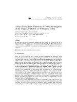
patients, the B2B Cohort (Fig. 1). The B2B Research
Program was established to support four core projects
that examine the lifestyle, pathological, and biologic factors associated with these debilitating lesions. The overall B2B Research Program is approved by the provincial
research ethics board (Health Research Ethics Board of
Alberta, HREBA) and the University of Calgary institutional ethics board (Conjoint Health Research Ethics
Board, CHREB).
Brockton et al. BMC Cancer (2015) 15:512
Page 3 of 15
Core Project 1: Vitamin
D, inflammation and bone
metastasis in breast
cancer survivors
Core Project 2: Primary
breast tumour RNA
expression and
bone metastasis
Clinical Data
Clinical Data
Interview/ Follow-up
Questionnaire data
Pre- Operative Blood
Core Project 3:
Nuclear Magnetic
Resonance (NMR)
spectroscopy and
metabolic markers
of bone metastasis
FFPE Tumour Tissue
B2B
Cohort
Clinical Data
Interview/ Follow-up
Questionnaire data
Frozen Tumour
Tissue
Core Project 4: The
role of breast cancer
stem cells in breast
cancer to bone
metastasis
Clinical Data
Fresh Tumour Tissue
Pre- Operative Blood
Fig. 1 B2B Research Program overview. Clinical data, questionnaire and interview responses, and biospecimens collected from the B2B Cohort are
used to support each of the four Core Projects
Core Project 1: Vitamin D, inflammation and bone
metastasis in breast cancer survivors
There is convincing evidence to support an inverse association between risk of breast cancer and both vitamin
D and calcium status (reviewed in [41]). Furthermore,
pre-clinical evidence, from animal models, suggests that
vitamin D may impede metastases to bone [42, 43].
However, the role of vitamin D in the development and
natural history of bone metastases, in humans, has not
yet been investigated. There are several plausible mechanisms by which vitamin D may reduce risk or retard
development of bone metastases. Vitamin D exhibits prodifferentiation and anti-proliferative properties [44, 45],
including the terminal differentiation of osteoclasts [46].
Accumulating evidence implicates sub-optimal vitamin D
status in the development of rheumatoid arthritis, diabetes
(types 1&2), multiple sclerosis, psoriasis, cardiovascular
disease, and cancer (reviewed in [47]). The etiology of
these chronic diseases all involve a suspected inflammatory
component compatible with the observed immunosuppressive and anti-inflammatory activity of 1,25-dihydroxyvitamin D, the active metabolite of Vitamin D, [48, 49].
The use of NSAID has recently been reported to reduce breast cancer recurrence [50] and improve survival
[51] and several of the genes identified in the bone
metastatic program, are associated with inflammatory
responses [31]. Therefore, chronic inflammation exacerbated by vitamin D inadequacy may potentiate the recruitment of disseminated breast cancer cells to the
bone and the initiation of osteolytic metastatic bone
lesions.
During summer in North America, up to 90 % of vitamin D is synthesized in the skin by ultraviolet B radiation
[UVB], with the remainder from food and supplements. In
the winter, especially for those living at latitudes above 42°
latitude (e.g., Boston, MA), diet and supplements are the
predominant sources of vitamin D. Therefore, both dietary and supplemental intake and sun exposure must be
considered when assessing vitamin D status in a Canadian
population. An estimated 25–39 % of all Canadians are
vitamin D deficient and the prevalence of vitamin D deficiency increases with age [52].
We will also measure 25-hydroxyvitamin D (25-OHD),
parathyroid hormone, calcium, creatinine, albumin, and
phosphate in serum, at baseline. Serum interleukin-1β (IL1B), Interleukin-6 (IL-6), interleukin-8 (IL-8), Interleukin11 (IL-11) and tumour necrosis factor-alpha (TNF-α) will
be measured as part of a 10-cytokine multiplex assay.
PTHrP expression will be quantified by automated immunohistochemistry (IHC) (HistoRx®) in the primary tumour.
This project is approved by both the University of Calgary
institutional research ethics boards (CHREB).
Brockton et al. BMC Cancer (2015) 15:512
Core Project 2: Primary breast tumour RNA expression and
bone metastasis
Previous studies have proposed primary tumour gene
expression patterns which appear to be candidate molecular pathways for migration to and successful growth
in the bone marrow [31, 32]. Some markers appear to be
particularly important in the process; these include:
CXCR4 (chemokine (C-X-C motif receptor 4), SDF1
(stromal cell-derived factor1, also known as CXCL12),
CTGF (connective tissue growth factor), FGF5 (fibroblast growth factor 5), MMP1 (matrix metallopeptidase
1), Il-11, PTHrP and osteopontin. These proteins have
acknowledged roles in cell recruitment, angiogenesis,
bone lysis, adhesion, migration [53–66] and are currently being evaluated as candidate therapeutic targets
for the prevention of metastasis. However, despite the
promising results in animal models, subsequent attempts
to identify a similarly informative signature in humans
have failed. It is likely that systemic factors interact with
tumour-specific factors to determine risk of bone metastases [34].
This core project will investigate whether the ability
for breast cancer to metastasize to bone is an intrinsic
characteristic of the primary breast tumour or if systemic factors are essential. Tumour protein marker expression will be evaluated on tissue microarrays (TMAs)
and quantified using fluorescence IHC and the HistoRx®
AQUAnalysis digital image analysis platform. Compartment specific analysis of protein expression will be accomplished by the use of compartment-specific stains
(4′,6-diamidino-2-phenylindole (DAPI) for nuclei, pancytokeratin for tumour cells and the tumour cytoplasm,
and vimentin for the non-malignant tumour-associated
stroma). In addition, RNA will be extracted from microdissected tumour-enriched tissues from each tumour
and multiplexed target gene expression will be assayed
on a Luminex 200 platform using a custom designed
Affymetrix QuantiGene® Plex 2.0 assay. Systemic factors
will be measured in corresponding serum samples by
multiplexed Luminex protein assays. This project is approved by the University of Calgary institutional research
ethics board (CHREB).
Core Project 3: Metabolic markers of bone metastasis in
breast cancer survivors
Metabolism in cancer cells is clearly distinct from that
in normal cells. The shift in energy metabolism from
mitochondrial oxidative phosphorylation to an enhanced
reliance on glycolysis is commonly referred to as the
Warburg effect [67]. Other key metabolic pathways are
also commonly dysregulated, including the pentose phosphate shunt, the tricarboxylic acid cycle, lipid and
phospholipid turnover, choline metabolism, various redox
pathways and nucleotide biosynthesis [68]. Metabolic
Page 4 of 15
profiles can be exploited through metabolomic approaches
as a potentially powerful method for cancer biomarker
discovery. The application of metabolic profiling towards
various cancers has been reviewed recently [68, 69] and
the use of large-scale metabolic analysis is gaining acceptance in multiple clinical settings [70]. To date, metabolic
profiling of serum or urine samples has been used, for example, to distinguish between cancerous and benign
growth in pancreatic cancer patients [71], for staging patients suffering from colon cancer [72], for studying the
effectiveness of bladder cancer treatments [73], and for
distinguishing between ER+ and ER- breast cancer tumours [74].
Recently, it has been suggested that ‘omics’ techniques
should be capable of predicting when metastasis to bone
in breast cancer patients will occur [75]. Indeed one
small-scale study already suggests that this type of prediction may be feasible using a metabolomics approach
[76]. The B2B Research Program is based on a larger,
prospective study with pre-surgical baseline blood collection and serial samples collected during extended
follow-up. We aim to derive a metabolic signature to
identify patients at highest risk of metastasis to bone,
potentially develop a test for early detection of bone
metastatic disease, and provide biologic insights into
both staging and transcriptional signatures and subtypes
within a single prospective cohort. This research brings
the prospect of a personalized treatment approach into
focus [77].
Our primary analytic platforms for metabolic profiling
are proton NMR spectroscopy and gas chromatography
time-of-flight mass spectrometry (GC-TOF-MS). These
are well-established methods that both provide quantitative results for polar metabolites [68, 69, 71, 74]. The
metabolite profiles will subsequently be analyzed using
standard chemometric and multivariate statistical methods
[78] to determine a signature associated with bone metastases. Serum samples are relatively non-invasive, provide an alternative to more invasive sampling techniques
[79, 80] and would be readily available for diagnostic and
prognostic studies in normal clinical settings for the prediction and early detection of metastatic disease and
treatment response monitoring. This project is approved
by the institutional research ethics board (CHREB).
Core Project 4: Breast cancer mediated osteoclast
differentiation and bone lysis
The detection of breast cancer cells in bone marrow
aspirates from breast cancer patients, even those diagnosed at an early stage of disease, suggests that dissemination of cancer cells is an early event in breast cancer
[15, 81]. However, only a subset of disseminated breast
cancer cells ever develop into overt metastases [82].
Many authors have suggested that only cells with stem-
Brockton et al. BMC Cancer (2015) 15:512
like properties can progress beyond micrometastases [83].
However, it is unclear whether these stem-like properties
are intrinsic or acquired at the metastatic site [84, 85]. Recently the importance of epithelial-mesenchymal transition (EMT) and its reversion to an epithelial phenotype
for metastatic colonization has been highlighted [86, 87].
There have also been reports of EMT inducing stem-like
properties in cancer cells [88, 89] although the link is not
necessarily direct [86].
Breast cancer cells communicate with resident osteoclasts and osteoblasts in the bone marrow to establish
predominantly osteolytic lesions. The primary treatments
of bone metastases are bisphosphonates and RANKL
inhibitors (e.g. Denosumab®, monoclonal antibody to
RANKL). We will focus on the contribution of RANKL
signalling and RANKL-independent osteoclast activation
in the context of breast cancer bone metastases. The interactions of cancer cells and cancer stem cells (tumourinitiating cells) with osteoclasts in the initiation and
progression of osteolytic lesions and the signaling pathways that control these cells are areas of intense current
research [34, 90]. Determining which disseminated tumour
cells can initiate overt metastases [82] and identifying the
factors that control their interactions are essential to developing effective therapeutic and preventive strategies.
Putative tumour-initiating cells will be enriched from
primary breast tumour tissue, cultured and characterized
to examine the signalling pathway interactions within
the metastatic microenvironment. Specific candidate signalling pathways will be interrogated for their ability to
influence lytic osteoclast formation. Co-culture experiments with breast cancer and bone marrow cells will facilitate interrogation of specific signalling pathways [91].
This project is approved by the Conjoint Health Research Ethics Board (CHREB).
Understanding which subset of breast cancer cells had
the potential to establish overt metastases and which signalling pathways contribute to the progression of these lesions will support the early detection and risk assessment
for metastases and the development of targeted therapeutics to manage or potentially eradicate bone metastases.
Methods
Study design
Population-based ascertainment
The population-based ascertainment of breast cancer
patients for the B2B Research Program was developed in
partnership with the Alberta Cancer Research Biobank
(ACRB) and the Alberta Cancer Registry (ACR). The
ACR is a province-wide cancer registry that has been
awarded a Gold Certification from the North American
Association of Central Cancer Registries since 1999, indicating the highest quality of completeness, accuracy, and
timeliness of cancer reporting. Prior to the establishment
Page 5 of 15
of the B2B Research Program, the ACRB focused predominantly on the collection of fresh-frozen tumour tissue
from breast cancer patients in addition to a limited collection of largely post-surgical blood samples. The need to
recruit a population-based cohort and collect pre-surgical
blood samples, led to the development of the Comprehensive Biospecimen Rapid Ascertainment (CoBRA) system
(Fig. 2). The CoBRA system is responsible for the ascertainment of patients and their recruitment into the ACRB.
The ACRB is approved by both the institutional and provincial research ethics boards (HREBA and CHREB,
respectively).
The CoBRA system was designed to identify all newly
diagnosed breast cancer patients, within the Calgary
area, through seven multiply-redundant mechanisms.
The primary mechanism for patient identification is the
pathology reports pertaining to their diagnostic (presurgical) fine needle and core biopsy. All biopsy reports
are submitted to the ACR; a copy of the report is also
submitted to dedicated ACRB personnel who are designated as affiliates of the ACR and bound by the code
of conduct and training required by all ACR personnel.
The additional six supplementary sources of patient ascertainment are outlined in Table 1. Each new patient
is recorded in the CoBRA database and all correspondence and patient contacts, pertaining to their informed
consent and biologic sample collection for the ACRB,
are managed within this system. Potential donors to
the ACRB are contacted only after their awareness of
their diagnosis has been confirmed. Written informed
consent is sought from each patient to donate a presurgery (or pre-neoadjuvant therapy, if applicable)
blood sample and tumour tissue at surgery if sufficient
tissue is available without compromising pathologic assessment or future clinical care. The informed consent
process for the ACRB consists of a Consent Information Brochure and a separate consent form on which
patients select to participate or not and indicate their
willingness to be contacted regarding future research.
The comprehensive population-based ascertainment of
breast cancer patients commenced in February 2010. All
potentially eligible participants for the B2B Cohort are
selected from patients who agreed to participate in
ACRB. The CoBRA procedures will continue to ascertain and recruit patients beyond the time frame of the
B2B Research Program, to support additional research
projects and biospecimen requests.
Study population
Patients with incident primary breast cancer are eligible
for recruitment into the B2B Cohort if they meet the following criteria as determined through the CoBRA database: (1) histologically-confirmed stage I to stage IIIc
breast cancer diagnosed between 2010 and 2015; (2)
Brockton et al. BMC Cancer (2015) 15:512
Page 6 of 15
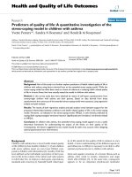
Fig. 2 B2B Research Program timeline. Recruitment for the B2B Cohort began in 2010, and steadily increased through successive operational
enhancements, key partnerships, and implementation of a centralized biospecimen ascertainment infrastructure
residents of Calgary, Alberta and the surrounding areas;
(3) females ≥18 and ≤80 years at initial diagnosis; (4) able
to provide informed, written consent and complete questionnaires and an in-person interview in English; and, (5)
no previous cancer diagnosis with the exception of cervical in-situ neoplasia (CIN) and non-melanoma skin cancer. Cohort participants must have donated a pre-surgical
blood sample to the ACRB (Fig. 3) and indicated willingness to be contacted for future research. The contact details of eligible patients are then exported to the B2B
Research Program database for recruitment into the B2B
Cohort. The B2B Cohort is restricted to the Calgary area
because of the feasibility of processing blood samples collected from community laboratories within 24 h.
Recruitment
The recruitment of patients into the B2B Cohort was harmonized with recruitment for the Alberta Moving beyond
Breast cancER (AMBER) study [92] because the potential
participants are drawn from the same population of breast
cancer patients (Fig. 4). If a patient agrees to be contacted
regarding future research, contact details for all eligible
patients are imported into the AMBER & B2B Recruitment
Database. Patients are first invited to participate in the
AMBER study so that their exercise assessments can be
completed prior to the delivery of systemic therapy [92].
Patients are invited to participate in the B2B Cohort approximately 6–8 weeks post-surgery, and after potential
recruitment into the AMBER study. The B2B Research
Program Study Coordinator contacts eligible women by
telephone to explain the research program. If the potential
participant verbally agrees to receive the recruitment
package, their information is imported into the B2B
Tracking Database and they are mailed a letter of invitation, consent information brochure, consent form and Pre
Interview Worksheets.
Brockton et al. BMC Cancer (2015) 15:512
Page 7 of 15
Table 1 Seven patient ascertainment and recruitment strategies to facilitate comprehensive population-based biospecimen accrual
and the potential for differential patient selection associated with each individual approach
Identification method
Description
Alberta Cancer Registrya
Pathological evidence of a positive cancer diagnosis provided Cancer registries may not capture 100 % of patient
by the Alberta Cancer Registry.
populations and/or may not identify patients with
sufficient time for recruitment prior to treatment.
Potential for selection bias
Direct Clinician Referral
Collaborations with key high-volume clinicians including
surgeons and oncologists pro-actively introduce the ACRB
to patients during pre-treatment consultations
Not all clinicians are supportive or have the time
and/or resources to support recruitment initiatives.
Surgical Booking Request
When a patient is diagnosed with a resectable cancer, a
surgical booking request is generated to secure a surgery
date and surgical suite.
Only includes patients scheduled for surgical
treatment for their cancer.
Pre-Admission Clinic
The pre-admission clinic ensures that patients are prepared
for a scheduled operation or procedure.
Over-representation of patients with significant
co-morbidities and/or are considered at high risk of
complications during a medical procedure.
Day Surgery Unit (DSU)
Patients are identified on the operating room slate and
encountered in the DSU just prior to their surgery on the
day of the operation.
Only includes patients treated for cancer with
surgery/excision.
Pre-treatment Patient Education Numerous programs are available to educate and inform
patients prior to treatment.
Patient education sessions are not mandatory;
only subsets of broader populations attend
these sessions.
Nurse Navigator Referral
Not all nurse navigators are prioritize research
recruitment and/or notify the ACRB of patients
entering their program.
Oncology nurses are assigned to patients to help them
navigate the continuum of cancer care. They may
introduce patients to the ACRB and/or notify the ACRB that
a patient has entered their program [113].
a
Additional ethical considerations involving the patient’s awareness of diagnosis must be addressed prior to contacting a patient to obtain informed consent
for biobanking
Consenting participants complete baseline worksheets,
an in-person interview and post-interview questionnaires
within six months of an initial breast cancer diagnosis,
with follow-up assessment occurring at 24, 48 and
72-month intervals post-diagnosis. Passive follow-up,
through chart abstraction of medical records, will
occur at 10 years following the completion of the active follow-up or for those who were lost to followup but did not withdraw consent. We anticipate that
the recruitment of the baseline B2B Cohort will be
completed by August 2015; recruitment of the baseline cohort of >600 participants will have taken a
total of 5.5 years. Analysis of data and biospecimens
will commence shortly afterwards.
Sample size
As a core infrastructure resource, the B2B Cohort sample size was not explicitly based on the power to address
a single hypothesis. However, to address our primary,
outcome–based hypotheses embedded within the core
projects, we will follow all cohort members (~600 patients) for a median of five years. Less than 60 % of
breast cancer recurrences are apparent within three
years of follow-up but ~80 % are evident after five years
of follow-up [3, 9, 10]. Within the 10-year time frame of
the B2B Research Program, we expect 70-80 women to
present with clinically evident bone metastases [3, 9, 10].
Using serum vitamin D concentrations as an example a
specific hypothesis to be tested, broad inter-quintile
ranges of serum [25-OHD] that are typical within North
American populations (Q1 < 14.9 ng/ml, Q5 > 35.3 ng/
ml [93]). Therefore, we anticipate that, for the vitamin D
and inflammation analyses, a 20 % difference in vitamin
D exposures and inflammatory status between women
with and without bone metastases will provide 80 %
power to detect a relative risk for bone metastases of 1.8
even with this modest sample size. We anticipate that
similar effect magnitudes will be observed in the other
core projects.
Data collection instruments
Pre-interview worksheets Each participant is mailed
pre-interview questionnaires including the Sun Exposure
Worksheets and Past Year Dietary Worksheets as part of
their recruitment package. Participants are asked to
complete these questionnaires and return them by mail.
The Sun Exposure Worksheets collect information on
residence, work history and vacation history for three
time periods: the 12 months prior to breast cancer diagnosis; the calendar year five years prior to breast cancer
diagnosis; and the calendar year 10 years prior to breast
cancer diagnosis. Although eligible participants must be
free of detectable distant metastases at diagnosis, breast
cancer cells can be disseminated early in tumour development. Capturing exposure data for an extended prediagnostic period, and during follow-up, ensures that
Brockton et al. BMC Cancer (2015) 15:512
Page 8 of 15
Alberta Cancer Registry
Alberta Cancer Research Biorepository (ACRB)
Comprehensive Biospecimen Rapid
Ascertainment (CoBRA) Database
Biospecimen Inventory (Freezerworks™) & Clinical Annotation Database
Pre-surgical/Pretreatment Blood sample
and questionnaire
+/- Tumor
specimen
Baseline
• Pre-interview
worksheets
• Baseline In person
interview
• DHQ
• PAQ
Blood sample
and blood
questionnaire
2 Year Follow-up
• DHQ
• PAQ
• 24 Month Follow-up
Questionnaire
Blood sample
and blood
questionnaire
4 year Follow-up
• DHQ
• PAQ
• 48 Month Follow-up
Questionnaire
Blood sample
and blood
questionnaire
6 year Follow-up
• DHQ
• PAQ
• 72 Month Follow-up
Questionnaire
10 year Follow-up
• Chart abstraction
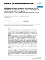
Fig. 3 Ascertainment recruitment, data and biospecimen collection and sharing scheme. Newly diagnosed cancer patients are identified through
the ACR, and are invited to donate biospecimen samples by the ACRB utilizing the CoBRA infrastructure. Clinical data and biospecimens are
stored by the ACRB, and contact information from eligible and consenting participants is sent to study coordinators of relevant research
programs. Subsequent blood samples and blood questionnaire data for routine study follow-up are collected by the ACRB and act as a shared
resource between the biorepository and research study team
exposure can be estimated for the entire period that a
patient was at risk of disease dissemination.
The Past Year Dietary Worksheets collect data regarding food intake and frequency in the 12 months prior to
breast cancer diagnosis, and was developed to assess intake of certain foods and supplements that have high
vitamin D and calcium content, consumed at a reasonable frequency by female study participants in Alberta
[94]. The sun exposure, dietary, and supplement data
will be used to estimate levels of vitamin D during the
relevant exposure period.
In-person interview The completed pre-interview questionnaires are scanned using TELEform®, an optical
character recognition software program, then verified by
study staff for completeness before being exported into
the Blaise® computer-assisted interviewing draw 1software program to pre-populate corresponding responses
in the B2B Baseline Interview. Furthermore, if the participant has completed the AMBER Baseline Health
Questionnaire (BHQ), those verified responses are also
used to pre-populate corresponding responses in the B2B
Baseline Interview. By pre-populating the interview with
information provided by the participants in the preinterview questionnaires and the AMBER BHQ, we reduce participant interview burden and expedite the inperson interview process.
One of the B2B Interviewers conducts the in-person
interview at a time and place convenient for the participant. The B2B Baseline Interview is typically an hour in
length, and collects information regarding pregnancy and
menstruation, menopausal status, hormone replacement
Brockton et al. BMC Cancer (2015) 15:512
Page 9 of 15
CoBRA
AMBER / B2B Eligible
AMBER / B2B
Recruitment
Database
AMBER Introduction
Phone call
B2B Introduction
Phone call
Yes
Yes
No
No
AMBER Tracking
Database
B2B Tracking
Database
AMBER Recruitment
B2B Recruitment
Fig. 4 B2B/AMBER participant recruitment. Eligible patients are identified by the ACRB through CoBRA processes, and the contact details of
consenting biospecimen donors are sent to a recruitment database shared by both the AMBER and B2B study coordinators. Patients are invited
to participate by each study, and if they agree, their information is then imported into the specific study database. Some information sharing
occurs between the AMBER and B2B study database, such as whether or not shared questionnaires have been completed
therapy, birth control and hormone contraceptive use,
personal health history/co-morbidity, medications (overthe-counter and prescription), vitamins, minerals and
herbal supplements, mobility and physical activity, sun exposure, diet history, family history of cancer, smoking
habits, alcohol consumption history, and demographic
information. Following the in-person interview, the participant is provided with a Canadian Diet History Questionnaire II (DHQ II) and Past Year Total Physical
Activity Questionnaire (PAQ) [95], which is to be completed and returned to the study office by mail. If a B2B
participant has already completed the DHQII and PAQ as
part of the AMBER study, these responses are made available to the B2B Research Program to further reduce participant burden.
Physical activity questionnaire The PAQ is administered at four time points throughout the study: baseline
(post-interview), 24, 48 and 72-month follow-up. The
PAQ is a self-administered questionnaire in which participants report their occupational, transportation, household
and recreational/leisure physical activities over the previous 12 months. Participants report the number of hours
spent in each activity per week, allowing for analysis of
each individual type of activity as well as a summation of
all four categories of activities to determine the participant’s total amount of physical activity over the past year
[95]. These measures are expressed as metabolic equivalents for each activity and are reported in total METhours/week/year of activity [95].
Diet history questionnaire The DHQ II [96] is also administered at four time-points during the study at baseline (post-interview), 24, 48 and 72-month follow-up. It
is a self-administered food frequency questionnaire developed initially by the National Institute of Health and
then adapted for use in the Canadian population [96].
This FFQ is a comprehensive assessment of dietary intake in the previous 12 months that has 164 questions
about 134 food items and includes seasonal intake of a
variety of foods, the portion size and frequency of intake
for each food item. Responses from the DHQII provide
Brockton et al. BMC Cancer (2015) 15:512
comprehensive information on dietary habits, output of
nutrients and the amount foods and food groups consumed. Additionally, the dose and frequency of vitamin
and mineral supplementation over the past year is also
obtained.
Biospecimen collection Each participant’s baseline blood
sample is collected as part of their ascertainment and upstream recruitment into the ACRB according to the
CoBRA procedures. Participants receive a blood requisition form and are asked to donate a blood sample at any
Calgary Laboratory Services location. The baseline collection consists of a 60 ml of blood sample collected in six
6 ml Red Top (clot activator) vacutainers and four 6 ml
Lavender Top (EDTA) vacutainers. The vacutainers are
transported to a central processing laboratory and fractionated by centrifugation to yield a total of 48 aliquots
comprised of 26 serum, 14 plasma, 4 buffy coat and 4 red
blood cells (400–500 μl per aliquot) in 1 ml Matrix® 2Dbarcoded tubes (Thermo Fisher Scientific Inc.). At the
time of blood collection, participants also complete a
short Blood Questionnaire that records information regarding their fasting status, recent smoking, medication,
supplement use, family history of cancer and menstrual
status.
Hematoxylin and eosin stained slides corresponding to
formalin-fixed paraffin embedded (FFPE) tissue blocks
are retrieved for all participants for whom tissue is available. Archived tissue blocks will be requested and retrieved according to the H&E slide pathology review; the
pathologist will mark the area of the block from which
triplicate 0.6 mm tissue cores should be collected for the
construction of TMAs. In addition to the collection of
tissue cores, 10 μm tissue scrolls will be collected for the
extraction of nucleic acids (DNA and RNA). The RNA
will be used for the transcript analysis in sub-project 2
and the DNA will be extracted at a later date for ancillary projects potentially investigating mutational analyses. All blood and tissue samples are stored within the
ACRB.
Participant follow-up at 24, 48 or 72-months Additional data and biospecimen collections occur at the
specified follow-up intervals of 24, 48 and 72-months
from the participant’s primary breast cancer diagnosis.
Each month, the Study Coordinator queries the B2B Recruitment Database to generate a list of participants eligible for follow-up. The Study Coordinator contacts
each participant by telephone to confirm their address
and willingness to continue their participation in the
B2B Research Program. If they agree, participants are
sent a follow-up package that includes a PAQ, DHQ II
and the appropriate Follow-Up Questionnaire (24, 48 or
72-month); these are self-administered questionnaires to
Page 10 of 15
be completed by the participants and returned by mail
to the study office. The 24, 48 or 72-month Follow-Up
Questionnaires request information on: personal health
history, breast cancer progression (only at 24 month follow up only), recurrence, contra-laterality and new primary diagnosis, medications (prescription and over the
counter), smoking habits, alcohol consumption, sun exposure, dietary intake, mobility and physical activity and
anthropometric measurements.
Follow-up blood samples are also collected at 24, 48
and 72 months. Each Follow-Up Package contains a Research Blood Requisition form and participants are asked
to donate a blood sample at any Calgary Lab Services
location. The follow-up blood collections consist of a
30 ml of blood sample collected using three 6 ml Red
Top (clot activator) vacutainers and two EDTA vacutainers. Again, the vacutainers are transported to a central
processing laboratory, fractionated by centrifugation to
yield serum, plasma red blood cells and buffy coat and
stored in 32 aliquots of 400–500 μl in 1 ml Matrix® 2Dbarcoded tubes (Table 1).
Vital status check The vital status of each participant is
checked before each follow-up contact at 24, 48 and 72
months through a record linkage done by the Department of Cancer Surveillance (Alberta Health Services).
Vital Statistics Alberta (VSA) provides information on
all deaths that occurred in the province to the ACR, on
request, with underlying cause of death provided by Statistics Canada to VSA. There is an average three-month
time lag between the actual death occurrence and
reporting to the ACR. Several mechanisms, such as reciprocal agreements between other provinces and record
linkages with the Canadian Mortality Database, exist to
capture the deaths of participants who left the province
of Alberta after their diagnosis. These agreements and
processes ensure that vital status can be determined for
over 95 % of participants. Cause and date of death will
also be obtained from this source.
Medical record abstraction Medical record abstraction
will occur in the final year of the B2B Program operation
(commencing August 2018). Health Record Technicians
from the ACR will use direct data entry to a medical
record abstraction form to collect data from the medical
records (both paper and electronic charts) for all participants in the B2B Cohort. The medical record abstraction
form was developed from standardized forms used in
our past physical activity and breast cancer cohort study
conducted in Alberta [97, 98].
Baseline pathologic data, including clinical stage and
pathologic stage (according to American Joint Committee on Cancer criteria [99], tumor size, grade, histology,
estrogen receptor status, progesterone receptor status
Brockton et al. BMC Cancer (2015) 15:512
human epidermal growth factor receptor 2 status, type
and results of computerized tomography or positron
emission tomography scans, status of margins (with breast
conserving surgery), and pathology of lymph nodes (if surgically sampled) are provided by the ACRB/CoBRA database and verified during the medical record abstraction.
Abstracted variables will include the type of surgery, and
all treatment and follow-up care including data on chemotherapy, radiation therapy, and hormone therapy. Treatment completion rates will be estimated for chemotherapy
and hormone therapy but not for radiation therapy since
few patients fail to complete radiation therapy. For
chemotherapy completion rate, we will estimate the average relative dose intensity (RDI) received for the originally
planned regimen based on standard formulae as we have
done in a previous RCT [100]. For hormone therapy, the
follow-up questionnaires ask participants to report if they
have stopped taking their prescribed hormone therapy at
any time before its intended completion and the reasons
for stopping.
Disease endpoints are defined according to the Standardized Definitions for Efficacy End Points in Adjuvant
Breast Cancer Trials [101]. Our primary endpoint of interest is bone metastasis or a skeletal-related event according
to the definition in the NSABP 34 trial (http://clinical
trials.gov/show/NCT00009945). We will also examine
other composite disease endpoints as secondary endpoints
including overall survival, distant disease-free survival, distant relapse-free survival, and distant recurrence-free
interval. Finally, we will examine the single disease endpoints of death from breast cancer and death from nonbreast cancer. For participants who have left the province
and who are not known to be deceased, the date of leaving
Alberta will be used as the censoring time.
Discussion
Bone metastases are the most common site of breast
cancer metastasis and there are currently no curative
treatments available. Consequently, predicting the risk of
bone metastasis, identifying modifiable lifestyle strategies
to reduce those risks, developing methods for early detection and understanding the fundamental biology and
potential therapeutic targets are of the highest priority.
The B2B Research Program seeks to address each of
these priorities via an integrated program of research
based on the population-based prospective B2B Cohort.
Each of the core projects (Fig. 1) focuses primarily on
one of these priorities; however, the integrated program
design and shared data and biospecimen resources
enables significant synergy between the projects and potential for additional future hypothesis generation and
testing.
A population-based prospective cohort of incident
breast cancer patients offers the ideal study design to
Page 11 of 15
address the priorities that we have identified. Framing
such research within existing randomised controlled trials
of treatment would be limited by the availability of biospecimens and likely lack the external validity afforded by the
population-based ascertainment [102, 103]. Also, large
disease-free prospective cohorts, such as the Canadian
Partnership for Tomorrow Project [104], could not deliver
the necessary number of outcomes within a reasonable
period and are not currently configured to collect epidemiologic data and biologic samples specifically during the
crucial peri-diagnostic period or conduct disease-specific
follow-up. Although previous studies have conducted
population-based recruitment of cancer patients in Alberta [94, 105], there was no existing mechanism to comprehensively identify, contact, obtain consent and track
breast cancer patients. Furthermore, the increasingly stringent privacy requirements demanded the development of
a system that could accomplish the recruitment and biospecimen collection targets whilst being sensitive to the
circumstances of the patients and complying with all relevant privacy regulations. The establishment of the B2B
Research Program, the co-development of the CoBRA
database and procedures and the partnership with the
ACRB all contributed to the creation of the infrastructure
to facilitate the current and future population-based prospective recruitment.
The collection of biospecimens is a critical component
of the B2B Research Program that required the development of new procedures and the implementation of new
technology. Adopting the 1 ml Matrix® 2D-barcoded
tubes (Thermo Fisher Scientific Inc.) and the 2D barcode scanner (Thermo Fisher Scientific Inc.) enabled a
large number of relatively low-volume aliquots to be collected and tracked for efficient inventory management
while avoiding excessive manual labelling and minimizing potential for human error. Two core projects within
the B2B Research Program are using serum samples to
investigate metabolomic and cytokine profiles associated
with the risk for bone metastases. Little has been published on the impact of surgery [106–110] or systemic
therapy [111, 112] on serum biomarkers, but the existing
literature clearly demonstrates that there are significant
changes in blood-based markers in response to both surgery and systemic therapy [106–110]. Consequently, the
collection of a pre-surgical blood sample is an eligibility
requirement for the B2B Cohort.
The detailed epidemiologic data collected on each participant are collected using several data collection instruments that have been adopted from previously published
research studies. The Pre-Interview Worksheets comprise
two short questionnaires developed for the OVarian cancer in ALberta (OVAL) Study [94] to improve the assessment of overall vitamin D exposure as well as dietary
calcium intake. The computer-assisted baseline In-Person
Brockton et al. BMC Cancer (2015) 15:512
interview (Blaise®) was adapted from the interviews created for the Alberta Endometrial Cancer Case-Control
Study [105] with modifications to address specific questions related to bone health and inflammation. The PAQ
that we use was developed to measure total physical activity in the previous year and has been tested for reliability
and validity [95]. The DHQ has also been specifically
modified to capture the food items available and consumed in Canada [96]. By using existing instruments or
modifying those that had previously been used in similar
settings, we have minimized the development costs, taken
advantage of existing reliability measures and maximized
the comparability of our data with existing and future
studies using those instruments.
The B2B Research Program will address several critical
aspects of breast cancer bone metastases including prediction, prevention, detection, and biology. Sub-clinical
vitamin D deficiency may be a prevalent, yet modifiable,
risk factor for breast cancer bone metastases. Optimal
vitamin D status may help prevent bone metastasis by a
fairly straightforward intervention through its reported
ability to attenuate inflammation and proliferation, while
promoting apoptosis and differentiation. The transcriptional features of the primary tumour and systemic response could provide a method to determine a patient’s
risk of bone metastases at diagnosis to direct appropriate
therapies or surveillance. Serum metabolomics offers the
potential for early detection of bone metastases. Furthermore, by combining these data with the epidemiologic
and clinical data, the impact of modifiable lifestyle factors on metabolites associated with bone metastases
might be determined. Finally, the in vitro and in vivo research can dissect the biologic mechanisms and identify
potential therapeutic targets.
Lessons and limitations
Lessons learned during the set-up and early operation of
the B2B Research Program have greatly enhanced the ascertainment and recruit of breast cancer patients in
Alberta, through the development of the CoBRA system
and refinement of study-specific processes. Translating
the comprehensive patient ascertainment into adequate
recruitment, and ultimately biospecimen collections, was
a significant challenge. However, continual development
of these processes and the supporting database has resulted in sustainable recruitment, and biospecimen collection, from ~75 % of breast cancer patients in the Calgary,
Alberta area.
We originally proposed a tiered sampling mechanism
to enrich the B2B Cohort for stage III cancers who are
most likely to develop bone metastases. However, our
initial rate of recruitment was insufficient to implement
this strategy. The most significant obstacle was achieving
patient contact with sufficient time remaining to obtain
Page 12 of 15
a pre-surgical blood sample. We introduced several
mechanisms to facilitate timely contact with patients
during clinic visits, through clinical care-related presentations and during the peri-operative period whilst maintaining the original correspondence-based procedures
(Fig. 2); these process enhancements improved our recruitment rates and we anticipate the full baseline cohort recruitment to be completed by August 2015. In
addition, we have extended the follow-up program over
a longer duration (six years of follow-up instead of the
three year period originally proposed) to off-set the
lower event rates in early stage patients, to capture a
greater number of events overall and maximize statistical power.
In 2013 we obtained approval from our institutional
ethics board to introduce email as a method of correspondence between B2B Cohort members and our research team. The pre-interview worksheet package has a
B2B Research Program email address with the B2B Research Program Study Coordinator contact details. B2B
Cohort members are invited to correspond with B2B
Program personnel through this email address and are
asked to provide permission for research personnel to
contact them through their personal email address. Email correspondence has been very popular and facilitates immediate engagement of new participants. It also
provides a mechanism to acknowledge receipt of study
materials, notify participants of their interviewer’s contact details and to thank participants for their contributions. The use of e-mail has resulted in much more
efficient communication with participants with less
time spent telephoning, greater accessibility for both
participants and interviewers, and a greater number of
participants completing the interview portion of the
study.
In summary, the B2B Research Program is establishing a population-based prospective cohort of breast
cancer patients in which we will conduct four initial
core research projects that will address key aspects of
bone metastasis in breast cancer survivors. The collection of pre-surgical blood samples and detailed epidemiologic data, at baseline and follow-up, will provide a
unique and rich resource to address current and future
research. To our knowledge, no equivalent resources
are currently available. The biospecimen and data resources established by the B2B Research Program will
also enable currently unanticipated important research
questions to be addressed in a timely manner. The ultimate goal of this research is to improve the prediction
of bone metastasis risk, identify modifiable lifestyle
strategies to reduce those risks, and improve our understanding of the fundamental biology and potential
therapeutic targets to reduce bone metastases in breast
cancer survivors.
Brockton et al. BMC Cancer (2015) 15:512
Abbreviations
25-OHD: 25-hydroxyvitamin D; ACR: Alberta Cancer Registry; ACRB: Alberta
Cancer Research Biobank; AMBER: Alberta Moving beyond Breast cancER;
B2B: Breast Cancer to Bone Metastasis; BHQ: Baseline health questionnaire;
CIN: Cervical in-situ neoplasia; CHREB: Conjoint health research ethics board;
CoBRA: Comprehensive Biospecimen Rapid Ascertainment; CTGF: Connective
tissue growth factor; CXCR4: Chemokine (C-X-C motif) receptor 4; DAPI: 4′,
6-diamidino-2-phenylindole; DHQ II: Diet history questionnaire II; DHQ: Diet
history questionnaire; DNA: Deoxyribonucleic acid; DSU: Day Surgery Unit;
EDTA: Ethylenediaminetetraacetic acid; EMT: Epithelial-mesenchymal
transition; FGF5: Fibroblast growth factor 5; HREBA: Health Research Ethics
Board of Alberta; IHC: Immunohistochemistry; IL-11: Interleukin-11;
IL-1B: Interleukin-1β; IL-6: Interleukin-6; IL-8: Interleukin-8; MMP1: Matrix
metallopeptidase 1; NMR: Nuclear magnetic resonance; NSAID: Non-steroidal
anti-inflammatory drugs; PAQ: Physical activity questionnaire;
PTHrP: Parathyroid hormone-related protein; RANKL: Receptor activator of
nuclear factor-κB ligand; RDI: Relative dose intensity; RNA: Ribonucleic acid;
SDF1: Stromal cell-derived factor1; TMAs: Tissue microarrays; TNF-α: Tumour
necrosis factor-alpha; TNM: Tumour Node Metastasis; UVB: Ultraviolet B;
VSA: Vital Statistics Alberta.
Page 13 of 15
5.
6.
7.
8.
9.
10.
11.
12.
Competing interests
The authors declare that they have no competing interests.
Authors’ contributions
NTB, LSC, AMM & AHGP conceived the study. NTB, CMF, AMM, LSC, AHGP,
HJV, CSS, DAH obtained research funding. NTB, LSC, AMM, DAH, SJG, SLL &
CMF developed the study methods. NTB, SJG, SLL and CMF drafted the
manuscript. NTB, LSC, CSS, HJV and AMM, each drafted the descriptions of
their specific sub-project components and edited the manuscript. AHGP
directed clinical details and edited the manuscript. DAH helped generate the
vitamin D assessment measures and edited the manuscript. All authors read
and approved the final manuscript.
Acknowledgments
This study is funded by a Translational Team Grant from the Alberta Cancer
Foundation. CMF is supported by an Alberta Innovates Health Solutions
Health Senior Scholar Award and by the Alberta Cancer Foundation
Weekend to End Women’s Cancers Breast Cancer Chair. HJV is supported by
the Alberta Cancer Foundation Lance Armstrong Chair in Molecular Cancer
Epidemiology.
Author details
Department of Cancer Epidemiology and Prevention Research,
CancerControl Alberta, Alberta Health Services, Room 515C, Holy Cross
Centre, 2210 2nd St, SW, Calgary, AB T2S 3C3, Canada. 2Department of
Oncology, Cumming School of Medicine, University of Calgary, Calgary,
Alberta, Canada. 3Department of Community Health Sciences, Cumming
School of Medicine, University of Calgary, Calgary, Alberta, Canada. 4Division
of Medical Oncology, Tom Baker Cancer Centre, Cancer Control Alberta,
Alberta Health Services, Calgary, Alberta, Canada. 5Division of Epidemiology,
Biostatistics and Preventive Medicine, Department of Internal Medicine,
University of New Mexico, Albuquerque, New Mexico, USA. 6Department of
Biological Sciences, Faculty of Science, University of Calgary, Calgary, Alberta,
Canada. 7Department of Medicine, Cumming School of Medicine, University
of Calgary, Calgary, Alberta, Canada. 8Department of Pathology, Moffitt
Cancer Center, Tampa, FL, USA.
13.
14.
15.
16.
17.
18.
19.
20.
1
21.
22.
23.
24.
25.
26.
Received: 1 April 2015 Accepted: 29 June 2015
27.
References
1. Siegel R, Ma J, Zou Z, Jemal A. Cancer statistics, 2014. CA Cancer J Clin.
2014;64(1):9–29.
2. Canadian Cancer Statistics 2014 [ />CW/cancer%20information/cancer%20101/Canadian%20cancer%20statistics/
Canadian-Cancer-Statistics-2014-EN.pdf]
3. Coleman RE, Rubens RD. The clinical course of bone metastases from
breast-cancer. Br J Cancer. 1987;55(1):61–6.
4. Manders K, van de Poll-Franse LV, Creemers GJ, Vreugdenhil G, van der
Sangen MJC, Nieuwenhuijzen GAP, et al. Clinical management of women
28.
29.
30.
31.
with metastatic breast cancer: a descriptive study according to age group.
BMC Cancer. 2006;6:179.
Glendenning J, Cook G. Imaging breast cancer bone metastases: current
status and future directions. Semin Nucl Med. 2013;43(4):317–23.
Jung SY, Rosenzweig M, Sereika SM, Linkov F, Brufsky A, Weissfeld JL.
Factors associated with mortality after breast cancer metastasis. Cancer
Causes Control. 2012;23(1):103–12.
Auerbach R. Patterns of tumor-metastasis - organ selectivity in the spread of
cancer-cells. Lab Investig. 1988;58(4):361–4.
Paget S. The distribution of secondary growths in cancer of the breast:
1889. Cancer Metastasis Rev. 1989;8(2):98–101.
Elder EE, Kennedy CW, Gluch L, Carmalt HL, Janu NC, Joseph MG, et al.
Patterns of breast cancer relapse. Ejso. 2006;32(9):922–7.
Jensen AO, Jacobsen JB, Norgaard M, Yong M, Fryzek JP, Sorensen HT.
Incidence of bone metastases and skeletal-related events in breast cancer
patients: a population-based cohort study in Denmark. BMC Cancer. 2011;11:29.
Sathiakumar N, Delzell E, Morrisey MA, Falkson C, Yong M, Chia V, et al.
Mortality following bone metastasis and skeletal-related events among
women with breast cancer: a population-based analysis of US Medicare
beneficiaries, 1999-2006. Breast Cancer Res Treat. 2012;131(1):231–8.
Masuda TA, Kataoka A, Ohno S, Murakami S, Mimori K, Utsunomiya T, et al.
Detection of occult cancer cells in peripheral blood and bone marrow by
quantitative RT-PCR assay for cytokeratin-7 in breast cancer patients. Int J
Oncol. 2005;26(3):721–30.
Braun S, Vogl FD, Naume B, Janni W, Osborne MP, Coombes RC, et al. A
pooled analysis of bone marrow micrometastasis in breast cancer. N Engl J
Med. 2005;353(8):793–802.
Baker M, Gillanders WE, Mikhitarian K, Mitas M, Cole DJ. The molecular
detection of micrometastatic breast cancer. Am J Surg. 2003;186(4):351–8.
Pantel K, Muller V, Auer M, Nusser N, Harbeck N, Braun S. Detection and
clinical implications of early systemic tumor cell dissemination in breast
cancer. Clin Cancer Res. 2003;9(17):6326–34.
Coleman R, Body JJ, Aapro M, Hadji P, Herrstedt J, Group EGW. Bone health
in cancer patients: ESMO Clinical Practice Guidelines. Ann Oncol.
2014;25 Suppl 3:iii124–37.
Roodman GD. Mechanisms of bone metastasis. N Engl J Med.
2004;350(16):1655–64.
Quattrocchi CC, Piciucchi S, Sammarra M, Santini D, Vincenzi B, Tonini G,
et al. Bone metastases in breast cancer: higher prevalence of osteosclerotic
lesions. Radiol Med. 2007;112(7):1049–59.
Rodan GA, Martin TJ. Therapeutic approaches to bone diseases. Science.
2000;289(5484):1508–14.
Eckhardt BL, Parker BS, van Laar RK, Restall CM, Natoli AL, Tavaria MD, et al.
Genomic analysis of a spontaneous model of breast cancer metastasis to
bone reveals a role for the extracellular matrix. Mol Cancer Res. 2005;3(1):1–13.
Yoneda T, Hiraga T. Crosstalk between cancer cells and bone microenvironment
in bone metastasis. Biochem Biophys Res Commun. 2005;328(3):679–87.
Mancino AT, Klimberg VS, Yamamoto M, Manolagas SC, Abe E. Breast cancer
increases osteoclastogenesis by secreting M-CSF and upregulating RANKL in
stromal cells. J Surg Res. 2001;100(1):18–24.
Roodman GD. Role of stromal-derived cytokines and growth factors in bone
metastasis. Cancer. 2003;97(3):733–8.
Guise T, Clines G, Mohammad K, Niewolna M, Mison A, McKenna R, et al.
Molecular mechanisms of osteoblastic bone metastases: rational for
targeting the endothelin axis. J Bone Miner Res. 2005;20:P2–3.
Mundy GR. Mechanisms of bone metastasis. Cancer. 1997;80(8 Suppl):1546–56.
Kozlow W, Guise TA. Breast cancer metastasis to bone: mechanisms of
osteolysis and implications for therapy. J Mammary Gland Biol Neoplasia.
2005;10(2):169–80.
Green JR, Clezardin P. Mechanisms of bisphosphonate effects on
osteoclasts, tumor cell growth, and metastasis. Am J Clin Oncol Cancer Clin
Trials. 2002;25(6):S3–9.
Horak CE, Steeg PS. Metastasis gets site specific. Cancer Cell. 2005;8(2):93–5.
Welch DR. Microarrays bring new insights into understanding of breast
cancer metastasis to bone. Breast Cancer Res. 2004;6(2):61–4.
Woelfle U, Cloos J, Sauter G, Riethdorf L, Janicke F, van Diest P, et al.
Molecular signature associated with bone marrow micrometastasis in
human breast cancer. Cancer Res. 2003;63(18):5679–84.
Kang Y, Siegel PM, Shu W, Drobnjak M, Kakonen SM, Cordon-Cardo C, et al.
A multigenic program mediating breast cancer metastasis to bone. Cancer
Cell. 2003;3(6):537–49.
Brockton et al. BMC Cancer (2015) 15:512
32. Smid M, Wang Y, Klijn JG, Sieuwerts AM, Zhang Y, Atkins D, et al. Genes
associated with breast cancer metastatic to bone. J Clin Oncol.
2006;24(15):2261–7.
33. Fazilaty H, Mehdipour P. Genetics of breast cancer bone metastasis: a
sequential multistep pattern. Clin Exp Metastasis. 2014;31(5):595–612.
34. Wilson C, Holen I, Coleman RE. Seed, soil and secreted hormones: potential
interactions of breast cancer cells with their endocrine/paracrine
microenvironment and implications for treatment with bisphosphonates.
Cancer Treat Rev. 2012;38(7):877–89.
35. Zhou X, Liu J. A computational model to predict bone metastasis in breast
cancer by integrating the dysregulated pathways. BMC Cancer. 2014;14:618.
36. Coleman RE, Gregory W, Marshall H, Wilson C, Holen I. The metastatic
microenvironment of breast cancer: clinical implications. Breast.
2013;22 Suppl 2:S50–6.
37. Krishnan AV, Swami S, Feldman D. Vitamin D and breast cancer: inhibition
of estrogen synthesis and signaling. J Steroid Biochem Mol Biol.
2010;121(1-2):343–8.
38. Ooi LL, Zheng Y, Stalgis-Bilinski K, Dunstan CR. The bone remodeling
environment is a factor in breast cancer bone metastasis. Bone.
2011;48(1):66–70.
39. Blair CK, Sweeney C, Anderson KE, Folsom AR. NSAID use and survival after
breast cancer diagnosis in post-menopausal women. Breast Cancer Res
Treat. 2007;101(2):191–7.
40. Li YL, Brasky TM, Nie J, Ambrosone CB, McCann SE, Shields PG, et al. Use of
nonsteroidal anti-inflammatory drugs and survival following breast cancer
diagnosis. Cancer Epidemiol Biomarkers Prev. 2012;21(1):239–42.
41. Cui Y, Rohan TE. Vitamin D, calcium, and breast cancer risk: a review. Cancer
Epidemiol Biomarkers Prev. 2006;15(8):1427–37.
42. Ooi LL, Zheng Y, Zhou H, Trivedi T, Conigrave AD, Seibel MJ, et al. Vitamin
D deficiency promotes growth of MCF-7 human breast cancer in a rodent
model of osteosclerotic bone metastasis. Bone. 2010;47(4):795–803.
43. Ooi LL, Zhou H, Kalak R, Zheng Y, Conigrave AD, Seibel MJ, et al. Vitamin d
deficiency promotes human breast cancer growth in a murine model of
bone metastasis. Cancer Res. 2010;70(5):1835–44.
44. Zehnder D, Bland R, Chana RS, Wheeler DC, Howie AJ, Williams MC, et al.
Synthesis of 1,25-dihydroxyvitamin D(3) by human endothelial cells is
regulated by inflammatory cytokines: a novel autocrine determinant of
vascular cell adhesion. J Am Soc Nephrol. 2002;13(3):621–9.
45. Yee YK, Chintalacharuvu SR, Lu J, Nagpal S. Vitamin D receptor modulators
for inflammation and cancer. Mini Rev Med Chem. 2005;5(8):761–78.
46. Ragab AA, Nalepka JL, Bi Y, Greenfield EM. Cytokines synergistically induce
osteoclast differentiation: support by immortalized or normal calvarial cells.
Am J Physiol Cell Physiol. 2002;283(3):C679–87.
47. Holick MF. High prevalence of vitamin D inadequacy and implications for
health. Mayo Clin Proc. 2006;81(3):353–73.
48. Cantorna MT, Zhu Y, Froicu M, Wittke A. Vitamin D status, 1,25-dihydroxyvitamin
D3, and the immune system. Am J Clin Nutr. 2004;80(6 Suppl):1717S–20.
49. Shibata T, Shira-Ishi A, Sato T, Masaki T, Sasaki A, Masuda Y, et al. Vitamin D
hormone inhibits osteoclastogenesis in vivo by decreasing the pool of
osteoclast precursors in bone marrow. J Bone Miner Res. 2002;17(4):622–9.
50. Kwan ML, Habel LA, Slattery ML, Caan B. NSAIDs and breast cancer
recurrence in a prospective cohort study. Cancer Causes Control.
2007;18(6):613–20.
51. Blair HC, Zaidi M. Osteoclastic differentiation and function regulated by old
and new pathways. Rev Endocr Metab Disord. 2006;7(1-2):23–32.
52. Rucker D, Allan JA, Fick GH, Hanley DA. Vitamin D insufficiency in a
population of healthy western Canadians. CMAJ. 2002;166(12):1517–24.
53. Dewan MZ, Ahmed S, Iwasaki Y, Ohba K, Toi M, Yamamoto N. Stromal cellderived factor-1 and CXCR4 receptor interaction in tumor growth and
metastasis of breast cancer. Biomed Pharmacother. 2006;60(6):273–6.
54. Kaifi JT, Yekebas EF, Schurr P, Obonyo D, Wachowiak R, Busch P, et al.
Tumor-cell homing to lymph nodes and bone marrow and CXCR4
expression in esophageal cancer. J Natl Cancer Inst. 2005;97(24):1840–7.
55. Russell HV, Hicks J, Okcu MF, Nuchtern JG. CXCR4 expression in
neuroblastoma primary tumors is associated with clinical presentation of
bone and bone marrow metastases. J Pediatr Surg. 2004;39(10):1506–11.
56. Darash-Yahana M, Pikarsky E, Abramovitch R, Zeira E, Pal B, Karplus R, et al.
Role of high expression levels of CXCR4 in tumor growth, vascularization,
and metastasis. Faseb J. 2004;18(9):1240.
57. Geminder H, Sagi-Assif O, Goldberg L, Meshel T, Rechavi G, Witz IP, et al. A
possible role for CXCR4 and its ligand, the CXC chemokine stromal cell-
Page 14 of 15
58.
59.
60.
61.
62.
63.
64.
65.
66.
67.
68.
69.
70.
71.
72.
73.
74.
75.
76.
77.
78.
79.
80.
81.
82.
derived factor-1, in the development of bone marrow metastases in
neuroblastoma. J Immunol. 2001;167(8):4747–57.
Lapidot T. Mechanism of human stem cell migration and repopulation of
NOD/SCID and B2mnull NOD/SCID mice - the role of SDF-1/CXCR4
interactions. 2001.
Shimo T, Kubota S, Yoshioka N, Ibaragi S, Isowa S, Eguchi T, et al.
Pathogenic role of connective tissue growth factor (CTGF/CCN2) in
osteolytic metastasis of breast cancer. J Bone Miner Res. 2006;21(7):1045–59.
Rangaswami H, Bulbule A, Kundu GC. Osteopontin: role in cell signaling and
cancer progression. Trends Cell Biol. 2006;16(2):79–87.
Wai PY, Kuo PC. The role of osteopontin in tumor metastasis. J Surg Res.
2004;121(2):228–41.
Standal T, Borset M, Sundan A. Role of osteopontin in adhesion, migration,
cell survival and bone remodeling. Exp Oncol. 2004;26(3):179–84.
Mi ZY, Guo HT, Wai PY, Gao CJ, Wei JP, Kuo PC. Differential osteopontin
expression in phenotypically distinct subclones of murine breast cancer
cells mediates metastatic behavior. J Biol Chem. 2004;279(45):46659–67.
Singhal H, Bautista DS, Tonkin KS, Omalley FP, Tuck AB, Chambers AF, et al.
Elevated plasma osteopontin in metastatic breast cancer associated with
increased tumor burden and decreased survival. Clin Cancer Res.
1997;3(4):605–11.
Bramwell VHC, Doig GS, Tuck AB, Wilson SM, Tonkin KS, Tomiak A, et al.
Serial plasma osteopontin levels have prognostic value in metastatic breast
cancer. Clin Cancer Res. 2006;12(11):3337–43.
Rittling SR, Chambers AF. Role of osteopontin in tumour progression. Br J
Cancer. 2004;90(10):1877–81.
Warburg O. On the origin of cancer cells. Science. 1956;123(3191):309–14.
Armitage EG, Barbas C. Metabolomics in cancer biomarker discovery: current
trends and future perspectives. J Pharm Biomed Anal. 2014;87:1–11.
Beger RD. A review of applications of metabolomics in cancer. Metabolites.
2013;3(3):552–74.
Nicholson JK, Holmes E, Kinross JM, Darzi AW, Takats Z, Lindon JC.
Metabolic phenotyping in clinical and surgical environments. Nature.
2012;491(7424):384–92.
Bathe OF, Shaykhutdinov R, Kopciuk K, Weljie AM, McKay A, Sutherland FR,
et al. Feasibility of identifying pancreatic cancer based on serum
metabolomics. Cancer Epidemiol Biomarkers Prev. 2011;20(1):140–7.
Farshidfar F, Weljie AM, Kopciuk K, Buie WD, Maclean A, Dixon E, et al.
Serum metabolomic profile as a means to distinguish stage of colorectal
cancer. Genome Med. 2012;4(5):42.
Wittmann BM, Stirdivant SM, Mitchell MW, Wulff JE, McDunn JE, Li Z, et al.
Bladder cancer biomarker discovery using global metabolomic profiling of
urine. PLoS One. 2014;9(12):e115870.
Budczies J, Brockmoller SF, Muller BM, Barupal DK, Richter-Ehrenstein C,
Kleine-Tebbe A, et al. Comparative metabolomics of estrogen receptor
positive and estrogen receptor negative breast cancer: alterations in
glutamine and beta-alanine metabolism. J Proteome. 2013;94:279–88.
Wood SL, Westbrook JA, Brown JE. Omic-profiling in breast cancer metastasis
to bone: implications for mechanisms, biomarkers and treatment. Cancer Treat
Rev. 2014;40(1):139–52.
Oakman C, Tenori L, Claudino WM, Cappadona S, Nepi S, Battaglia A, et al.
Identification of a serum-detectable metabolomic fingerprint potentially
correlated with the presence of micrometastatic disease in early breast
cancer patients at varying risks of disease relapse by traditional prognostic
methods. Ann Oncol. 2011;22(6):1295–301.
Palmnas MS, Vogel HJ. The future of NMR metabolomics in cancer therapy:
towards personalizing treatment and developing targeted drugs?
Metabolites. 2013;3(2):373–96.
Madsen R, Lundstedt T, Trygg J. Chemometrics in metabolomics–a review
in human disease diagnosis. Anal Chim Acta. 2010;659(1-2):23–33.
Martineau E, Tea I, Akoka S, Giraudeau P. Absolute quantification of
metabolites in breast cancer cell extracts by quantitative 2D (1) H
INADEQUATE NMR. NMR Biomed. 2012;25(8):985–92.
Giskeodegard GF, Grinde MT, Sitter B, Axelson DE, Lundgren S, Fjosne HE,
et al. Multivariate modeling and prediction of breast cancer prognostic
factors using MR metabolomics. J Proteome Res. 2010;9(2):972–9.
Husemann Y, Geigl JB, Schubert F, Musiani P, Meyer M, Burghart E, et al.
Systemic spread is an early step in breast cancer. Cancer Cell.
2008;13(1):58–68.
Slade MJ, Coombes RC. The clinical significance of disseminated tumor cells
in breast cancer. Nat Clin Pract Oncol. 2007;4(1):30–41.
Brockton et al. BMC Cancer (2015) 15:512
83. Braun S, Auer D, Marth C. The prognostic impact of bone marrow
micrometastases in women with breast cancer. Cancer Invest.
2009;27(6):598–603.
84. Balic M, Lin H, Young L, Hawes D, Giuliano A, McNamara G, et al. Most early
disseminated cancer cells detected in bone marrow of breast cancer
patients have a putative breast cancer stem cell phenotype. Clin Cancer
Res. 2006;12(19):5615–21.
85. D'Amico L, Patane S, Grange C, Bussolati B, Isella C, Fontani L, et al. Primary
breast cancer stem-like cells metastasise to bone, switch phenotype and
acquire a bone tropism signature. Br J Cancer. 2013;108(12):2525–36.
86. Ocana OH, Corcoles R, Fabra A, Moreno-Bueno G, Acloque H, Vega S, et al.
Metastatic colonization requires the repression of the epithelialmesenchymal transition inducer Prrx1. Cancer Cell. 2012;22(6):709–24.
87. Tsai JH, Donaher JL, Murphy DA, Chau S, Yang J. Spatiotemporal regulation
of epithelial-mesenchymal transition is essential for squamous cell
carcinoma metastasis. Cancer Cell. 2012;22(6):725–36.
88. Mani SA, Guo W, Liao MJ, Eaton EN, Ayyanan A, Zhou AY, et al. The
epithelial-mesenchymal transition generates cells with properties of stem
cells. Cell. 2008;133(4):704–15.
89. Andres A, Majno PE, Morel P, Rubbia-Brandt L, Giostra E, Gervaz P, et al.
Improved long-term outcome of surgery for advanced colorectal liver
metastases: reasons and implications for management on the basis of a
severity score. Ann Surg Oncol. 2008;15(1):134–43.
90. Klein-Goldberg A, Maman S, Witz IP. The role played by the
microenvironment in site-specific metastasis. Cancer Lett. 2014;352(1):54–8.
91. Lau YS, Danks L, Sun SG, Fox S, Sabokbar A, Harris A, et al. RANKLdependent and RANKL-independent mechanisms of macrophageosteoclast differentiation in breast cancer. Breast Cancer Res Treat.
2007;105(1):7–16.
92. Courneya KS, Vallance JK, Culos-Reed SN, McNeely ML, Bell GJ, Mackey JR,
et al. The Alberta moving beyond breast cancer (AMBER) cohort study: a
prospective study of physical activity and health-related fitness in breast
cancer survivors. BMC Cancer. 2012;12:525.
93. Feskanich D, Ma J, Fuchs CS, Kirkner GJ, Hankinson SE, Hollis BW, et al.
Plasma vitamin D metabolites and risk of colorectal cancer in women.
Cancer Epidemiol Biomarkers Prev. 2004;13(9):1502–8.
94. Cook LS, Moon BL, Dong Y, Neilson HK. Reliability of self-reported sun
exposure in Canadian women and estimation of lifetime exposure to
vitamin D from sun and diet. Public Health Nutr. 2014;17(4):747–55.
95. Friedenreich CM, Courneya KS, Neilson HK, Matthews CE, Willis G, Irwin M,
et al. Reliability and validity of the past year total physical activity
questionnaire. Am J Epidemiol. 2006;163(10):959–70.
96. Csizmadi I, Kahle L, Ullman R, Dawe U, Zimmerman TP, Friedenreich CM,
et al. Adaptation and evaluation of the National Cancer Institute's Diet
History Questionnaire and nutrient database for Canadian populations.
Public Health Nutr. 2007;10(1):88–96.
97. Neilson HK, Friedenreich CM, Brockton NT, Millikan RC. Physical activity and
postmenopausal breast cancer: proposed biologic mechanisms and areas
for future research. Cancer Epidemiol Biomarkers Prev. 2009;18(1):11–27.
98. Friedenreich CM, Gregory J, Kopciuk KA, Mackey JR, Courneya KS.
Prospective cohort study of lifetime physical activity and breast cancer
survival. Int J Cancer. 2009;124(8):1954–62.
99. Greene FL, Page DL, Fleming ID, Fritz AG, Balch CM, Haller DG, et al. AJCC
Cancer springer manual, 6th edition. 6th ed. New York: Springer; 2002.
100. Courneya KS, Segal RJ, Mackey JR, Gelmon K, Reid RD, Friedenreich CM,
et al. Effects of aerobic and resistance exercise in breast cancer patients
receiving adjuvant chemotherapy: a multicenter randomized controlled trial.
J Clin Oncol. 2007;25(28):4396–404.
101. Hudis CA, Barlow WE, Costantino JP, Gray RJ, Pritchard KI, Chapman JA, et al.
Proposal for standardized definitions for efficacy end points in adjuvant
breast cancer trials: the STEEP system. J Clin Oncol. 2007;25(15):2127–32.
102. Ransohoff DF. Bias as a threat to the validity of cancer molecular-marker
research. Nat Rev Cancer. 2005;5(2):142–9.
103. Ransohoff DF. Cultivating cohort studies for observational translational
research. Cancer Epidemiol Biomarkers Prev. 2013;22(4):481–4.
104. Borugian MJ, Robson P, Fortier I, Parker L, McLaughlin J, Knoppers BM, et al.
The Canadian partnership for tomorrow project: building a pan-Canadian
research platform for disease prevention. CMAJ. 2010;182(11):1197–201.
105. Friedenreich CM, Cook LS, Magliocco AM, Duggan MA, Courneya KS. Casecontrol study of lifetime total physical activity and endometrial cancer risk.
Cancer Causes Control. 2010;21(7):1105–16.
Page 15 of 15
106. Samy N, Abd El-Maksoud MD, Mousa TE, El-Mezayen HA, Shaalan M. Potential
role of serum level of soluble CD44 and IFN-gamma in B-cell chronic
lymphocytic leukemia. Med Oncol. 2011;28 Suppl 1:S471–5.
107. Kong FM, Anscher MS, Murase T, Abbott BD, Iglehart JD, Jirtle RL. Elevated
plasma transforming growth factor-beta 1 levels in breast cancer patients
decrease after surgical removal of the tumor. Ann Surg. 1995;222(2):155–62.
108. Rocca A, Cancello G, Bagnardi V, Sandri MT, Torrisi R, Zorzino L, et al.
Perioperative serum VEGF and extracellular domains of EGFR and HER2 in
early breast cancer. Anticancer Res. 2009;29(12):5111–9.
109. Hormbrey E, Han C, Roberts A, McGrouther DA, Harris AL. The relationship
of human wound vascular endothelial growth factor (VEGF) after breast
cancer surgery to circulating VEGF and angiogenesis. Clin Cancer Res.
2003;9(12):4332–9.
110. Perez-Rivas LG, Jerez JM, Fernandez-De Sousa CE, de Luque V, Quero C,
Pajares B, et al. Serum protein levels following surgery in breast cancer
patients: a protein microarray approach. Int J Oncol. 2012;41(6):2200–6.
111. Jacot W, Pouderoux S, Thezenas S, Chapelle A, Bleuse JP, Romieu G, et al.
Increased prevalence of vitamin D insufficiency in patients with breast
cancer after neoadjuvant chemotherapy. Breast Cancer Res Treat.
2012;134(2):709–17.
112. Gupta N, Goswami B, Mittal P. Effect of standard anthracycline based
neoadjuvant chemotherapy on circulating levels of serum IL-6 in patients of
locally advanced carcinoma breast - a prospective study. Int J Surg.
2012;10(10):638–40.
113. McMullen L. Oncology nurse navigators and the continuum of cancer care.
Semin Oncol Nurs. 2013;29(2):105–17.
Submit your next manuscript to BioMed Central
and take full advantage of:
• Convenient online submission
• Thorough peer review
• No space constraints or color figure charges
• Immediate publication on acceptance
• Inclusion in PubMed, CAS, Scopus and Google Scholar
• Research which is freely available for redistribution
Submit your manuscript at
www.biomedcentral.com/submit