Targeting mTOR/p70S6K/glycolysis signaling pathway restores glucocorticoid sensitivity to 4E-BP1 null Burkitt Lymphoma
Bạn đang xem bản rút gọn của tài liệu. Xem và tải ngay bản đầy đủ của tài liệu tại đây (2.49 MB, 12 trang )
Gu et al. BMC Cancer (2015) 15:529
DOI 10.1186/s12885-015-1535-z
RESEARCH ARTICLE
Open Access
Targeting mTOR/p70S6K/glycolysis
signaling pathway restores glucocorticoid
sensitivity to 4E-BP1 null Burkitt Lymphoma
Ling Gu1*†, Liping Xie2†, Chuan Zuo3†, Zhigui Ma1, Yanle Zhang1, Yiping Zhu1 and Ju Gao1
Abstract
Background: Increasing evidence indicates that rapamycin could be used as a potential glucocorticoid (GC)
sensitizer in lymphoblastic malignancies via genetic prevention of 4E-BP1 phosphorylation. Interestingly, we
found that combined rapamycin with dexamethasone can effectively reverse GC resistance in 4E-BP1 null
lymphoma cells. In this study, we investigated the potential link between mTOR/p70S6K signaling pathway,
glycolysis, autophagy and GC resistance.
Methods: Antitumor effects of the combination of rapamycin and dexamethasone were evaluated on cell viability by
MTT assay and in vivo studies, on cell cycle and apoptosis by flow cytometry, on autophagy by western blot,
MDC staining and transmission electron microscopy and on cell signaling by western blot. Moreover, to test
whether inhibiting glycolysis is the core mechanism in rapamycin restoring GC sensitivity, we took glycolysis
inhibitor 2-deoxyglucose to replace rapamycin and then evaluated the antitumor effects in vitro.
Results: Raji cells are resistant to rapamycin (IC50 > 1000 nM) or dexamethasone (IC50 > 100 μM) treatment alone.
The combination of rapamycin and dexamethasone synergistically inhibited the viability of Raji cells in vitro and in vivo
by inducing caspase-dependent and -independent cell death and G0/G1 cell cycle arrest. These effects were achieved
by the inhibition of mTOR/p70S6K signaling pathway, which led to the inhibition of glycolysis and the induction of
autophagy. Pretreatment with pan-caspase inhibitor z-VAD-fmk or autophagy inhibitor 3-MA failed to protect the cells
from combined treatment-induced death. Glycolysis inhibitor combined with dexamethasone produced a similar
antitumor effects in vitro.
Conclusions: Inhibition of mTOR/p70S6K/glycolysis signaling pathway is the key point of therapy in reversing GC
resistant in Burkitt lymphoma patients.
Keywords: Rapamycin, Mammalian target of rapamycin, p70S6 kinase, Glucocorticoid, Resistance, Raji, Burkitt
lymphoma, Glycolysis
Background
Glucocorticoids (GCs) induce cell cycle arrest and
apoptosis in lymphoblastic cells and therefore constitute a central component in the treatment of lymphoid
malignancies. GC resistance is a therapeutic problem
with an unclear molecular mechanism [1]. We have
* Correspondence:
†
Equal contributors
1
Laboratory of Hematology/Oncology, Department of Pediatric Hematology/
Oncology, Key Laboratory of Birth Defects and Related Diseases of Women
and Children (Ministry of Education), West China Second University Hospital,
Sichuan University, Chengdu 610041, China
Full list of author information is available at the end of the article
demonstrated that rapamycin (Rap), a mammalian target
of rapamycin (mTOR) inhibitor, can effectively sensitize
anaplastic lymphoma kinase-positive lymphoid cells to
dexamethasone (Dex)-induced apoptosis [2]. Rap could
be used as a potential GC sensitizer in hematological
malignancies [3–7]. mTOR is a serine-threonine protein
kinase that belongs to the phosphoinositide 3-kinase
(PI3K)-related kinase family. The inhibition of mTOR
kinase leads to dephosphorylation of its two major downstream signaling components, p70S6 kinase (p70S6K), a
kinase implicated in cell proliferation, and eukaryotic initiation factor 4E binding protein 1 (4E-BP1), a protein that
© 2015 Gu et al. This is an Open Access article distributed under the terms of the Creative Commons Attribution License
( which permits unrestricted use, distribution, and reproduction in any medium,
provided the original work is properly credited. The Creative Commons Public Domain Dedication waiver (http://
creativecommons.org/publicdomain/zero/1.0/) applies to the data made available in this article, unless otherwise stated.
Gu et al. BMC Cancer (2015) 15:529
Page 2 of 12
inhibits the translation of 5’-cap mRNAs [8]. A previous
study has reported that genetic prevention of 4E-BP1
phosphorylation (p-4E-BP1) but not p70S6K phosphorylation (p-p70S6K) enhances Dex-induced apoptosis in multiple myeloma cells [4]. In addition, 4E-BP1 expression
correlates with resistance to mTOR inhibitors [9, 10].
The GC-resistant Raji cell line, established in 1963
from the left maxilla of a 12-year-old African boy with
Burkitt lymphoma [11], with 4E-BP1-null [12], t(8;14),
and high c-Myc expression, is a Rap-resistant cell line
[9, 13]. Unexpectedly, our data showed that Rap effectively potentiates Dex-induced apoptosis in the 4E-BP1null Raji cells. There should have other underlying
mechanisms for the association between mTOR activation and GC resistance.
An increasing number of studies have reported that increased aerobic glycolysis is a hallmark of cancer and
plays a role in the chemoresistance of different tumor
cells [14–16]. Interestingly, in addition to being a key
mediator that regulates cell survival, S6K is also a critical
mediator of glycolytic metabolism in mTOR-activated
cells [17]. Targeting glycolysis sensitizes tumor cells to
chemotherapy [18, 19]. Inhibition of the mTOR pathway
sensitizes leukemia cells to aurora inhibitors by suppression of the glycolytic metabolism [20]. More interestingly,
mTOR is also a master negative regulator of autophagy
[21]. Bonapace [22] reported that induction of autophagydependent necroptosis is required for childhood acute
lymphoblastic leukemia cells to overcome GC resistance.
There should have potential links among the mTOR/
p70S6K signaling pathway, glycolysis, autophagy and GC
resistance.
In our current study, we have shown that the combination of Rap with Dex effectively inhibited the mTOR/
p70S6K/glycolysis signaling pathway and induced autophagy, which led to the restoration of GC sensitivity
in Rap- and Dex-resistant Raji cells in vitro and in vivo.
Therefore, the combination of an mTOR inhibitor with
Dex is a promising therapeutic approach for GC-resistant
Burkitt lymphoma. More importantly, the study provides
further insight into the molecular mechanisms involved in
Rap reversing GC resistance. Components of mTOR/
p70S6K/glycolysis signaling network could be targeted
for the reversion of GC resistance.
Reagents and antibodies
Methods
Cell viability assay
Cell line and culture conditions
MTT assays were performed as described previously.
Briefly, cells were seeded in 96-well plates (100,000/ml)
and incubated for 24 or 48 h. Next, 0.5 mg/ml MTT
(final concentration) was added to each well for 4 h at
37 °C. Then, solubilization buffer (10 % SDS in 0.01 M
HCl) was added to each well, and the plates were further
incubated for 24 h at 37 °C. The spectrophotometric absorbance was measured at 570 nm (reference 690 nm)
The Burkitt lymphoma cell line Raji was purchased from
the Shanghai Institute Cell Resources Bank. Raji cells
were maintained in RPMI 1640 (Hyclone, Logan, USA)
supplemented with 10 % fetal bovine serum, 2 mM Lglutamine (Hyclone) and antibiotics (100 U/ml penicillin
and 50 μg/ml streptomycin) at 37 °C in a humidified 5 %
CO2 in-air atmosphere.
As described previously [2], Rap (Calbiochem, San Diego,
CA, USA) was dissolved in dimethyl sulfoxide (DMSO,
Sigma, St. Louis, MO, USA) and used at a concentration of
10 nM. Dex (Sigma) was dissolved in ethanol and used at a
concentration of 1 μM. The final concentrations of DMSO
and ethanol in the medium were 0.05 % and 0.01 %,
respectively, at which cell proliferation or viability was not
obviously altered. Propidium iodide (PI), 3-methyladenine
(3-MA), 2-deoxyglucose (2-DG) and 3-(4,5-dimethylthiazol-2-yl)-2,5-diphenyltetrazolium bromide (MTT)
were purchased from Sigma. The pan-caspase inhibitor z-VAD-fmk was purchased from R&D Systems
(Minneapolis, MN, USA). The Annexin V-PI Kit was
purchased from Roche (Mannheim, Germany). Antibodies
to phospho-glucocorticoid receptor (p-GR) (Ser211),
p70S6K, p-p70S6K (Thr421/Ser424), 4E-BP1, p-4E-BP1
(Thr37/46), AMP-activated protein kinase (AMPK),
phospho-AMPK (p-AMPK) (Thr172), Cyclin D, p27,
Bax, Mcl-1, and Bcl-2 were purchased from Cell Signaling Technology (Beverly, MA, USA). The antibody for
p21 was purchased from BD Bioscience (San Jose, CA,
USA). Antibodies to extracellular signal-regulated kinase (ERK) and phospho-ERK (p-ERK) were purchased
from Upstate/Millipore (Billerica, MA, USA). Antibody
to LC3 was purchased from Sigma. Antibodies to GR,
Bim, Cyclin A, horseradish peroxidase (HRP)–conjugated donkey anti-rabbit antibody and HRP-conjugated
sheep anti-mouse antibodies were obtained from Santa
Cruz Biotech (Santa Cruz, CA, USA). The actin antibody was obtained from Kangchen Bio-Tech (Shanghai,
China).
Cell treatment
Logarithmically growing cells were harvested and plated
in 96-well sterile plastic culture plates and 25-cm2 flasks
(Corning Inc.), to which various concentrations of Rap
or Dex, specifically 10 nM Rap (Rap group), 1 μM Dex
(Dex group), 10 nM Rap plus 1 μM Dex (Rap + Dex
group) and 0.05 % DMSO plus 0.01 % ethanol (Control
group), were added. At the end of the incubation period,
cells were transferred to sterile centrifuge tubes, pelleted
by centrifugation at 400 g at room temperature for
5 min, and prepared for analysis as described below.
Gu et al. BMC Cancer (2015) 15:529
using a multi-plate reader (Multiskan Spectrum, Thermo
Electron Co., Waltham, MA, USA). Values were obtained by comparing the experimental cells with their
respective controls. Mean values were calculated from
triplicate cultures. Coefficient of drug interaction (CDI)
was used to analyze the effects of drug combinations.
The CDI is calculated as follows: CDI = AB/(A × B). According to the absorbance of each group, AB is the ratio
of the combination groups to control group; A or B is the
ratio of the single agent group to control group. Thus,
a CDI value <1, =1 or >1 indicates that the drugs are
synergistic, additive or antagonistic, respectively.
Cell cycle analysis
For each analysis, 106 cells were harvested 48 h after
treatment and fixed overnight in 70 % ethanol at 4 °C.
Cells were then washed and stained with 5 μg/ml PI in
the presence of DNAse-free RNAse (Sigma). After 30 min
at room temperature, the cells were analyzed via flow
cytometry (Beckman Coulter Inc., Miami, FL, USA), acquiring 30,000 events.
Page 3 of 12
In vivo studies
All animal studies were conducted in accordance with
the guidelines established by the internal Institutional
Animal Care and Use Committee and Ethics Committee
guidelines of Sichuan University. All animals were kept
under specific pathogen-free conditions in Laboratory
Center of West China Second Hospital, Sichuan University. Female Balb/c (nu/nu) mice (Laboratory Animal
Center of Sichuan University, Chengdu, China), 5–6
weeks of age, 16-18 g of weight, were inoculated with
3 × 10⁶ Raji cells subcutaneously (s.c.) in the right flank
with an inoculation volume of 0.2 ml. Tumor size was
measured by calipers every 2 days. The approximate tumor
volume was calculated using the equation V = (length ×
width × depth)/2. Once palpable tumors were established
(tumor volume reaching 30–40 mm3), animals were randomized into 4 groups, each containing 6 mice. Mice were
injected intraperitoneally daily with 3 mg/kg/d Rap (Rap
group), 15 mg/kg/d Dex (Dex group), 3 mg/kg/d Rap plus
15 mg/kg/d Dex (Rap + Dex group) or PBS (Control
group). All animals were ear-tagged and monitored individually throughout the experiment.
Apoptosis assay
The samples were washed with phosphate-buffered saline (PBS) twice and stained with annexin V-FLUOS and
PI using Annexin-V-FLUOS staining kit (Roche) according to the manufacturer protocol. The percentages of
annexin-V single positive cells were determined by flow
cytometry (Beckman Coulter), as the percentages of cells
in the early stages of apoptosis.
Mitochondrial membrane potential detection
The mitochondrial membrane potential (Δψm) was measured using Rhodamine 123 (Rh123) staining. In brief,
Rh123 (10 μM) was loaded into cells for 20 min at 37 °C.
The fluorescence intensity of cells was analyzed by flow
cytometry (Beckman Coulter) with an excitation wavelength at 488 nm and an emission wavelength at 525 nm.
Glucose consumption assay
Glucose consumption was measured with a Glucose
(HK) Assay Kit (Sigma). Briefly, 1 × 10⁶ cells were grown
in 10 ml RPMI containing 2 g/l glucose. After 48 h, the
medium was collected by centrifugation to remove the
cells. Medium from each condition was incubated for
30 min with the glucose assay reagent. Spectrophotometric
absorbance was measured at 340 nm using a multi-plate
reader (Multiskan Spectrum). Values were obtained by
comparing with a glucose standard solution.
Lactic acid assay
Lactic acid production was measured with a Lactic Acid
Assay Kit (Jiancheng, Nanjing, China). Briefly, 1 × 10⁶
cells were grown in 10 ml RPMI. After 48 h, the medium
was collected by centrifugation to remove the cells.
Medium from each condition was incubated with the
lactic acid assay reagent according to the manufacturer
protocol. Spectrophotometric absorbance was measured
at 530 nm using a multi-plate reader (Multiskan Spectrum).
Values were obtained by comparing with a lactic acid standard solution.
Analysis of autophagy with MDC staining
The cells were suspended in 0.05 mM Monodansylcadaverine (MDC, Sigma) and incubated at 37 °C for 40 min. Then,
the fluorescent changes were observed by fluorescence
microscopy (Olympus, Tokyo, Japan) with the emission
wavelength at 525 nm.
Transmission electron microscopy
Cells were harvested after treatment. Following fixation
in 2 % paraformaldehyde/2.5 % glutaraldehyde, pellets
were rinsed and post-fixed in 1 % osmium tetroxide/
1.25 % potassium ferrocyanide. Samples were dehydrated
in a graded series of ethanol, followed by propylene
oxide and infiltrated and embedded in Polybed 812 resin.
Ultrathin 70-nm-thick sections were taken from areas selected by light microscopy, mounted on 200 mesh copper
grids, and stained with uranyl acetate and lead citrate.
These were observed and photographed using a Jeol J
EM-1200EX transmission electron microscope (Jeol Ltd.,
Tokyo, Japan).
Gu et al. BMC Cancer (2015) 15:529
Western blotting analysis
Cells (106) were washed twice in cold PBS and then
lysed by Laemmli sample buffer (Bio-Rad). Samples were
boiled for 5 min at 100 °C. Proteins were separated by
10 % or 15 % SDS–polyacrylamide gel electrophoresis and
transferred onto nitrocellulose membranes (0.22 μm or
0.45 μm, Millipore). Non-specific binding sites were
blocked with 5 % non-fat dry milk dissolved in TBS
(10 mM Tris–HCl, pH 7.6, 137 mM NaCl) with 0.1 %
Tween 20 (TTBS) for 2 h at room temperature, followed
by incubation with primary antibody for 2 h at room
temperature or at 4 °C overnight. The membranes were
then washed 3 times in TTBS and incubated for 2 h at
room temperature with secondary HRP–conjugated donkey anti-rabbit antibody or HRP-conjugated sheep antimouse antibody (Santa Cruz) diluted 1:5000 in TTBS with
5 % non-fat milk. Proteins were visualized by incubation
with ECL plus (Millipore). All experiments were carried
out independently at least 3 times. The level of Actin protein was used as a control for the amount of protein
loaded into each lane.
Statistical analysis
All assays were performed in triplicate, and data are
expressed as mean values ± SD. One-way ANOVA was
used to compare two groups. A p-value < 0.05 was considered to be significant.
Results
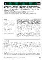
Raji cells are resistant to Rap or Dex treatment alone
Raji cells were treated with various concentrations of Rap
or Dex alone for 48 h, followed by assessment of cell
viability using MTT assays. No significant concentrationdependent decrease in cell viability was observed in response to Rap or Dex treatment (Fig. 1a and b). After 48 h
Page 4 of 12
treatment, Rap inhibited the viability of Raji cells slightly
at a regular concentration (10 nM); when the concentration was increased to 1000 nM, the cells exhibited a 30 %
viability inhibition (Fig. 1a). Thus, the IC50 (concentration
that inhibits 50 %) of Rap is higher than 1000 nM in Raji
cells. Additionally, 1 μM Dex alone showed almost no
effect on cell viability. When the concentration was increased to 100 μM, the cell line exhibited a 25 % viability
inhibition at 48 h (Fig. 1b).
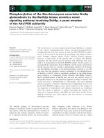
The combination of Rap with Dex effectively inhibits the
growth of Rap- and Dex-resistant Raji cells in vitro and
in vivo
We incubated Raji cells with 10 nM Rap and/or 1 μM
Dex for 48 h. Rap alone induced an approximately 13 %
reduction in cell viability, and Dex alone induced an approximately 9 % reduction in cell viability. However,
when provided in combination, Rap and Dex achieved
more than a 40 % cell reduction (Fig. 2a). Rap and Dex
combination treatment inhibited viability of Raji cells
synergistically, with a CDI of 0.75 ± 0.04. Using a light
microscope, we found that the cell size decreased and
that cell aggregation was obviously reduced in the Rap +
Dex group. Flow cytometric analysis showed that 48 h
treatment with 10 nM Rap clearly reduced cell size as
seen by the leftward shift of the mean forward scatter
(FS), but combining Rap with 1 μM Dex made the cell
size smaller and Dex alone did not affect the cell size
(Fig. 2b).
Having shown that combined treatment induced cell
viability inhibition in Raji cells in vitro, we examined the
in vivo efficacy of the two drugs given intraperitoneally
in Raji xenografts in nude mice. As shown in Fig. 2c,
3 mg/kg/d Rap or 15 mg/kg/d Dex used alone showed
almost no antitumor effect, whereas combined treatment
Fig. 1 Raji cells are resistant to Rap or Dex treatment alone. a Raji cells were cultured with various concentrations of Rap (ranging from 0.1 to
1000 nM) for 24 h and 48 h. The viability rates of the cells were evaluated with an MTT assay. The experiments were performed in triplicate.
b Raji cells were cultured with various concentrations of Dex (ranging from 0.01 to 100 μM) for 24 h and 48 h. The viability rates of the cells
were evaluated with an MTT assay. The experiments were performed in triplicate
Gu et al. BMC Cancer (2015) 15:529
Page 5 of 12
Fig. 2 The combination of Rap with Dex effectively inhibits the growth of Rap- and Dex-resistant Raji cells in vitro and in vivo. a Raji cells were
incubated with Rap (10 nM) and/or Dex (1 μM) for 48 h. The viability of the cells were evaluated with an MTT assay. b Flow cytometric analysis
showed the cell size (as seen by FS) after 48 h treatment with 10 nM Rap and/or 1 μM Dex. c Combined treatment significantly inhibited tumor
growth in Raji cell xenografts in nude mice (n = 6 per group, age 5 ~ 6 weeks, average-weight 16.9 ~ 17.1 g). All animal procedures were carried
out in accordance with ARRIVE guidelines (Additional file 1). *: p < 0.01 versus the control group, Dex group, or Rap group
significantly inhibited tumor growth when compared to
the Rap, Dex, and control groups (P < 0.001).
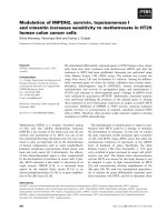
and p27 (especially p27) and reduced Cyclin A and Cyclin
D1 levels.
Combination of Rap with Dex arrests Raji cells in G0/G1
phase of the cell cycle
Rap sensitizes Raji cells to Dex-induced apoptosis
Rap, at regular dosages, inhibits cell growth of hematological
malignancies by inducing a G0/G1 arrest without inducing
apoptosis [2, 4, 8]. Dex inhibits tumor cell growth
mainly by inducing apoptosis. Flow cytometric analysis
showed that 48 h treatment with 10 nM Rap or 1 μM
Dex alone did not induce G0/G1 arrest in Raji cells.
Interestingly, combined treatment clearly induced G0/G1
arrest (Fig. 3a). To evaluate the molecular basis underlying
the cell cycle arrest, we investigated the expression of cell
cycle regulatory proteins. As shown in Fig. 3b and c, after
48 h treatment, combined treatment induced the expression of cyclin-dependent kinase (CDK) inhibitors of p21
The main mechanism of Dex in the treatment of lymphoid malignancies is to induce apoptotic cell death. We
used Annexin V-PI staining to determine the early stage
of apoptosis. Single treatment with 1 μM Dex or 10 nM
Rap had no apoptotic effect on Raji cells; however, when
used in combination, a remarkable increase in cell apoptosis was observed (Fig. 4a). Therefore, Rap can effectively
sensitize Raji cells to Dex-induced apoptosis. Bcl-2 family
members play an important role in GC-induced apoptosis
[23]. We then examined the expression of Bcl-2, Bax,
Bim-EL, Mcl-1 and caspase-3. Bim was clearly induced
in Rap, Dex, and Rap + Dex group, Bax was elevated
slightly in the three group, Mcl-1 was induced in Dex
Gu et al. BMC Cancer (2015) 15:529
Page 6 of 12
Fig. 3 The combination of Rap with Dex arrests Raji cells in the G0/G1 phase of the cell cycle. a Raji cells were incubated for 48 h with
Rap (10 nM) and/or Dex (1 μM), and the cell cycle progression was analyzed by PI staining. For all experiments, values are presented as
the mean ± SD (n = 3) *: p < 0.01 versus the control group, Dex group, or Rap group. b After 48 h exposure to Rap and/or Dex, cells were
lysed and extracts were analyzed by western blotting. β-Actin was used as an internal control. c Bar graphs show the normalized intensity
of the different proteins. Values are the results of 3 determinations. R, Rap; D, Dex; RD, Rap + Dex, and C, control
group only, Bcl-2 and caspase-3 was cleaved only in
combined treatment group (Fig. 4b). These data support that, at least in part, Rap reverses GC resistance
via activation of the intrinsic apoptotic program. Next,
we analyzed the changes in Δψm. As shown in Fig. 4c,
Rap and Dex alone or in combination dissipated Δψm,
and there were no significant differences between them.
To determine whether the apoptosis triggered by Rap
and Dex was caspase-dependent or caspase-independent,
the cells were pretreated with the pan-caspase inhibitor
z-VAD-fmk. The cell viability did not change in response
to Rap or Dex in cells pre-treated with 20 μM z-VAD-fmk
but was induced slightly in the Rap + Dex group compared
with the control group (p < 0.05) (Fig. 4d). And the cell
apoptosis rate did not change in response to Rap or Dex
pre-treatment with 20 μM z-VAD-fmk but reduced in
the Rap + Dex group compared with the control group
(p < 0.05) (Fig. 4e). Pretreatment with z-VAD-fmk failed
to fully protect Raji cells from apoptosis and cell death.
These findings suggest that combined treatment induces cell death through both caspase-dependent and
caspase-independent mechanisms in Raji cells.
Combination of Rap with Dex induces autophagic cell
death
By far, the potentially most-studied of the caspaseindependent cell death mechanisms is autophagic cell
death [24]. Rap is known to be an inducer of autophagy.
Although Raji cells are resistant to Rap treatment, Rap
alone induced the formation of autophagosomes and the
generation of LC3-II, Dex alone slightly induced autophagy, and the combined treatment strongly induced autophagy (Fig. 5a, b and c). Because autophagy can result
in both cell survival and death, we next determined
whether Rap- and Dex-induced autophagy is protective.
Pre-incubation with the autophagy inhibitor 3-MA abolished autophagosome formation (Fig. 5c) and reduced
the cell viability in Rap and Dex alone groups (Fig. 5d).
Therefore, autophagy promoted survival in the cells
treated with Rap or Dex alone. However, in the combined group, inhibiting autophagy did not affect the
cell viability by inducing apoptosis (Fig. 5d and e).
Similarly, in the combined group, when caspase-dependent
apoptosis was blocked, autophagy was strongly induced
(Fig. 5c). These findings implicate autophagy as a part
Gu et al. BMC Cancer (2015) 15:529
Page 7 of 12
Fig. 4 Rap treatment sensitizes Raji cells to GC treatment by inducing apoptosis. a Raji cells were incubated for 48 h with Rap (10 nM)
and/or Dex (1 μM), and the early stage of apoptosis was detected by Annexin V-FLUOS/PI staining (Annexin V-FLUOS positive/PI negative).
For all experiments, values of triplicate experiments are shown as the mean ± SD; *: p < 0.01 versus the control group or Dex group or
Rap group. b After 48 h exposure to Rap and/or Dex, cells were lysed and extracts were analyzed by western blotting for Bcl-2 family
proteins and caspase-3. The experiments were performed in triplicate. β-Actin was used as an internal control. c Δψm was detected by
Rh123 staining. d The cells were pretreated with 20 μM z-VAD-fmk or 0.1 % DMSO as a control for 2 h in four groups. The viability rates
of the cells were evaluated by MTT assays. e The early stage of apoptosis was detected by Annexin V-FLUOS/PI staining. *: p < 0.05 versus
the control in the RD group. R, Rap; D, Dex; RD, Rap + Dex and C, control
of a cell death mechanism for GC resensitization by
Rap.
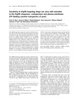
The combination of Rap with Dex acts synergistically on the
dephosphorylation of p70S6K and inhibition of glycolysis
Previous articles have reported that p-p70S6K is a critical
mediator of autophagy [25]. Rap inhibits cell growth
by dephosphorylation of p70S6K and 4E-BP1 [8, 26],
and dephosphorylation of 4E-BP1 is the key mechanism to reverse GC resistance [4]. We performed western
blotting analysis using antibodies specific for p-p70S6K
(Thr421/Ser424) and p-4E-BP1 (Thr37/46). As expected,
Raji cells are null for p-4E-BP1 (data not shown),
and Rap alone inhibited p-p70S6K (Fig. 6a). A stronger synergistic inhibition of p-p70S6K was detected
in combined group (Fig. 6a). Our results suggest that
inhibition of p-p70S6K may potentiate the cytotoxic effect of Dex.
In addition, p-p70S6K is a critical mediator of glycolytic
metabolism [17]. Our data showed that along with
the inhibition of p-p70S6K, Rap combined with Dex
strongly inhibited the cell glucose consumption and
lactic acid production (Fig. 6b). The inhibition of glycolysis
leads to a decrease in intracellular ATP concentration.
AMPK has been proposed as a physiological cellular
energy sensor [27]. We detected the expression of AMPK
Gu et al. BMC Cancer (2015) 15:529
Page 8 of 12
Fig. 5 The combination of Rap with Dex induces autophagic cell death. a After 24 h exposure to Rap and/or Dex, ultrastructural changes
were examined by transmission electron microscopy. Arrows indicate the autophagic vacuoles in the R and RD groups (8000× magnification).
b Cells were lysed and extracts were analyzed by western blotting for LC3. The experiments were repeated three times and the data show the
representative results. c MDC staining revealed the formation of autophagosomes (1000× magnification). d The cells were pretreated with 1 mM 3-MA
or 0.1 % DMSO as a control for 2 h in four groups. The viability rates of the cells were evaluated by MTT assays. *: p < 0.05 versus control in the R and D
groups. e Apoptosis was detected by Annexin V-FLUOS/PI staining (Annexin V-FLUOS positive/PI negative). R, Rap; D, Dex; RD, Rap + Dex and C, control
phosphorylated at Thr172. Rap combined with Dex
strongly induced the expression of p-AMPK (Fig. 6a).
Together, these results indicate that the combination
of Rap with Dex clearly inhibited p-p70S6K expression
and then inhibited glycolysis in Raji cells.
To test whether inhibiting glycolysis is the core mechanism for Rap-mediated restoration of GC sensitivity, we
used the glycolysis inhibitor 2-DG to replace Rap and
achieved similar results. 2-DG alone inhibited glucose
uptake and lactic acid production, and when combined
with Dex, showed a much stronger inhibitory effect on
glycolysis (Fig. 6c) and inhibited cell viability (Fig. 6d) by
inducing apoptosis (Fig. 6e) and arresting the cell cycle
in Raji cells (Fig. 6f ).
Gu et al. BMC Cancer (2015) 15:529
Page 9 of 12
Fig. 6 Inhibition of the p70S6K/glycolysis pathway plays an important role in Dex re-sensitization by Rap. a After 48 h exposure to Rap and/or Dex, cells
were lysed and extracts were analyzed by western blotting for p-p70S6K and p-AMPK. Quantification is shown as a ratio of phospho-protein to total
protein. b After 48 h exposure to Rap and/or Dex, glucose consumption and lactic acid production were measured with the Glucose (HK) Assay Kit and
Lactic Acid Assay Kit, respectively. c After 48 h exposure to 2-DG and/or Dex, glucose consumption and lactic acid production were measured with the
Glucose (HK) Assay Kit and Lactic Acid Assay Kit, respectively. d The viability rates of the cells were evaluated by MTT assays after 48 h exposure to 2-DG
and/or Dex. e The early stage of apoptosis was detected by Annexin V-FLUOS/PI staining (Annexin V-FLUOS positive/PI negative) after 48 h exposure to
2-DG and/or Dex. f The cell cycle phases were analyzed by PI staining after 48 h exposure to 2-DG and/or Dex. *: p < 0.01 versus the control group, Dex
group, or Rap group. R, Rap; D, Dex; RD, Rap + Dex and C, control
The combination of Rap with Dex acts synergistically on
the phosphorylation of glucocorticoid receptor and
dephosphorylation of ERK
The ability to up-regulate GR expression upon GC exposure has been demonstrated in various lymphoid leukemia
cell lines and has been described as essential for GCinduced apoptosis [28]. In Raji cells, we found no obvious
change in GR expression after treatment with Rap or Dex
singly or in combination (Fig. 7a and b). However, p-GR
at Ser211 was strongly induced by combined treatment.
There was also little change in ERK but an obvious
reduction of p-ERK in response to combined treatment
(Fig. 7a and b).
Discussion
Despite the good outcomes with intensive chemotherapy,
GC resistance remains a major obstacle to successful treatment of lymphoblastic malignancies. Novel and less toxic
Gu et al. BMC Cancer (2015) 15:529
Page 10 of 12
Fig. 7 The combination of Rap with Dex acts synergistically on the phosphorylation of GR and dephosphorylation of ERK. Western blot analysis of
GR, p-GR, ERK and p-ERK protein levels in Raji cells after 48 h exposure to Rap and/or Dex. Bar graphs show the ratio of phospho-protein to total
protein. R, Rap; D, Dex; RD, Rap + Dex and C, control
treatment strategies are needed, especially for pediatric
patients. Recently, the mTOR signaling pathway has received much attention as a potential target in hematological
malignancies [29–31]. However, there are still some tumor
cells that are resistant to Rap, for example, the Burkitt
lymphoma cell line Raji. The Raji cell line possesses several
Rap-resistant characteristics described by Houghton,
such as the 4E-BP1-null mutation, a high level of capindependent c-Myc expression, and the association of
a4 with PP2Ac [9]. Furthermore, Raji cells are also resistant to GC. Surprisingly, the present study provides
evidence that Rap combined with Dex, both at clinically
achievable concentrations, interacted synergistically to
inhibit Raji cell viability. This effect was found not only
in vivo but also in vitro.
To unveil the underlying mechanism, we further studied
the effect of the combined treatment on the cell cycle. Rap
or Dex alone had no effect on the cell cycle progression
of Raji cells. Combined treatment, similar to those Rapsensitive cells, can induce G0/G1 cycle arrest in Raji cells.
The down-regulation of Cyclin D1 and Cyclin A along
with the up-regulation of CDK inhibitors p21 and p27 has
previously been suggested to be the mechanism behind
mTOR inhibitor-induced cell cycle arrest in Rap-sensitive
cells [2, 32]. We achieved similar results in the combined
group: a strong induction of p27, a slight up-regulation
of p21, and down-regulation of Cyclin D1 and Cyclin
A. Therefore, combined treatment successfully restored
the sensitivity to Rap.
According to the results of the apoptosis assays, combined treatment restored the sensitivity of Raji cells to
GC. Bcl-2 family members are critical regulators of the
intrinsic apoptotic pathway and play critical roles in
GC-induced apoptosis [23]. Members of this family can
be divided into two groups: the anti-apoptotic proteins,
such as Bcl-2 and Mcl-1, and the pro-apoptotic proteins,
such as Bax and Bim. Published papers have verified that
Rap restores GC sensitivity and induces apoptosis through
the intrinsic apoptotic pathway [2–7]. Our studies showed
that in Raji cells, Rap combined with Dex obviously
cleaved Bcl-2 and caspase-3. Unlike the reported results [2–7], Rap and Dex alone or combined induced
Bim expression clearly, and combined treatment had
little effect on Bax and did not affect Mcl-1expression.
The changes on bcl-2 related proteins may correlate
with GC resistance in Raji cells, which need confirmation by further research. In Burkitt lymphoma cells,
enhanced apoptosis in response to chemotherapeutic
agents is independent of p53 and Bax [33]. Δψm dissipation is an early event in apoptosis activated through
the mitochondrial pathway [34]. However, there are
emerging data suggesting that depending on the cell
system under investigation and the apoptotic stimuli
used, the dissipation of Δψm may or may not be an
early event in the apoptotic pathway [35]. In our study,
there were no significant differences between Rap and
Dex alone or combination in dissipation of Δψm. Further
study indicated that the pan-caspase inhibitor z-VAD-fmk
only partially interfered with the GC-sensitizing effect
of Rap, whereas z-VAD-fmk blocked the cytotoxic effect
of Dex in GC-sensitive cells [22]. The data proved that
combined treatment triggers a caspase-independent
cell death in Raji cells. Autophagic cell death is the
most studied caspase-independent cell death [24]. Induction of autophagy-dependent necroptosis is a potential
mechanism for childhood ALL cells to overcome GC
resistance [22].
As Raji cells lack the expression of 4E-BP1, Rap treatment only reduced the expression of p-p70S6K and cannot arrest the cell cycle. Fortunately, dephosphorylation
of p70S6K can effectively induce cell autophagy [36].
Our results reconfirmed that Rap treatment alone
inhibited p-p70S6K expression and induced autophagy
in 4E-BP1-null Raji cells; combining Rap with Dex increased these effects. While it is clear that autophagy is
a protective mechanism at times of cellular stress, the
contribution of autophagy in regulating cancer cell death
or survival remains controversial [37]. In our study, the
Gu et al. BMC Cancer (2015) 15:529
autophagy inhibitor 3-MA inhibited the viability of Raji
cells in the Rap and Dex treatment alone groups. However,
3-MA did not affect the cytotoxicity of the combination
treatment by inducing apoptosis. z-VAD-fmk has been reported to induce cell death via autophagy [38], which may
explain why z-VAD-fmk did not fully protect the cells
from the combined treatment. Our data showed that Rap
combined with Dex induced cell killing depended on
caspase-dependent apoptosis and caspase-independent
autophagy cell death in Raji cells. Importantly, once the
cytotoxicity of the combined treatment is triggered, the
cancer cells will not be protected by the inhibition of
apoptosis or autophagy.
How can Rap restore Dex-induced apoptosis in 4EBP1-null Raji cells? Notably, S6K is the core regulator of
glycolysis [17]. Ninety years ago, Otto Warburg [39] discovered that enhanced aerobic glycolysis distinguishes
cancer from normal tissues (also known as the Warburg
effect). Upregulation of the cellular metabolism (including glycolytic and oxidative phosphorylative pathways)
and proliferation is an important aspect of GC resistance
in ALL and may contribute to patient outcome [40]. GC
resistance is directly associated with a glycolytic phenotype [41] and the activation of glycolysis has suppressive
effects on the apoptotic potential [42]. The inhibition of
glycolysis can reverse the GC resistance by inducing
apoptosis in ALL cells [41, 43]. It is noteworthy that although Raji cells are resistant to Rap, Rap treatment
alone can diminish p-p70S6K, dissipate Δψm and inhibit
glycolysis in Raji cells. There may be a potential link
among p70S6K, glycolysis and GC resistance. In support
of this hypothesis, our data indicated that Rap combined
with Dex clearly inhibited glycolysis, and the glycolysis
inhibitor 2-DG effectively took the place of Rap. When
2-DG was combined with Dex, it recapitulated the effect
of Rap combined with Dex by inducing apoptosis and
arresting the cell cycle. We got the same results in Rapsensitive T-ALL and B-ALL cell lines (data not shown).
GC resistance may be caused by a lack of GR upregulation upon GC exposure in leukemia cell lines
[44]. However, there is evidence that GC resistance in
childhood ALL cannot be attributed to an inability of
resistant cells to up-regulate the expression of the GR
upon GC exposure, nor to differences in the GR promoter
usage [45]. Another study demonstrated that the Ser211
phosphorylation site is a key regulator of GR transcriptional activation and repression [46]. Treatment with Dex
results in the phosphorylation of GR at Ser211 with increased GR expression in Dex-sensitive CEM clones,
whereas in Dex-resistant CEM clones, Rap + Dex elevates
p-GR (Ser211) expression with no increase in GR protein
[47]. Our study showed that Rap combined Dex induced
the expression of p-GR (Ser211) with no increase in GR
expression in Raji cells. Meanwhile, combined treatment
Page 11 of 12
did not influence the expression of ERK but inhibited the
ERK signaling pathway by reducing p-ERK levels. Garza
[5] found that the Dex-resistant cell lines have high basal
levels of p-ERK relative to Dex-sensitive CEM-C7-14 cells.
The p-ERK protects against GC-evoked apoptosis in sensitive T-ALL cells [47]. The inhibition of ERK also restores
GC sensitivity in resistant T-ALL cells [48]. The induction
of p-GR (Ser211) and reduction of p-ERK verified directly
that combined treatment restored the GC sensitivity in
Raji cells.
Conclusions
Taken together, inhibition of the p70S6K/glycolysis signaling pathway plays an essential role in reversing GC
resistance in Raji cells, which provides new insight into
the molecular mechanisms involved in Rap reversing
GC resistance. Targeting mTOR/p70S6K/glycolysis signaling pathway warrants further investigation as an attractive
new therapeutic approach for GC-resistant lymphoblastic
malignancies.
Additional file
Additional file 1: The ARRIVE Guidelines Checklist.
Abbreviations
Dex: Dexamethasone; GC: Glucocorticoid; mTOR: Mammalian target
of rapamycin; p70S6K: p70S6 kinase; p-p70S6K: p70S6K phosphorylation;
p-4E-BP1: Phospho-4E-BP1; Rap: Rapamycin; 2-DG: 2-deoxyglucose;
3-MA: 3-methyladenine; 4E-BP1: Eukaryotic initiation factor 4E binding
protein 1.
Competing interests
The authors declare that they have no competing interests.
Authors’ contributions
LG designed the research, performed a part of the research, analyzed the
data and wrote the paper. LPX and CZ participated in the molecular and
animal experiments and helped to draft the manuscript. ZGM helped to
design the research and contributed essential tools. YLZ performed a part
of the research. YPZ and JG contributed essential tools. All authors read
and approved the final manuscript.
Acknowledgements
This work was supported by National Natural Science Foundation of China
(Grant No.81270602 and No.30800494); Ph.D. Programs Foundation of
Ministry of Education of China (Grant No. 20090181120115); Applied Basic
Research Programs of Science and Technology Commission Foundation of
Sichuan Province, China (Grant No. 2011JY0017) and Program of Changjiang
Scholars and Innovative Research Team in University (IRT0935).
Author details
1
Laboratory of Hematology/Oncology, Department of Pediatric Hematology/
Oncology, Key Laboratory of Birth Defects and Related Diseases of Women
and Children (Ministry of Education), West China Second University Hospital,
Sichuan University, Chengdu 610041, China. 2Department of Hematology,
West China University Hospital, Sichuan University, Chengdu 610041, China.
3
Department of Rheumatology, West China University Hospital, Sichuan
University, Chengdu 610041, China.
Received: 24 September 2014 Accepted: 7 July 2015
Gu et al. BMC Cancer (2015) 15:529
References
1. Lewis-Tuffin LJ, Cidlowski JA. The physiology of human glucocorticoid receptor
beta (hGRbeta) and glucocorticoid resistance. Ann N Y Acad Sci. 2006;1069:1–9.
2. Gu L, Gao J, Li Q, Zhu YP, Jia CS, Fu RY, et al. Rapamycin reversesNPM-ALKinduced glucocorticoid resistance in lymphoid tumor cells by inhibiting
mTOR signaling pathway, enhancing G1 cell cycle arrest and apoptosis.
Leukemia. 2008;22:2091–6.
3. Stromberg T, Dimberg A, Hammarberg A, Carlson K, Osterborg A, Nilsson K,
et al. Rapamycin sensitizes multiple myeloma cells to apoptosis induced by
dexamethasone. Blood. 2004;103:3138–47.
4. Yan H, Frost P, Shi Y, Hoang B, Sharma S, Fisher M, et al. Mechanism by which
mammalian target of rapamycin inhibitors sensitize multiple myeloma cells to
dexamethasone-induced apoptosis. Cancer Res. 2006;66:2305–13.
5. Garza AS, Miller AL, Johnson BH, Thompson EB. Converting cell lines representing
hematological malignancies from glucocorticoid-resistant to glucocorticoidsensitive: signaling pathway interactions. Leukemia Res. 2009;33:717–27.
6. Gu L, Zhou C, Liu H, Gao J, Li Q, Mu D, et al. Rapamycin sensitizes T-ALL cells to
dexamethasone-induced apoptosis. J Exp Clin Cancer Res. 2010;29:150.
7. Wei G, Twomey D, Lamb J, Schlis K, Agarwal J, Stam RW, et al. Gene
expression-based chemical genomics identifies rapamycin as a modulator
of MCL1 and glucocorticoid resistance. Cancer Cell. 2006;10:331–42.
8. Fingar DC, Richardson CJ, Tee AR, Cheatham L, Tsou C, Blenis J. mTOR controls
cell cycle progression through its cell growth effectors S6K1 and 4E-BP1/
eukaryotic translation initiation factor 4E. Mol Cell Biol.2004;24:200–16.
9. Kurmasheva R, Huang S, Houghton P. Predicted mechanisms of resistance
to mTOR inhibitors. Br J Cancer. 2006;95:955–60.
10. Dilling MB, Germain GS, Dudkin L, Jayaraman AL, Zhang X, Harwood FC,
et al. 4E-binding proteins, the suppressors of eukaryotic initiation factor 4E,
are down-regulated in cells with acquired or intrinsic resistance to rapamycin.
J Biol Chem. 2002;277:13907–17.
11. Pulvertaft JV. Cytology of Burkitt’s tumor (African lymphoma). Lancet.
1964;1:238–40.
12. Wlodarski P, Kasprzycka M, Liu X, Marzec M, Robertson ES, Slupianek A, et al.
Activation of mammalian target of rapamycin in transformed B lymphocytes
is nutrient dependent but independent of Akt, mitogen-activated protein
kinase/extracellular signal-regulated kinase kinase, insulin growth factor-I,
and serum. Cancer Res. 2005;65:7800–8.
13. Inui S, Sanjo H, Maeda K, Yamamoto H, Miyamoto E, Sakaguchi N. Ig
receptor binding protein 1 (alpha4) is associated with a rapamycin-sensitive
signal transduction in lymphocytes through direct binding to the catalytic
subunit of protein phosphatase 2A. Blood. 1998;92:539–46.
14. Zhou M, Zhao Y, Ding Y, Liu H, Liu Z, Fodstad O, et al. Warburg effect in
chemosensitivity: targeting lactate dehydrogenase-A re-sensitizes
taxol-resistant cancer cells to taxol. Mol Cancer. 2010;9:33.
15. Bhattacharya B, Low SH, Soh C, Kamal Mustapa N, Beloueche-Babari M, Koh
KX, et al. Increased drug resistance associated with reduced glucose levels
and an enhanced glycolysis phenotype. Br J Pharmacol. 2014;171:3255–67.
16. Zhou Y, Tozzi F, Chen J, Fan F, Xia L, Wang J, et al. Intracellular ATP levels
are a pivotal determinant of chemoresistance in colon cancer cells. Cancer
Res. 2012;72:304–14.
17. Tandon P, Gallo CA, Khatri S, Barger JF, Yepiskoposyan H, Plas DR. Requirement
for ribosomal protein S6 kinase 1 to mediate glycolysis and apoptosis resistance
induced by Pten deficiency. Proc Natl Acad Sci U S A. 2011;108:2361–5.
18. Aries IM, Hansen BR, Koch T, van den Dungen R, Evans WE, Pieters R, et al.
The synergism of MCL1 and glycolysis on pediatric acute lymphoblastic
leukemia cell survival and prednisolone resistance. Haematologica.
2013;98:1905–11.
19. Ganapathy-Kanniappan S, Geschwind JF. Tumor glycolysis as a target for
cancer therapy: progress and prospects. Mol Cancer. 2013;12:152.
20. Liu LL, Long ZJ, Wang LX, Zheng FM, Fang ZG, Yan M, et al. Inhibition of
mTOR pathway sensitizes acute myeloid leukemia cells to aurora inhibitors
by suppression of glycolytic metabolism. Mol Cancer Res. 2013;11:1326–36.
21. Jung C, Ro S, Cao J, Otto N, Kim D. mTOR regulation of autophagy. FEBS
Lett. 2010;584:1287–95.
22. Bonapace L, Bornhauser BC, Schmitz M, Cario G, Ziegler U, Niggli FK, et al.
Induction of autophagy-dependent necroptosis is required for childhood
acute lymphoblastic leukemia cells to overcome glucocorticoid resistance. J
Clin Invest. 2010;120:1310–23.
23. Almawi WY, Melemedjian OK, Jaoude MM. On the link between Bcl-2
family proteins and glucocorticoid-induced apoptosis. J Leukoc Biol.
2004;76:7–14.
Page 12 of 12
24. Lockshin RA, Zakeri Z. Caspase-independent cell deaths. Curr Opin Cell Biol.
2002;14:727–33.
25. Shinojima N, Yokoyama T, Kondo Y, Kondo S. Roles of the Akt/mTOR/
p70S6K and ERK1/2 signaling pathways in curcumin-induced autophagy.
Autophagy. 2007;3:635–7.
26. Hay N, Sonenberg N. Upstream and downstream of mTOR. Genes Dev.
2004;18:1926–45.
27. Hardie DG. AMP-activated protein kinase: an energy sensor that regulates all
aspects of cell function. Genes Dev. 2011;25:1895–908.
28. Ramdas J, Liu W, Harmon JM. Glucocorticoid-induced cell death requires
autoinduction of glucocorticoid receptor expression in human leukemic T
cells. Cancer Res. 1999;59:1378–85.
29. Wang M, Popplewell LL, Collins Jr RH, Winter JN, Goy A, Kaminski MS, et al.
Everolimus for patients with mantle cell lymphoma refractory to or intolerant of
bortezomib: multicentre, single-arm, phase 2 study .Br J Haematol. 2014;165:510–8.
30. Witzig TE, Reeder CB, LaPlant BR, Gupta M, Johnston PB, Micallef IN, et al. A
phase II trial of the oral mTOR inhibitor everolimus in relapsed aggressive
lymphoma. Leukemia. 2011;25:341–7.
31. Younes A, Samad N. Utility of mTOR inhibition in hematologic malignancies.
Oncologist. 2011;16:730–41.
32. Vega F, Medeiros LJ, Leventaki V, Atwell C, Cho-Vega JH, Tian L, et al.
Activation of mammalian target of rapamycin signaling pathway
contributes to tumor cell survival in anaplastic lymphoma kinase-positive
anaplastic large cell lymphoma. Cancer Res. 2006;66:6589–97.
33. Kanda K, Wong W, Boxer LM. Enhanced Apoptosis to Chemotherapeutic Agents
Is Dependent on NF{kappa}B and Bcl2-Related Proteins but Is Independent of
P53 and Bax in Burkitt's Lymphoma Cells [abstract]. Blood. 2004;104:1541.
34. Chandra D, Liu JW, Tang DG. Early mitochondrial activation and cytochrome
c up-regulation during apoptosis. J Biol Chem. 2002;277:50842–54.
35. Nicholls DG, Ward MW. Mitochondrial membrane potential and neuronal
glutamate excitotoxicity: mortality and millivolts. Trends Neurosci. 2000;23:166–74.
36. Blommaart EF, Luiken JJ, Blommaart PJ, van Woerkom GM, Meijer AJ.
Phosphorylation of ribosomal protein S6 is inhibitory for autophagy in
isolated rat hepatocytes. J Biol Chem. 1995;270:2320–6.
37. Maycotte P, Thorburn A. Autophagy and cancer therapy. Cancer Biol Ther.
2011;11:127–37.
38. Chen SY, Chiu LY, Maa MC, Wang JS, Chien CL, Lin WW. zVAD-induced
autophagic cell death requires c-Src-dependent ERK and JNK activation
and reactive oxygen species generation. Autophagy. 2011;7:217–28.
39. Warburg O. On the origin of cancer cells. Science. 1956;123:309–14.
40. Beesley AH, Firth MJ, Ford J, Weller RE, Freitas JR, Perera KU, et al.
Glucocorticoid resistance in T-lineage acute lymphoblastic leukaemia is
associated with a proliferative metabolism. Br J Cancer. 2009;100:1926–36.
41. Samuels AL, Heng JY, Beesley AH, Kees UR. Bioenergetic modulation
overcomes glucocorticoid resistance in T-lineage acute lymphoblastic
leukaemia. Br J Haematol. 2014;165:57–66.
42. Rathmell JC, Fox CJ, Plas DR, Hammerman PS, Cinalli RM, Thompson CB.
Akt-directed glucose metabolism can prevent Bax conformation change and
promote growth factor-independent survival. Mol Cell Biol. 2003;23:7315–28.
43. Hulleman E, Kazemier KM, Holleman A, VanderWeele DJ, Rudin CM,
Broekhuis MJ, et al. Inhibition of glycolysis modulates prednisolone
resistance in acute lymphoblastic leukemia cells. Blood. 2009;113:2014–21.
44. Schmidt S, Irving JA, Minto L, Matheson E, Nicholson L, Ploner A, et al.
Glucocorticoid resistance in two key models of acute lymphoblastic leukemia
occurs at the level of the glucocorticoid receptor. FASEB J. 2006;20:2600–2.
45. Tissing WJ, Meijerink JP, Brinkhof B, Broekhuis MJ, Menezes RX, den Boer ML,
et al. Glucocorticoid-induced glucocorticoid-receptor expression and
promoter usage is not linked to glucocorticoid resistance in childhood ALL.
Blood. 2006;108:1045–9.
46. Chen W, Dang T, Blind RD, Wang Z, Cavasotto CN, Hittelman AB, et al.
Glucocorticoid receptor phosphorylation differentially affects target gene
expression. Mol Endocrinol. 2008;22:1754–66.
47. Miller AL, Webb MS, Copik AJ, Wang Y, Johnson BH, Kumar R, et al. p38
Mitogen-activated protein kinase (MAPK) is a key mediator in
glucocorticoid-induced apoptosis of lymphoid cells: correlation between
p38 MAPK activation and site-specific phosphorylation of the human
glucocorticoid receptor at serine 211. Mol Endocrinol.
2005;19:1569–83.
48. Miller AL, Garza AS, Johnson BH, Thompson EB. Pathway interactions
between MAPKs, mTOR, PKA, and the glucocorticoid receptor in lymphoid
cells. Cancer Cell Int. 2007;7:3.